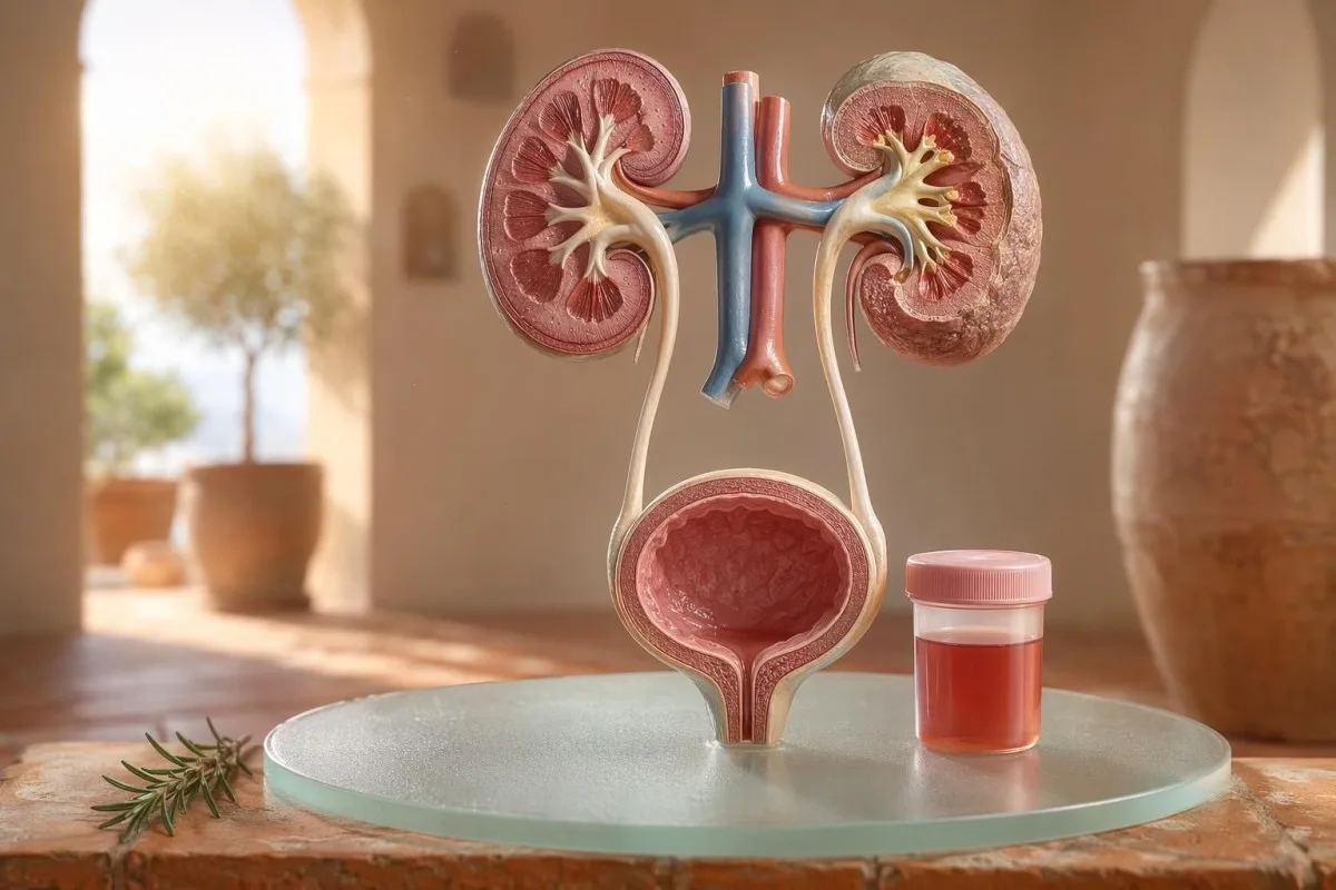
Gabay na nakatuon sa pasyente para sa nakikita at mikroskopikong hematuria, kabilang ang kung bakit ang dugo sa dipstick ay hindi pareho sa mga pulang selula sa ilalim ng mikroskopyo.


- Dugo sa ihi ibig sabihin ay nakikitang pulang ihi, ihi na kulay-tsaa o kulay-cola, o mikroskopikong hematuria na natuklasan sa pagsusuri; ang nakikitang dugo ay laging nararapat sa medikal na follow-up.
- Microscopic hematuria ay karaniwang tinutukoy bilang 3 o higit pang RBC bawat high-power field sa maayos na nakolektang sample ng urine microscopy.
- Dugo sa dipstick nakakakita ng heme activity, kaya maaari itong maging positibo mula sa buo pang RBC, libreng hemoglobin o myoglobin pagkatapos ng pinsala sa kalamnan.
- Mga pahiwatig ng UTI kabilang ang pananakit/pagkapaso, madalas na pag-ihi, leukocyte esterase, nitrites at mga puting selula; dapat muling suriin ang hematuria matapos mawala ang impeksiyon.
- Urine sediment mga natuklasan tulad ng dysmorphic red cells, red cell casts o makabuluhang protina ay tumuturo sa sakit ng kidney filtering-unit.
- Urine casts ay mga hinulma na materyal mula sa mga tubule ng bato; ang mga red cell cast ay isang senyales ng agarang pag-aalala sa nephrology sa loob ng parehong linggo, lalo na kung may mataas na presyon ng dugo o bumababang eGFR.
- Mga babalang palatandaan ng kanser kabilang ang walang sakit na nakikitang hematuria, mga namuong dugo, edad na higit sa 45, kasaysayan ng paninigarilyo, pagkakalantad sa mga kemikal sa trabaho, at paulit-ulit na hindi maipaliwanag na mga yugto.
- Hematuria na dulot ng ehersisyo kadalasang humuhupa sa loob ng 48-72 oras; ang patuloy na pagdurugo matapos magpahinga ay hindi dapat sisihin sa pagtakbo o pagbibisikleta.
- Agarang pagsusuri ay kailangan para sa dugo sa ihi na may lagnat, pananakit sa tagiliran, mga namuong dugo na nagdudulot ng bara sa pag-ihi, pagbubuntis, trauma, o mga sintomas ng matinding anemia.
Ano ang ibig sabihin ng dugo sa ihi bago ka mag-panic
Dugo sa ihi ay maaaring magmula sa impeksiyon, mga bato, sakit sa pagsala ng bato, mga kondisyon ng prostate o pantog, mga gamot, ehersisyo, o kontaminasyon mula sa regla. Ang praktikal na tuntunin ay simple: ang nakikitang dugo, paulit-ulit na microscopic hematuria, o hematuria na may pananakit, lagnat, mga namuong dugo, mataas na presyon ng dugo, protina, o nabawasang paggana ng bato ay nangangailangan ng agarang pagsusuri kaysa sa watchful waiting.
Ang isang solong kulay-rosas na mangkok ng banyo pagkatapos ng beetroot o bagong supplement ay hindi kaparehong klinikal na problema ng ihi na kulay cola pagkatapos ng namamagang lalamunan. Sa aking pagsasanay, ang kuwento ng kulay ay madalas nagsasabi sa akin kung saan unang titingnan: ang maliwanag na pula na may mga namuong dugo ay kadalasang mula sa mababang urinary tract, habang ang kayumanggi o kulay-tsaa na ihi ay maaaring nangangahulugang mas lumang heme pigments mula sa mga salaan ng bato.
Ang hematuria ay hindi diagnosis; ito ay isang senyales. Nakikitang hematuria ibig sabihin ay nakikita mo ang pagbabago ng kulay, habang microscopic hematuria ibig sabihin ay mukhang normal ang ihi ngunit sa microscopy ay may nakikitang mga pulang selula, kadalasan sa 3 o higit pang red blood cells kada high-power field.
Si Kantesti ay isang AI blood test analyzer na tumutulong sa mga pasyente na ikonekta ang mga natuklasan sa ihi sa mga marker ng bato tulad ng creatinine, eGFR, CRP, hemoglobin at platelets sa iisang timeline. Kung nakakalito ang kulay, ang aming gabay sa kulay ng ihi ay nagpapaliwanag kung bakit hindi lahat ng pulang, kayumanggi, kahel, at madilim na dilaw na ihi ay nangangahulugang pareho.
Ang dugo sa dipstick ay hindi pareho sa RBC sa mikroskopyo
Ang urine dipstick na minarkahan ng blood positive ay nakakakita ng heme-like na aktibidad ng kemikal, hindi kinakailangang buo ang mga red blood cells. Sinasagot ng microscopy ang susunod na tanong: may mga aktuwal bang RBC sa sediment ng ihi, at kung mayroon, ano ang hitsura nila?
Mabilis ang pagsusuri sa dipstick dahil ang reagent pad ay tumutugon sa peroxidase activity ng heme. Ibig sabihin, maaaring lumabas na positibo kahit may buo pang RBC, libreng hemoglobin matapos masira ang mga red cell, o myoglobin matapos ang makabuluhang pinsala sa kalamnan.
Mas mabagal ang microscopy ngunit mas tiyak. Ang maingat na laboratoryo ay i-centrifuge ang ihi, susuriin ang urine sediment, at mag-uulat ng mga red cells kada high-power field; maraming clinician ang gumagamit ng 0-2 RBC/hpf bilang normal at 3 o higit pang RBC/hpf bilang abnormal sa mga adulto.
Mahalaga ang hindi pagkakatugma. Ang dipstick na may 3+ blood at 0-2 RBC/hpf ay magpapausisa sa akin tungkol sa rhabdomyolysis, hemolysis, mga oxidizing contaminants, at paghawak ng specimen—hindi agad sa bladder disease; ang pagkakaibang ito ay katulad ng lohika sa aming paghahambing ng urinalysis at culture.
Kapag ang mga pahiwatig ng UTI ay nagpapaliwanag ng hematuria, at kapag hindi
Ang impeksyon sa urinary tract ay maaaring magdulot ng dugo sa ihi, lalo na kung may hapdi, pagka-madali ng pag-ihi, dalas ng pag-ihi, kakulangan sa pelvic, leukocyte esterase, nitrites, at mga white cells. Ngunit dapat mawala ang hematuria matapos ang paggamot; ang patuloy na mga pulang selula pagkatapos ng UTI ay nangangailangan ng pangalawang pagsusuri.
Sa isang tuwirang lower UTI, inaasahan kong magkakaroon ng kumpol: dysuria, dalas ng pag-ihi, positibo ang leukocyte esterase, ang pyuria ay madalas na higit sa 5-10 white cells/hpf, at minsan ay nitrites. Ang tradisyunal na culture positivity ay 100,000 CFU/mL, ngunit sa mga babaeng may sintomas, ang mas mababang bilang gaya ng 1,000-10,000 CFU/mL ay maaari pa ring maging klinikal na makabuluhan.
Ang leukocyte esterase ay isang enzyme mula sa white cells, hindi patunay ng bakterya. Ang positibong resulta na may hematuria ay maaaring mangyari dahil sa impeksyon, bato, kontaminasyon, o pamamaga ng bato, kaya ang aming gabay sa leukocyte esterase ay naghihiwalay ng mga kapaki-pakinabang na pahiwatig mula sa mga karaniwang maling positibo.
Ang nitrites ay mas tiyak ngunit hindi gaanong sensitibo dahil hindi lahat ng bakterya ay nagko-convert ng nitrate sa nitrite. Kung klasik ang mga sintomas ngunit negatibo ang nitrites, maaari pa ring maging positibo ang culture; ang mga praktikal na detalye ay nasa aming paliwanag sa resulta ng nitrite.
Nakikitang hematuria: mga pahiwatig sa kulay, namuong dugo at timing
Ang nakikitang hematuria ay hindi kailanman isang bagay na basta ko na lang pinapabayaan, kahit nangyari ito minsan lang. Ang matingkad na pulang ihi, mga namuong dugo, o pagdurugo na walang sakit ay nangangailangan ng maagap na pagtatasa dahil mas nagiging malamang ang mga sanhi sa lower urinary tract habang tumataas ang edad at pagkakalantad sa paninigarilyo.
Ang timing ay maaaring maging kapaki-pakinabang nang nakakagulat. Ang dugo na naroon lamang sa simula ng pag-ihi ay maaaring magpahiwatig ng urethra; ang dugo sa buong daloy ay maaaring mula sa pantog o pinagmumulan sa bato; at ang terminal na dugo malapit sa dulo ay minsan ay tumuturo sa bahagi ng leeg ng pantog.
Karaniwang nangangahulugan ang mga namuong dugo na ang pinagmumulan ng pagdurugo ay nasa ibaba ng mga filter ng bato, dahil hindi madaling nabubuo ang mga namuong dugo matapos dumaan sa glomeruli at tubules. Ang namuong humaharang sa daloy ng ihi ay kagyat: ang pagkapuno ng pantog (bladder distension) ay maaaring maging masakit sa loob ng ilang oras, at maaaring kailanganin ang pangangalaga sa catheter.
Minsan ipinapalagay ng mga lalaki na ang nakikitang hematuria pagkatapos ng mga sintomas sa pag-ihi ay simpleng pangangati lang ng prostate. Totoo iyon sa ilang kaso, pero kung ang PSA ay nasuri habang o kaagad pagkatapos ng impeksiyon, maaaring maging magulo ang interpretasyon; ang aming Gabay sa PSA pagkatapos ng UTI nagpapaliwanag kung bakit maraming clinician ang naghihintay ng 6-8 linggo bago ulitin ang PSA.
Mga threshold ng mikroskopikong hematuria na ginagamit ng mga doktor
Ang microscopic hematuria ay karaniwang tinutukoy bilang 3 o higit pang RBC bawat high-power field sa maayos na nakolektang sample ng ihi. Ginagamit ng alituntunin ng AUA/SUFU ang threshold na ito at inirerekomenda ang pagsusuring nakabatay sa panganib kaysa sa paggamot sa bawat pasyente na magkapareho (Barocas et al., 2020).
Ang dipstick lamang ay hindi dapat gamitin upang mag-diagnose ng microscopic hematuria. Sa alituntunin ng Barocas et al. 2020 AUA/SUFU, ang depinisyon ay nakadepende sa microscopy dahil maaaring maging positibo ang dipstick mula sa heme pigments kahit walang buo na red cells.
Ang pag-uulit ng pagsusuri ay hindi kahinaan; ito ay mabuting medisina. Kung may 3-10 RBC/hpf ang isang tao pagkatapos ng mabigat na ehersisyo, lagnat, pakikipagtalik, regla, o kontaminadong pagkolekta, kadalasan gusto ko ang malinis na ulit na sample bago mag-utos ng mga scan, gaya ng ipinapayo natin para sa gabay sa pag-uulit ng abnormal na mga pagsusuri.
Binabago ng panganib ang daanan ng pagsusuri. Ang 24-anyos na may 4 RBC/hpf pagkatapos ng kalahating marathon ay hindi katulad ng 68-anyos na dating naninigarilyo na may 25 RBC/hpf sa dalawang specimen, kahit na parehong teknikal na pumapasa sa depinisyon ng hematuria.
Urine sediment, protina at urine casts na nagbabago sa pagsusuri
Urine sediment ay maaaring magpahiwatig kung malamang na ang hematuria ay nagmumula sa mga filter ng bato. Ang mga dysmorphic na pulang selula, red cell casts, at makabuluhang pagtaas/pagbabago ng protina ay inililipat ang pag-aalala tungo sa glomerular disease kaysa sa simpleng impeksiyon sa pantog.
Ang mga red cells na mukhang hindi pantay, may bleb o hugis-singsing ay tinatawag na dysmorphic RBC, at iminumungkahi nito ang pagdaan sa isang inflamed na glomerular filter. Ang mga Acanthocytes na nasa itaas ng humigit-kumulang 5% ng urinary red cells ay madalas itinuturing na malakas na pahiwatig ng glomerular, bagama’t nag-iiba ang mga lab kung gaano sila kaingat sa pag-uulat nito.
Ang protina ang ikalawang pahiwatig na hindi ko pinapabayaan. Ang urine albumin-creatinine ratio na higit sa 30 mg/g, o mga 3 mg/mmol, ay abnormal, at ang nakikitang hematuria kasama ang protina ay nararapat sa mas mabilis na pagsusuri na nakatuon sa bato; ang aming gabay sa protina sa ihi ay nagbibigay ng mga threshold na nakabatay sa antas ng pasyente.
Urine casts ay maliliit na hulma (molds) na nabubuo sa loob ng kidney tubules. Ang hyaline casts ay maaaring lumitaw pagkatapos ng dehydration o ehersisyo, pero ang red cell casts ay abnormal at kadalasang tumuturo sa glomerulonephritis; para sa mas malawak na sanggunian sa urinalysis, tingnan ang aming kumpletong gabay sa pagsusuri ng ihi.
Mga bato, ehersisyo at pansamantalang sanhi ng hematuria
Ang mga bato at masinsing ehersisyo ay maaaring magdulot ng hematuria, pero magkaiba ang timeline nila. Ang hematuria na may kaugnayan sa bato ay madalas may kasamang pananakit sa tagiliran (flank pain) o mga kristal, habang ang hematuria sa ehersisyo ay karaniwang humuhupa sa loob ng 48-72 oras ng pahinga.
Klasikong nagdudulot ang kidney stones ng mga alon ng flank pain, pagduduwal, at microscopic o nakikitang hematuria. Ang calcium oxalate crystals ay hindi nagpapatunay ng bato, pero pinapataas nito ang hinala kapag ipinares sa colicky pain; ang aming gabay sa calcium oxalate crystals ay nagpapaliwanag sa pahiwatig sa urine microscopy.
Totoo ang exercise hematuria. Nakakita na ako ng mga marathon runner na may 10-20 RBC/hpf kinabukasan ng karera, normal ang culture, walang protina, at ganap na normal ang ulit na urinalysis 3 araw pagkatapos.
Ang panganib ay sisihin ang lahat sa training. Kung ang hematuria ay nagpapatuloy lampas sa 72 oras, lumilitaw kasama ang madilim na cola na kulay ng ihi, o kasabay ng mataas na CK, pagtaas ng creatinine, o pananakit ng kalamnan, tumingin nang higit pa sa pantog; ang aming gabay sa creatinine sa ehersisyo sumasaklaw sa overlap ng bato at kalamnan.
Mga “red flag” sa bato na hindi binabalewala ng mga doktor
Ang hematuria na may protina, mataas na presyon ng dugo, pamamaga, nabawasang eGFR, red cell casts, o mabilis na pagtaas ng creatinine ay isang red flag sa bato. Ipinapahiwatig ng mga natuklasang ito na ang mga yunit ng pagsasala ay maaaring namamaga, at ang paghihintay ng mga buwan ay maaaring makapinsala sa paggana ng bato.
Tinutukoy ng KDIGO 2024 ang chronic kidney disease sa pamamagitan ng mga marker ng pinsala sa bato, tulad ng albuminuria, o eGFR na mas mababa sa 60 mL/min/1.73 m² nang hindi bababa sa 3 buwan. Sa mga acute na sitwasyon, gayunpaman, ang hematuria kasama ang pagtaas ng creatinine sa loob ng mga araw o linggo ay sapat para i-escalate bago matugunan ang 3-buwang kahulugan.
Ang albuminuria ay madalas ang maagang babalang senyales. Ang ACR na 30-300 mg/g ay katamtamang nadagdagan, ang higit sa 300 mg/g ay malubhang nadagdagan, at ang pagsasama ng alinman sa mga ito sa hematuria ay nagbabago sa usapan; ang aming gabay sa kidney ACR ang nagpapaliwanag kung bakit ang urine albumin ay nakakakita ng pinsala bago pa man lumitaw ang mga sintomas.
Mahalaga rito ang presyon ng dugo. Ang bagong BP na 160/100 mmHg na may cola urine at pamamaga ng bukung-bukong pagkatapos ng impeksiyon sa lalamunan ay nagpapaisip sa akin ng glomerulonephritis, hindi cystitis; kung hindi sigurado ang eGFR, ang aming gabay sa muling pag-check ng GFR ang nagpapaliwanag kung kailan makatutulong ang cystatin C upang linawin ang paggana ng bato.
Mga pahiwatig ng panganib sa kanser nang walang panic
Walang sakit na nakikitang hematuria, lalo na pagkatapos ng edad 45, ay nararapat sa urgent na pagsusuri dahil ang mga kanser sa pantog at itaas na urinary tract ay maaaring magpakita sa ganitong paraan. Inirerekomenda ng NICE NG12 ang referral para sa pinaghihinalaang kanser para sa hindi maipaliwanag na nakikitang hematuria sa mga nasa hustong gulang na 45 at pataas, o nakikitang hematuria na nagpapatuloy matapos ang paggamot sa UTI.
Karamihan sa hematuria ay hindi kanser. Gayunman, ang mga kaso na naaalala ko ay ang mga tahimik: walang sakit, walang lagnat, walang hapdi, dalawang yugto lang ng pulang ihi sa isang 56-anyos na naninigarilyo na halos hindi pinansin iyon.
Ang risk ay naipon, hindi binary. Ang edad na higit sa 45-50, kasaysayan ng paninigarilyo, pagkakalantad sa cyclophosphamide, pelvic radiotherapy, paulit-ulit na nakikitang hematuria, occupational aromatic amine exposure, at mga namuong dugo ay lahat nagpapataas ng pangangailangan para sa cystoscopy o imaging.
Ang mga prostate marker ay hindi nag-aalis ng mga sanhi mula sa pantog. Kung may urinary symptoms, ang mga pagbabago sa PSA at hematuria ay nagkakapatong, maingat na suriin ang timing at katayuan ng impeksiyon; ang aming gabay sa prostate marker ipinaliliwanag kung bakit ang PSA ay isa lamang sa bahagi ng pagtatasa ng ihi ng lalaki.
Mga gamot, anticoagulants at mga maling alarma
Ang mga pampanipis ng dugo ay maaaring magpahiwatig ng pagdurugo, ngunit hindi dapat agad sisihin ang mga ito sa hematuria. Ang hematuria na kaugnay ng anticoagulant ay kailangan pa ring suriin dahil maaaring mailantad ng gamot ang mga bato, impeksiyon, sakit sa bato o isang paglaki sa urinary tract.
Ang warfarin, apixaban, rivaroxaban, aspirin at clopidogrel ay maaaring maging mas halata ang pagdurugo. Kung ang INR ay nasa itaas ng target range, mahalaga ang pagwawasto nito, ngunit ang normal na INR ay hindi ginagawang hindi nakakapinsala ang nakikitang hematuria.
Maraming sanhi na hindi nagdudulot ng pagdurugo ang kahawig ng hematuria. Ang rifampicin ay maaaring gawing orange-red ang ihi, ang phenazopyridine ay maaaring magpatingkad sa maliwanag na orange, ang beetroot ay maaaring mantsahan ang ihi sa mga taong madaling kapitan, at ang dehydration ay maaaring magdilim ng pigment nang sapat para matakot ang mga pasyente; ang pagsubaybay sa anticoagulant ay sakop sa aming mga safety labs para sa pampanipis ng dugo.
Maaari ring malinlang ang dipstick pagkatapos ng matinding ehersisyo o pinsala sa kalamnan. Ang positibong heme pad na may kaunti o walang RBC ay nagtutulak sa pag-iisip ko patungo sa myoglobin, lalo na kung ang CK ay higit sa 1,000 IU/L at madilim ang ihi.
Mga bata, pagbubuntis at kontaminasyon mula sa regla
Ang hematuria sa mga bata, pagbubuntis, o sa paligid ng regla ay nangangailangan ng mas malinis na pagkuha ng sample at mas mababang threshold para sa pagsusuri. Ang parehong resulta ng ihi ay maaaring mangahulugan ng magkaibang bagay depende sa edad, katayuan sa pagbubuntis, lagnat, blood pressure at kalidad ng pagkolekta.
Karaniwan ang kontaminasyon mula sa regla at walang dapat ikahiya. Kung hindi inaasahan ang resulta, ulitin ang midstream clean-catch sample nang hindi bababa sa 48 oras matapos tumigil ang pagdurugo; kung tuloy-tuloy ang 3 o higit pang RBC/hpf, mas nagiging makabuluhan.
Madalas kailangan ng ibang pagtingin ang mga bata. Ang hematuria pagkatapos ng viral illness ay maaaring pansamantala, ngunit ang hematuria na may pamamaga, mataas na blood pressure, protein, o nabawasang output ng ihi ay nangangailangan ng agarang pagsusuri ng pediatric.
Tumataas ang halaga ng pagsusuri sa pagbubuntis dahil maaaring mag-overlap ang UTI, mga bato, stress sa bato na may kaugnayan sa preeclampsia at kontaminasyon. Para sa mga pattern ng babala sa parehong araw na pagbubuntis na kinasasangkutan ng kidney, liver, platelet at urine findings, tingnan ang aming pregnancy lab red flags.
Mga pagsusuri na karaniwang ipinapagawa pagkatapos ng hematuria
Ang karaniwang susunod na mga pagsusuri pagkatapos ng hematuria ay paulit-ulit na urinalysis na may microscopy, urine culture kung posibleng may impeksiyon, mga pagsusuri sa dugo para sa kidney function, urine protein o ACR, pagsukat ng blood pressure, at imaging o cystoscopy na nakabatay sa risk. Ang eksaktong pagkakasunod-sunod ay nakadepende sa mga sintomas at risk.
Para sa sintomas ng uncomplicated cystitis, maaaring mauna ang culture at paggamot, kasunod ang paulit-ulit na urine microscopy matapos mawala ang sintomas. Para sa painless na nakikitang hematuria, maraming clinician ang mas maagang lumilipat sa imaging at cystoscopy dahil ang paghihintay sa paulit-ulit na episode ay maaaring maantala ang diagnosis.
Ang mga pagsusuri sa dugo ay nagdaragdag ng konteksto na hindi kayang ibigay ng ihi lamang. Maaaring angkop ang creatinine, eGFR, urea o BUN, electrolytes, CBC, CRP, complement C3/C4 at mga autoimmune marker kapag pinaghihinalaan ang pamamaga ng kidney; ang aming gabay sa ratio ng BUN/creatinine ay nagpapaliwanag ng isang karaniwang pattern sa kidney.
Ang Kantesti AI ay maaaring ayusin ang mga resultang iyon sa trend view, ngunit nananatiling mahalaga ang pagsusuri ng manggagamot kapag may mga red flags. Ang aming clinical safety approach ay inilarawan sa Medikal na Pagpapatunay, kabilang ang kung paano hinahawakan ang mga abnormal na kumpol kaysa sa iisang numero lamang.
Paano nakakatulong ang Kantesti na maayos ang konteksto ng hematuria
Si Kantesti ay isang serbisyo ng AI lab test interpretation na nagbabasa ng mga urine-adjacent na blood markers sa konteksto, gaya ng creatinine, eGFR, CRP, hemoglobin, platelets at glucose. Hindi nito dinidiyagnose ang hematuria, ngunit tinutulungan nito ang mga pasyente na makapaghanda ng mas malinis at mas kapaki-pakinabang na buod para sa kanilang clinician.
Kapag nire-review ko ang hematuria, bihira akong tumingin sa isang resulta lang. Ang normal na creatinine na 0.82 mg/dL, stable na eGFR na higit sa 90, walang protein at negatibong culture ay ibang-iba sa hematuria na may eGFR 48 at CRP 65 mg/L.
Si Kantesti ay isang platform ng interpretasyon ng biomarker ng AI na kayang magproseso ng mga na-upload na PDF ng blood test o mga larawan at iugnay ang mga marker ng kidney, inflammation at anemia na nasa labas ng saklaw sa timing ng mga sintomas. Ang mga pamamaraan sa likod ng pattern reading na iyon ay inilarawan sa aming Technology Guide, at ang mas malawak na saklaw ng mga marker ay nakabalangkas sa gabay sa mga biomarker.
Isang praktikal na tip: bago mag-upload, tiyaking tama ang mga unit, petsa at mga identifier ng pasyente. Ang mga OCR error ay maaaring gawing 0.9 ang 9.0, na ganap na magkakaibang kuwento sa kidney; ang aming PDF upload checklist ipinapakita ang mga pagkakamali na hinihiling kong mahuli muna ng mga pasyente.
Mga tala sa pananaliksik, pamamahala at ang huling checklist
Noong Hulyo 11, 2026, ang pinakaligtas na plano para sa pasyente ay kumpirmahin kung ang hematuria ay totoo, tukuyin ang mga palatandaan ng impeksiyon, hanapin ang mga red flag sa bato, at i-escalate ang nakikita o paulit-ulit na hematuria. Huwag umasa lamang sa kulay ng ihi.
Ako si Thomas Klein, MD, at ang payo ko ay sinadyang konserbatibo dahil ang napalampas na hematuria ay maaaring mahalaga. Ang medikal na nilalaman ng Kantesti ay nire-review na may klinikal na pangangasiwa mula sa aming Medical Advisory Board, at ang aming papel ay tulungan ang mga pasyente na magtanong nang mas mabuti, hindi palitan ang kanilang doktor.
Si Kantesti ay isang AI-powered tool para sa pagsusuri ng dugo ginagamit ng milyun-milyong tao sa 127 bansa, kaya kailangang maisama ang privacy at klinikal na kalinawan sa workflow, hindi idinagdag pa mamaya. Maaari mong basahin pa ang tungkol sa Kantesti LTD, ang aming istrukturang pangkumpanya sa UK at klinikal na misyon sa Tungkol sa Amin.
Kantesti AI Clinical Research Group. (2026). AI Blood Test Analyzer: 2.5M Pagsusuri ng Pagsusuri ng Dugo | Pandaigdigang Ulat sa Kalusugan 2026. Zenodo. DOI: https://doi.org/10.5281/zenodo.18175532. ResearchGate: https://www.researchgate.net/. Academia.edu: https://www.academia.edu/.
Kantesti AI Clinical Research Group. (2026). RDW Blood Test: Complete Guide to RDW-CV, MCV & MCHC. Zenodo. DOI: https://doi.org/10.5281/zenodo.18202598. ResearchGate: https://www.researchgate.net/. Academia.edu: https://www.academia.edu/.
Mga Madalas Itanong
Laging seryoso ba ang dugo sa ihi?
Ang dugo sa ihi ay hindi laging seryoso, ngunit dapat itong kumpirmahin at ipaliwanag. Ang isang beses na microscopic na resulta na 3-5 RBC/hpf pagkatapos ng ehersisyo o regla ay maaaring mawala sa paulit-ulit na pagsusuri, samantalang ang nakikitang dugo, mga namuong dugo, lagnat, pananakit sa tagiliran, protina, mataas na presyon ng dugo o nabawasang eGFR ay nangangailangan ng agarang pagsusuri. Sa mga nasa hustong gulang na higit sa 45, ang hindi maipaliwanag na nakikitang hematuria ay nararapat sa kagyat na pagtatasa kahit na walang pananakit.
Ano ang pagkakaiba ng dipstick blood at RBCs sa ihi?
Ang dipstick na pagsusuri sa dugo ay nakakakita ng aktibidad ng kemikal na kahawig ng heme, habang ang urine microscopy ay binibilang ang aktuwal na red blood cells. Ang dipstick ay maaaring maging positibo mula sa buo na RBC, libreng hemoglobin, o myoglobin pagkatapos ng pinsala sa kalamnan, kaya hindi ito kapareho ng kumpirmadong hematuria. Ang microscopic hematuria ay karaniwang tinutukoy bilang 3 o higit pang RBC bawat high-power field sa maayos na nakolektang sample.
Maaari bang magdulot ang UTI ng dugo sa ihi?
Oo, ang UTI ay maaaring magdulot ng dugo sa ihi, lalo na kapag may panununog, madalas na pag-ihi, at pagkaapurahan, at kapag naroroon din ang leukocyte esterase, nitrites, at mga puting selula. Ang tradisyunal na positibong urine culture ay kadalasang 100,000 CFU/mL, ngunit ang mas mababang bilang ay maaari pa ring mahalaga sa mga pasyenteng may sintomas. Ang hematuria ay karaniwang dapat muling suriin pagkatapos ng paggamot dahil ang patuloy na pagdurugo ay maaaring magpahiwatig ng mga bato, sakit sa bato, o iba pang sanhi ng karamdaman sa urinary tract.
Kailan ako dapat pumunta sa urgent care para sa hematuria?
Magpatingin agad sa urgent care kung may dugo sa ihi na may lagnat, matinding pananakit sa tagiliran (flank pain), pagsusuka, pagbubuntis, kamakailang trauma, mga namuong dugo (clots), kawalan ng kakayahang makaihi, pagkahimatay, o mga sintomas ng matinding anemia. Ang pagsusuri sa loob ng linggong ito ay angkop din para sa hematuria na may protina, red cell casts, bagong mataas na blood pressure, o pagtaas ng creatinine. Ang pagbara ng daloy ng ihi dahil sa mga namuong dugo ay maaaring maging masakit at delikado sa loob ng ilang oras.
Maaari bang ang ehersisyo ay magdulot ng mikroskopikong hematuria?
Ang masinsinang ehersisyo ay maaaring magdulot ng mikroskopikong hematuria, lalo na pagkatapos ng long-distance running, matinding pagbibisikleta, o mga isports na may kontak. Ang hematuria na kaugnay ng ehersisyo ay karaniwang nawawala sa loob ng 48–72 oras ng pahinga at hindi dapat may kasamang patuloy na protein, pagbaba ng eGFR, o matinding pananakit ng kalamnan. Kung positibo ang dugo sa dipstick ngunit ang mikroskopya ay nagpapakita ng kakaunting RBC, maaaring suriin ng mga clinician ang CK dahil ang myoglobin mula sa pinsala sa kalamnan ay maaaring mag-trigger ng dipstick.
Ano ang ibig sabihin ng urine casts kapag may nakitang dugo?
Ang mga urine cast ay mga nakamold na materyal na nabubuo sa loob ng mga kidney tubules, at ang uri nito ay nagbabago sa kahulugan ng hematuria. Ang hyaline casts ay maaaring lumitaw sa dehydration o ehersisyo, ngunit ang red cell casts ay abnormal at nagmumungkahi ng pamamaga ng glomerular na bahagi ng kidney. Ang dugo sa ihi na may red cell casts, protina, at mataas na blood pressure ay dapat gamutin bilang isang kidney red flag kaysa sa simpleng UTI.
Maaari bang magmukhang dugo ang ihi dahil sa dehydration?
Kadalasang ginagawang madilim na dilaw o amber ang ihi dahil sa dehydration, ngunit hindi ito karaniwang lumilikha ng mga pulang selula sa ihi. Uminom ng tubig at magpa-schedule ng pagsusuri sa ihi kung ang kulay ay pink, pula, kayumanggi, o parang cola, lalo na kung nagpapatuloy ito pagkatapos ng hydration o may kasamang pananakit.
Kumuha ng AI-Powered Blood Test Analysis Ngayon
Sumali sa mahigit 2 milyong user sa buong mundo na nagtitiwala sa Kantesti para sa agarang at tumpak na pagsusuri ng lab test. I-upload ang iyong resulta ng blood test at makakuha ng komprehensibong interpretasyon ng mga biomarker ng 15,000+ sa loob ng ilang segundo.
📚 Mga Sanggunian na Publikasyon sa Pananaliksik
Klein, T., Mitchell, S., & Weber, H. (2026). AI Blood Test Analyzer: 2.5M na mga pagsusuri ang sinuri | Global Health Report 2026. Kantesti AI Medical Research.
Klein, T., Mitchell, S., & Weber, H. (2026). RDW Blood Test: Kumpletong Gabay sa RDW-CV, MCV & MCHC. Kantesti AI Medical Research.
📖 Mga Panlabas na Sanggunian sa Medikal
National Institute for Health and Care Excellence (2015). Pinaghihinalaang kanser: pagkilala at pagrerefer. NICE guideline NG12. NICE Guideline.
Kidney Disease: Improving Global Outcomes CKD Work Group (2024). KDIGO 2024 Clinical Practice Guideline para sa Pagtatasa at Pamamahala ng Malalang Sakit sa Bato. Kidney International.
⚕️ Pagtatanggi sa Medikal
Ang artikulong ito ay para sa mga layuning pang-edukasyon lamang at hindi ito bumubuo ng medikal na payo. Palaging kumonsulta sa isang kwalipikadong tagapagbigay ng pangangalagang pangkalusugan para sa mga desisyon sa diagnosis at paggamot.
Mga Signal ng Tiwala ng E-E-A-T
Karanasan
Pinamumunuan ng manggagamot na klinikal na pagsusuri ng mga daloy ng interpretasyon ng mga pagsusuri sa laboratoryo.
Kadalubhasaan
Pokus sa laboratoryong medisina kung paano kumikilos ang mga biomarker sa kontekstong klinikal.
Pagka-awtoridad
Isinulat ni Dr. Thomas Klein na may pagsusuri ni Dr. Sarah Mitchell at Prof. Dr. Hans Weber.
Pagiging Mapagkakatiwalaan
Interpretasyong nakabatay sa ebidensya na may malinaw na mga susunod na hakbang upang mabawasan ang pag-aalarma.