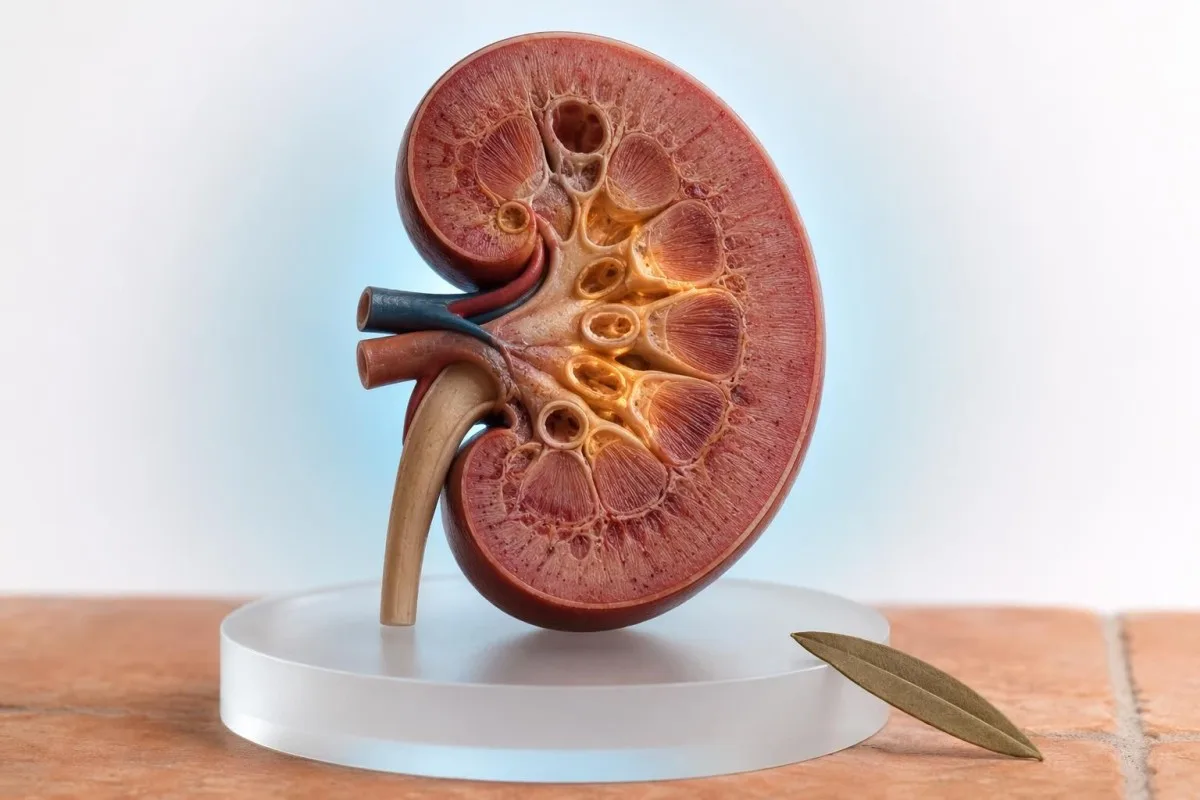
A small number of granular casts may be temporary after concentrated urine or strenuous exercise, especially when kidney blood tests are stable. Dense muddy-brown casts, rising creatinine, reduced urine output, or protein and blood in urine need prompt kidney-focused assessment.


- Granular casts in urine are cylindrical particles formed inside kidney tubules; one isolated finding is not a diagnosis.
- Muddy-brown casts are strongly associated with acute tubular injury, particularly when creatinine rises by 0.3 mg/dL or more within 48 hours.
- Temporary concentration from fever, vomiting, dehydration, or hard exercise can increase casts, but a repeat sample should be collected after 24-72 hours of recovery.
- Urine sediment microscopy is time-sensitive: casts can break down if a sample sits at room temperature for more than 2 hours.
- Urine ACR of 30 mg/g or higher suggests albumin leakage and makes granular casts more clinically meaningful.
- AKI criteria include urine output below 0.5 mL/kg/hour for 6 hours, a creatinine rise of 0.3 mg/dL in 48 hours, or 1.5 times baseline within 7 days.
- Red flags include very little urine, swelling, breathlessness, confusion, severe weakness, or cola-colored urine after exertion.
- Next tests usually include creatinine, eGFR, potassium, bicarbonate, urine ACR, repeat microscopy, and creatine kinase when muscle injury is possible.
What granular casts in urine actually mean
Granular casts in urine are particles molded in the kidney's distal tubules and collecting ducts, usually from uromodulin protein mixed with cellular debris. A few fine casts in a concentrated sample can be temporary; repeated coarse or muddy-brown casts alongside abnormal kidney tests suggest tubular stress or injury and should not be brushed off.
Casts are not debris that drift in from the bladder. They take the shape of a renal tubule before being washed into urine, which is why their presence localizes the finding to the kidneys rather than the lower urinary tract. The protein scaffold is largely uromodulin, formerly called Tamm-Horsfall protein, and it gels more readily in acidic, slow-flowing, concentrated urine.
In practice, I care far more about the pattern than the word “granular” alone: appearance, quantity, protein, red cells, urine concentration, creatinine trend, medicines, and symptoms all change the meaning. Kantesti is an AI blood test interpretation platform that places kidney markers such as creatinine, urea, potassium, and albumin in that clinical context; it cannot replace a properly reviewed urine sediment.
Dr. Thomas Klein's rule of thumb is simple: a single automated urinalysis flag without a microscopy description deserves confirmation, not panic. Ask whether the laboratory saw fine granular, coarse granular, or muddy-brown casts and whether the specimen was examined promptly. Comparing the result with hyaline casts in urine often helps because hyaline casts are much more often a concentration finding.
How urine sediment microscopy identifies casts
Urine sediment microscopy identifies casts after a laboratory professional concentrates urine and examines the sediment under magnification. The result is only as reliable as the sample handling, because casts and cells deteriorate quickly in dilute or delayed specimens.
Most laboratories centrifuge roughly 10 to 15 mL of mixed urine, remove most of the supernatant, and inspect the concentrated sediment at low and high power. Casts are commonly reported per low-power field, but there is no universal worldwide numerical reference interval. One laboratory's “occasional” may be another laboratory's “0-2 per low-power field.”
A sample examined within 1 to 2 hours at room temperature is preferable; refrigeration slows breakdown but can precipitate crystals that complicate interpretation. Very alkaline urine, often above pH 8.0, and very dilute urine can dissolve or fragment casts, producing a falsely reassuring microscopy result. This is one reason a dipstick alone cannot answer the granular casts urine meaning question.
Do not confuse a urinalysis with a uric acid test, despite the shared abbreviation “UA.” The distinction matters when requesting follow-up, as explained in our guide to urinalysis versus uric acid results. A repeat first-morning, midstream sample is often the cleanest way to settle an uncertain result.
Fine, coarse and muddy-brown casts are not equivalent
Fine granular casts may reflect mild, nonspecific tubular protein aggregation, whereas coarse muddy-brown casts raise greater concern for acute tubular injury. The darker and more densely granular the cast, the more urgently I look for a creatinine change, low urine output, medication exposure, or a recent serious illness.
The phrase muddy brown casts usually describes coarse, deeply pigmented granular cylinders that contain degenerated tubular cells and protein. Nephrologists consider this a classic urine clue for acute tubular injury, formerly called acute tubular necrosis, although microscopy is supportive rather than perfectly diagnostic. The same appearance can be missed when the sample is collected after kidney function has already started recovering.
Fine granular casts can appear after a long run, a fever, short-lived low fluid intake, or a transient fall in kidney perfusion. That does not mean exercise “causes kidney failure.” It means the result deserves a calmer repeat after normal drinking and 24 to 72 hours without hard training, provided there are no warning symptoms.
Urine concentration changes the odds. A specific gravity above 1.025 indicates concentrated urine in many laboratories and makes transient casts more plausible, while a low specific gravity around 1.005 during suspected kidney injury can signal impaired concentrating ability. Our explanation of urine specific gravity helps put that number beside the sediment finding.
When dehydration or exercise can explain a result
Dehydration and strenuous exercise can temporarily increase granular casts when urine is concentrated and kidney filtration returns to baseline after recovery. They are less reassuring explanations when creatinine rises, urine stays dark or scant, protein is present, or casts persist in a fresh repeat sample.
A 52-year-old marathon runner with an AST of 89 IU/L, creatinine 1.28 mg/dL, specific gravity 1.030, and rare granular casts should not automatically be labeled as having chronic kidney disease. In my experience, repeat testing 48 to 72 hours after rest, normal meals, and ordinary hydration often shows a lower creatinine. Our guide to creatinine after exercise explains why muscle mass and recent activity muddy the picture.
The sensible goal is normal hydration, not force-feeding liters of water. People with heart failure, advanced kidney disease, cirrhosis, or prescribed fluid limits can become unwell from excessive fluid intake; a clinician should set their target. Pale-yellow urine is not a kidney clearance test, and clear urine does not prove that the sediment has normalized.
Kantesti AI can highlight a creatinine or potassium change across uploaded laboratory reports, but a blood panel cannot identify cast type. A short recovery interval is reasonable only if you feel well, urine output is normal, and the clinician who ordered the test agrees. Stop intense training and seek assessment sooner if muscle pain, weakness, or brown urine develops.
Patterns that make granular casts more concerning
Granular casts become concerning when they occur with a rising creatinine, albuminuria, hematuria, hyperkalemia, or reduced urine output. This combination suggests active kidney stress rather than a one-off concentration effect and usually merits repeat testing within days, not months.
A urine albumin-to-creatinine ratio, or ACR, below 30 mg/g is considered normal to mildly increased in adults; 30 to 300 mg/g is moderately increased, and above 300 mg/g is severely increased. A dipstick reading of 1+ protein does not quantify albumin and can be falsely positive in concentrated urine. Protein in urine needs confirmation with ACR or protein-to-creatinine ratio.
Creatinine must be interpreted against a personal baseline. A rise from 0.70 to 1.05 mg/dL may be clinically meaningful even though 1.05 falls inside some laboratory reference ranges, while 1.30 mg/dL may be stable for a muscular person. The trend is the story, not the red flag color on a single report.
Microscopy becomes especially useful when it narrows the anatomical source of injury. Red-cell casts point toward glomerular bleeding, white-cell casts suggest kidney inflammation or upper urinary infection, and renal tubular epithelial-cell casts support tubular damage. For broader urine chemistry context, our complete urinalysis research guide covers how dipstick and microscopy answer different questions.
Kidney thresholds doctors use for prompt follow-up
Acute kidney injury is defined by a creatinine rise of at least 0.3 mg/dL within 48 hours, a rise to 1.5 times baseline within 7 days, or urine output below 0.5 mL/kg/hour for 6 hours. Muddy-brown casts in this setting warrant same-day clinical contact, even if you feel relatively well.
The 2012 KDIGO Acute Kidney Injury guideline uses those thresholds because serum creatinine rises late relative to the original renal insult; damage can already be underway before a result looks dramatic. Potassium above 5.5 mmol/L, bicarbonate below 18 mmol/L, or a rapidly increasing creatinine adds urgency because these changes can affect heart rhythm and acid-base balance (KDIGO AKI Work Group, 2012).
A basic renal work-up commonly includes creatinine, eGFR, urea or BUN, sodium, potassium, bicarbonate, calcium, phosphate, urinalysis with microscopy, and urine ACR. A basic metabolic panel covers several of these blood markers, though the exact components differ by country and laboratory.
Dr. Thomas Klein has seen patients reassured by a “normal eGFR” printed from a single test despite a meaningful acute creatinine increase from their usual level. eGFR equations are less reliable during rapidly changing kidney function because they assume relative stability. In suspected AKI, repeat creatinine timing and urine output often tell us more than one estimated filtration number.
Common causes of muddy-brown casts and tubular injury
Muddy-brown casts most often accompany reduced kidney perfusion, medication-related toxicity, severe infection, pigment injury from muscle breakdown, or prolonged obstruction. The next step is identifying the trigger quickly, because several causes improve when treated early.
Low kidney perfusion can follow vomiting, diarrhea, fever, major fluid loss, low blood pressure, heart failure, or a severe systemic illness. Non-steroidal anti-inflammatory drugs such as ibuprofen and naproxen can compound this risk by altering blood flow into the kidney, particularly during dehydration. A medication list should include supplements, contrast exposures, antibiotics, and recent dose changes.
Aminoglycoside antibiotics, some chemotherapy agents, calcineurin inhibitors, iodinated contrast in selected high-risk settings, and certain antiviral drugs can injure tubules. The finding does not prove that a drug caused the problem, but it changes the risk-benefit conversation. Kantesti, an AI lab test interpretation service, can organize medication-relevant blood-test trends, while our AI technology guide explains why any algorithmic alert needs clinician review.
Obstruction is a sometimes-overlooked cause, especially with a solitary kidney, enlarged prostate, pelvic mass, stone symptoms, or inability to pass urine. A renal ultrasound is often considered when AKI has no obvious reversible explanation. Visible blood or clots should be assessed through a structured hematuria work-up, not assumed to be from casts.
Why sediment microscopy can outperform a dipstick alone
Urine sediment microscopy adds anatomical clues that a dipstick cannot provide: casts indicate renal tubular origin, while dipstick protein and blood are chemical signals without a precise location. A normal dipstick does not reliably exclude early tubular injury, particularly when urine is very dilute.
Perazella and colleagues found that a sediment score based on renal tubular epithelial cells and granular casts correlated with AKI severity and worsening among hospitalized patients (Perazella et al., 2010). This does not mean every outpatient with one cast has AKI. It means a careful microscopy result can add real clinical signal when the blood tests and illness history already raise concern.
Automated analyzers are useful screeners but may misclassify mucus threads, debris, and fragmented casts, so manual review matters when the report says “pathological casts,” “muddy brown,” or “cellular casts.” A laboratory can sometimes perform a reflex microscopic examination from the original sample, but only if it was stored appropriately. Delayed review after 24 hours is often less informative.
A urine sediment is a snapshot, not a kidney biopsy. Cast burden can fluctuate over hours with hydration and urine flow, while albuminuria may vary with exercise, fever, and blood pressure. Readers who want the longer-term framework should review chronic kidney disease stages, which require persistence for at least 3 months.
Other urine findings that change the differential
Red-cell casts, white-cell casts, fatty casts, and waxy casts point to different kidney processes than uncomplicated granular casts. Their presence should shift the work-up toward glomerular disease, kidney inflammation, nephrotic protein loss, or chronic low-flow tubular disease.
Red-cell casts plus proteinuria, high blood pressure, swelling, or a rising creatinine are concerning for glomerulonephritis and may require urgent nephrology input. Dysmorphic red cells support a glomerular source, but that distinction depends on experienced microscopy. A positive dipstick for blood without red cells can instead occur with hemoglobin or myoglobin.
White-cell casts can occur in pyelonephritis or acute interstitial nephritis, especially with fever, flank discomfort, rash, eosinophilia, or a newly started medicine. They are not synonymous with a routine bladder UTI. A urine culture is useful when symptoms and leukocytes support infection, but it does not diagnose every cause of sterile white cells.
Fatty casts and oval fat bodies suggest substantial protein leakage, often in nephrotic-range disease; waxy casts are broader, more rigid, and may appear with advanced chronic kidney impairment. A first-morning urine ACR test is more reproducible than random dipstick protein when deciding whether albumin leakage is persistent.
Hard exercise, rhabdomyolysis and pigment-related kidney stress
Severe muscle injury can produce granular casts and acute kidney injury because myoglobin stresses kidney tubules. Urgent evaluation is needed after extreme exercise, crush injury, seizures, heat illness, or drug exposure when dark urine, muscle pain, weakness, or reduced urine output occur.
Rhabdomyolysis is usually suspected when creatine kinase, or CK, is at least 5 times the laboratory upper limit, although no single CK value predicts kidney injury perfectly. CK can peak 24 to 72 hours after muscle injury, so an early “not too high” result may need repeating. Our review of dangerous CK elevations outlines the practical warning pattern.
A dipstick may read strongly positive for blood because it detects heme pigment, while microscopy shows few or no red cells; that mismatch raises the possibility of myoglobinuria. Granular or pigmented casts add concern but are not mandatory for diagnosis. Potassium, phosphate, calcium, creatinine, bicarbonate, and CK should be checked together in a concerning presentation.
Do not try to manage possible rhabdomyolysis by drinking large amounts of water at home and waiting for urine color to improve. Intravenous fluid decisions depend on heart function, kidney function, electrolytes, and urine output. Anyone with chest symptoms, confusion, fainting, severe weakness, or very little urine should seek emergency care.
Medicines and supplements worth reviewing with a clinician
Recent NSAID use, diuretics, ACE inhibitors or ARBs during dehydration, antibiotics, contrast exposure, and unregulated supplements can contribute to a tubular-injury pattern. Do not stop prescribed medicines abruptly, but bring every product and dose to the clinician reviewing muddy-brown casts.
The risk is often a combination rather than one culprit: a diuretic, vomiting illness, an ACE inhibitor or ARB, and ibuprofen can reduce the kidney's ability to adapt to low circulating volume. This is sometimes informally called the “triple whammy,” though the actual risk varies widely by age, baseline eGFR, and dose. Your prescriber should decide which medicines, if any, to pause during an acute illness.
Creatine can raise measured creatinine without necessarily reducing filtration, while very high-dose vitamin C may raise urinary oxalate in susceptible people. Herbal products deserve specific names rather than a vague “natural supplement” label because ingredients and contamination vary. A low threshold for repeat testing is sensible if potassium changes by more than 0.5 mmol/L or creatinine is climbing.
Kantesti AI compares laboratory trends across reports and can make a changing creatinine easier to spot, but it does not determine medication safety. Our clinical methods are subject to medical validation and oversight, and a pharmacist or prescribing clinician remains the right person to assess drug interactions. For kidney context beyond creatinine, cystatin C testing may be helpful in selected cases.
A practical repeat-testing plan after an unexpected result
An isolated granular-cast result in a well person is often rechecked with a fresh first-morning urine sample and kidney blood tests within 48 to 72 hours. Faster testing is appropriate when casts are muddy-brown, symptoms are present, or creatinine, potassium, or urine output is abnormal.
Before a planned repeat, avoid vigorous exercise for 24 to 48 hours, drink normally unless fluid restricted, and collect a clean-catch midstream sample. Deliver it quickly, ideally within 1 hour, and tell the laboratory about menstruation, recent exercise, fever, antibiotics, and supplements. These details change how a microscopist reads ambiguous sediment.
Ask for creatinine, eGFR, electrolytes, bicarbonate, urine dipstick with microscopy, and ACR when protein is suspected. A urea-to-creatinine relationship can offer clues about volume status but cannot diagnose dehydration by itself; our BUN and creatinine ratio guide explains the limitations. Creatinine should be compared with at least one prior result whenever available.
KDIGO's 2024 CKD guideline defines chronic kidney disease as abnormalities of kidney structure or function present for at least 3 months, not one off-day urine result (KDIGO CKD Work Group, 2024). Kantesti is an AI biomarker interpretation platform that can preserve serial blood-test context, including dates and prior creatinine values. Persistent casts, ACR at or above 30 mg/g, or eGFR below 60 mL/min/1.73 m² should be discussed with a clinician rather than self-monitored indefinitely.
When a culture, ultrasound, or specialist referral helps
Urine culture helps when urinary symptoms and white cells suggest infection, while ultrasound helps when obstruction or an unexplained acute kidney injury is possible. Neither test is routinely needed for every rare fine granular cast in an otherwise normal repeat sample.
Burning urination, frequency, fever, flank pain, nitrites, and white cells make culture more useful; granular casts alone do not prove infection. Mixed bacterial growth can reflect collection contamination rather than a true urinary infection, especially when epithelial cells are abundant. Our urine culture results guide explains why the organism and colony pattern matter.
Ultrasound is commonly used to look for hydronephrosis, kidney size, stones, or impaired bladder emptying when obstruction is on the table. It may be normal in early tubular injury, so a normal scan does not erase an abnormal creatinine trend. In a person with one functioning kidney or known urinary obstruction, the threshold for same-day assessment should be lower.
Nephrology referral is reasonable for unresolved AKI, persistent ACR above 300 mg/g, recurrent cellular casts, suspected glomerulonephritis, difficult electrolyte changes, or eGFR below 30 mL/min/1.73 m². Referral urgency depends on trajectory: a creatinine that doubles over 48 hours is very different from a stable mildly reduced eGFR over 5 years.
Symptoms and results that need same-day or emergency care
Seek same-day medical advice for muddy-brown casts plus falling urine output, swelling, new vomiting, fever, or a rising creatinine. Seek emergency care for severe breathlessness, chest pain, confusion, fainting, seizures, inability to pass urine, or weakness with suspected high potassium.
Urine output below 0.5 mL/kg/hour for 6 hours is a formal AKI threshold; for a 70 kg adult, that is less than 210 mL over 6 hours. Measuring output is imperfect outside hospital, but a noticeable drop in urination combined with illness is actionable. Sudden ankle swelling or rapid weight gain can indicate fluid retention even before blood results return.
Potassium above 6.0 mmol/L, severe acidosis, or progressive fluid overload may require hospital-level treatment because dangerous complications can develop without pain. An ECG is often used when potassium is significantly elevated, but a normal ECG does not make a high potassium result safe to ignore. Never take potassium supplements or salt substitutes to “correct” fatigue during a possible kidney problem.
A 24-hour urine collection is not usually the first response to granular casts, and it is easy to collect incorrectly. It can become useful for selected stone, protein, or electrolyte questions once the acute issue is stable; see our practical 24-hour urine collection guide. Bring the original microscopy report rather than relying on a remembered phrase.
Questions to take to your next kidney-focused appointment
The most useful question is not “Are granular casts bad?” but “Do my casts fit with a change in kidney function, urine protein, medications, and symptoms?” A focused conversation can prevent both unnecessary alarm and dangerous delay.
Ask whether the sample was fresh, whether microscopy was manual or automated, how many casts were seen, and whether they were fine, coarse, or muddy-brown. Ask for your exact creatinine today and baseline, eGFR, potassium, bicarbonate, urine ACR, specific gravity, and dipstick blood result. These are more useful than a generic “abnormal urine” label.
Bring a dated list of diarrheal illness, fever, workouts, heat exposure, new medicines, NSAID use, supplements, imaging contrast, and urine-output changes from the prior 7 days. If protein or blood persists, ask whether repeat microscopy, culture, ultrasound, autoimmune tests, or nephrology referral is appropriate. There is no shame in requesting a copy of the sediment report.
Kantesti's physician-led review standards are overseen by our Medical Advisory Board, but neither our tools nor this article can diagnose acute kidney injury from a screenshot. As of July 21, 2026, the safest approach remains pattern-based: confirm the sample, compare the trend, find the trigger, and escalate promptly when red flags appear.
Frequently Asked Questions
Are granular casts in urine always serious?
Granular casts in urine are not always serious, because rare fine granular casts can appear with dehydration, fever, or strenuous exercise. The result becomes more concerning when casts are coarse or muddy-brown, persist on a fresh repeat sample, or occur with a creatinine rise of 0.3 mg/dL within 48 hours. Urine protein, blood, specific gravity, symptoms, medication exposure, and prior kidney results determine the real level of concern. A clinician should review any cast result accompanied by reduced urine output or abnormal potassium.
What do muddy-brown casts mean?
Muddy-brown casts usually indicate degenerated tubular cells and protein within kidney tubules and are a classic clue for acute tubular injury. They are most meaningful when creatinine rises to 1.5 times baseline within 7 days, urine output falls below 0.5 mL/kg/hour for 6 hours, or potassium and bicarbonate are abnormal. Muddy-brown casts do not identify the cause by themselves; dehydration, low blood pressure, medicines, severe illness, pigment injury, and obstruction all require consideration. Same-day clinical contact is sensible when this phrase appears on a urine microscopy report.
Can exercise cause granular casts in urine?
Hard exercise can temporarily increase granular casts in urine by concentrating urine and briefly stressing kidney tubules, especially after endurance events or heat exposure. A repeat sample collected after 24 to 72 hours of rest and normal hydration often clarifies whether the finding was transient. Exercise is not an adequate explanation for dark urine, severe muscle pain, low urine volume, or a CK level at least 5 times the laboratory upper limit, because those features can suggest rhabdomyolysis. Creatinine should be compared with a pre-exercise baseline whenever possible.
How many granular casts are normal?
There is no single international normal range for granular casts because laboratories use different collection, centrifugation, and reporting methods. Some laboratories describe rare or occasional fine granular casts without attaching a number, while others report casts per low-power field. No casts or rare hyaline casts are generally more reassuring than persistent granular casts, but quantity alone cannot diagnose kidney disease. A fresh specimen analyzed within 1 to 2 hours gives the most useful comparison.
What tests should I ask for after granular casts are found?
After granular casts are found, clinicians commonly check creatinine, eGFR, urea or BUN, potassium, bicarbonate, urinalysis with microscopy, urine albumin-to-creatinine ratio, and blood pressure. An ACR of 30 mg/g or higher indicates increased albumin leakage and adds significance to an abnormal sediment result. CK should be measured when severe exercise, muscle pain, heat illness, or pigment-positive urine raises concern for rhabdomyolysis. Urine culture, ultrasound, autoimmune testing, or nephrology referral depends on the accompanying pattern rather than casts alone.
Can dehydration cause muddy-brown casts?
Severe dehydration can contribute to muddy-brown casts when reduced kidney blood flow progresses to acute tubular injury, but simple mild dehydration more often produces concentrated urine and occasional hyaline or fine granular casts. The distinction matters because muddy-brown casts with a creatinine increase of 0.3 mg/dL in 48 hours meet a pattern that needs prompt assessment. Drinking water is not a safe substitute for evaluation if urine output is falling, vomiting continues, swelling develops, or potassium is abnormal. People with prescribed fluid restrictions should not increase fluids without medical advice.
Can a urinary tract infection cause granular casts?
A bladder infection alone does not usually create casts, because casts form inside kidney tubules rather than the bladder. Infection symptoms with white blood cell casts, fever, or flank pain can suggest kidney involvement and deserve prompt medical evaluation.
Get AI-Powered Blood Test Analysis Today
Join over 2 million users worldwide who trust Kantesti for instant, accurate lab test analysis. Upload your blood test results and receive comprehensive interpretation of 15,000+ biomarkers in seconds.
📚 Referenced Research Publications
Klein, T., Mitchell, S., & Weber, H. (2026). Urobilinogen in Urine Test: Complete Urinalysis Guide 2026. Kantesti AI Medical Research.
Klein, T., Mitchell, S., & Weber, H. (2026). Iron Studies Guide: TIBC, Iron Saturation & Binding Capacity. Kantesti AI Medical Research.
📖 External Medical References
⚕️ Medical Disclaimer
This article is for educational purposes only and does not constitute medical advice. Always consult a qualified healthcare provider for diagnosis and treatment decisions.
E-E-A-T Trust Signals
Experience
Physician-led clinical review of lab interpretation workflows.
Expertise
Laboratory medicine focus on how biomarkers behave in clinical context.
Authoritativeness
Written by Dr. Thomas Klein with review by Dr. Sarah Mitchell and Prof. Dr. Hans Weber.
Trustworthiness
Evidence-based interpretation with clear follow-up pathways to reduce alarm.