A small number of hyaline casts is commonly a temporary concentration effect, not kidney disease. The deciding factor is what else appears in the urine sediment test and kidney panel.


- Typical finding: 0-2 hyaline casts per low-power field is commonly accepted as normal when urine protein, blood, and kidney function are normal.
- Dehydration clue: Concentrated urine with specific gravity above 1.030 can make hyaline casts more likely without indicating kidney damage.
- Exercise effect: Strenuous endurance exercise can temporarily increase hyaline casts and urine protein; a repeat sample after 24-48 hours of recovery is often more informative.
- Repeat trigger: Recheck a urine sediment test if hyaline casts persist after normal fluid intake or occur with protein, blood, or raised creatinine.
- Kidney threshold: An eGFR below 60 mL/min/1.73 m² for 3 months or more meets a chronic kidney disease criterion.
- Albumin check: A urine albumin-creatinine ratio of 30 mg/g or higher needs clinical follow-up when confirmed for at least 3 months.
- Urgent pattern: Red-cell casts, rapidly reduced urine output, swelling, high blood pressure, or dark cola-coloured urine need prompt medical assessment.
- Context first: Hyaline casts alone do not diagnose kidney disease; the accompanying protein, cells, symptoms, and trend matter more than the count.
When hyaline casts are harmless and when they need follow-up
A small number of hyaline casts in urine—often 0-2 per low-power field (LPF)—is usually harmless when you are well, hydrated, and have no protein, blood, or reduced kidney function. Repeat testing is sensible when casts persist, rise after recovery, or accompany abnormal urine protein, red cells, swelling, high blood pressure, or a creatinine increase. In my clinical experience, the cast count by itself is rarely the deciding fact.
Hyaline casts are clear, protein-based cylinders formed in the distal tubules and collecting ducts. They can appear after a hot day, vomiting, fever, an intense workout, or taking a diuretic because slower, more concentrated urine gives uromodulin protein time to gel. Dr. Thomas Klein sees this pattern most often in people whose urine specific gravity is high and whose repeat result settles within 48 hours.
The reassuring combination is simple: isolated hyaline casts, negative or trace protein, no urine blood, normal blood pressure, and a stable creatinine. A urine sediment test is more meaningful than a dipstick alone because microscopy can distinguish hyaline material from red-cell, white-cell, granular, and waxy casts. Our complete urinalysis guide explains why a dipstick result and a microscopic result can point in different directions.
Kantesti is an AI lab test interpretation service that places creatinine, eGFR, urea, albumin, electrolytes, and urinalysis findings into one follow-up view rather than treating an isolated cast count as a diagnosis. If a result was collected after a marathon, gastroenteritis, sauna use, or poor fluid intake, that timing should be recorded; it can change the next clinical step substantially.
What hyaline casts are on a urine sediment test
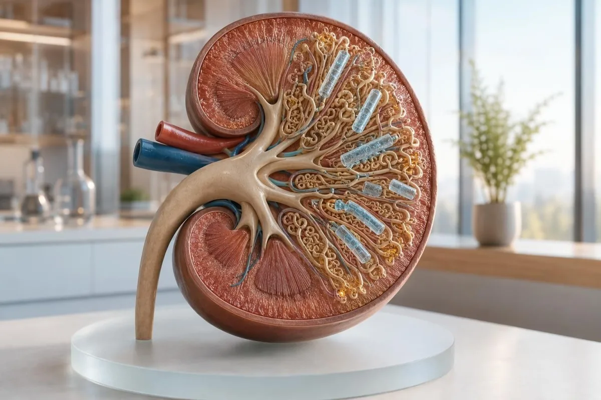
Hyaline casts are transparent moulds of the kidney tubules made mainly from uromodulin, formerly called Tamm-Horsfall protein. They are not cells, bacteria, or crystals, and their presence alone does not prove kidney injury.
The kidney normally releases uromodulin into urine, particularly from the thick ascending limb of the loop of Henle. When urine becomes concentrated, acidic, or slow-moving, that protein can aggregate into a cylindrical cast which is then flushed out. Simerville, Maxted, and Pahira described hyaline casts as a finding that can occur in healthy people in their 2005 review of urinalysis.
A laboratory scientist usually reports casts per LPF, meaning a low-power microscopic field, whereas red and white cells are often counted per high-power field. This mismatch causes avoidable anxiety: “0-2/LPF” is not equivalent to “0-2 cells/HPF,” and neither number should be read without the laboratory’s own reference interval. For basics on terminology, see our guide to urinalysis versus uric acid.
Freshness matters. Casts can dissolve in alkaline or dilute urine, while delayed testing can alter cell appearance; ideally, microscopy occurs within 2 hours of collection or the sample is refrigerated according to the laboratory protocol. A negative cast report therefore does not always mean that no casts were present at the time of urination.
Are hyaline casts normal on a urinalysis report?
Many laboratories consider 0-2 hyaline casts per LPF compatible with a normal result, especially when every other urine marker is unremarkable. There is no internationally validated cast number that independently defines kidney disease, so the wording and reference range printed by your own laboratory take priority.
Some laboratories report “none seen,” “occasional,” or “rare” instead of a numeric count. Others use automated digital microscopy and then confirm uncertain particles manually, which can shift how a faint cast is classified. A report of 3-5/LPF is not automatically dangerous, but it deserves context and often a properly prepared repeat sample if it was unexpected.
I get more concerned when a cast result travels with proteinuria, haematuria, glucose, leukocyte esterase, or an eGFR trend that is falling. Urine specific gravity normally runs roughly from 1.005 to 1.030, and a value above 1.030 supports concentration as one possible explanation—not proof—when hyaline casts are isolated. Our urine specific gravity guide covers the common reporting differences.
One practical trap: a highly concentrated first-morning sample may contain more casts than a daytime sample, yet it can be the best specimen for confirming albumin loss. Rather than deliberately overdrinking before a repeat, aim for your usual fluid intake for 24 hours; extreme water loading can dilute protein and obscure a clinically useful result.
Common benign causes of hyaline casts in urine
Dehydration, temporary low urine flow, fever, and physical exertion are the commonest benign causes of hyaline casts in urine. These triggers usually resolve after the underlying stress ends and fluid balance returns to normal.
Fluid loss does not need to be dramatic. A 1-2 kg fall in body weight during a long walk, diarrhoeal illness, or a humid workday can concentrate urine enough to make casts easier to see. Dark-yellow urine and a specific gravity near 1.030 support this explanation, although urine colour can also change with foods, vitamins, and medicines.
Fever can cause transient hyaline casts through both reduced intake and higher insensible fluid loss. In a person with a temperature of 38.5°C, I would usually repeat the test after recovery rather than label the finding as chronic kidney disease from one sample—provided there is no protein, blood, flank pain, or creatinine change. The urine osmolality result can sometimes clarify whether the kidneys are concentrating appropriately.
Short-lived low blood pressure, such as after a stomach virus, can also reduce kidney perfusion briefly. That is a reason to check a basic metabolic panel sooner in older adults or anyone taking ACE inhibitors, ARBs, NSAIDs, or diuretics, because the same event can expose limited kidney reserve.
How exercise, heat, fever, and medicines change cast results
Hard exercise and heat exposure can produce transient hyaline casts because filtration changes and urine becomes concentrated. In most healthy adults, repeating the test after 24-48 hours without strenuous training gives a clearer baseline.
A long run, heavy resistance session, or endurance event can also cause temporary albumin leakage and microscopic blood, particularly when sampling occurs soon afterwards. I have seen a 52-year-old recreational marathoner with hyaline casts, 1+ protein, and creatinine of 1.32 mg/dL the morning after a race; all three normalised on a rested repeat 72 hours later. For athlete-specific context, read our marathon recovery lab guide.
Diuretics can increase urine concentration, while NSAIDs can reduce kidney blood flow in susceptible people—especially during dehydration. Do not stop a prescribed medicine merely because casts appeared, but tell the clinician reviewing the result the exact drug, dose, and date of the last dose. A medication list often explains more than a second microscope slide.
Creatine supplements and unusually high cooked-meat intake may raise serum creatinine without directly causing casts, which can make a benign cast finding look more ominous. In that situation, cystatin C, urine ACR, blood pressure, and repeat creatinine after recovery may offer a fairer picture of filtration.
Urine findings that make hyaline casts more concerning
Hyaline casts matter more when they occur with albumin or protein, blood, kidney cells, or a reduced eGFR. The combination suggests a kidney process more strongly than an isolated hyaline cast ever can.
A urine albumin-creatinine ratio, or ACR, below 30 mg/g is category A1; 30-300 mg/g is A2; and above 300 mg/g is A3. KDIGO 2024 uses persistent ACR of 30 mg/g or more, confirmed for at least 3 months, as evidence of chronic kidney damage even when eGFR is 60 mL/min/1.73 m² or higher (KDIGO CKD Work Group, 2024). See our practical guide to protein in urine.
Dipstick blood should be confirmed with microscopy because myoglobin after heavy exercise, menstrual contamination, and oxidising agents can create misleading results. By contrast, persistent red cells—particularly dysmorphic red cells or red-cell casts—shift the question toward glomerular bleeding and deserve timely clinician review. Our blood in urine guide explains the usual evaluation pathway.
Kantesti AI interprets kidney-related lab results by comparing the urine finding with eGFR, creatinine, potassium, bicarbonate, glucose, blood pressure history, and prior values. That is clinically safer than reading “hyaline casts present” as either automatically normal or automatically serious.
How hyaline casts differ from other casts in urine
Hyaline casts are the least specific common cast type; red-cell, white-cell, granular, waxy, fatty, and epithelial-cell casts carry different diagnostic weight. A laboratory should identify the cast type rather than report only “casts present.”
Red-cell casts strongly support a glomerular source of bleeding, while white-cell casts may occur with kidney inflammation or upper urinary tract infection. Granular casts can follow tubular stress, and broad waxy casts can appear in advanced chronic kidney impairment, although microscopy must be interpreted by trained staff. Perazella’s 2015 review explains why sediment morphology can function as a kidney-disease biomarker when the clinical setting fits.
Calcium oxalate crystals are not casts and are often confused with them by patients reading a portal report. Crystals are geometric mineral deposits; casts are tubular protein moulds that carry material formed within the kidney nephron. If crystals were also reported, our explanation of calcium oxalate crystals may be useful.
A vague report such as “casts: present” is a reasonable reason to ask for clarification, not a reason to panic. Ask whether the finding was hyaline only, how many per LPF, whether the sample was fresh, and whether protein, red cells, or white cells were seen on the same examination.
How to repeat a urine sediment test properly
Repeat an unexpected isolated hyaline-cast result after 24-48 hours of usual hydration and no strenuous exercise, unless you have red-flag symptoms. A clean, promptly processed midstream sample reduces avoidable false alarms.
Use a clean container and collect a midstream sample after cleaning the genital area according to local instructions. Deliver it within 2 hours where possible; if transport will take longer, use the laboratory’s refrigeration guidance. Do not collect during heavy menstrual flow if the purpose is to clarify blood or protein in urine, because contamination can make the result difficult to interpret.
Avoid hard training, sauna exposure, deliberate fasting, and unusually high-protein meals for 24-48 hours before a non-urgent repeat. Continue prescribed medicines unless your clinician gives different advice, and write down recent fever, diarrhoea, supplements, and exercise. Urinalysis versus culture testing is helpful here: a culture answers a different question and does not replace microscopy.
As Dr. Thomas Klein, I advise patients to preserve the original report and place the two results side by side. A fall from 5-10 casts/LPF after normal recovery is clinically more reassuring than a single “negative” result obtained after drinking several litres of water just before collection.
Kidney tests to ask about if hyaline casts persist
Persistent hyaline casts should prompt a focused kidney check: creatinine with eGFR, urine ACR, blood pressure, and repeat microscopy are the core tests. The need for ultrasound, autoimmune testing, or nephrology referral depends on those results and symptoms.
Creatinine is affected by muscle mass, meat intake, creatine use, and hydration, so one mildly high value should usually be compared with a previous baseline. An eGFR below 60 mL/min/1.73 m² is concerning when present for 3 months or longer; a sudden rise in creatinine may instead suggest acute kidney injury and needs a faster response. Our chronic kidney disease stages guide sets out the ACR-eGFR grid clinicians use.
Urea or BUN can rise with dehydration and high protein intake, which is why the BUN-creatinine ratio is a clue rather than a diagnosis. Kantesti is an AI blood test analyzer that can display the ratio alongside sodium, potassium, bicarbonate, albumin, and prior creatinine values, helping users prepare precise questions for a clinician. Our BUN-to-creatinine guide describes its limits.
Cystatin C can be helpful when creatinine may be misleading because of unusually high or low muscle mass. It is not needed for every person with an isolated cast result, but it can help confirm filtration estimates near decision thresholds, especially where eGFR is 45-59 mL/min/1.73 m² without other markers of kidney damage.
Who needs a lower threshold for kidney follow-up
People with diabetes, high blood pressure, known kidney disease, systemic autoimmune disease, or pregnancy need earlier review of persistent hyaline casts. In these groups, even a bland urine sediment can coexist with early kidney injury.
Diabetes and hypertension are leading causes of chronic kidney disease, so annual ACR and eGFR testing is standard even if a routine urinalysis looks broadly normal. A single ACR of 30 mg/g or above should be repeated, because exercise, infection, high glucose, and uncontrolled blood pressure can transiently elevate albumin. Explore the kidney ACR test for collection and interpretation details.
During pregnancy, proteinuria, rising blood pressure, headache, visual symptoms, or upper abdominal pain must be assessed urgently rather than attributed to dehydration. Hyaline casts alone are not diagnostic of pre-eclampsia, but a urine protein result of 300 mg or more in 24 hours, or a protein-creatinine ratio of 0.3 or more, changes the obstetric assessment. Our guide to pregnancy lab red flags gives a safety-focused overview.
Older adults can have less renal reserve despite a creatinine value that sits inside a laboratory reference range. In an 80-year-old with low muscle mass, an eGFR trend, ACR, potassium level, and medication list generally tell me more than creatinine alone.
Symptoms and result patterns that need prompt medical review
Seek same-day medical advice for hyaline casts plus new swelling, markedly reduced urine, visible blood, fever with flank pain, or rapidly rising blood pressure. Emergency assessment is appropriate for breathlessness, confusion, chest pain, severe weakness, or almost no urine output.
New ankle or eyelid swelling matters because it can reflect sodium retention or protein loss, although it also has heart, liver, venous, and medication causes. If swelling is paired with foamy urine, 2+ or 3+ dipstick protein, and rising weight over several days, ask for urine ACR, creatinine, albumin, and blood pressure review. Our swelling blood test guide outlines the related tests.
Dark brown or cola-coloured urine after intense exercise can signal myoglobin release, while red or pink urine may be blood—both require timely assessment when persistent or combined with low output. A creatine kinase above 5 times the laboratory upper limit after severe muscle symptoms raises concern for rhabdomyolysis, where kidney injury risk is higher.
Fever, urinary burning, urgency, and flank pain may need urinalysis and culture, particularly in pregnancy, diabetes, or immunosuppression. Hyaline casts do not diagnose a urinary tract infection; white cells, nitrites, symptoms, and culture results carry much more weight.
Why urine cast results can be inconsistent
Cast results can differ between samples because casts form and dissolve rapidly, and microscopy methods vary between laboratories. One unexpected hyaline-cast result should therefore be verified before it drives a long-term kidney label, unless other abnormalities are present.
Hyaline casts are nearly colourless and can be missed in dilute urine or mistaken for mucus threads, fibre, or slide artefact. Automated imaging systems improve workflow but may send ambiguous particles for manual review; this is a sensible quality step, not a sign that the laboratory has failed. A sample left warm for several hours is less reliable for cellular sediment interpretation.
A positive culture with “mixed growth” may reflect collection contamination rather than a kidney infection, especially if there are many squamous epithelial cells and no symptoms. That is why I separate the question “Was there a urinary pathogen?” from “What did the kidney sediment show?” when discussing a report. Our guide to mixed urine culture results helps decode this language.
Kantesti's neural network can organise uploaded laboratory values, but it cannot inspect the physical slide, verify sample age, or replace a laboratory professional’s microscopy judgement. Readers can use our AI result accuracy checklist to check dates, units, reference ranges, and possible transcription errors before acting.
How to track a hyaline-cast result over time
A trend of urine findings, blood pressure, ACR, and eGFR is more informative than a single hyaline-cast count. Record the collection conditions so that a clinician can distinguish a changing kidney pattern from ordinary day-to-day variation.
Keep the date and time, whether the sample was first-morning, recent exercise within 48 hours, fluid losses, fever, menstrual status, and medicines taken. Add the numeric cast result, specific gravity, urine protein, blood, ACR, creatinine, eGFR, and blood pressure if available. A stable eGFR over 12 months is often more reassuring than small day-to-day creatinine movement.
A useful clinician question is: “Were these only hyaline casts, and did microscopy show red cells, white cells, granular casts, or dysmorphic red cells?” Another is: “Should I repeat ACR first-morning, and when?” Our blood test trend guide shows how to record changes without overreacting to one flag.
Kantesti AI can help users place several kidney panels in chronological order and flag changes for discussion, but the best interpretation still incorporates symptoms and examination findings. Pairing the report with a concise lab result tracker makes a short appointment far more productive.
What the evidence supports—and what a cast result cannot tell you
The evidence supports using urine sediment as one part of kidney assessment, not as a stand-alone screening diagnosis. As of July 20, 2026, persistent albuminuria, eGFR trajectory, blood pressure, symptoms, and the specific cast type remain the evidence-based anchors for follow-up.
KDIGO 2024 recommends confirming chronicity for at least 3 months before diagnosing chronic kidney disease unless there is clear evidence of longstanding damage or an acute presentation requires immediate care (KDIGO CKD Work Group, 2024). That interval prevents a dehydrated sample, short illness, or temporary medication effect from becoming a lifelong diagnosis. It does not mean waiting 3 months if creatinine rises quickly, potassium is high, or urine output falls.
The evidence is honestly mixed on whether a higher isolated hyaline-cast count predicts future disease in otherwise well adults, partly because laboratories use different collection, imaging, and reporting methods. Dr. Thomas Klein’s practical approach is to repeat a clean sample and test ACR/eGFR before escalating, while treating casts with protein, blood, or systemic symptoms as a different category entirely. Our clinical validation standards explain how we keep AI interpretation within that clinical context.
Kantesti is an AI biomarker interpretation platform designed to identify patterns across laboratory reports, not to diagnose glomerulonephritis or acute kidney injury from a photograph. If the pattern remains unclear, a clinician can review the original laboratory record and decide on nephrology input; our Medical Advisory Board supports that physician-led standard.
Frequently Asked Questions
Are 0-2 hyaline casts per LPF normal?
Yes, 0-2 hyaline casts per low-power field is commonly reported as normal or clinically insignificant when urine protein, blood, white cells, creatinine, eGFR, and blood pressure are normal. Hyaline casts can form in healthy kidneys when urine is concentrated after exercise, heat exposure, or minor dehydration. Laboratories differ in microscopy methods and reference wording, so the reference interval printed on your report remains the appropriate comparator. A repeat test is reasonable if the finding is new, persistent, or accompanied by protein or blood.
Can dehydration cause hyaline casts in urine?
Yes, dehydration is a common cause of hyaline casts in urine because concentrated, lower-flow urine promotes uromodulin protein aggregation inside kidney tubules. A specific gravity near or above 1.030 can support concentrated urine, although it cannot prove dehydration by itself. Most isolated dehydration-related casts disappear after 24-48 hours of ordinary fluid intake and recovery. Persistent casts after recovery should be reviewed with urine ACR, creatinine, and eGFR rather than assumed to be harmless.
Do hyaline casts mean kidney disease?
No, hyaline casts alone do not mean kidney disease and can occur in people with normal kidney function. The more concerning pattern is hyaline casts plus urine albumin-creatinine ratio of 30 mg/g or higher, persistent blood in urine, abnormal cellular casts, eGFR below 60 mL/min/1.73 m² for 3 months or more, or worsening blood pressure. Cast type matters: red-cell and waxy casts generally require more urgent clinical interpretation than isolated hyaline casts. A single result should be compared with symptoms and prior kidney results.
Should I drink lots of water before repeating a urine test for casts?
No, avoid deliberately drinking excessive water before a repeat urine test because overhydration can dilute urine protein and alter sediment visibility. Aim for your usual fluid intake for 24 hours, avoid strenuous exercise for 24-48 hours, and use a clean midstream collection. Deliver the sample within about 2 hours or follow the laboratory’s refrigeration instructions. This produces a more clinically interpretable urine sediment test than trying to force a negative result.
When should hyaline casts be checked with blood tests?
Hyaline casts should be checked with creatinine, eGFR, electrolytes, and urine ACR when they persist on repeat testing or occur with protein, blood, swelling, diabetes, hypertension, known kidney disease, or symptoms of reduced urine output. An ACR below 30 mg/g is category A1, while 30-300 mg/g is moderately increased albuminuria and should be confirmed. A sudden creatinine rise, high potassium, or eGFR decline needs prompt clinical review rather than routine monitoring. Blood testing is less urgent for a single isolated 0-2/LPF finding after obvious dehydration or strenuous exercise.
Can exercise cause both protein and hyaline casts in urine?
Yes, strenuous exercise can temporarily cause hyaline casts, mild proteinuria, and sometimes microscopic blood in urine through concentrated urine and transient changes in kidney filtration. In healthy people, these changes often improve after 24-72 hours of rest, normal eating, and usual hydration. Protein that persists on a rested first-morning urine ACR is more clinically meaningful than protein in a sample collected immediately after an endurance event. Severe muscle pain, dark brown urine, weakness, or low urine output after exercise requires same-day medical assessment.
Can a urinary tract infection cause hyaline casts?
A simple bladder infection usually causes white blood cells, bacteria, leukocyte esterase, or nitrites rather than isolated hyaline casts. Fever, flank pain, or white-cell casts may suggest kidney involvement and should be assessed promptly.
Get AI-Powered Blood Test Analysis Today
Join over 2 million users worldwide who trust Kantesti for instant, accurate lab test analysis. Upload your blood test results and receive comprehensive interpretation of 15,000+ biomarkers in seconds.
📚 Referenced Research Publications
Klein, T., Mitchell, S., & Weber, H. (2026). Kantesti LTD. (2026). RDW Blood Test: Complete Guide to RDW-CV, MCV & MCHC. Zenodo. https://doi.org/10.5281/zenodo.18202598. Kantesti AI Medical Research.
Klein, T., Mitchell, S., & Weber, H. (2026). Kantesti LTD. (2026). BUN/Creatinine Ratio Explained: Kidney Function Test Guide. Zenodo. https://doi.org/10.5281/zenodo.18207872. Kantesti AI Medical Research.
📖 External Medical References
Simerville JA et al. (2005). Urinalysis: A Comprehensive Review. American Family Physician.
Perazella MA (2015). The Urine Sediment as a Biomarker of Kidney Disease. American Journal of Kidney Diseases.
⚕️ Medical Disclaimer
This article is for educational purposes only and does not constitute medical advice. Always consult a qualified healthcare provider for diagnosis and treatment decisions.
E-E-A-T Trust Signals
Experience
Physician-led clinical review of lab interpretation workflows.
Expertise
Laboratory medicine focus on how biomarkers behave in clinical context.
Authoritativeness
Written by Dr. Thomas Klein with review by Dr. Sarah Mitchell and Prof. Dr. Hans Weber.
Trustworthiness
Evidence-based interpretation with clear follow-up pathways to reduce alarm.