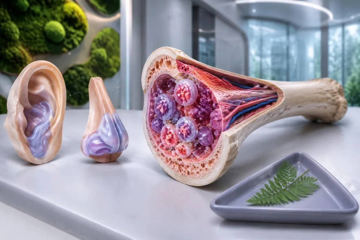
VEXAS синдромът е придобито възпалително състояние, причинено от соматични мутации в UBA1 в кръвотворните клетки. Повтарящата се температура, подуването на хрущяла на ухото или носа и необяснимата макроцитна анемия налагат консултация със специалист; диагнозата изисква подходящо генетично изследване, а не рутинен автоимунен панел.

Основни изводи
- VEXAS синдром обикновено започва след 50-годишна възраст и съчетава системно възпаление с аномалии в кръвотворните клетки.
- Подуването на хрущяла може да засегне ушите, носа или дихателните пътища; възпалението на ушния хрущял често щади меката ушна висулка.
- Macrocytosis означава MCV над приблизително 100 fL при възрастни, но само по себе си тази находка не диагностицира VEXAS.
- Ниски тромбоцити под 150 × 10^9/L може да засили съмнението, когато е съчетано с повтарящо се възпаление и необяснима анемия.
- CRP обикновено има горна референтна граница от 5 mg/L, въпреки че някои лаборатории използват 10 mg/L; няма гранична стойност на CRP, която да потвърждава VEXAS.
- Изследването за мутация в UBA1 търси придобит патогенен вариант, обикновено чрез изследване на периферна кръв или костен мозък, а не с рутинен панел за наследствен риск.
- Автоимунните панели като ANA, ревматоиден фактор и ANCA не могат да потвърдят или изключат синдрома VEXAS.
- Спешни симптоми включват новопоявил се задух, болка в гърдите, затруднено дишане или температура 38°C или по-висока при значима имуносупресия.
Какво представлява VEXAS синдромът и защо кръвните показатели са важни?
VEXAS синдром е придобито автовъзпалително заболяване, причинено от соматична мутация в UBA1 в кръвотворните клетки. Съчетанието от повтарящи се възпаления, подут хрущял и необясними отклонения в кръвната картина — особено макроцитна анемия при човек над 50 години — трябва да породи съмнение, докато диагнозата зависи от установяването на подходящ патогенен вариант на UBA1 в клиничния контекст.
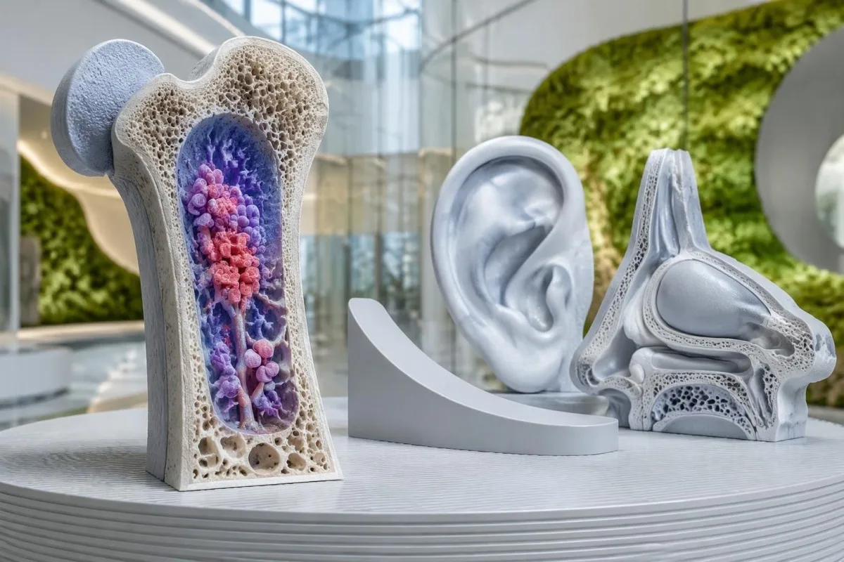
Наименованието описва 5 характеристики: вакуоли, ензим E1, свързан с X-хромозомата, автовъзпалително и соматично. Това са насоки към биологията на заболяването, а не контролен списък, на който всеки трябва да отговаря; видими вакуоли в костния мозък, хрущялни симптоми и изразена анемия не е задължително да се проявят едновременно при първия преглед.
Beck et al. описват за първи път VEXAS в New England Journal of Medicine през 2020 г., като свързват възпалението с начало в зряла възраст с придобити варианти на UBA1. Това откритие обяснява защо човек може преди това да е получил повече от 1 диагноза, например рецидивиращ полихондрит и заболяване на костния мозък, преди да бъде разпозната общата причина.
Единичен повишен резултат за CRP не насочва конкретно към VEXAS, а MCV от 103 fL често има по-чести обяснения. Подозрението се засилва, когато възпалителните пристъпи се повтарят наред с персистиращи или влошаващи се цитопении, тъй като тази картина насочва към възпалителен процес, свързан с нарушено образуване на кръвни клетки, а не към изолирано лабораторно отклонение.
Кантести е един AI кръвен анализатор който може да обясни как се съчетават хемоглобинът, MCV, тромбоцитите и CRP, като диагностиката на VEXAS се оставя на специалистите. Нашите организация и цел са описани отделно; 1 качена CBC не може да разкрие мутация в UBA1 или да замени клиничния преглед.
Кой развива VEXAS и наследява ли се мутацията в UBA1?
Синдромът VEXAS засяга най-често възрастни мъже, обикновено със симптоми, започващи след 50-годишна възраст. Причиняващата заболяването мутация в UBA1 обикновено се придобива в популация от кръвотворни клетки, вместо да се унаследява от родител или да присъства във всяка клетка от раждането.
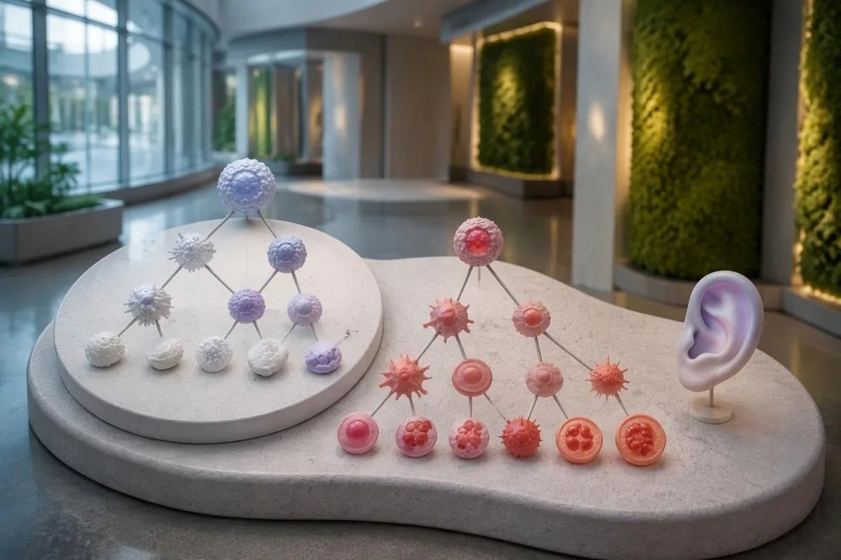
UBA1 се намира на X-хромозомата, а повечето засегнати мъже имат 1 X-хромозома. VEXAS се среща и при жени, понякога във връзка със загуба на X-хромозома в засегнатите клетки, но полът сам по себе си не бива да възпрепятства оценката, когато клиничната картина силно насочва към заболяването.
В проучване в JAMA сред 163,096 участници в кохорта от здравната система на САЩ Beck et al. установяват 11 души с варианти на UBA1, свързани със заболяването. Изчислената честота сред мъжете над 50 години е около 1 на 4,269; това е конкретна кохорта, предимно с европейски произход, а не оценка за разпространението в световен мащаб.
Обикновено липсва фамилна обремененост, тъй като съответната мутация възниква в течение на живота на човека. Това се различава от наследствените синдроми на рецидивираща треска, разгледани в нашия обяснение за MEFV изследването; изследването на 2 роднини за фамилен вариант не е стандартна първа стъпка при типичен соматичен VEXAS.
Думата „генетичен“ не означава автоматично „наследствен“, а ген, свързан с X-хромозомата, не предполага автоматично предаване на децата. Ако резултатът насочва към конституционен вариант или необичайна проява преди 50-годишна възраст, специалист по клинична генетика може да уточни дали са подходящи допълнителни изследвания на тъкани или генетично консултиране на семейството.
Кои симптоми на VEXAS синдрома трябва да породят съмнение?
Симптоми на синдрома VEXAS обикновено включват повтаряща се треска, възпалителни кожни промени, умора, засягане на белите дробове и възпаление на хрущяла. Подуването на хрущяла на ухото или носа става особено показателно, когато е съчетано с необяснима анемия или други отклонения в кръвната картина при възрастен над 50 години.
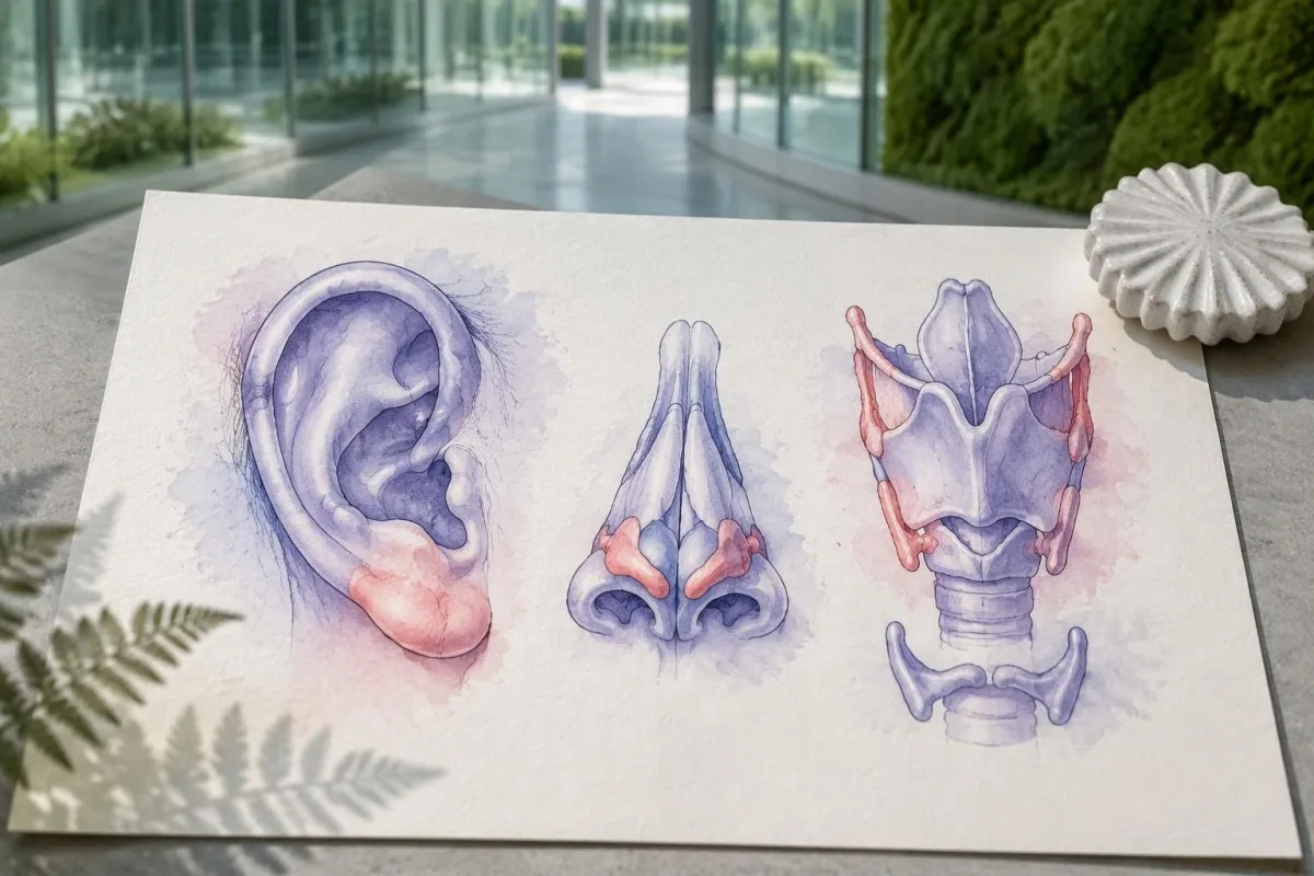
Хондритът на ухото често засяга твърдата горна част на ухото, като щади меката ушна висулка, в която няма хрущял. Това анатомично разграничение може да помогне за точното описание на пристъп, но 1 зачервено ухо все пак изисква оценка за по-чести причини като локална инфекция, травма или дерматит.
Кожните прояви може да включват болезнени надигнати участъци, плаки или виолетови промени; само по външния вид причината не може да се установи. Изследването на тъкан може да помогне за разграничаване на неутрофилна дерматоза, васкулит и други процеси, а снимки от 2 отделни епизода без лечение може да помогнат на специалиста да разпознае повтаряща се картина.
Inflammatory attacks can resemble established rheumatological conditions, including relapsing polychondritis or polymyalgia rheumatica. Our explanation of PMR laboratory limitations describes why inflammatory markers are not disease-specific; persistent macrocytosis above 100 fL or falling platelets should prompt a broader assessment rather than automatic acceptance of the first label.
New hoarseness, noisy breathing or difficulty breathing needs urgent assessment because airway cartilage can be involved. A useful symptom record includes 3 details for each episode: the affected site, its duration and whether fever or a medication change accompanied it; do not wait for another flare if breathing is affected.
Какъв модел на кръвните показатели може да насочи към VEXAS синдром?
Макроцитна анемия, sometimes accompanied by low platelets or other cytopenias, is a major laboratory clue to VEXAS. Adult MCV is commonly about 80–100 fL, but neither an MCV above 100 fL nor an elevated CRP result can confirm the condition.
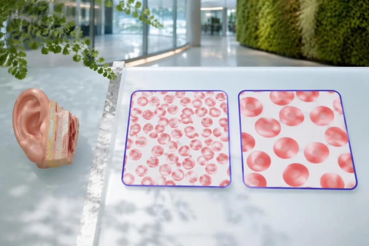
Hemoglobin measures anemia, while MCV measures average red-cell size; the 2 results answer different questions. An MCV of 108 fL with hemoglobin of 10.2 g/dL deserves a different assessment from an isolated MCV of 101 fL with stable hemoglobin, particularly when inflammatory symptoms are also present.
Many laboratories use a platelet reference interval of approximately 150–450 × 10^9/L. A falling platelet count can matter before it crosses the lower limit, and our borderline MCV explanation shows why persistent trends are often more useful than treating one near-boundary number as a diagnosis.
CRP is often markedly elevated during active VEXAS, but infection and many other inflammatory disorders can produce the same result. A hypothetical CRP of 80 mg/L is 16 times an upper limit of 5 mg/L; that comparison describes inflammatory intensity, not the probability of a UBA1 mutation.
RDW describes variation in red-cell size and can help explain a mixed CBC pattern, but it is not a VEXAS-specific biomarker. Our RDW и MCV водич explains how 2 overlapping processes, such as iron deficiency and macrocytosis, can produce a deceptively ordinary average MCV.
| Ниво | Диапазон | Какво означава |
|---|---|---|
| Typical adult MCV interval | 80–100 fL | Use the reporting laboratory's interval; an in-range result does not exclude early or masked macrocytosis. |
| Macrocytosis | >100 fL | Investigate nutritional, medication, liver, thyroid and marrow causes; this is not a VEXAS diagnostic threshold. |
| Тромбоцитопения | <150 × 10^9/L | Low platelets can support concern when combined with persistent inflammation and unexplained anemia. |
| CRP above the laboratory limit | Often >5 mg/L; some laboratories use >10 mg/L | Indicates an inflammatory response without identifying its cause. |
Защо рутинните автоимунни панели не могат да потвърдят VEXAS?
Routine autoimmune panels cannot confirm VEXAS syndrome because they measure antibodies or related immune markers, not acquired UBA1 mutations. ANA, rheumatoid factor, ANCA and complement results may help assess alternative or overlapping conditions, but no combination substitutes for appropriate molecular testing.
ANA testing detects antibodies directed against nuclear components; it does not read the UBA1 gene. A positive ANA at 1:160 may deserve interpretation in its own clinical context, while a negative ANA does not make unexplained fever, chondritis and macrocytic anemia safe to dismiss.
Repeating several antibody panels can create incidental positives without resolving the central question. Our ANA result explanation describes why 1 positive result is not equivalent to a disease diagnosis; the same caution applies when an antibody is found during a suspected VEXAS evaluation.
Complement testing can be useful when clinicians suspect an immune-complex disorder, but normal C3 and C4 cannot exclude VEXAS. The complement and antibody guide explains these 2 proteins; their biological role differs from the acquired enzyme dysfunction caused by a disease-associated UBA1 variant.
Autoinflammatory and autoimmune are related but not interchangeable descriptions: VEXAS is driven principally by abnormal innate immune activity associated with mutated blood-forming cells. Clinicians may still order 2 types of investigation in parallel—antibody testing for alternatives and molecular testing for VEXAS—because another diagnosis can coexist rather than disappear.
Какви други причини трябва да се проверят, преди да се обвини VEXAS?
Vitamin B12 or folate deficiency, medication effects, alcohol use, liver disease, hypothyroidism and marrow disorders can cause macrocytosis. An MCV above 100 fL should trigger evaluation of these possibilities, while recurrent chondritis or unexplained inflammatory attacks may justify investigating VEXAS at the same time.
Vitamin B12 and folate results need interpretation alongside symptoms, treatment history and the CBC. A borderline B12 result may require additional assessment rather than immediate attribution of an MCV of 107 fL to VEXAS; our B12 versus folate explanation also explains why folate treatment can obscure part of a B12 deficiency picture.
Copper deficiency can cause cytopenias and vacuolated marrow precursors, making it an especially relevant mimic. Reviewing high-dose zinc supplements is therefore useful, and our ръководство за оценка на нисък мед explains why 2 supplement ingredients—zinc and copper—may matter even when a product is marketed for general immune support.
Myelodysplastic syndrome can cause macrocytic anemia and may coexist with VEXAS rather than simply compete with it as an explanation. Our MDS confirmation guide distinguishes CBC clues from marrow evidence; 1 abnormal count cannot establish either diagnosis or determine whether both are present.
Medication and infection reviews are essential because treatment can change the very numbers being investigated. For example, glucocorticoids may increase neutrophils while lowering CRP, and a recent transfusion can alter MCV; bring the dates of these events, not just 2 laboratory reports that appear to disagree.
Как изследването за соматична мутация в UBA1 подпомага диагностицирането на VEXAS?
VEXAS syndrome diagnosis is established by a disease-associated somatic UBA1 variant in a compatible clinical setting. Peripheral blood is commonly used for testing, while marrow or a more sensitive method may be appropriate after an inconclusive result; many recognized variants affect methionine at position 41.
The request should specify suspected VEXAS and testing for somatic UBA1 variants rather than a general hereditary disease panel. These are 2 different diagnostic tasks: an inherited-risk assay may not include the relevant gene regions, cell source or detection sensitivity needed to identify an acquired clone.
Beck et al. linked UBA1 variants to altered ubiquitin-activating enzyme function in their 2020 New England Journal of Medicine report. Position 41 is a major hotspot, but a hotspot-only test is not equivalent to comprehensive relevant UBA1 analysis, because disease-associated noncanonical variants have also been described.
A negative result needs the laboratory method attached to it before its meaning is clear. An assay validated to detect a 5% variant allele fraction may miss a smaller clone; ask which regions were covered, the reported detection limit and whether the sample was suitable for somatic testing.
Кантести е един Платформа за кръвни изследвания тълкуване с AI that can help explain accompanying CBC and inflammatory-marker results, but it cannot infer a UBA1 sequence from 1 routine report. Our обяснение на AI технологията describes report interpretation; molecular diagnosis still requires an accredited laboratory and specialist interpretation.
Как трябва да разчетете резултат от изследване за мутация в UBA1?
A UBA1 report should state the variant, classification, sample type and testing limitations, often including variant allele fraction. A pathogenic or likely pathogenic disease-associated variant can support VEXAS, but a variant of uncertain significance is not diagnostic, and a 10% allele fraction is not a 10% severity score.
Variant allele fraction is the proportion of analyzed sequence reads carrying a particular change at that location. In a simple 1-X-chromosome model it may approximate the proportion of affected sampled cells, but cell mixtures, chromosome changes and laboratory methods complicate that estimate; it does not directly measure organ damage.
A hypothetical report showing a recognized disease-associated variant at 12% allele fraction may be meaningful even though most reads are unchanged. By contrast, a novel uncertain variant at 40% still requires expert assessment, because abundance cannot turn an unproven genetic finding into an established cause of symptoms.
Different clonal disorders require different molecular or cellular tests, even when anemia and thrombosis overlap. Our PNH clone testing guide explains a separate disorder assessed by flow cytometry; 1 negative PNH test does not evaluate UBA1, and a UBA1 result does not replace PNH testing when indicated.
Kantesti cannot determine whether an uncertain UBA1 finding is disease-causing simply by matching it to an elevated CRP of 70 mg/L. Ask the specialist whether additional sequencing, another specimen or formal variant review is needed, and keep the complete report rather than a screenshot containing only the word positive.
Достатъчни ли са вакуолите в костния мозък за диагностициране на VEXAS?
Bone marrow vacuoles are a clue, not proof of VEXAS syndrome. Vacuoles may appear in early myeloid and erythroid precursors, but they are neither completely specific nor universally present; an adult with an MCV above 100 fL still needs the broader clinical and molecular assessment.
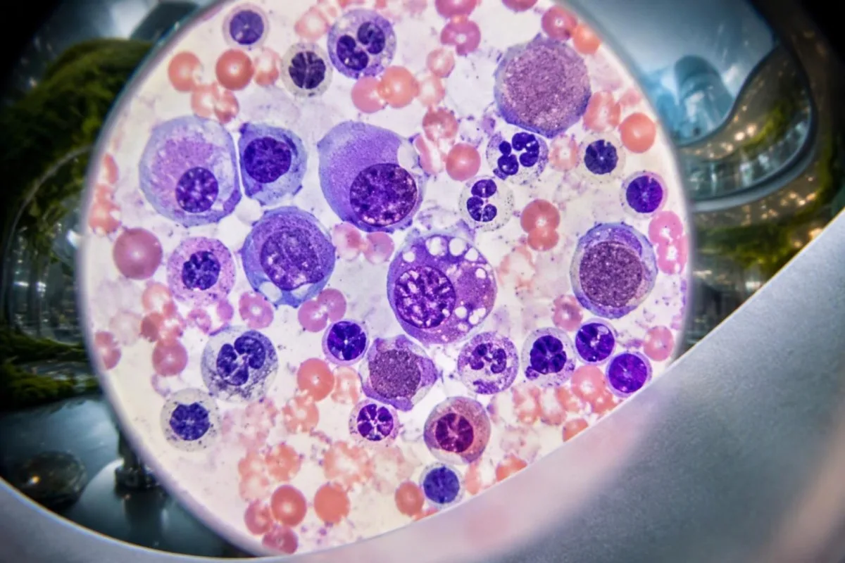
Vacuoles are small clear spaces visible within cells under microscopy, not holes that a patient can see on an ordinary CBC. Their absence on 1 marrow examination does not automatically exclude VEXAS, particularly when specimen quality, disease stage or the specific UBA1 variant affects the appearance.
Copper deficiency, alcohol-related effects and other conditions can produce vacuolated precursors. That overlap is why 2 complementary investigations may be needed: laboratory assessment for reversible mimics and somatic UBA1 testing, rather than treating a descriptive pathology finding as the final diagnosis.
Marrow examination also assesses cellularity, dysplasia and other abnormalities that may influence treatment decisions. Our explanation of teardrop cells and marrow clues describes a different microscopy finding; neither teardrop cells nor vacuoles can independently explain a platelet count of 95 × 10^9/L.
A marrow procedure is not automatically necessary for every isolated MCV of 102 fL, and a positive molecular result may already establish VEXAS in the right context. Hematology decides whether marrow assessment is needed to investigate persistent cytopenias, suspected MDS or another process that would change management.
Кога е подходяща консултация с ревматолог или хематолог?
Specialist assessment is appropriate when recurrent unexplained inflammation accompanies macrocytic anemia, falling platelets or other persistent cytopenias. Suspicion is higher after age 50, especially with chondritis, inflammatory skin findings or lung involvement; rheumatology and hematology often need to evaluate the pattern together.
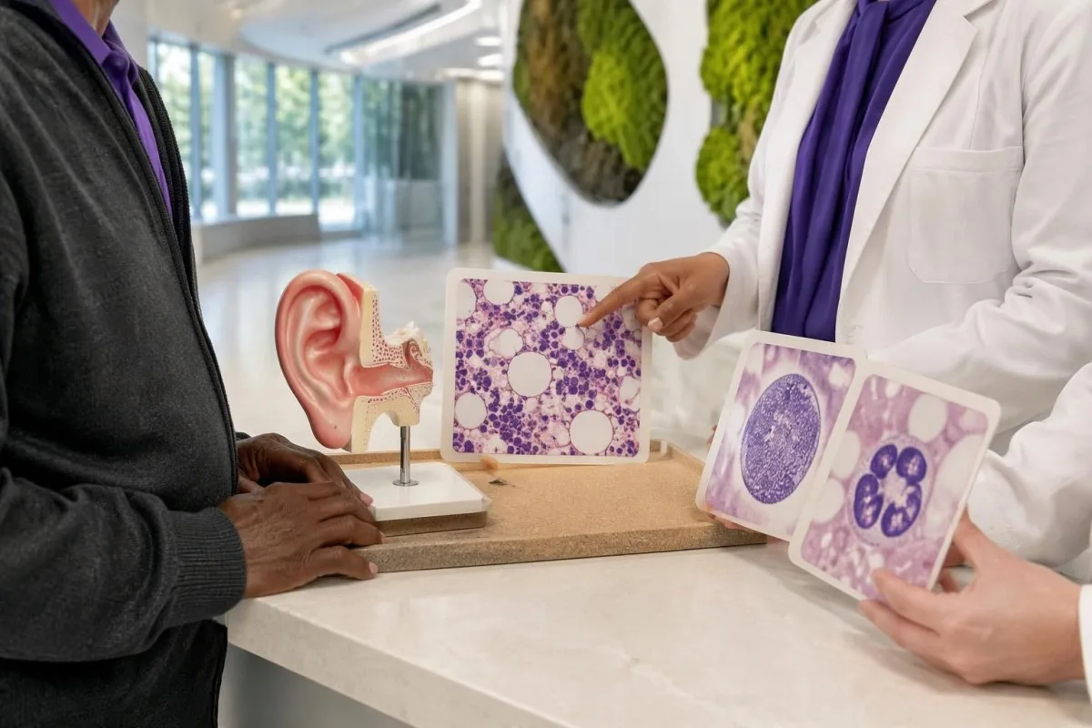
There is no universal CRP level, age boundary or number of attacks that automatically mandates UBA1 testing. A person aged 68 with recurrent ear chondritis and unexplained anemia has a different pretest probability from a person aged 28 with 1 viral fever and an otherwise stable CBC.
Steroid-responsive inflammation that repeatedly returns during dose reduction deserves reassessment, particularly when blood counts remain abnormal. Improvement over 2 days may show that symptoms are inflammatory, but it does not distinguish VEXAS from several other disorders and should not become the only justification for ongoing treatment.
Unexplained pain needs a broader workup when it accompanies constitutional symptoms or changing counts. Our ръководство за тестване при необяснима болка explains why inflammatory markers are only part of that workup; an MCV above 100 fL adds a marrow-related question that a pain score cannot answer.
A referral should highlight 3 elements: documented inflammatory episodes, the CBC trend and the results of common-cause investigations. If symptoms are progressing rapidly, new organs are involved or transfusions are being considered, ask the referring clinician whether the appointment needs to be expedited rather than waiting for another routine repeat test.
Разгледан пример: повтарящо се подуване на ухото и променяща се пълна кръвна картина
A hypothetical combination of recurrent chondritis, macrocytic anemia and low platelets warrants specialist evaluation, not an AI-generated diagnosis. Consider a 68-year-old man with hemoglobin 10.2 g/dL, MCV 108 fL, platelets 112 × 10^9/L and CRP 86 mg/L during a third episode of upper-ear swelling.

The first reasoning step is to separate symptom severity from diagnostic specificity: CRP of 86 mg/L confirms a substantial inflammatory response but does not identify its cause. The MCV of 108 fL and platelet count of 112 × 10^9/L add evidence that blood-cell production also needs investigation.
Suppose the person's B12, folate and thyroid assessments do not reveal an explanation, and medication, alcohol and liver reviews are unrevealing. A negative ANA would not close the case; the combination of 3 inflammatory episodes and persistent unexplained cytopenias would justify discussing somatic UBA1 testing with hematology and rheumatology.
Now suppose CRP falls to 12 mg/L after prescribed glucocorticoids, but hemoglobin falls to 9.8 g/dL. Those 2 trends are not contradictory: inflammatory activity may improve while marrow dysfunction or another anemia mechanism persists, so treatment response cannot be used as a substitute for investigating the falling hemoglobin.
Kantesti can help explain why those 2 trends deserve separate attention, but cannot establish VEXAS or choose immunosuppressive treatment. Our наръчник за анализ на тенденциите в лекарствата explains why dose changes, transfusions and intercurrent illness should be recorded beside each result; the example here is entirely hypothetical.
Как специалистите лекуват потвърдения VEXAS синдром?
VEXAS treatment aims to control inflammation, manage cytopenias and reduce treatment-related harm. Glucocorticoids often suppress symptoms but may be difficult to taper; other options include selected immunomodulators, marrow-directed treatment and allogeneic stem-cell transplantation, with no single regimen appropriate for every adult over 50.
A falling CRP does not by itself establish treatment success if anemia, thrombosis or lung disease is worsening. Specialists follow at least 2 dimensions—clinical inflammatory activity and hematological status—and weigh these against cumulative glucocorticoid exposure, because prolonged treatment can cause infections, diabetes, osteoporosis and muscle weakness.
JAK inhibitors and IL-6-directed therapy are among approaches used by specialists, while azacitidine may be considered in selected patients, particularly with associated marrow disease. Evidence and access vary, and much published experience is observational; a response lasting 6 months in one cohort does not reliably predict an individual's longer-term benefit.
Allogeneic stem-cell transplantation can potentially eliminate the abnormal blood-forming clone and produce sustained remission in selected patients. It also carries substantial risks, including graft-versus-host disease and serious infection, so 2 people of the same age may receive different recommendations because fitness, organ involvement and donor availability differ.
Infection prevention deserves planning before immunosuppression, not only after fever develops. Depending on exposures and the proposed regimen, clinicians may consider screening or prevention measures described in our Strongyloides testing explanation; even 1 relevant past exposure can matter before corticosteroids, and prescribed steroids should never be stopped abruptly without guidance.
Кои симптоми изискват спешна медицинска помощ?
New breathing difficulty, chest pain, airway symptoms, neurological deficits or significant bleeding require urgent assessment in someone with suspected or confirmed VEXAS. Fever of 38°C or higher during substantial immunosuppression also needs prompt clinical advice, particularly when neutrophils are low.
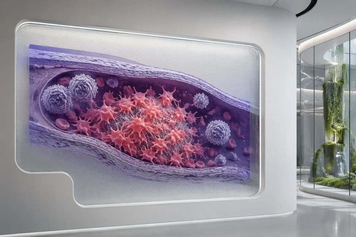
VEXAS is associated with venous thrombosis, so a painful swollen leg or sudden breathlessness should not be dismissed as another inflammatory flare. Normal routine clotting times do not exclude a clot; our ограничения на коагулационния панел explain why 2 normal tests, PT and aPTT, cannot provide that reassurance.
An absolute neutrophil count below 0.5 × 10^9/L represents severe neutropenia and increases concern when fever occurs. Treatment can also blunt the temperature response, so feeling acutely unwell with shaking chills, confusion or rapidly worsening weakness can require urgent assessment even when a thermometer reads below 38°C.
Breathlessness can reflect lung inflammation, infection, pulmonary embolism or worsening anemia, and these require different treatments. Our breathlessness blood test guide explains why hemoglobin of 8 g/dL may contribute without proving the entire cause; new chest pain or sudden breathing difficulty should not wait for an uploaded report.
Persistent nose or gum bleeding, black stools or a severe new headache needs urgent advice, especially if platelets are very low. A count below 20 × 10^9/L generally warrants prompt clinical review, but the urgency depends on symptoms and context; significant bleeding is an emergency even above that number.
Проследяване, ограничения на ИИ и научни публикации
Useful VEXAS follow-up combines symptoms, treatment history, CBC trends and inflammatory markers, with molecular and marrow findings interpreted by specialists. An AI explanation of 1 laboratory report can support preparation for care, but it cannot diagnose VEXAS, exclude infection or establish whether a UBA1 variant is pathogenic.

Bring original CBC reports, inflammatory markers and the complete UBA1 result, including specimen type and detection limit. Record 3 timeline features beside them: flare dates, medication changes and transfusions; this makes it easier to distinguish a true disease trend from a treatment effect or a change in testing conditions.
Кантести е един Инструмент за анализ на кръвни изследвания с AI that can explain laboratory patterns without converting an MCV above 100 fL into a rare-disease diagnosis. Genetic reports can reveal sensitive information, so review our информация за поверителността и данните before sharing records, and include only documents you are authorized to upload.
Kantesti's 2 DOI-linked publications below concern broader blood-test interpretation and RDW education, not clinical trials of VEXAS diagnosis or UBA1 sequencing. They should therefore be read separately from the disease-specific medical studies listed in the external references; neither publication establishes VEXAS-specific diagnostic sensitivity or treatment effectiveness.
Нашите методология и клинични стандарти explain the distinction between report interpretation and medical diagnosis. Before the next appointment, write 2 questions: whether the laboratory method adequately assessed somatic UBA1 variants, and whether persistent cytopenias need marrow evaluation; these are more actionable than asking an AI to calculate an unsupported VEXAS probability.
Често задавани въпроси
Какви са първите симптоми на синдрома VEXAS?
Синдромът VEXAS често започва с повтаряща се необяснима температура, възпалителни кожни промени, умора или подуване на хрущяла на ухото или носа, обикновено след 50-годишна възраст. Макроцитната анемия или други отклонения в кръвната картина могат да се появят преди, заедно с или след тези симптоми. MCV над приблизително 100 fL при възрастен е насочващ признак, а не диагноза. Повтарящите се възпалителни симптоми, съчетани с персистиращи необясними цитопении, дават основание да се обсъди оценка от специалисти по ревматология и хематология.
Кой тест потвърждава синдрома VEXAS?
Синдромът VEXAS се диагностицира чрез установяване на свързан със заболяването соматичен вариант в UBA1 при съответстваща клинична картина. Обикновено за изследването се използва периферна кръв, въпреки че в избрани случаи може да са необходими костен мозък или по-чувствителен метод. Много от установените варианти засягат метионина на позиция 41, но ограниченото изследване на горещи точки може да пропусне други свързани със заболяването варианти. Резултатът трябва да се интерпретира с оглед на класификацията на варианта, покритието на гена и границата на откриване.
Може ли нормален тест за АНФ или автоимунен панел да изключи VEXAS?
Нормалният ANA или рутинният панел за автоимунни заболявания не може да изключи синдрома VEXAS, тъй като тези изследвания не откриват соматични мутации в UBA1. Положителният ANA при титър 1:160 също не потвърждава VEXAS и не установява непременно друго автоимунно заболяване. Изследването на антитела може да помогне при проучването на алтернативни диагнози, докато показателите от CBC, симптомите и молекулярните изследвания дават отговори на отделни въпроси. Персистиращата макроцитна анемия и повтарящото се възпаление на хрущяла не бива да се пренебрегват само защото тестовете за антитела са отрицателни.
Синдромът VEXAS унаследява ли се и трябва ли децата ми да бъдат изследвани?
Типичният синдром VEXAS е придобит, а не наследствен, затова по правило не се налага рутинно изследване на децата. Съответната мутация в UBA1 обикновено възниква в кръвотворните клетки в зряла възраст, често с прояви след 50-годишна възраст. Макар UBA1 да се намира на X-хромозомата, това не означава, че соматичният вариант при VEXAS автоматично се предава по наследство. Необичайните находки или тези, които е възможно да са конституционни, трябва да бъдат прегледани от специалист по клинична генетика.
Могат ли жените да развият синдрома VEXAS?
Жените могат да развият синдром на VEXAS, въпреки че повечето разпознати случаи са при по-възрастни мъже. UBA1 се намира в Х-хромозомата, а загубата на 1 Х-хромозома в засегнатите клетки е един от механизмите, описани при жени. Проучване на здравната система, публикувано в JAMA през 2023 г., установява 2 жени сред 11 участници с асоциирани със заболяването варианти на UBA1. Самият пол не бива да възпрепятства оценката от специалист, когато рецидивиращото възпаление и необяснимите отклонения в кръвната картина са силно показателни.
Изключва ли отрицателен тест за UBA1 синдрома VEXAS?
Отрицателният тест за UBA1 намалява вероятността за VEXAS само доколкото пробата и методът на изследване са били подходящи. Тест с праг на откриване на вариантна алелна фракция от 5% може да пропусне по-малък клон, а изследването само за горещи точки може да пропусне варианти извън обхванатия регион. Силното клинично подозрение може да обоснове преглед от специалист на обхвата, чувствителността и вида на пробата. Правилно извършеният отрицателен резултат следва също да подтикне към внимателна преоценка на алтернативните диагнози, а не към повторно изследване без ясна причина.
Има ли лечение за синдрома VEXAS?
0 Алогенната трансплантация на стволови клетки може потенциално да доведе до трайна ремисия чрез заместване на абнормния кръвотворен клон, но е подходяща само за подбрани пациенти и крие значителни рискове. Други лечения имат за цел да овладеят възпалението, да контролират цитопениите и да намалят експозицията на глюкокортикоиди, вместо надеждно да елиминират клона. При възрастни над 50 години физическата пригодност, засягането на органи и съпътстващото заболяване на костния мозък могат съществено да повлияят на избора на лечение. Не е доказано, че хранителна добавка, диетичен режим или интерпретация на кръвни изследвания чрез ИИ лекува VEXAS.
Получете обяснение на резултатите от кръвните си изследвания
Качете лабораторния си доклад и AI на Kantesti ще обясни всеки биомаркер на разбираем език за секунди. Използва се от над 2 милиона души по света.
Референции
- Klein, T., Mitchell, S., & Weber, H. (2026). AI анализатор на кръвни изследвания: 2,5M изследвания анализирани | Global Health Report 2026. Медицински изследвания с Kantesti AI. ДОИ
- Klein, T., Mitchell, S., & Weber, H. (2026). Кръвен тест RDW: Пълно ръководство за RDW-CV, MCV и MCHC. Медицински изследвания с Kantesti AI. ДОИ
- Beck DB et al. (2020). Somatic Mutations in UBA1 and Severe Adult-Onset Autoinflammatory Disease. New England Journal of Medicine. ДОИ
- Beck DB et al. (2023). Estimated Prevalence and Clinical Manifestations of VEXAS Syndrome in a Large US Health Care System. JAMA. ДОИ
- 2 милиона+Анализирани тестове
- 127+Държави
- 100+Езици
За медицинския екип


Сигнали за доверие E-E-A-T
Опит
Медицински преглед, воден от лекар, на работните процеси за интерпретация на лабораторни резултати.
Експертиза
Фокус в лабораторната медицина върху това как се държат биомаркерите в клиничен контекст.
Авторитетност
Медицинска рецензия от д-р Thomas Klein, с принос от д-р Sarah Mitchell и проф. д-р Hans Weber.
Надеждност
Интерпретация, основана на доказателства, с ясни последващи стъпки за намаляване на тревогата.
Медицински отказ от отговорност
Тази статия е само с образователна цел и не представлява медицински съвет. Винаги се консултирайте с квалифициран медицински специалист за решения относно диагностика и лечение.
Публикувано · Свържете се с редакционния екип
Издава се от Kantesti Ltd, регистрирана в Англия и Уелс, дружество №. 17090423, Лондон, Обединено кралство.
