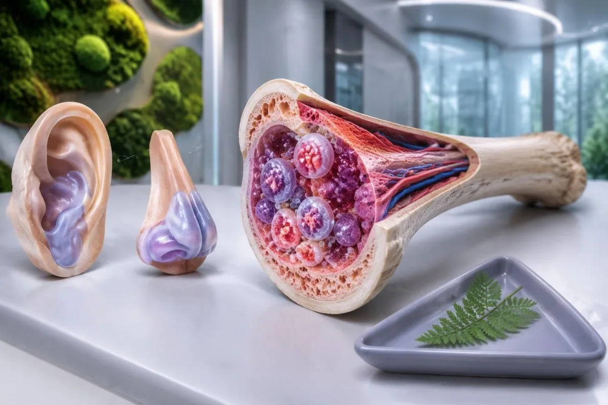
Сіндром VEXAS — набытае запаленчае захворванне, выкліканае саматычнымі мутацыямі UBA1 у крыватворных клетках. Перыядычная ліхаманка, ацёк храсткоў вуха або носа і невытлумачальная макрацытарная анемія павінны стаць падставай для кансультацыі спецыяліста; для дыягностыкі неабходнае адпаведнае генетычнае тэставанне, а не звычайная панэль аналізаў на аўтаімунныя захворванні.

Асноўныя высновы
- Сіндром VEXAS звычайна пачынаецца пасля 50 гадоў і спалучае сістэмнае запаленне са зменамі ў крыватворных клетках.
- Ацёк храстка можа закранаць вушы, нос або дыхальныя шляхі; пры запаленні храстка вуха мяккая мочка вуха часта не закранаецца.
- Макрацытоз азначае MCV вышэй за прыблізна 100 fL у дарослых, але адна гэтая прыкмета не пацвярджае дыягназ VEXAS.
- Нізкія трамбацыты ніжэй за 150 × 10^9/L могуць узмацніць падазрэнне, калі суправаджаюцца перыядычным запаленнем і невытлумачальнай анеміяй.
- СРБ звычайна мае верхнюю мяжу рэферэнтнага дыяпазону 5 mg/L, хоць некаторыя лабараторыі выкарыстоўваюць 10 mg/L; не існуе парогавага значэння CRP, якое пацвярджала б VEXAS.
- Тэставанне на мутацыі UBA1 дазваляе выявіць набыты патагенны варыянт, звычайна з выкарыстаннем перыферычнай крыві або касцявога мозгу, а не звычайнай панэлі для ацэнкі спадчыннай рызыкі.
- Аўтаімунныя панэлі такія як ANA, рэўматоідны фактар і ANCA, не могуць пацвердзіць або выключыць сіндром VEXAS.
- Неадкладныя сімптомы уключаюць новую дыхавіцу, боль у грудзях, цяжкасць дыхання або тэмпературу 38°C і вышэй падчас значнай імунасупрэсіі.
Што такое сіндром VEXAS і чаму важныя паказчыкі аналізу крыві?
Сіндром VEXAS — гэта набытае аўтазапаленчае захворванне, выкліканае саматычнай мутацыяй UBA1 у крыватворных клетках. Спалучэнне рэцыдывавальнага запалення, ацёку храстка і невытлумачальных адхіленняў у паказчыках крыві — асабліва макрацытарнай анеміі ў чалавека старэйшага за 50 гадоў — павінна выклікаць падазрэнне, а дыягназ залежыць ад выяўлення адпаведнага патагеннага варыянта UBA1 у клінічным кантэксце.
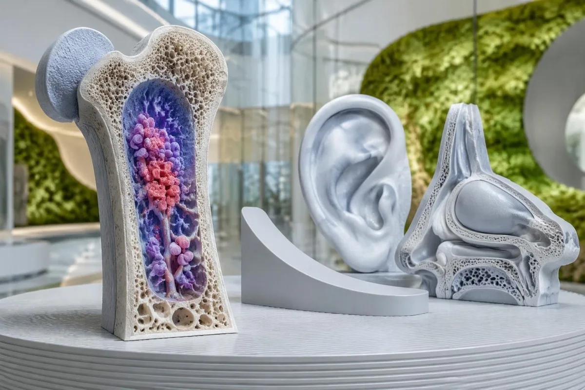
Назва апісвае 5 характарыстык: вакуолі, фермент E1, звязаны з X-храмасомай, аўтазапаленчы і саматычны. Гэта падказкі да біялогіі захворвання, а не спіс крытэрыяў, якім павінен адпавядаць кожны чалавек; бачныя вакуолі ў касцявым мозгу, сімптомы з боку храстка і выяўленая анемія неабавязкова з'яўляюцца разам падчас першага прыёму.
Beck et al. упершыню апісалі VEXAS у New England Journal of Medicine ў 2020 годзе, звязаўшы запаленне з пачаткам у дарослым узросце з набытымі варыянтамі UBA1. Гэта адкрыццё тлумачыць, чаму чалавеку маглі раней паставіць больш за 1 дыягназ, напрыклад рэцыдывавальны паліхандрыт і захворванне касцявога мозгу, перш чым выявілі агульную прычыну.
Адзіны павышаны вынік CRP не ўказвае канкрэтна на VEXAS, а значэнне MCV 103 fL часта мае больш распаўсюджаныя тлумачэнні. Падазрэнне ўзрастае, калі прыступы запалення паўтараюцца адначасова з устойлівымі або пагаршальнымі цытапеніямі, бо такая карціна сведчыць пра запаленчы працэс, звязаны з парушанай прадукцыяй клетак крыві, а не пра ізаляванае ваганне лабараторнага паказчыка.
Кантэсці — гэта Аналізатар крыві са штучным інтэлектам які можа растлумачыць, як суадносяцца гемаглабін, MCV, трамбацыты і CRP, пакідаючы дыягностыку VEXAS спецыялістам. Наша арганізацыя і мэта апісваюцца асобна; 1 загружаны CBC не можа выявіць мутацыю UBA1 або замяніць клінічны агляд.
У каго развіваецца VEXAS і ці перадаецца мутацыя UBA1 па спадчыне?
Сіндром VEXAS найчасцей сустракаецца ў мужчын старэйшага ўзросту, звычайна сімптомы пачынаюцца пасля 50 гадоў. Мутацыя UBA1, якая выклікае захворванне, звычайна набываецца ў папуляцыі крыватворных клетак, а не перадаецца ў спадчыну ад аднаго з бацькоў і не прысутнічае ва ўсіх клетках ад нараджэння.
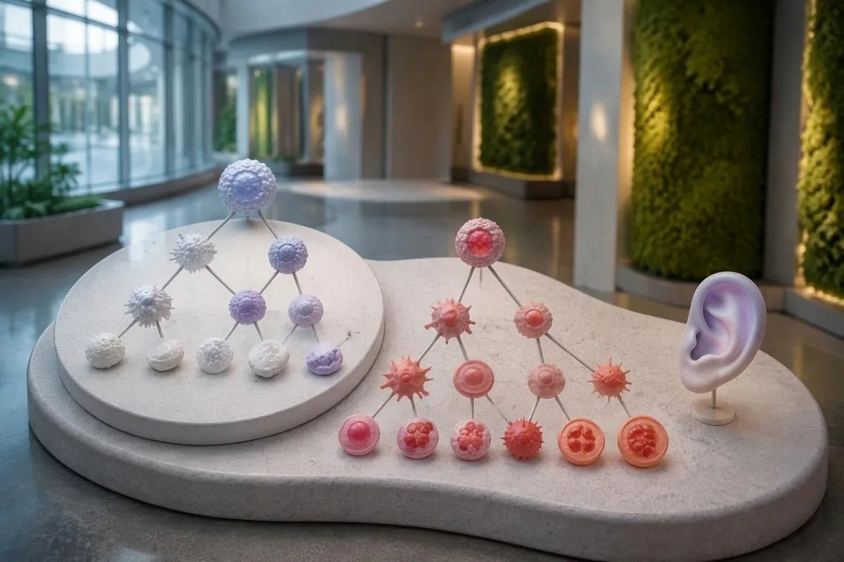
UBA1 размешчаны на X-храмасоме, і ў большасці мужчын з гэтым захворваннем ёсць 1 X-храмасома. VEXAS сустракаецца таксама ў жанчын, часам у сувязі са стратай X-храмасомы ў здзіўленых клетках, але пол сам па сабе не павінен спыняць абследаванне, калі клінічная карціна выразна паказвае на гэта захворванне.
У даследаванні JAMA з удзелам 163,096 чалавек з кагорты сістэмы аховы здароўя ЗША Beck et al. выявілі 11 чалавек з варыянтамі UBA1, звязанымі з захворваннем. Разліковая частата сярод мужчын старэйшых за 50 гадоў складала каля 1 на 4,269; гэта была канкрэтная кагорта, пераважна еўрапейскага паходжання, а не ацэнка распаўсюджанасці ва ўсім свеце.
Сямейны анамнез звычайна адсутнічае, бо адпаведная мутацыя ўзнікае на працягу жыцця чалавека. Гэта адрозніваецца ад спадчынных захворванняў з рэцыдывавальнай ліхаманкай, якія разглядаюцца ў нашым тлумачэнне аналізу MEFV; абследаванне 2 сваякоў на наяўнасць сямейнага варыянта не з'яўляецца стандартным першым крокам пры тыповым саматычным VEXAS.
Слова «генетычны» не азначае аўтаматычна «спадчынны», а ген, звязаны з X-храмасомай, не абавязкова перадаецца дзецям. Калі ў заключэнні ўзгадваецца канстытуцыйны варыянт або нетыповая праява ва ўзросце да 50 гадоў, спецыяліст па клінічнай генетыцы можа вызначыць, ці патрэбныя дадатковыя даследаванні тканак або генетычнае кансультаванне сям'і.
Якія сімптомы сіндрому VEXAS павінны выклікаць падазрэнне?
Сімптомы сіндрому VEXAS звычайна ўключаюць рэцыдывавальную ліхаманку, запаленчыя змены скуры, стомленасць, паражэнне лёгкіх і запаленне храстка. Ацёк храстка вуха або носа асабліва інфарматыўны, калі ўзнікае разам з невытлумачальнай анеміяй або іншымі адхіленнямі ў паказчыках крыві ў дарослага старэйшага за 50 гадоў.
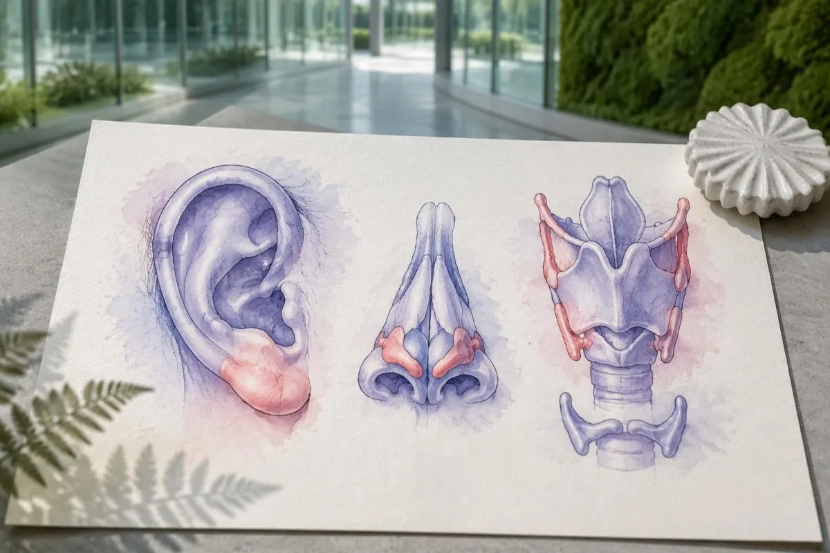
Хандрыт вуха часта закранае цвёрдую верхнюю частку вуха, пакідаючы непашкоджанай мяккую мочку, у якой няма храстка. Гэта анатамічнае адрозненне можа дапамагчы дакладна апісаць эпізод, але пачырваненне 1 вуха ўсё роўна патрабуе ацэнкі больш распаўсюджаных прычын, такіх як мясцовая інфекцыя, траўма або дэрматыт.
Праявы на скуры могуць уключаць балючыя ўзвышаныя ўчасткі, бляшкі або пурпурныя змены; толькі па знешнім выглядзе вызначыць прычыну немагчыма. Даследаванне тканкі можа дапамагчы адрозніць нейтрафільны дэрматоз, васкуліт і іншыя працэсы, а фатаграфіі 2 асобных эпізодаў без лячэння могуць дапамагчы спецыялісту распазнаць рэцыдывавальны характар.
Inflammatory attacks can resemble established rheumatological conditions, including relapsing polychondritis or polymyalgia rheumatica. Our explanation of PMR laboratory limitations describes why inflammatory markers are not disease-specific; persistent macrocytosis above 100 fL or falling platelets should prompt a broader assessment rather than automatic acceptance of the first label.
New hoarseness, noisy breathing or difficulty breathing needs urgent assessment because airway cartilage can be involved. A useful symptom record includes 3 details for each episode: the affected site, its duration and whether fever or a medication change accompanied it; do not wait for another flare if breathing is affected.
Якія змены ў аналізе крыві могуць сведчыць пра сіндром VEXAS?
Макрацытарная анемія, sometimes accompanied by low platelets or other cytopenias, is a major laboratory clue to VEXAS. Adult MCV is commonly about 80–100 фл, but neither an MCV above 100 fL nor an elevated CRP result can confirm the condition.
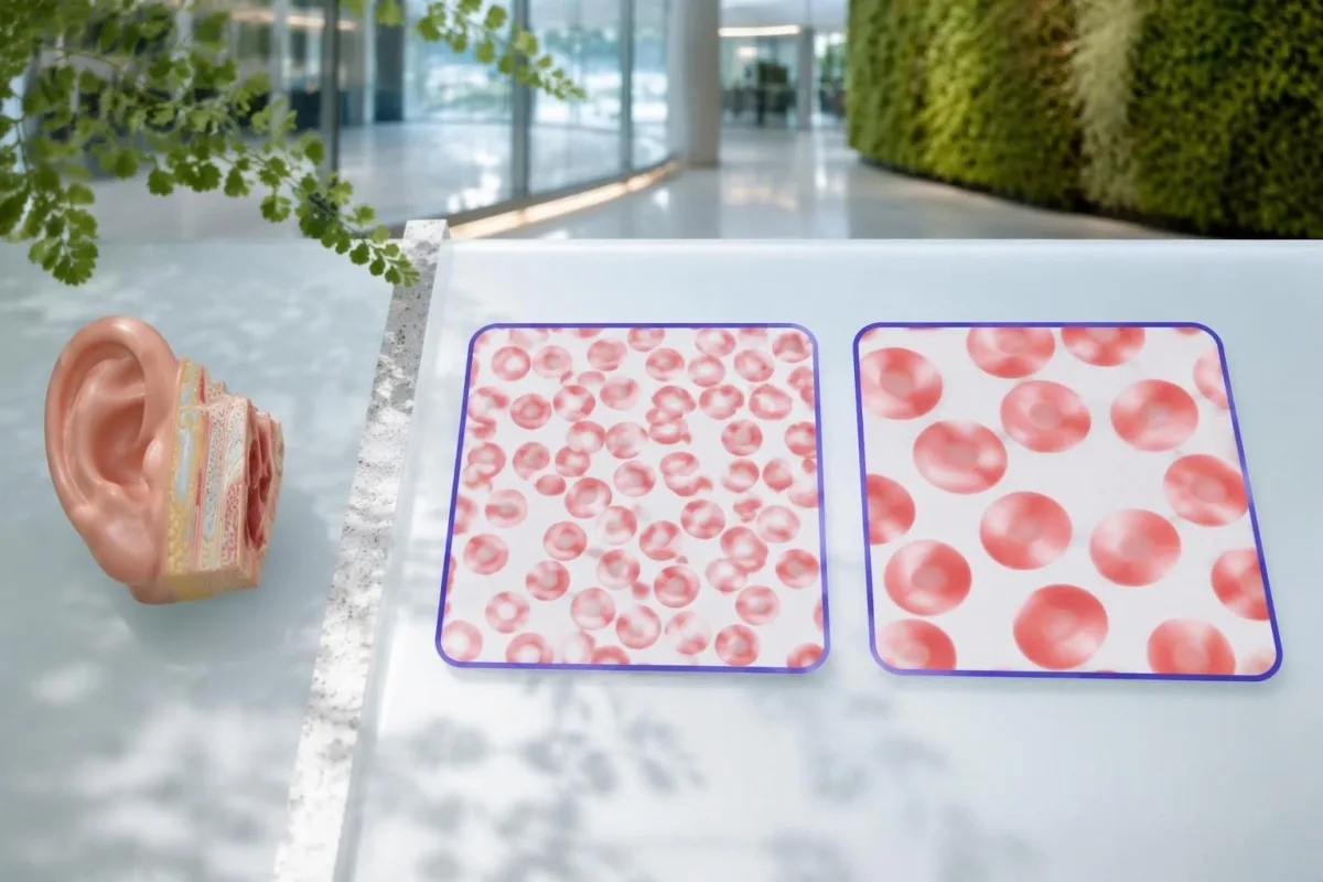
Hemoglobin measures anemia, while MCV measures average red-cell size; the 2 results answer different questions. An MCV of 108 fL with hemoglobin of 10.2 g/dL deserves a different assessment from an isolated MCV of 101 fL with stable hemoglobin, particularly when inflammatory symptoms are also present.
Many laboratories use a platelet reference interval of approximately 150–450 × 10^9/L. A falling platelet count can matter before it crosses the lower limit, and our borderline MCV explanation shows why persistent trends are often more useful than treating one near-boundary number as a diagnosis.
CRP is often markedly elevated during active VEXAS, but infection and many other inflammatory disorders can produce the same result. A hypothetical CRP of 80 mg/L is 16 times an upper limit of 5 mg/L; that comparison describes inflammatory intensity, not the probability of a UBA1 mutation.
RDW describes variation in red-cell size and can help explain a mixed CBC pattern, but it is not a VEXAS-specific biomarker. Our Кіраўніцтва па RDW і MCV explains how 2 overlapping processes, such as iron deficiency and macrocytosis, can produce a deceptively ordinary average MCV.
| Узровень | Дыяпазон | Што гэта значыць |
|---|---|---|
| Typical adult MCV interval | 80–100 фл | Use the reporting laboratory's interval; an in-range result does not exclude early or masked macrocytosis. |
| Макрацытоз | >100 fL | Investigate nutritional, medication, liver, thyroid and marrow causes; this is not a VEXAS diagnostic threshold. |
| Тромбацытапенія | <150 × 10^9/L | Low platelets can support concern when combined with persistent inflammation and unexplained anemia. |
| CRP above the laboratory limit | Often >5 mg/L; some laboratories use >10 mg/L | Indicates an inflammatory response without identifying its cause. |
Чаму звычайныя панэлі аналізаў на аўтаімунныя захворванні не могуць пацвердзіць VEXAS?
Routine autoimmune panels cannot confirm VEXAS syndrome because they measure antibodies or related immune markers, not acquired UBA1 mutations. ANA, rheumatoid factor, ANCA and complement results may help assess alternative or overlapping conditions, but no combination substitutes for appropriate molecular testing.
ANA testing detects antibodies directed against nuclear components; it does not read the UBA1 gene. A positive ANA at 1:160 may deserve interpretation in its own clinical context, while a negative ANA does not make unexplained fever, chondritis and macrocytic anemia safe to dismiss.
Repeating several antibody panels can create incidental positives without resolving the central question. Our ANA result explanation describes why 1 positive result is not equivalent to a disease diagnosis; the same caution applies when an antibody is found during a suspected VEXAS evaluation.
Complement testing can be useful when clinicians suspect an immune-complex disorder, but normal C3 and C4 cannot exclude VEXAS. The complement and antibody guide explains these 2 proteins; their biological role differs from the acquired enzyme dysfunction caused by a disease-associated UBA1 variant.
Autoinflammatory and autoimmune are related but not interchangeable descriptions: VEXAS is driven principally by abnormal innate immune activity associated with mutated blood-forming cells. Clinicians may still order 2 types of investigation in parallel—antibody testing for alternatives and molecular testing for VEXAS—because another diagnosis can coexist rather than disappear.
Якія іншыя прычыны неабходна праверыць, перш чым звязваць сімптомы з VEXAS?
Vitamin B12 or folate deficiency, medication effects, alcohol use, liver disease, hypothyroidism and marrow disorders can cause macrocytosis. An MCV above 100 fL should trigger evaluation of these possibilities, while recurrent chondritis or unexplained inflammatory attacks may justify investigating VEXAS at the same time.
Vitamin B12 and folate results need interpretation alongside symptoms, treatment history and the CBC. A borderline B12 result may require additional assessment rather than immediate attribution of an MCV of 107 fL to VEXAS; our B12 versus folate explanation also explains why folate treatment can obscure part of a B12 deficiency picture.
Copper deficiency can cause cytopenias and vacuolated marrow precursors, making it an especially relevant mimic. Reviewing high-dose zinc supplements is therefore useful, and our кіраўніцтва па ацэнцы нізкага ўтрымання медзі explains why 2 supplement ingredients—zinc and copper—may matter even when a product is marketed for general immune support.
Myelodysplastic syndrome can cause macrocytic anemia and may coexist with VEXAS rather than simply compete with it as an explanation. Our MDS confirmation guide distinguishes CBC clues from marrow evidence; 1 abnormal count cannot establish either diagnosis or determine whether both are present.
Medication and infection reviews are essential because treatment can change the very numbers being investigated. For example, glucocorticoids may increase neutrophils while lowering CRP, and a recent transfusion can alter MCV; bring the dates of these events, not just 2 laboratory reports that appear to disagree.
Як саматычнае тэставанне UBA1 дапамагае дыягнаставаць VEXAS?
VEXAS syndrome diagnosis is established by a disease-associated somatic UBA1 variant in a compatible clinical setting. Peripheral blood is commonly used for testing, while marrow or a more sensitive method may be appropriate after an inconclusive result; many recognized variants affect methionine at position 41.
The request should specify suspected VEXAS and testing for somatic UBA1 variants rather than a general hereditary disease panel. These are 2 different diagnostic tasks: an inherited-risk assay may not include the relevant gene regions, cell source or detection sensitivity needed to identify an acquired clone.
Beck et al. linked UBA1 variants to altered ubiquitin-activating enzyme function in their 2020 New England Journal of Medicine report. Position 41 is a major hotspot, but a hotspot-only test is not equivalent to comprehensive relevant UBA1 analysis, because disease-associated noncanonical variants have also been described.
A negative result needs the laboratory method attached to it before its meaning is clear. An assay validated to detect a 5% variant allele fraction may miss a smaller clone; ask which regions were covered, the reported detection limit and whether the sample was suitable for somatic testing.
Кантэсці — гэта Платформа AI для расшыфроўкі аналізу крыві that can help explain accompanying CBC and inflammatory-marker results, but it cannot infer a UBA1 sequence from 1 routine report. Our тлумачэнне тэхналогіі ШІ describes report interpretation; molecular diagnosis still requires an accredited laboratory and specialist interpretation.
Як інтэрпрэтаваць заключэнне аб мутацыі UBA1?
A UBA1 report should state the variant, classification, sample type and testing limitations, often including variant allele fraction. A pathogenic or likely pathogenic disease-associated variant can support VEXAS, but a variant of uncertain significance is not diagnostic, and a 10% allele fraction is not a 10% severity score.
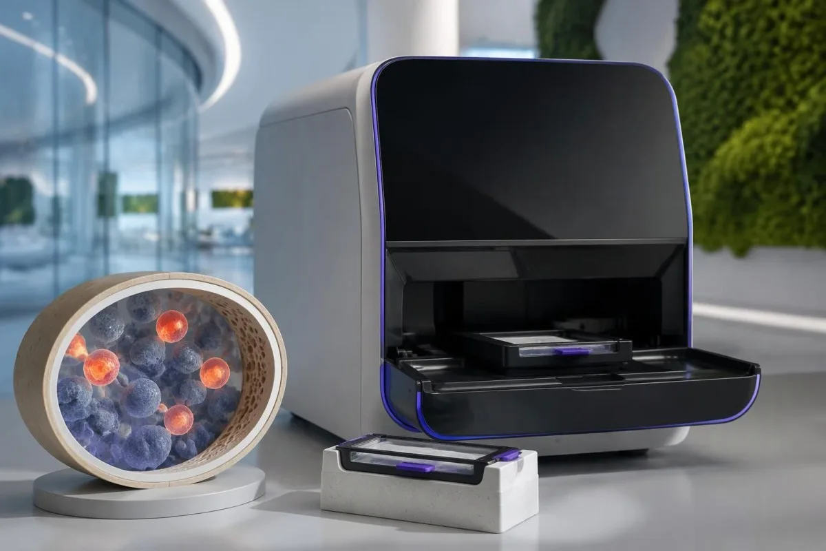
Variant allele fraction is the proportion of analyzed sequence reads carrying a particular change at that location. In a simple 1-X-chromosome model it may approximate the proportion of affected sampled cells, but cell mixtures, chromosome changes and laboratory methods complicate that estimate; it does not directly measure organ damage.
A hypothetical report showing a recognized disease-associated variant at 12% allele fraction may be meaningful even though most reads are unchanged. By contrast, a novel uncertain variant at 40% still requires expert assessment, because abundance cannot turn an unproven genetic finding into an established cause of symptoms.
Different clonal disorders require different molecular or cellular tests, even when anemia and thrombosis overlap. Our PNH clone testing guide explains a separate disorder assessed by flow cytometry; 1 negative PNH test does not evaluate UBA1, and a UBA1 result does not replace PNH testing when indicated.
Kantesti cannot determine whether an uncertain UBA1 finding is disease-causing simply by matching it to an elevated CRP of 70 mg/L. Ask the specialist whether additional sequencing, another specimen or formal variant review is needed, and keep the complete report rather than a screenshot containing only the word positive.
Ці дастаткова вакуоляў у касцявым мозгу для дыягностыкі VEXAS?
Bone marrow vacuoles are a clue, not proof of VEXAS syndrome. Vacuoles may appear in early myeloid and erythroid precursors, but they are neither completely specific nor universally present; an adult with an MCV above 100 fL still needs the broader clinical and molecular assessment.
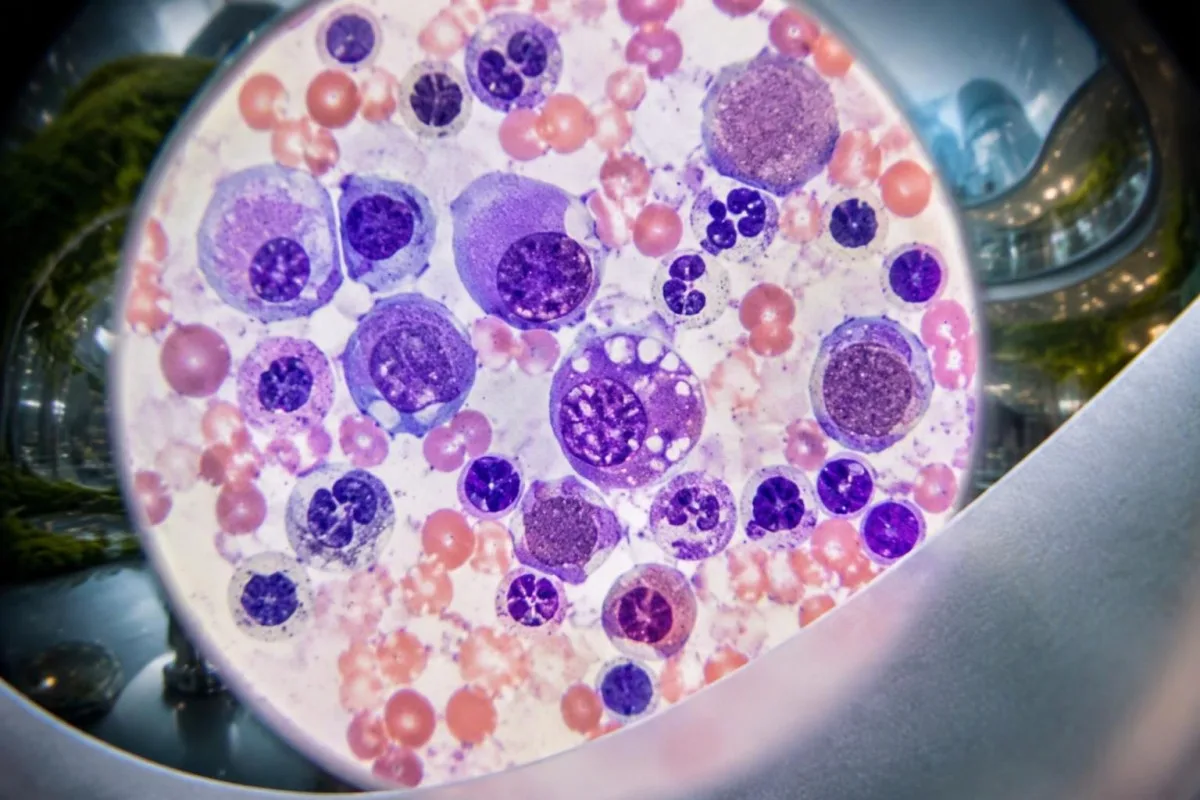
Vacuoles are small clear spaces visible within cells under microscopy, not holes that a patient can see on an ordinary CBC. Their absence on 1 marrow examination does not automatically exclude VEXAS, particularly when specimen quality, disease stage or the specific UBA1 variant affects the appearance.
Copper deficiency, alcohol-related effects and other conditions can produce vacuolated precursors. That overlap is why 2 complementary investigations may be needed: laboratory assessment for reversible mimics and somatic UBA1 testing, rather than treating a descriptive pathology finding as the final diagnosis.
Marrow examination also assesses cellularity, dysplasia and other abnormalities that may influence treatment decisions. Our explanation of teardrop cells and marrow clues describes a different microscopy finding; neither teardrop cells nor vacuoles can independently explain a platelet count of 95 × 10^9/L.
A marrow procedure is not automatically necessary for every isolated MCV of 102 fL, and a positive molecular result may already establish VEXAS in the right context. Hematology decides whether marrow assessment is needed to investigate persistent cytopenias, suspected MDS or another process that would change management.
Калі мэтазгодна звярнуцца да рэўматолага або гематолага?
Specialist assessment is appropriate when recurrent unexplained inflammation accompanies macrocytic anemia, falling platelets or other persistent cytopenias. Suspicion is higher after age 50, especially with chondritis, inflammatory skin findings or lung involvement; rheumatology and hematology often need to evaluate the pattern together.
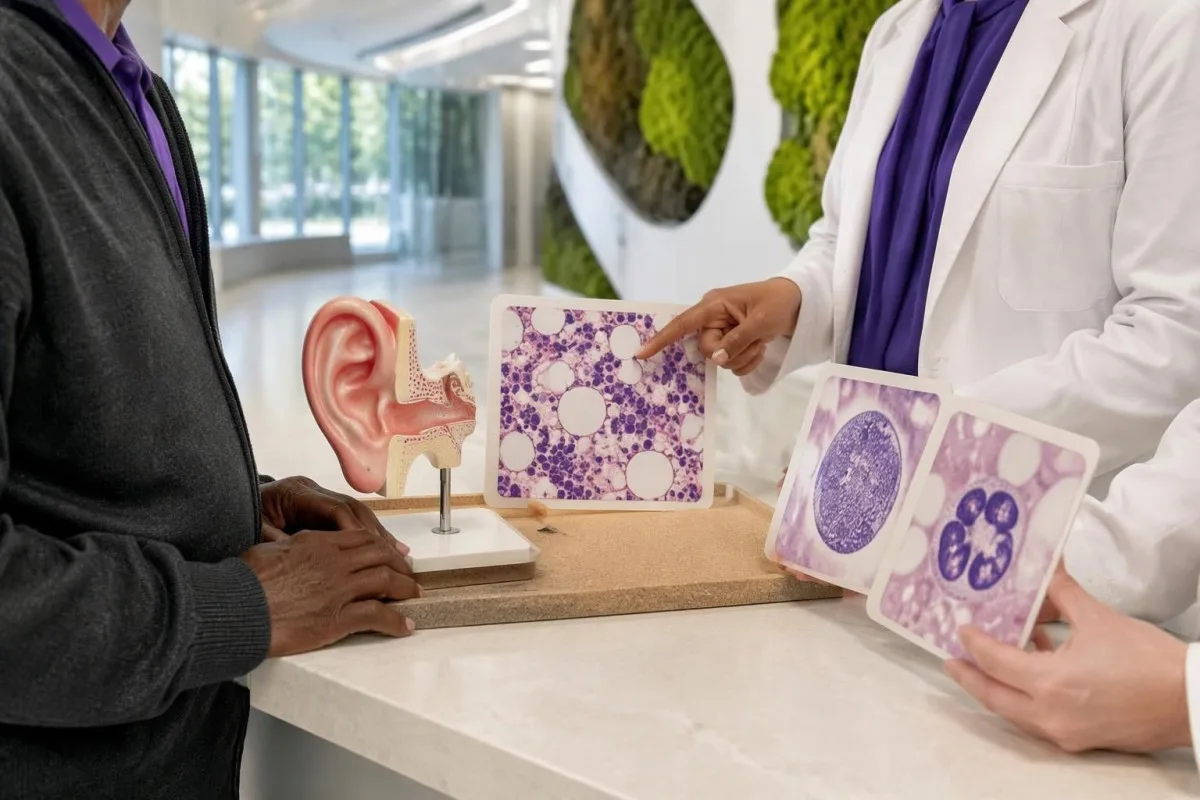
There is no universal CRP level, age boundary or number of attacks that automatically mandates UBA1 testing. A person aged 68 with recurrent ear chondritis and unexplained anemia has a different pretest probability from a person aged 28 with 1 viral fever and an otherwise stable CBC.
Steroid-responsive inflammation that repeatedly returns during dose reduction deserves reassessment, particularly when blood counts remain abnormal. Improvement over 2 days may show that symptoms are inflammatory, but it does not distinguish VEXAS from several other disorders and should not become the only justification for ongoing treatment.
Unexplained pain needs a broader workup when it accompanies constitutional symptoms or changing counts. Our кіраўніцтва па тэсціраванні невытлумачальнай болю explains why inflammatory markers are only part of that workup; an MCV above 100 fL adds a marrow-related question that a pain score cannot answer.
A referral should highlight 3 elements: documented inflammatory episodes, the CBC trend and the results of common-cause investigations. If symptoms are progressing rapidly, new organs are involved or transfusions are being considered, ask the referring clinician whether the appointment needs to be expedited rather than waiting for another routine repeat test.
Практычны прыклад: перыядычны ацёк вуха і змяненне паказчыкаў CBC
A hypothetical combination of recurrent chondritis, macrocytic anemia and low platelets warrants specialist evaluation, not an AI-generated diagnosis. Consider a 68-year-old man with hemoglobin 10.2 g/dL, MCV 108 fL, platelets 112 × 10^9/L and CRP 86 mg/L during a third episode of upper-ear swelling.

The first reasoning step is to separate symptom severity from diagnostic specificity: CRP of 86 mg/L confirms a substantial inflammatory response but does not identify its cause. The MCV of 108 fL and platelet count of 112 × 10^9/L add evidence that blood-cell production also needs investigation.
Suppose the person's B12, folate and thyroid assessments do not reveal an explanation, and medication, alcohol and liver reviews are unrevealing. A negative ANA would not close the case; the combination of 3 inflammatory episodes and persistent unexplained cytopenias would justify discussing somatic UBA1 testing with hematology and rheumatology.
Now suppose CRP falls to 12 mg/L after prescribed glucocorticoids, but hemoglobin falls to 9.8 g/dL. Those 2 trends are not contradictory: inflammatory activity may improve while marrow dysfunction or another anemia mechanism persists, so treatment response cannot be used as a substitute for investigating the falling hemoglobin.
Kantesti can help explain why those 2 trends deserve separate attention, but cannot establish VEXAS or choose immunosuppressive treatment. Our кіраўніцтва па аналізе тэндэнцый лекаў explains why dose changes, transfusions and intercurrent illness should be recorded beside each result; the example here is entirely hypothetical.
Як спецыялісты лечаць пацверджаны сіндром VEXAS?
VEXAS treatment aims to control inflammation, manage cytopenias and reduce treatment-related harm. Glucocorticoids often suppress symptoms but may be difficult to taper; other options include selected immunomodulators, marrow-directed treatment and allogeneic stem-cell transplantation, with no single regimen appropriate for every adult over 50.
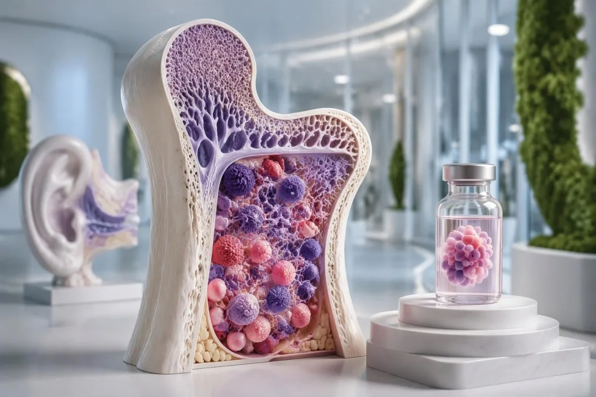
A falling CRP does not by itself establish treatment success if anemia, thrombosis or lung disease is worsening. Specialists follow at least 2 dimensions—clinical inflammatory activity and hematological status—and weigh these against cumulative glucocorticoid exposure, because prolonged treatment can cause infections, diabetes, osteoporosis and muscle weakness.
JAK inhibitors and IL-6-directed therapy are among approaches used by specialists, while azacitidine may be considered in selected patients, particularly with associated marrow disease. Evidence and access vary, and much published experience is observational; a response lasting 6 months in one cohort does not reliably predict an individual's longer-term benefit.
Allogeneic stem-cell transplantation can potentially eliminate the abnormal blood-forming clone and produce sustained remission in selected patients. It also carries substantial risks, including graft-versus-host disease and serious infection, so 2 people of the same age may receive different recommendations because fitness, organ involvement and donor availability differ.
Infection prevention deserves planning before immunosuppression, not only after fever develops. Depending on exposures and the proposed regimen, clinicians may consider screening or prevention measures described in our Strongyloides testing explanation; even 1 relevant past exposure can matter before corticosteroids, and prescribed steroids should never be stopped abruptly without guidance.
Пры якіх сімптомах патрэбна неадкладная або экстраная медыцынская дапамога?
New breathing difficulty, chest pain, airway symptoms, neurological deficits or significant bleeding require urgent assessment in someone with suspected or confirmed VEXAS. Fever of 38°C or higher during substantial immunosuppression also needs prompt clinical advice, particularly when neutrophils are low.
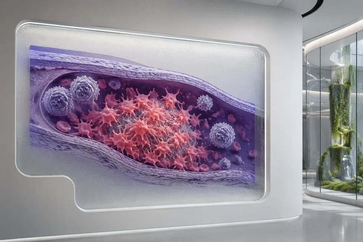
VEXAS is associated with venous thrombosis, so a painful swollen leg or sudden breathlessness should not be dismissed as another inflammatory flare. Normal routine clotting times do not exclude a clot; our абмежаванні каагулаграмы explain why 2 normal tests, PT and aPTT, cannot provide that reassurance.
An absolute neutrophil count below 0.5 × 10^9/L represents severe neutropenia and increases concern when fever occurs. Treatment can also blunt the temperature response, so feeling acutely unwell with shaking chills, confusion or rapidly worsening weakness can require urgent assessment even when a thermometer reads below 38°C.
Breathlessness can reflect lung inflammation, infection, pulmonary embolism or worsening anemia, and these require different treatments. Our breathlessness blood test guide explains why hemoglobin of 8 g/dL may contribute without proving the entire cause; new chest pain or sudden breathing difficulty should not wait for an uploaded report.
Persistent nose or gum bleeding, black stools or a severe new headache needs urgent advice, especially if platelets are very low. A count below 20 × 10^9/L generally warrants prompt clinical review, but the urgency depends on symptoms and context; significant bleeding is an emergency even above that number.
Назіранне, абмежаванні ШІ і навуковыя публікацыі
Useful VEXAS follow-up combines symptoms, treatment history, CBC trends and inflammatory markers, with molecular and marrow findings interpreted by specialists. An AI explanation of 1 laboratory report can support preparation for care, but it cannot diagnose VEXAS, exclude infection or establish whether a UBA1 variant is pathogenic.

Bring original CBC reports, inflammatory markers and the complete UBA1 result, including specimen type and detection limit. Record 3 timeline features beside them: flare dates, medication changes and transfusions; this makes it easier to distinguish a true disease trend from a treatment effect or a change in testing conditions.
Кантэсці — гэта Інструмент аналізу вынікаў аналізу крыві з падтрымкай AI that can explain laboratory patterns without converting an MCV above 100 fL into a rare-disease diagnosis. Genetic reports can reveal sensitive information, so review our інфармацыю пра прыватнасць і даныя before sharing records, and include only documents you are authorized to upload.
Kantesti's 2 DOI-linked publications below concern broader blood-test interpretation and RDW education, not clinical trials of VEXAS diagnosis or UBA1 sequencing. They should therefore be read separately from the disease-specific medical studies listed in the external references; neither publication establishes VEXAS-specific diagnostic sensitivity or treatment effectiveness.
Наш метадалогія і клінічныя стандарты explain the distinction between report interpretation and medical diagnosis. Before the next appointment, write 2 questions: whether the laboratory method adequately assessed somatic UBA1 variants, and whether persistent cytopenias need marrow evaluation; these are more actionable than asking an AI to calculate an unsupported VEXAS probability.
Часта задаваныя пытанні
Якія першыя сімптомы сіндрому VEXAS?
Сіндром VEXAS часта пачынаецца з перыядычнай ліхаманкі невядомага паходжання, запаленчых змен скуры, стомленасці або ацёку храстка вуха ці носа, звычайна пасля 50 гадоў. Макрацытарная анемія або іншыя адхіленні ў паказчыках крыві могуць з’явіцца да гэтых сімптомаў, адначасова з імі або пасля іх. MCV у дарослага вышэй за прыкладна 100 fL — гэта падказка, а не дыягназ. Паўторныя запаленчыя сімптомы ў спалучэнні з устойлівымі цытопеніямі невядомага паходжання — падстава абмеркаваць абследаванне ў рэўматолага і гематолага.
Які аналіз пацвярджае сіндром VEXAS?
Сіндром VEXAS дыягнастуюць шляхам выяўлення звязанага з захворваннем саматычнага варыянта UBA1 у адпаведным клінічным кантэксце. Звычайна для даследавання выкарыстоўваюць перыферычную кроў, хоць у асобных выпадках могуць спатрэбіцца касцявы мозг або больш адчувальны метад аналізу. Многія вядомыя варыянты закранаюць метыянін у пазіцыі 41, але абмежаванае даследаванне гарачых кропак можа не выявіць іншыя звязаныя з захворваннем варыянты. Вынік трэба інтэрпрэтаваць з улікам класіфікацыі варыянта, ахопу гена і мяжы выяўлення.
Ці могуць нармальны аналіз на антыядзерныя антыцелы (ANA) або панэль на аутаімунныя захворванні выключыць VEXAS?
Нармальны аналіз на ANA або стандартная панэль аналізаў на аўтаімунныя захворванні не дазваляюць выключыць сіндром VEXAS, бо гэтыя тэсты не выяўляюць саматычныя мутацыі UBA1. Станоўчы вынік аналізу на ANA ў тытры 1:160 таксама не пацвярджае VEXAS і не абавязкова сведчыць пра наяўнасць іншага аўтаімуннага захворвання. Аналіз на антыцелы можа дапамагчы даследаваць альтэрнатыўныя варыянты, а паказчыкі CBC, сімптомы і малекулярнае тэставанне даюць адказы на розныя пытанні. Не варта адкідаць персістуючую макрацытарную анемію і рэцыдывавальнае запаленне храсткоў толькі таму, што аналізы на антыцелы адмоўныя.
Ці перадаецца сіндром VEXAS па спадчыне і ці варта абследаваць маіх дзяцей?
Тыповы сіндром VEXAS з’яўляецца набытым, а не спадчынным, таму звычайна няма паказанняў да руціннага абследавання дзяцей. Адпаведная мутацыя UBA1 звычайна ўзнікае ў крывятворных клетках у дарослым узросце, часта пасля 50 гадоў з’яўляюцца сімптомы. Хоць ген UBA1 знаходзіцца ў Х-храмасоме, гэта не азначае, што саматычны варыянт VEXAS аўтаматычна перадаецца ў спадчыну. Незвычайныя або, магчыма, канстытуцыянальныя вынікі варта абмеркаваць са спецыялістам па клінічнай генетыцы.
Ці можа ў жанчын развіцца сіндром VEXAS?
У жанчын можа развіцца сіндром VEXAS, хоць большасць выяўленых выпадкаў назіраецца ў мужчын старэйшага ўзросту. Ген UBA1 размешчаны ў X-храмасоме, і страта адной X-храмасомы ў здзіўленых клетках — адзін з механізмаў, апісаных у жанчын. У даследаванні сістэмы аховы здароўя, апублікаваным у JAMA ў 2023 годзе, сярод 11 удзельнікаў з варыянтамі UBA1, звязанымі з захворваннем, былі выяўлены дзве жанчыны. Пол сам па сабе не павінен быць падставай адмаўляцца ад кансультацыі спецыяліста, калі рэцыдывавальнае запаленне і невытлумачальныя змены паказчыкаў агульнага аналізу крыві выклікаюць сур’ёзнае падазрэнне.
Ці выключае адмоўны вынік тэсту UBA1 сіндром VEXAS?
Адмоўны вынік тэсту UBA1 зніжае верагоднасць VEXAS толькі ў той ступені, у якой узор і аналіз былі належнымі. Тэст з мяжой выяўлення варыянтнай алельнай частаты 5% можа не выявіць меншы клон, а даследаванне толькі гарачых кропак можа прапусціць варыянты па-за межамі ахопленага рэгіёна. Высокая клінічная падазронасць можа служыць падставай для экспертнай ацэнкі ахопу, адчувальнасці і тыпу ўзору. Адмоўны вынік, атрыманы належным чынам, таксама павінен стаць падставай для ўважлівай пераацэнкі альтэрнатыўных дыягназаў, а не для паўторнага тэставання без дакладнай прычыны.
Ці існуе лячэнне сіндрому VEXAS?
Аллагенная трансплантацыя ствалавых клетак патэнцыйна можа забяспечыць працяглую рэмісію, замяніўшы анамальны крывятворны клон, але падыходзіць толькі асобным пацыентам і нясе значныя рызыкі. Іншыя метады лячэння накіраваны на кантроль запалення, лячэнне цытапеній і зніжэнне ўздзеяння глюкакартыкоідаў, а не на надзейнае знішчэнне клона. У дарослых старэйшых за 50 гадоў фізічны стан, паражэнне органаў і спадарожныя захворванні касцявога мозгу могуць істотна ўплываць на выбар лячэння. Ніводная дабаўка, план харчавання або інтэрпрэтацыя аналізу крыві з дапамогай ШІ не даказалі здольнасці вылечваць VEXAS.
Атрымайце тлумачэнне вынікаў аналізу крыві
Загрузіце свой лабараторны справаздачу, і AI Kantesti за некалькі секунд простай мовай растлумачыць кожны біямаркер. Ім карыстаюцца больш за 2 мільёны чалавек па ўсім свеце.
Спасылкі
- Кляйн, Т., Мітчэл, С., і Вебер, Х. (2026). AI Blood Test Analyzer: 2,5 млн. тэстаў прааналізавана | Global Health Report 2026. Kantesti AI Medical Research. DOI
- Кляйн, Т., Мітчэл, С., і Вебер, Х. (2026). Аналіз крыві на RDW: поўнае кіраўніцтва па RDW-CV, MCV і MCHC. Kantesti AI Medical Research. DOI
- Beck DB et al. (2020). Somatic Mutations in UBA1 and Severe Adult-Onset Autoinflammatory Disease. New England Journal of Medicine. DOI
- Beck DB et al. (2023). Estimated Prevalence and Clinical Manifestations of VEXAS Syndrome in a Large US Health Care System. JAMA. DOI
- 2 млн+Прааналізаваныя тэсты
- 127+Краіны
- 100+Мовы
Пра медыцынскую каманду


Сігналы даверу E-E-A-T
Вопыт
Клінічны агляд, які вядзе лекар, працэсаў інтэрпрэтацыі лабараторных паказчыкаў.
Экспертыза
Фокус лабараторнай медыцыны на тым, як біямаркеры паводзяць сябе ў клінічным кантэксце.
Аўтарытэтнасць
Медыцынскую рэцэнзію правёў доктар Томас Кляйн пры ўдзеле доктара Сары Мітчэл і прафесара, доктара Ганса Вэбера.
Надзейнасць
Інтэрпрэтацыя на аснове доказаў з выразнымі шляхамі далейшых дзеянняў, каб паменшыць трывогу.
Медыцынская адмова ад адказнасці
Гэты артыкул прызначаны толькі для адукацыйных мэт і не з’яўляецца медычнай парадай. Заўсёды кансультуйцеся з кваліфікаваным спецыялістам аховы здароўя для прыняцця рашэнняў па дыягностыцы і лячэнні.
Апублікавана · Звязацца з рэдакцыйнай камандай
Апублікавана кампаніяй Kantesti Ltd, зарэгістраванай у Англіі і Уэльсе, нумар кампаніі. 17090423, Лондан, Злучанае Каралеўства.
