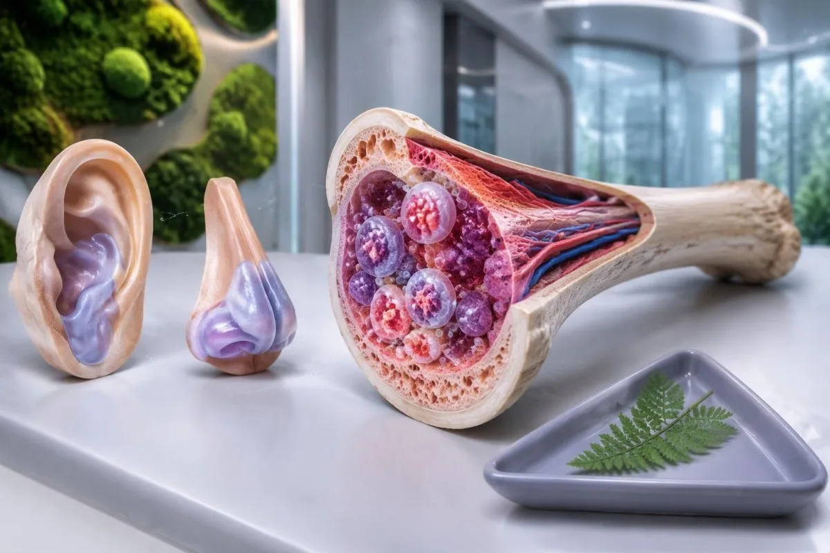
VEXAS සින්ඩ්රෝමය යනු රුධිරය නිපදවන සෛලවල ඇති වන ශාරීරික UBA1 විකෘති හේතුවෙන් ඇතිවන අත්පත් කරගත් ප්රදාහකාරී තත්ත්වයකි. නැවත නැවත ඇතිවන උණ, කන් හෝ නාසයේ කාටිලේජ ඉදිමීම සහ පැහැදිලි කළ නොහැකි මැක්රොසයිටික් රක්තහීනතාව සඳහා විශේෂඥ ඇගයීමක් සිදු කළ යුතුය; රෝග විනිශ්චය සඳහා සාමාන්ය ස්වයංප්රතිශක්තිකරණ පැනලයක් නොව, සුදුසු ජාන පරීක්ෂණ අවශ්ය වේ.

ප්රධාන කරුණු
- VEXAS සින්ඩ්රෝමය සාමාන්යයෙන් වයස අවුරුදු 50ට පසු ආරම්භ වන අතර, පද්ධතිමය ප්රදාහය රුධිරය නිපදවන සෛලවල අසාමාන්යතා සමඟ එක් කරයි.
- කාටිලේජ ඉදිමීම කන්, නාසය හෝ ශ්වසන මාර්ගයට බලපෑ හැකිය; කන් කාටිලේජයේ ප්රදාහය බොහෝ විට මෘදු කන්පෙත්තට බලපාන්නේ නැත.
- මැක්රොසයිටෝසිස් (Macrocytosis) යනු වැඩිහිටියන්ගේ ආසන්න වශයෙන් 100 fLට වැඩි MCV අගයකි, නමුත් මෙම සොයාගැනීමෙන් පමණක් VEXAS රෝග විනිශ්චය කළ නොහැක.
- අඩු පට්ටිකා 150 × 10^9/Lට අඩු අගයන්, නැවත නැවත ඇතිවන ප්රදාහය සහ පැහැදිලි කළ නොහැකි රක්තහීනතාව සමඟ පවතින විට සැකය තවත් වැඩි කළ හැකිය.
- සී.ආර්.පී. සාමාන්යයෙන් ඉහළ සන්දර්භ සීමාව 5 mg/L වන නමුත් සමහර රසායනාගාර 10 mg/L භාවිත කරයි; කිසිදු CRP සීමාවකින් VEXAS තහවුරු නොවේ.
- UBA1 විකෘති පරීක්ෂණය අත්පත් කරගත් රෝගකාරක ප්රභේදයක් සොයයි; සාමාන්යයෙන් සාමාන්ය උරුම අවදානම් පැනලයක් වෙනුවට පර්යන්ත රුධිරය හෝ අස්ථි මිදුළුව භාවිත කරයි.
- ස්වයංප්රතිශක්තිකරණ පරීක්ෂණ පැනල ANA, රූමටොයිඩ් සාධකය සහ ANCA වැනි දේවලට VEXAS සින්ඩ්රෝමය තහවුරු කිරීමට හෝ බැහැර කිරීමට නොහැක.
- හදිසි ලක්ෂණ සැලකිය යුතු ප්රතිශක්ති මර්දනයක් පවතින අතරතුර ඇතිවන නව හුස්ම හිරවීම, පපුවේ වේදනාව, ශ්වසන මාර්ගයේ අපහසුතාව හෝ 38°C හෝ ඊට වැඩි උණ ඇතුළත් වේ.
VEXAS සින්ඩ්රෝමය යනු කුමක්ද, රුධිර ගණනයන් වැදගත් වන්නේ ඇයි?
VEXAS සින්ඩ්රෝමය සෝමැටික් විකෘතියක් හේතුවෙන් ඇතිවන අත්පත් කරගත් ස්වයං-ප්රදාහකාරී ආබාධයකි UBA1 විකෘතියක් රුධිරය නිපදවන සෛලවල. නැවත නැවත ඇතිවන ප්රදාහය, ඉදිමුණු කාටිලේජය සහ පැහැදිලි කළ නොහැකි අසාමාන්ය රුධිර ගණන්—විශේෂයෙන් වයස අවුරුදු 50ට වැඩි අයෙකුගේ මැක්රොසයිටික් රක්තහීනතාව—සැකයක් මතු කළ යුතු අතර, රෝග විනිශ්චය රඳා පවතින්නේ සායනික පසුබිම තුළ ගැළපෙන රෝගකාරක UBA1 ප්රභේදයක් හඳුනා ගැනීම මතය.
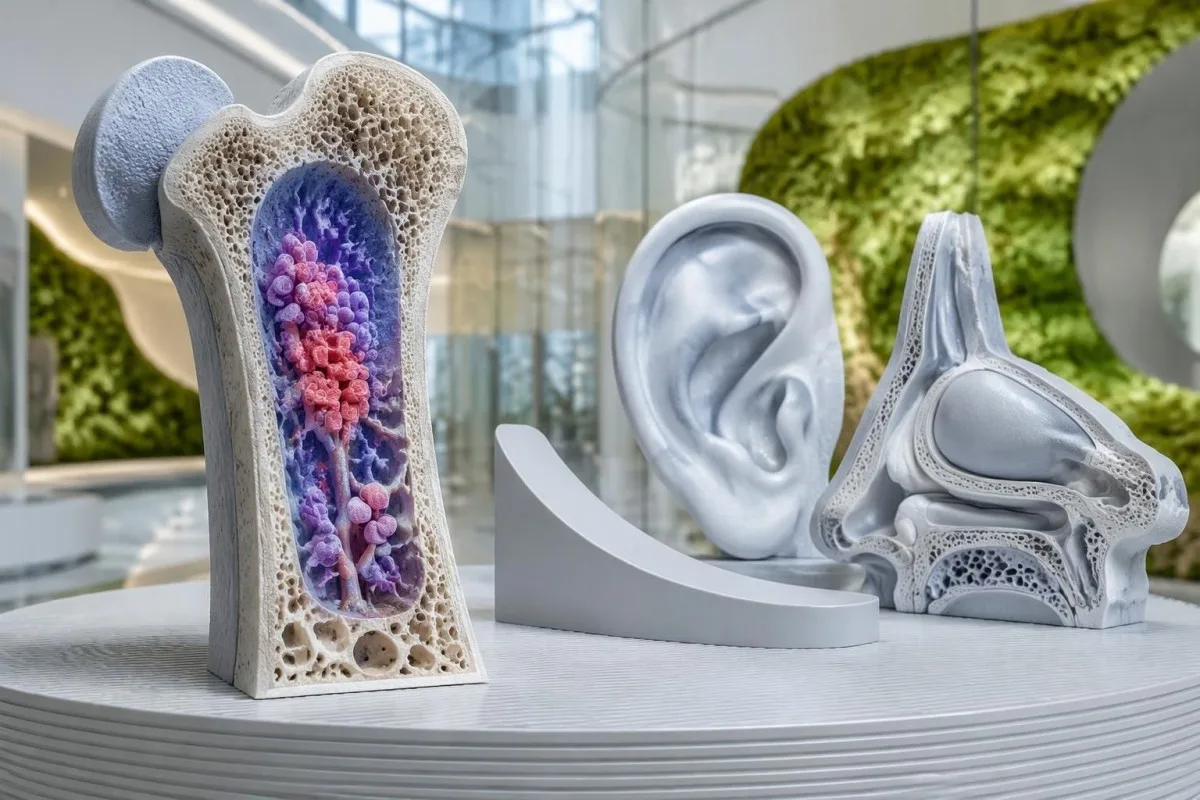
නාමයෙන් ලක්ෂණ 5ක් විස්තර වේ: රික්තක, E1 එන්සයිමය, X-සම්බන්ධිත, ස්වයං-ප්රදාහකාරී සහ සෝමැටික්. මේවා ජීව විද්යාත්මක ක්රියාවලියට ඉඟි මිස, සෑම පුද්ගලයෙකුම සපුරාලිය යුතු පරීක්ෂණ ලැයිස්තුවක් නොවේ; අස්ථි मज්ජාවේ පෙනෙන රික්තක, කාටිලේජ රෝග ලක්ෂණ සහ දැඩි රක්තහීනතාව පළමු හමුවීමේදීම එකට නොපෙනිය හැක.
Beck සහ සගයන් 2020දී New England Journal of Medicine හි VEXAS ප්රථමයෙන් විස්තර කරමින්, වැඩිහිටි වියේදී ආරම්භ වන ප්රදාහය අත්පත් කරගත් UBA1 ප්රභේද සමඟ සම්බන්ධ කළහ. මෙම සොයාගැනීමෙන් පැහැදිලි වන්නේ, පොදු හේතුව හඳුනාගැනීමට පෙර පුද්ගලයෙකුට නැවත නැවත ඇතිවන පොලිකොන්ඩ්රයිටිස් සහ අස්ථි मज්ජා ආබාධයක් වැනි රෝග විනිශ්චය 1කට වඩා ලැබී තිබිය හැක්කේ මන්ද යන්නයි.
CRP අගය ඉහළ බව පෙන්වන තනි ප්රතිඵලයක් VEXAS සඳහා විශේෂිතව ඉඟි නොකරන අතර, MCV අගය 103 fL වීම බොහෝවිට වඩාත් සාමාන්ය හේතු නිසා වේ. රුධිර සෛල අඩුවීම් අඛණ්ඩව පවතින හෝ නරක අතට හැරෙන අතරතුර ප්රදාහකාරී ප්රහාර නැවත ඇති වුවහොත් සැලකිල්ල වැඩි වේ; එම රටාවෙන් හුදකලා රසායනාගාර උච්චාවචනයකට වඩා, අසාමාන්ය රුධිර සෛල නිෂ්පාදනයට සම්බන්ධ ප්රදාහකාරී ක්රියාවලියක් යෝජනා වන බැවිනි.
කන්ටෙස්ටි යනු AI රුධිර පරීක්ෂණ විශ්ලේෂකය හිමොග්ලොබින්, MCV, පට්ටිකා සහ CRP එකිනෙකට සම්බන්ධ වන ආකාරය පැහැදිලි කළ හැකි අතර, VEXAS රෝග විනිශ්චය විශේෂඥයන්ට ඉතිරි කරයි. අපගේ සංවිධානය සහ අරමුණ වෙන වෙනම විස්තර කෙරේ; උඩුගත කළ CBC 1කින් UBA1 විකෘතියක් හඳුනාගත නොහැකි අතර සායනික පරීක්ෂණයක් වෙනුවටද භාවිත කළ නොහැක.
VEXAS වැළඳෙන්නේ කාටද, UBA1 විකෘතිය උරුමයෙන් ලැබෙන්නේද?
VEXAS සින්ඩ්රෝමය බොහෝවිට වැඩිහිටි පිරිමින්ට බලපායි, සාමාන්යයෙන් රෝග ලක්ෂණ වයස අවුරුදු 50ට පසු ආරම්භ වේ. රෝගය ඇති කරන UBA1 විකෘතිය සාමාන්යයෙන් උරුම වී දෙමාපියෙකුගෙන් ලැබෙන හෝ උපතේ සිට සෑම සෛලයකම පවතින එකක් නොව, රුධිරය නිපදවන සෛල සමූහයක් තුළ අත්පත් කරගත් එකකි.
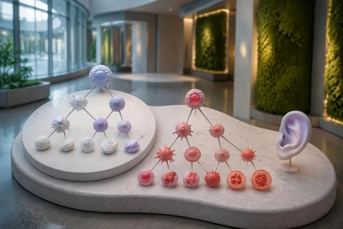
UBA1 පිහිටා ඇත්තේ X වර්ණදේහයේ වන අතර, බලපෑමට ලක් වූ බොහෝ පිරිමින්ට X වර්ණදේහ 1ක් ඇත. VEXAS කාන්තාවන්ටද ඇති වේ; ඇතැම්විට බලපෑමට ලක් වූ සෛලවල X වර්ණදේහයක් අහිමි වීම සමඟ සම්බන්ධව එය ඇති විය හැක. එහෙත් සායනික රටාව ප්රබල ලෙස සැක උපදවන විට, ලිංගය පමණක් ඇගයීම නතර කිරීමට හේතුවක් නොවිය යුතුය.
එක්සත් ජනපද සෞඛ්ය පද්ධති කණ්ඩායමක සහභාගිවන්නන් 163,096ක් පිළිබඳ JAMA අධ්යයනයකදී, Beck සහ සගයන් රෝගය සමඟ සම්බන්ධ UBA1 ප්රභේද ඇති පුද්ගලයන් 11 දෙනෙකු හඳුනා ගත්හ. වයස අවුරුදු 50ට වැඩි පිරිමින් අතර ඇස්තමේන්තුගත සංඛ්යාතය 4,269කින් 1ක් පමණ විය; මෙය ප්රධාන වශයෙන් යුරෝපීය සම්භවයක් ඇති විශේෂිත කණ්ඩායමක් මිස, ලොව පුරා ව්යාප්තිය පිළිබඳ ඇස්තමේන්තුවක් නොවේ.
අදාළ විකෘතිය පුද්ගලයෙකුගේ ජීවිත කාලය තුළ ඇතිවන බැවින් පවුල් ඉතිහාසයක් සාමාන්යයෙන් නොමැත. මෙය අපගේ MEFV පරීක්ෂණය පිළිබඳ පැහැදිලි කිරීම; හි ආවරණය වන උරුමයෙන් ලැබෙන නැවත නැවත ඇතිවන උණ ආබාධවලට වඩා වෙනස්ය; පවුලේ ප්රභේදයක් සඳහා ඥාතීන් 2 දෙනෙකු පරීක්ෂා කිරීම, සාමාන්ය සෝමැටික් VEXAS සඳහා සම්මත ආරම්භක පියවර නොවේ.
ජානමය යන්නෙන් ස්වයංක්රීයව උරුමයෙන් ලැබෙන බව අදහස් නොවන අතර, X-සම්බන්ධිත ජානයක් යන්නෙන් දරුවන්ට සම්ප්රේෂණය වන බව ස්වයංක්රීයව අදහස් නොවේ. වාර්තාවක ව්යවස්ථාපිත ප්රභේදයක් හෝ වයස අවුරුදු 50ට පෙර ඇතිවන අසාමාන්ය රෝගී තත්ත්වයක් සඳහන් වේ නම්, අමතර පටක පරීක්ෂාවක් හෝ පවුල් උපදේශනයක් සුදුසුද යන්න සායනික ජාන විද්යා විශේෂඥයෙකුට පැහැදිලි කළ හැක.
සැකයක් ඇති කළ යුතු VEXAS සින්ඩ්රෝමයේ රෝග ලක්ෂණ මොනවාද?
VEXAS සින්ඩ්රෝමයේ රෝග ලක්ෂණ අතර සාමාන්යයෙන් නැවත නැවත ඇතිවන උණ, සමේ ප්රදාහකාරී වෙනස්කම්, තෙහෙට්ටුව, පෙනහළු සම්බන්ධ වීම සහ කාටිලේජ ප්රදාහය ඇතුළත් වේ. කන් හෝ නාසයේ කාටිලේජ ඉදිමීම, වයස අවුරුදු 50ට වැඩි වැඩිහිටියෙකුගේ පැහැදිලි කළ නොහැකි රක්තහීනතාවක් හෝ වෙනත් අසාමාන්ය රුධිර ගණන් සමඟ ඇති වූ විට විශේෂයෙන් වැදගත් වේ.
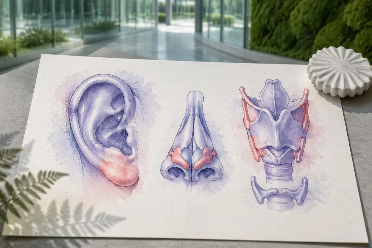
කන් කාටිලේජ ප්රදාහය බොහෝවිට කනේ තද ඉහළ කොටසට බලපාන අතර, කාටිලේජ නොමැති මෘදු කන් පෙත්තට බලපාන්නේ නැත. එම ව්යුහ විද්යාත්මක වෙනස සිදුවීමක් නිවැරදිව විස්තර කිරීමට උපකාරී විය හැකි නමුත්, රතු වූ කනක් 1ක් වුවද ස්ථානීය ආසාදනය, තුවාලය හෝ චර්ම ප්රදාහය වැනි වඩාත් සාමාන්ය හේතු සඳහා ඇගයිය යුතුය.
සමේ ලක්ෂණවලට ස්පර්ශයට වේදනාකාරීව උස් වූ ප්රදේශ, පැල්ලම් හෝ දම් පැහැති වෙනස්කම් ඇතුළත් විය හැක; පෙනුමෙන් පමණක් හේතුව හඳුනාගත නොහැක. පටක පරීක්ෂාවකින් නියුට්රොෆිලික් චර්ම රෝගය, රුධිර නාල ප්රදාහය සහ වෙනත් ක්රියාවලීන් වෙන්කර හඳුනා ගැනීමට උපකාරී විය හැකි අතර, ප්රතිකාර නොකළ වෙන වෙනම සිදුවීම් 2ක ඡායාරූප විශේෂඥයෙකුට නැවත නැවත ඇතිවන රටාව හඳුනාගැනීමට උපකාර විය හැක.
Inflammatory attacks can resemble established rheumatological conditions, including relapsing polychondritis or polymyalgia rheumatica. Our explanation of PMR laboratory limitations describes why inflammatory markers are not disease-specific; persistent macrocytosis above 100 fL or falling platelets should prompt a broader assessment rather than automatic acceptance of the first label.
New hoarseness, noisy breathing or difficulty breathing needs urgent assessment because airway cartilage can be involved. A useful symptom record includes 3 details for each episode: the affected site, its duration and whether fever or a medication change accompanied it; do not wait for another flare if breathing is affected.
VEXAS සින්ඩ්රෝමය සැක කළ හැකි රුධිර පරීක්ෂණ රටාව කුමක්ද?
Macrocytic anemia, sometimes accompanied by low platelets or other cytopenias, is a major laboratory clue to VEXAS. Adult MCV is commonly about 80–100 fL, but neither an MCV above 100 fL nor an elevated CRP result can confirm the condition.
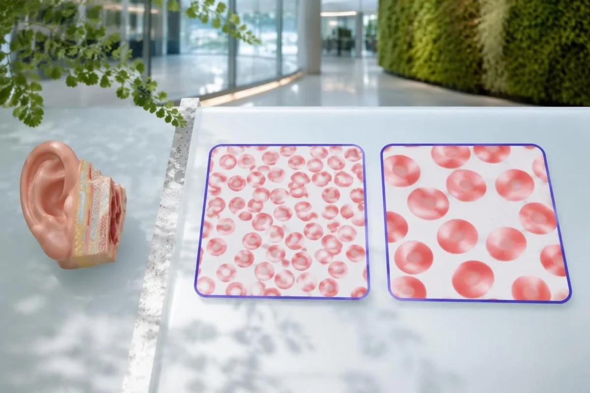
Hemoglobin measures anemia, while MCV measures average red-cell size; the 2 results answer different questions. An MCV of 108 fL with hemoglobin of 10.2 g/dL deserves a different assessment from an isolated MCV of 101 fL with stable hemoglobin, particularly when inflammatory symptoms are also present.
Many laboratories use a platelet reference interval of approximately 150–450 × 10^9/L. A falling platelet count can matter before it crosses the lower limit, and our borderline MCV explanation shows why persistent trends are often more useful than treating one near-boundary number as a diagnosis.
CRP is often markedly elevated during active VEXAS, but infection and many other inflammatory disorders can produce the same result. A hypothetical CRP of 80 mg/L is 16 times an upper limit of 5 mg/L; that comparison describes inflammatory intensity, not the probability of a UBA1 mutation.
RDW describes variation in red-cell size and can help explain a mixed CBC pattern, but it is not a VEXAS-specific biomarker. Our RDW සහ MCV මාර්ගෝපදේශය explains how 2 overlapping processes, such as iron deficiency and macrocytosis, can produce a deceptively ordinary average MCV.
| මට්ටම | පරාසය | එහි අර්ථය |
|---|---|---|
| Typical adult MCV interval | 80–100 fL | Use the reporting laboratory's interval; an in-range result does not exclude early or masked macrocytosis. |
| මැක්රොසයිටෝසිස් (Macrocytosis) | >100 fL | Investigate nutritional, medication, liver, thyroid and marrow causes; this is not a VEXAS diagnostic threshold. |
| ත්රොම්බොසයිටොපීනියාව | <150 × 10^9/L | Low platelets can support concern when combined with persistent inflammation and unexplained anemia. |
| CRP above the laboratory limit | Often >5 mg/L; some laboratories use >10 mg/L | Indicates an inflammatory response without identifying its cause. |
සාමාන්ය ස්වයංප්රතිශක්තිකරණ පැනලවලින් VEXAS තහවුරු කළ නොහැක්කේ ඇයි?
Routine autoimmune panels cannot confirm VEXAS syndrome because they measure antibodies or related immune markers, not acquired UBA1 mutations. ANA, rheumatoid factor, ANCA and complement results may help assess alternative or overlapping conditions, but no combination substitutes for appropriate molecular testing.
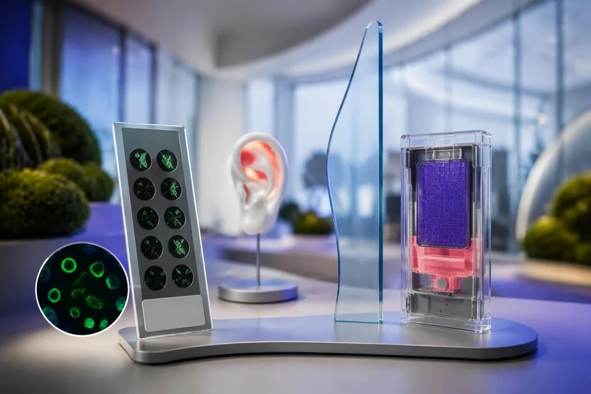
ANA testing detects antibodies directed against nuclear components; it does not read the UBA1 gene. A positive ANA at 1:160 may deserve interpretation in its own clinical context, while a negative ANA does not make unexplained fever, chondritis and macrocytic anemia safe to dismiss.
Repeating several antibody panels can create incidental positives without resolving the central question. Our ANA result explanation describes why 1 positive result is not equivalent to a disease diagnosis; the same caution applies when an antibody is found during a suspected VEXAS evaluation.
Complement testing can be useful when clinicians suspect an immune-complex disorder, but normal C3 and C4 cannot exclude VEXAS. The complement and antibody guide explains these 2 proteins; their biological role differs from the acquired enzyme dysfunction caused by a disease-associated UBA1 variant.
Autoinflammatory and autoimmune are related but not interchangeable descriptions: VEXAS is driven principally by abnormal innate immune activity associated with mutated blood-forming cells. Clinicians may still order 2 types of investigation in parallel—antibody testing for alternatives and molecular testing for VEXAS—because another diagnosis can coexist rather than disappear.
VEXAS හේතුව ලෙස සැලකීමට පෙර පරීක්ෂා කළ යුතු වෙනත් හේතු මොනවාද?
Vitamin B12 or folate deficiency, medication effects, alcohol use, liver disease, hypothyroidism and marrow disorders can cause macrocytosis. An MCV above 100 fL should trigger evaluation of these possibilities, while recurrent chondritis or unexplained inflammatory attacks may justify investigating VEXAS at the same time.
Vitamin B12 and folate results need interpretation alongside symptoms, treatment history and the CBC. A borderline B12 result may require additional assessment rather than immediate attribution of an MCV of 107 fL to VEXAS; our B12 versus folate explanation also explains why folate treatment can obscure part of a B12 deficiency picture.
Copper deficiency can cause cytopenias and vacuolated marrow precursors, making it an especially relevant mimic. Reviewing high-dose zinc supplements is therefore useful, and our අඩු තඹ තක්සේරු මාර්ගෝපදේශය explains why 2 supplement ingredients—zinc and copper—may matter even when a product is marketed for general immune support.
Myelodysplastic syndrome can cause macrocytic anemia and may coexist with VEXAS rather than simply compete with it as an explanation. Our MDS confirmation guide distinguishes CBC clues from marrow evidence; 1 abnormal count cannot establish either diagnosis or determine whether both are present.
Medication and infection reviews are essential because treatment can change the very numbers being investigated. For example, glucocorticoids may increase neutrophils while lowering CRP, and a recent transfusion can alter MCV; bring the dates of these events, not just 2 laboratory reports that appear to disagree.
ශාරීරික UBA1 පරීක්ෂණය VEXAS රෝග විනිශ්චයට සහාය වන්නේ කෙසේද?
VEXAS syndrome diagnosis is established by a disease-associated somatic UBA1 variant in a compatible clinical setting. Peripheral blood is commonly used for testing, while marrow or a more sensitive method may be appropriate after an inconclusive result; many recognized variants affect methionine at position 41.
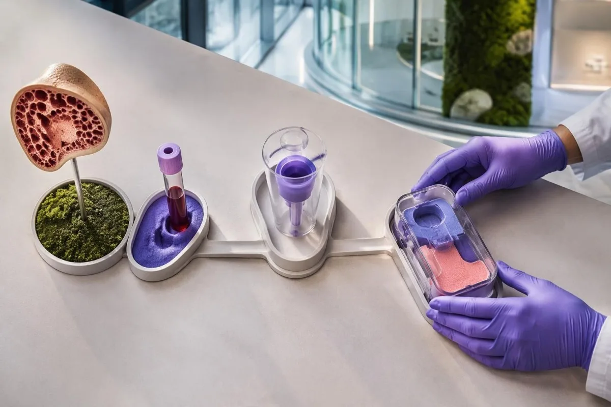
The request should specify suspected VEXAS and testing for somatic UBA1 variants rather than a general hereditary disease panel. These are 2 different diagnostic tasks: an inherited-risk assay may not include the relevant gene regions, cell source or detection sensitivity needed to identify an acquired clone.
Beck et al. linked UBA1 variants to altered ubiquitin-activating enzyme function in their 2020 New England Journal of Medicine report. Position 41 is a major hotspot, but a hotspot-only test is not equivalent to comprehensive relevant UBA1 analysis, because disease-associated noncanonical variants have also been described.
A negative result needs the laboratory method attached to it before its meaning is clear. An assay validated to detect a 5% variant allele fraction may miss a smaller clone; ask which regions were covered, the reported detection limit and whether the sample was suitable for somatic testing.
කන්ටෙස්ටි යනු AI රුධිර පරීක්ෂණ ප්රතිඵල වේදිකාව that can help explain accompanying CBC and inflammatory-marker results, but it cannot infer a UBA1 sequence from 1 routine report. Our AI තාක්ෂණ පැහැදිලි කිරීම describes report interpretation; molecular diagnosis still requires an accredited laboratory and specialist interpretation.
UBA1 විකෘති වාර්තාවක් කියවිය යුත්තේ කෙසේද?
A UBA1 report should state the variant, classification, sample type and testing limitations, often including variant allele fraction. A pathogenic or likely pathogenic disease-associated variant can support VEXAS, but a variant of uncertain significance is not diagnostic, and a 10% allele fraction is not a 10% severity score.
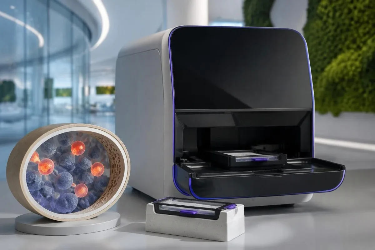
Variant allele fraction is the proportion of analyzed sequence reads carrying a particular change at that location. In a simple 1-X-chromosome model it may approximate the proportion of affected sampled cells, but cell mixtures, chromosome changes and laboratory methods complicate that estimate; it does not directly measure organ damage.
A hypothetical report showing a recognized disease-associated variant at 12% allele fraction may be meaningful even though most reads are unchanged. By contrast, a novel uncertain variant at 40% still requires expert assessment, because abundance cannot turn an unproven genetic finding into an established cause of symptoms.
Different clonal disorders require different molecular or cellular tests, even when anemia and thrombosis overlap. Our PNH clone testing guide explains a separate disorder assessed by flow cytometry; 1 negative PNH test does not evaluate UBA1, and a UBA1 result does not replace PNH testing when indicated.
Kantesti cannot determine whether an uncertain UBA1 finding is disease-causing simply by matching it to an elevated CRP of 70 mg/L. Ask the specialist whether additional sequencing, another specimen or formal variant review is needed, and keep the complete report rather than a screenshot containing only the word positive.
VEXAS රෝග විනිශ්චය කිරීමට අස්ථි මිදුළුවල ඇති වැකුයෝල් ප්රමාණවත්ද?
Bone marrow vacuoles are a clue, not proof of VEXAS syndrome. Vacuoles may appear in early myeloid and erythroid precursors, but they are neither completely specific nor universally present; an adult with an MCV above 100 fL still needs the broader clinical and molecular assessment.
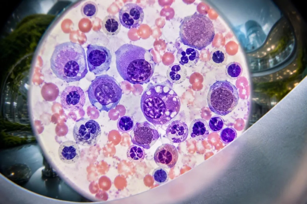
Vacuoles are small clear spaces visible within cells under microscopy, not holes that a patient can see on an ordinary CBC. Their absence on 1 marrow examination does not automatically exclude VEXAS, particularly when specimen quality, disease stage or the specific UBA1 variant affects the appearance.
Copper deficiency, alcohol-related effects and other conditions can produce vacuolated precursors. That overlap is why 2 complementary investigations may be needed: laboratory assessment for reversible mimics and somatic UBA1 testing, rather than treating a descriptive pathology finding as the final diagnosis.
Marrow examination also assesses cellularity, dysplasia and other abnormalities that may influence treatment decisions. Our explanation of teardrop cells and marrow clues describes a different microscopy finding; neither teardrop cells nor vacuoles can independently explain a platelet count of 95 × 10^9/L.
A marrow procedure is not automatically necessary for every isolated MCV of 102 fL, and a positive molecular result may already establish VEXAS in the right context. Hematology decides whether marrow assessment is needed to investigate persistent cytopenias, suspected MDS or another process that would change management.
රූමැටෝලොජි හෝ රක්තවේද ඇගයීම සුදුසු වන්නේ කවදාද?
Specialist assessment is appropriate when recurrent unexplained inflammation accompanies macrocytic anemia, falling platelets or other persistent cytopenias. Suspicion is higher after age 50, especially with chondritis, inflammatory skin findings or lung involvement; rheumatology and hematology often need to evaluate the pattern together.
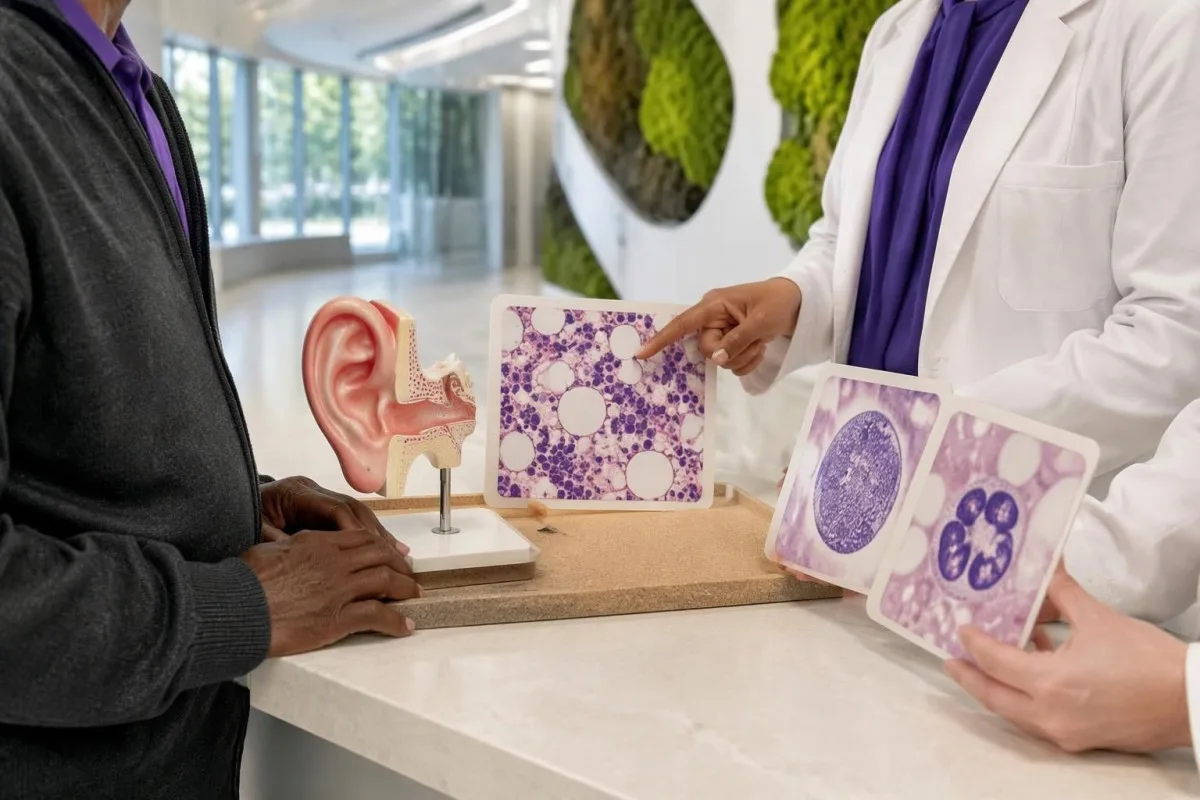
There is no universal CRP level, age boundary or number of attacks that automatically mandates UBA1 testing. A person aged 68 with recurrent ear chondritis and unexplained anemia has a different pretest probability from a person aged 28 with 1 viral fever and an otherwise stable CBC.
Steroid-responsive inflammation that repeatedly returns during dose reduction deserves reassessment, particularly when blood counts remain abnormal. Improvement over 2 days may show that symptoms are inflammatory, but it does not distinguish VEXAS from several other disorders and should not become the only justification for ongoing treatment.
Unexplained pain needs a broader workup when it accompanies constitutional symptoms or changing counts. Our පැහැදිලි කළ නොහැකි වේදනාව පරීක්ෂා කිරීමේ මාර්ගෝපදේශය explains why inflammatory markers are only part of that workup; an MCV above 100 fL adds a marrow-related question that a pain score cannot answer.
A referral should highlight 3 elements: documented inflammatory episodes, the CBC trend and the results of common-cause investigations. If symptoms are progressing rapidly, new organs are involved or transfusions are being considered, ask the referring clinician whether the appointment needs to be expedited rather than waiting for another routine repeat test.
වැඩ කළ උදාහරණයක්: නැවත නැවත කන් ඉදිමීම සහ වෙනස් වන CBC
A hypothetical combination of recurrent chondritis, macrocytic anemia and low platelets warrants specialist evaluation, not an AI-generated diagnosis. Consider a 68-year-old man with hemoglobin 10.2 g/dL, MCV 108 fL, platelets 112 × 10^9/L and CRP 86 mg/L during a third episode of upper-ear swelling.
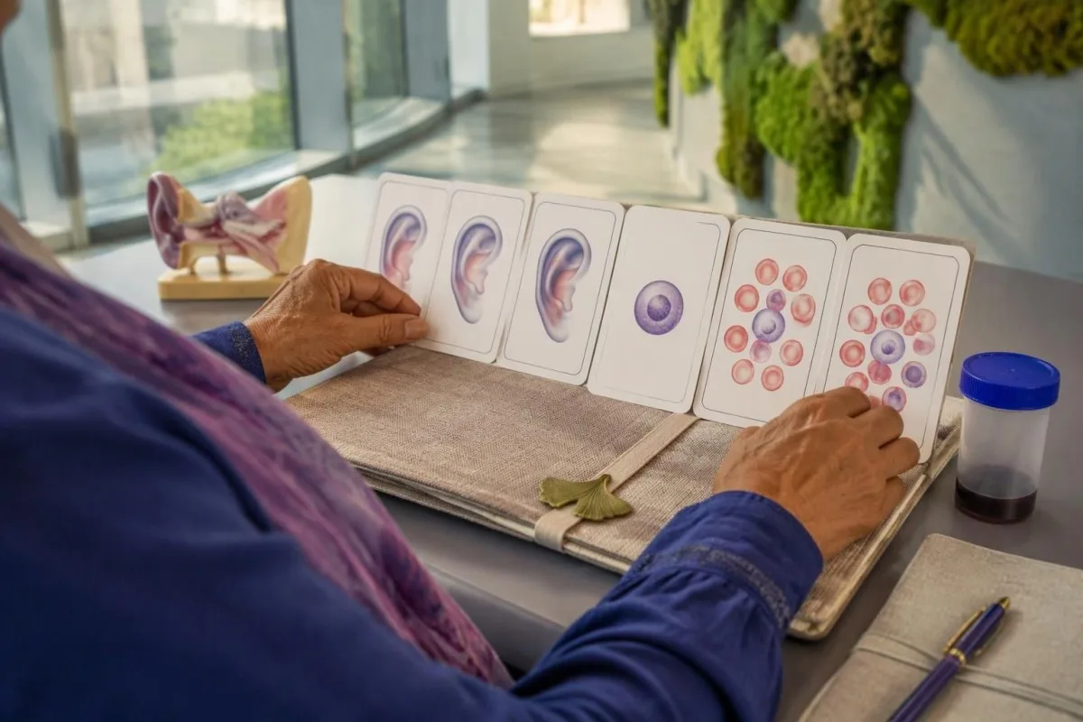
The first reasoning step is to separate symptom severity from diagnostic specificity: CRP of 86 mg/L confirms a substantial inflammatory response but does not identify its cause. The MCV of 108 fL and platelet count of 112 × 10^9/L add evidence that blood-cell production also needs investigation.
Suppose the person's B12, folate and thyroid assessments do not reveal an explanation, and medication, alcohol and liver reviews are unrevealing. A negative ANA would not close the case; the combination of 3 inflammatory episodes and persistent unexplained cytopenias would justify discussing somatic UBA1 testing with hematology and rheumatology.
Now suppose CRP falls to 12 mg/L after prescribed glucocorticoids, but hemoglobin falls to 9.8 g/dL. Those 2 trends are not contradictory: inflammatory activity may improve while marrow dysfunction or another anemia mechanism persists, so treatment response cannot be used as a substitute for investigating the falling hemoglobin.
Kantesti can help explain why those 2 trends deserve separate attention, but cannot establish VEXAS or choose immunosuppressive treatment. Our ඖෂධ ප්රවණතා විශ්ලේෂණ මාර්ගෝපදේශය explains why dose changes, transfusions and intercurrent illness should be recorded beside each result; the example here is entirely hypothetical.
තහවුරු කළ VEXAS සින්ඩ්රෝමයට විශේෂඥයන් ප්රතිකාර කරන්නේ කෙසේද?
VEXAS treatment aims to control inflammation, manage cytopenias and reduce treatment-related harm. Glucocorticoids often suppress symptoms but may be difficult to taper; other options include selected immunomodulators, marrow-directed treatment and allogeneic stem-cell transplantation, with no single regimen appropriate for every adult over 50.
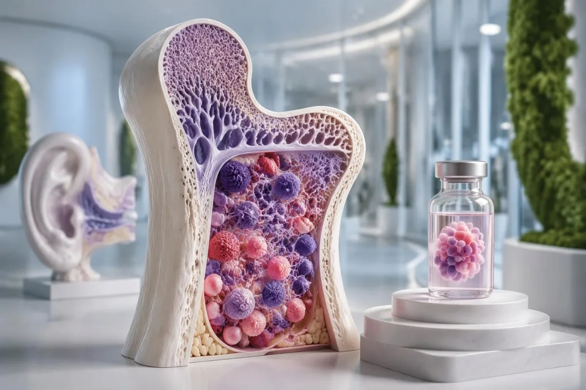
A falling CRP does not by itself establish treatment success if anemia, thrombosis or lung disease is worsening. Specialists follow at least 2 dimensions—clinical inflammatory activity and hematological status—and weigh these against cumulative glucocorticoid exposure, because prolonged treatment can cause infections, diabetes, osteoporosis and muscle weakness.
JAK inhibitors and IL-6-directed therapy are among approaches used by specialists, while azacitidine may be considered in selected patients, particularly with associated marrow disease. Evidence and access vary, and much published experience is observational; a response lasting 6 months in one cohort does not reliably predict an individual's longer-term benefit.
Allogeneic stem-cell transplantation can potentially eliminate the abnormal blood-forming clone and produce sustained remission in selected patients. It also carries substantial risks, including graft-versus-host disease and serious infection, so 2 people of the same age may receive different recommendations because fitness, organ involvement and donor availability differ.
Infection prevention deserves planning before immunosuppression, not only after fever develops. Depending on exposures and the proposed regimen, clinicians may consider screening or prevention measures described in our Strongyloides testing explanation; even 1 relevant past exposure can matter before corticosteroids, and prescribed steroids should never be stopped abruptly without guidance.
කුමන රෝග ලක්ෂණ සඳහා හදිසි හෝ ඉතා හදිසි වෛද්ය ප්රතිකාර අවශ්යද?
New breathing difficulty, chest pain, airway symptoms, neurological deficits or significant bleeding require urgent assessment in someone with suspected or confirmed VEXAS. Fever of 38°C or higher during substantial immunosuppression also needs prompt clinical advice, particularly when neutrophils are low.
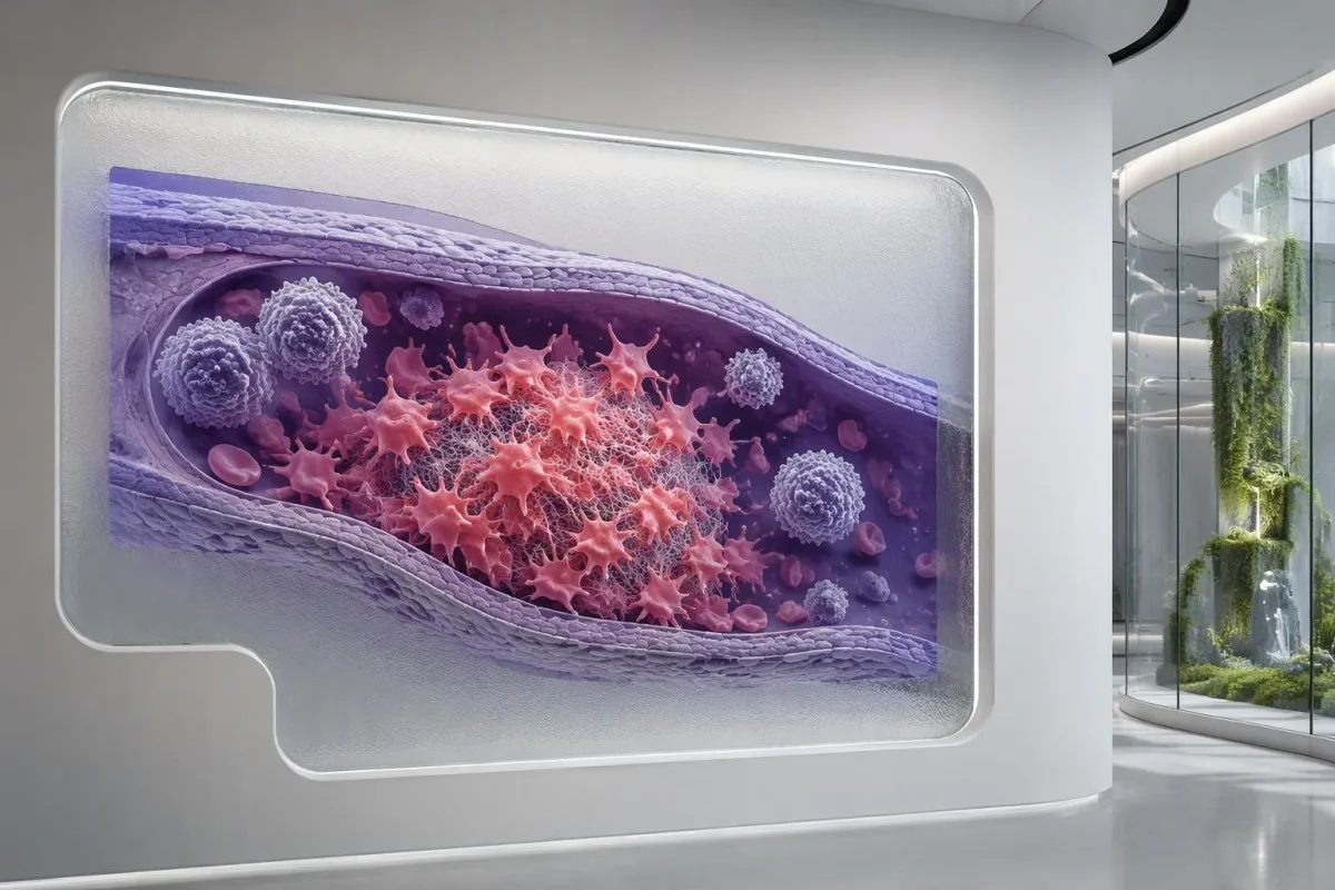
VEXAS is associated with venous thrombosis, so a painful swollen leg or sudden breathlessness should not be dismissed as another inflammatory flare. Normal routine clotting times do not exclude a clot; our කැටිගැසීමේ පරීක්ෂණ පැනලයේ සීමාවන් explain why 2 normal tests, PT and aPTT, cannot provide that reassurance.
An absolute neutrophil count below 0.5 × 10^9/L represents severe neutropenia and increases concern when fever occurs. Treatment can also blunt the temperature response, so feeling acutely unwell with shaking chills, confusion or rapidly worsening weakness can require urgent assessment even when a thermometer reads below 38°C.
Breathlessness can reflect lung inflammation, infection, pulmonary embolism or worsening anemia, and these require different treatments. Our breathlessness blood test guide explains why hemoglobin of 8 g/dL may contribute without proving the entire cause; new chest pain or sudden breathing difficulty should not wait for an uploaded report.
Persistent nose or gum bleeding, black stools or a severe new headache needs urgent advice, especially if platelets are very low. A count below 20 × 10^9/L generally warrants prompt clinical review, but the urgency depends on symptoms and context; significant bleeding is an emergency even above that number.
පසු විපරම, AI සීමාවන් සහ පර්යේෂණ ප්රකාශන
Useful VEXAS follow-up combines symptoms, treatment history, CBC trends and inflammatory markers, with molecular and marrow findings interpreted by specialists. An AI explanation of 1 laboratory report can support preparation for care, but it cannot diagnose VEXAS, exclude infection or establish whether a UBA1 variant is pathogenic.

Bring original CBC reports, inflammatory markers and the complete UBA1 result, including specimen type and detection limit. Record 3 timeline features beside them: flare dates, medication changes and transfusions; this makes it easier to distinguish a true disease trend from a treatment effect or a change in testing conditions.
කන්ටෙස්ටි යනු AI මගින් බලගැන්වූ රුධිර පරීක්ෂණ විශ්ලේෂණ මෙවලම that can explain laboratory patterns without converting an MCV above 100 fL into a rare-disease diagnosis. Genetic reports can reveal sensitive information, so review our පෞද්ගලිකත්වය සහ දත්ත පිළිබඳ තොරතුරු before sharing records, and include only documents you are authorized to upload.
Kantesti's 2 DOI-linked publications below concern broader blood-test interpretation and RDW education, not clinical trials of VEXAS diagnosis or UBA1 sequencing. They should therefore be read separately from the disease-specific medical studies listed in the external references; neither publication establishes VEXAS-specific diagnostic sensitivity or treatment effectiveness.
අපගේ ක්රමවේදය සහ සායනික ප්රමිතීන් explain the distinction between report interpretation and medical diagnosis. Before the next appointment, write 2 questions: whether the laboratory method adequately assessed somatic UBA1 variants, and whether persistent cytopenias need marrow evaluation; these are more actionable than asking an AI to calculate an unsupported VEXAS probability.
නිතර අසනු ලබන ප්රශ්න
VEXAS සහලක්ෂණයේ පළමු රෝග ලක්ෂණ මොනවාද?
VEXAS සින්ඩ්රෝමය බොහෝ විට ආරම්භ වන්නේ නැවත නැවත ඇතිවන, හේතුව පැහැදිලි නොවන උණ, සමේ ප්රදාහමය වෙනස්කම්, තෙහෙට්ටුව හෝ කනේ හෝ නාසයේ කාටිලේජ ඉදිමීම සමඟය; සාමාන්යයෙන් වයස අවුරුදු 50න් පසුවය. මෙම ලක්ෂණවලට පෙර, ඒවා සමඟ හෝ පසුව මැක්රෝසයිටික් රක්තහීනතාව හෝ වෙනත් අසාමාන්ය රුධිර ගණනයන් මතුවිය හැකිය. වැඩිහිටියෙකුගේ MCV අගය ආසන්න වශයෙන් 100 fL ඉක්මවා තිබීම රෝග විනිශ්චයක් නොව, ඒ පිළිබඳ ඉඟියකි. නැවත නැවත ඇතිවන ප්රදාහමය ලක්ෂණ සමඟ අඛණ්ඩව පවතින, හේතුව පැහැදිලි නොවන සයිටෝපීනියා තිබේ නම් රූමැටොලොජි සහ රක්තවේද ඇගයීමක් ගැන සාකච්ඡා කිරීම සුදුසුය.
VEXAS සින්ඩ්රෝමය තහවුරු කරන පරීක්ෂණය කුමක්ද?
VEXAS සින්ඩ්රෝමය හඳුනාගනු ලබන්නේ ගැළපෙන සායනික පසුබිමකදී රෝගයට සම්බන්ධ සොමැටික් UBA1 ප්රභේදයක් හඳුනාගැනීමෙනි. පරීක්ෂණ සඳහා සාමාන්යයෙන් පර්යන්ත රුධිරය භාවිත කළද, තෝරාගත් අවස්ථාවලදී අස්ථි मज්ජාව හෝ වඩාත් සංවේදී පරීක්ෂණයක් අවශ්ය විය හැකිය. හඳුනාගෙන ඇති බොහෝ ප්රභේද 41 වන ස්ථානයේ ඇති මෙතියොනීන්ට බලපාන නමුත්, සීමිත හොට්ස්පොට් පරීක්ෂණවලින් රෝගයට සම්බන්ධ වෙනත් ප්රභේද මඟහැරිය හැකිය. වාර්තාව එහි ප්රභේද වර්ගීකරණය, ජාන ආවරණය සහ හඳුනාගැනීමේ සීමාව සමඟ අර්ථකථනය කළ යුතුය.
සාමාන්ය ANA පරීක්ෂණයකින් හෝ ස්වයංප්රතිශක්තිකරණ පරීක්ෂණ මණ්ඩලයකින් VEXAS බැහැර කළ හැකිද?
සාමාන්ය ANA පරීක්ෂණයක් හෝ සාමාන්ය ස්වයංප්රතිශක්තික පරීක්ෂණ පැනලයක් මඟින් VEXAS සින්ඩ්රෝමය බැහැර කළ නොහැක, මන්ද මෙම පරීක්ෂණ මඟින් දේහසෛලීය UBA1 විකෘති හඳුනා නොගනී. 1:160 මට්ටමේ ධනාත්මක ANA ප්රතිඵලයක් ද VEXAS තහවුරු නොකරන අතර වෙනත් ස්වයංප්රතිශක්තික රෝගයක් ඇති බව අනිවාර්යයෙන්ම තහවුරු නොකරයි. විකල්ප හේතු විමර්ශනය කිරීමට ප්රතිදේහ පරීක්ෂණ උපකාරී විය හැකි අතර, CBC රටා, රෝග ලක්ෂණ සහ අණුක පරීක්ෂණ මඟින් වෙන වෙනම ප්රශ්නවලට පිළිතුරු සපයයි. දිගටම පවතින මැක්රොසයිටික් රක්තහීනතාව සහ නැවත නැවත ඇතිවන කාටිලේජ ප්රදාහය, ප්රතිදේහ පරීක්ෂණ ඍණාත්මක වූ පමණින් නොසලකා හැරිය යුතු නොවේ.
VEXAS සින්ඩ්රෝමය පරම්පරාවෙන් උරුම වනවාද, සහ මගේ දරුවන් පරීක්ෂා කළ යුතුද?
සාමාන්යයෙන් VEXAS සින්ඩ්රෝමය උරුම වූවක් නොව අත්පත් කරගත් තත්ත්වයකි; එබැවින් දරුවන් සාමාන්යයෙන් පරීක්ෂා කිරීම අවශ්ය නොවේ. අදාළ UBA1 විකෘතිය සාමාන්යයෙන් වැඩිහිටි වියේදී රුධිර සෛල නිපදවන සෛල තුළ වර්ධනය වන අතර, රෝග ලක්ෂණ බොහෝවිට වයස අවුරුදු 50 ඉක්මවූ පසු මතු වේ. UBA1 X වර්ණදේහයේ පිහිටා තිබුණද, එයින් සෝමැටික් VEXAS ප්රභේදයක් ස්වයංක්රීයව සම්ප්රේෂණය වන බවක් අදහස් නොවේ. අසාමාන්ය හෝ ව්යවස්ථාපිත විය හැකි සොයාගැනීම් සායනික ජාන විද්යා විශේෂඥයකු විසින් සමාලෝචනය කළ යුතුය.
කාන්තාවන්ට VEXAS සින්ඩ්රෝමය ඇති විය හැකිද?
කාන්තාවන්ට VEXAS සින්ඩ්රෝමය වර්ධනය විය හැකි නමුත්, හඳුනාගෙන ඇති බොහෝ සිද්ධි වයස්ගත පිරිමින් අතර දක්නට ලැබේ. UBA1 X වර්ණදේහයේ පිහිටා ඇති අතර, බලපෑමට ලක් වූ සෛලවල X වර්ණදේහ 1ක් අහිමි වීම කාන්තාවන් තුළ වාර්තා වී ඇති එක් යාන්ත්රණයකි. 2023 JAMA සෞඛ්ය පද්ධති අධ්යයනයකදී රෝගය හා සම්බන්ධ UBA1 ප්රභේද ඇති සහභාගිවන්නන් 11 දෙනා අතර කාන්තාවන් 2 දෙනෙකු හඳුනා ගන්නා ලදී. නැවත නැවත ඇතිවන ප්රදාහය සහ පැහැදිලි කළ නොහැකි රුධිර ගණන්වල අසාමාන්යතා රෝගය තදින්ම සැක කිරීමට හේතු වන විට, ස්ත්රී පුරුෂ භාවය පමණක් විශේෂඥ ඇගයීම වැළැක්වීමට හේතු නොවිය යුතුය.
UBA1 පරීක්ෂණ ප්රතිඵලය ඍණ වීමෙන් VEXAS සින්ඩ්රෝමය බැහැර වේද?
ඍණ UBA1 පරීක්ෂණයක් VEXAS ඇතිවීමේ සම්භාවිතාව අඩු කරන්නේ, නියැදිය සහ පරීක්ෂණ ක්රමය යෝග්ය වූ තරමට පමණි. 5% ප්රභේද ඇලීල භාග හඳුනාගැනීමේ සීමාවක් සහිත පරීක්ෂණයකින් කුඩා ක්ලෝනයක් හඳුනාගැනීමට නොහැකි විය හැකි අතර, හොට්ස්පොට් පමණක් පරීක්ෂා කිරීමෙන් ආවරණය වන කලාපයෙන් පිටත ප්රභේද මඟහැරිය හැකිය. ප්රබල සායනික සැකයක් පවතින්නේ නම් ආවරණය, සංවේදීතාව සහ නියැදි වර්ගය පිළිබඳ විශේෂඥ සමාලෝචනයක් සාධාරණීකරණය කළ හැකිය. නිසි ලෙස සිදු කළ ඍණ ප්රතිඵලයක්, පැහැදිලි හේතුවක් නොමැතිව නැවත නැවත පරීක්ෂා කිරීම වෙනුවට, විකල්ප රෝග විනිශ්චයන් ප්රවේශමෙන් නැවත තක්සේරු කිරීමට ද හේතු විය යුතුය.
VEXAS සින්ඩ්රෝමය සුව කළ හැකි ප්රතිකාරයක් තිබේද?
අසාමාන්ය රුධිර සෛල නිපදවන සෛල ක්ලෝනය ප්රතිස්ථාපනය කිරීමෙන් සමජාතීය ප්රාථමික සෛල බද්ධ කිරීමෙන් දිගුකාලීන සුවය ලබාගත හැකි නමුත්, එය සුදුසු වන්නේ තෝරාගත් රෝගීන්ට පමණක් වන අතර සැලකිය යුතු අවදානම් දරයි. අනෙකුත් ප්රතිකාරවල අරමුණ වන්නේ ක්ලෝනය විශ්වාසදායක ලෙස තුරන් කිරීමට වඩා, ප්රදාහය පාලනය කිරීම, රුධිර සෛල හිඟතා කළමනාකරණය කිරීම සහ ග්ලූකෝකෝටිකොයිඩ් භාවිතය අඩු කිරීමයි. වයස අවුරුදු 50ට වැඩි වැඩිහිටියන් සම්බන්ධයෙන්, ශාරීරික යෝග්යතාව, අවයවවලට ඇති බලපෑම සහ ඒ ආශ්රිත ඇටමිදුළු රෝග ප්රතිකාර තේරීම් කෙරෙහි දැඩි ලෙස බලපෑ හැකිය. කිසිදු අතිරේකයක්, ආහාර සැලැස්මක් හෝ AI මගින් රුධිර පරීක්ෂණ අර්ථකථනය කිරීමක් VEXAS සුව කරන බව පෙන්වා දී නැත.
ඔබේ රුධිර පරීක්ෂණ ප්රතිඵල කියවා ගන්න
ඔබේ රසායනාගාර වාර්තාව උඩුගත කරන්න; Kantesti හි AI තත්පර කිහිපයකින් එක් එක් ජෛව සලකුණ සරල භාෂාවෙන් පැහැදිලි කරයි. ලොව පුරා මිලියන 2කට අධික පිරිසක් භාවිත කරති.
ආශ්රිත
- ක්ලයින්, ටී., මිචෙල්, එස්., සහ වෙබර්, එච්. (2026). AI රුධිර පරීක්ෂණ විශ්ලේෂකය: 2.5M පරීක්ෂණ විශ්ලේෂණය කරන ලදී | ගෝලීය සෞඛ්ය වාර්තාව 2026. Kantesti AI වෛද්ය පර්යේෂණය. ඩීඕඅයි
- ක්ලයින්, ටී., මිචෙල්, එස්., සහ වෙබර්, එච්. (2026). RDW රුධිර පරීක්ෂණය: RDW-CV, MCV සහ MCHC පිළිබඳ සම්පූර්ණ මාර්ගෝපදේශය. Kantesti AI වෛද්ය පර්යේෂණය. ඩීඕඅයි
- Beck DB et al. (2020). Somatic Mutations in UBA1 and Severe Adult-Onset Autoinflammatory Disease. New England Journal of Medicine. ඩීඕඅයි
- Beck DB et al. (2023). Estimated Prevalence and Clinical Manifestations of VEXAS Syndrome in a Large US Health Care System. JAMA. ඩීඕඅයි
- මි2+විශ්ලේෂණය කළ පරීක්ෂණ
- 127+රටවල්
- 100+භාෂා
වෛද්ය කණ්ඩායම පිළිබඳව


E-E-A-T විශ්වාස සංඥා
අත්දැකීම්
වෛද්යවරයා විසින් මෙහෙයවන ලද රසායනාගාර අර්ථකථන ක්රියාවලි පිළිබඳ සමාලෝචනය.
ප්රවීණතාව
සායනික සන්දර්භය තුළ ජෛව සලකුණු (biomarkers) හැසිරෙන ආකාරය පිළිබඳ රසායනාගාර වෛද්ය විද්යා අවධානය.
අධිකාරීත්වය
වෛද්ය Thomas Klein විසින් වෛද්යමය වශයෙන් සමාලෝචනය කරන ලද අතර, Dr. Sarah Mitchell සහ Prof. Dr. Hans Weber දායක වූහ.
විශ්වසනීයත්වය
අනතුරු ඇඟවීම් අඩු කිරීමට පැහැදිලි පසුකැඳවීම් මාර්ග සහිත සාක්ෂි-පාදක අර්ථකථනය.
වෛද්ය වියාචනය
මෙම ලිපිය අධ්යාපනික අරමුණු සඳහා පමණක් වන අතර වෛද්ය උපදෙස් ලෙස නොසැලකේ. රෝග නිદાન සහ ප්රතිකාර තීරණ සඳහා සෑම විටම සුදුසුකම් ලත් සෞඛ්ය සේවා සපයන්නෙකුගෙන් උපදෙස් ලබාගන්න.
ප්රකාශයට පත් කරන ලදී · කර්තෘ මණ්ඩලය අමතන්න
Kantesti Ltd විසින් ප්රකාශයට පත් කරන ලද අතර, එංගලන්තයේ සහ වේල්සයේ ලියාපදිංචි සමාගමකි, සමාගම් අංකය. 17090423, ලන්ඩන්, එක්සත් රාජධානිය.
