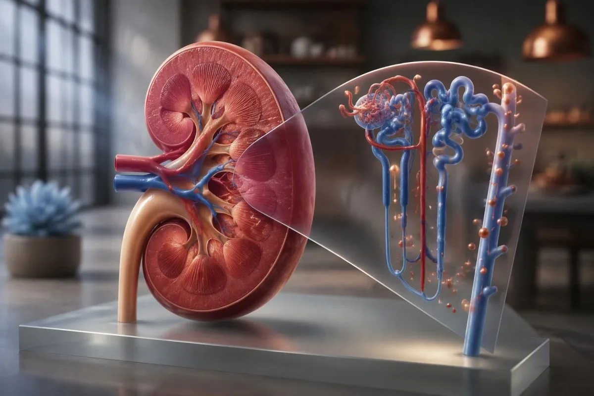
Renal tubular acidosis usually causes a normal-anion-gap, high-chloride metabolic acidosis. The decisive clues are potassium, urine pH, urine ammonium surrogates and whether kidney function is otherwise preserved.


- Core pattern: RTA usually produces bicarbonate below 22 mmol/L, chloride elevation, and a normal serum anion gap.
- Urine pH: In untreated metabolic acidosis, urine pH persistently above 5.5 supports distal (type 1) RTA.
- Potassium clue: Type 1 and type 2 RTA often lower potassium; type 4 RTA usually raises it above 5.0 mmol/L.
- Urine anion gap: A positive urine anion gap during acidosis suggests inadequate ammonium excretion; a negative value favors diarrhea.
- Type 2 test: Fractional bicarbonate excretion above 15% during bicarbonate loading supports proximal RTA.
- Type 4 priority: Hyperkalemia with low bicarbonate needs medication, diabetes, adrenal and kidney-function review promptly.
- Do not diagnose from CO2 alone: A chemistry-panel CO2 result is a bicarbonate estimate and should be confirmed with a blood gas when the clinical picture is unclear.
- Urgent threshold: Bicarbonate below 15 mmol/L, potassium at or above 6.0 mmol/L, confusion, arrhythmia symptoms or rapid breathing merits urgent assessment.
Start With the Core Acid-Base Pattern
Renal tubular acidosis labs typically show low bicarbonate, high chloride and a normal anion gap despite relatively preserved filtration. The first practical question is whether the low chemistry-panel CO2 truly represents metabolic acidosis, rather than a sample delay, respiratory alkalosis, or a mixed disorder.
On a standard metabolic panel, total CO2 is usually a close surrogate for bicarbonate; most adult laboratories report roughly 22-29 mmol/L as the reference interval. A value of 18 mmol/L with chloride of 112 mmol/L is a more meaningful pattern than either value alone. Review basic metabolic panel kidney clues before assuming a rare tubular diagnosis.
Calculate the serum anion gap as sodium minus chloride minus bicarbonate: Na − (Cl + HCO3−). With sodium 140, chloride 112 and bicarbonate 18 mmol/L, the gap is 10 mmol/L, which fits hyperchloremic or normal-gap acidosis. If albumin is low, add about 2.5 mmol/L to the gap for every 1 g/dL albumin below 4.0 g/dL.
Kantesti is an AI blood test analyzer that reads bicarbonate, chloride, potassium, creatinine and albumin together rather than flagging one isolated result. In my experience, a sudden CO2 fall from 25 to 18 mmol/L deserves a repeat sample and medication review even when eGFR looks reassuring. Thomas Klein, MD, would not label this RTA without that confirmation.
Confirm acidosis before classifying it
A venous blood gas with pH below 7.35 and reduced calculated bicarbonate confirms metabolic acidosis. A low bicarbonate with a low PaCO2 can also reflect chronic respiratory alkalosis, so blood-gas context prevents an avoidable diagnostic detour.
Separate RTA From Other Low Bicarbonate Causes
Diarrhea is the commonest mimic of RTA because both cause normal-gap metabolic acidosis. The kidney should increase ammonium excretion during gastrointestinal bicarbonate loss; it cannot do that adequately in most forms of RTA.
A negative urine anion gap, usually below −20 mmol/L, suggests high urinary ammonium and therefore an appropriate renal response to diarrhea. A positive result, often above 0 mmol/L, supports impaired ammonium excretion and raises RTA or advanced chronic kidney disease. Kraut and Madias describe this systematic distinction in CJASN (Kraut and Madias, 2012).
Do not overread the urine anion gap. It becomes unreliable with urine sodium below 25 mmol/L, unusual ammonium salts, ketoacid anions, toluene exposure, or recent diuretics. Urine osmolal gap can estimate ammonium more directly, although it is technically less tidy and varies by laboratory method.
Chronic kidney disease itself can lower bicarbonate once eGFR falls, particularly below 30 mL/min/1.73 m², without being classic RTA. Compare creatinine, cystatin C when available, and albuminuria; our guide to chronic kidney disease stages explains why one eGFR value needs context.
Use Potassium to Narrow the RTA Type
Low potassium points toward distal or proximal RTA, while high potassium strongly favors type 4 RTA. Potassium is not merely an accompanying abnormality: it reflects the transport defect and determines immediate safety.
Type 1 distal RTA often produces potassium below 3.5 mmol/L, nephrolithiasis, and urine that remains inappropriately alkaline. Type 2 proximal RTA can also cause hypokalemia, especially after alkali treatment increases distal sodium delivery. Severe potassium depletion may cause weakness, constipation or palpitations before acid-base symptoms are obvious.
Type 4 RTA usually presents with potassium above 5.0 mmol/L and bicarbonate commonly 17-22 mmol/L, rather than the very low bicarbonate sometimes seen in untreated distal disease. Diabetes, reduced renin production, adrenal insufficiency, trimethoprim, ACE inhibitors, ARBs, NSAIDs and potassium-sparing diuretics are frequent contributors. See slightly elevated potassium guidance for urgent thresholds.
Potassium at 6.0 mmol/L or higher, new muscle weakness, fainting, chest discomfort, or an abnormal ECG needs urgent medical evaluation. A hemolyzed sample can falsely elevate potassium, but it should never be casually dismissed; repeat testing and ECG timing matter, as explained in potassium draw-error review.
Recognize Distal Type 1 RTA on Labs
Distal type 1 RTA causes normal-gap acidosis with urine pH persistently above 5.5, often low potassium, and a positive urine anion gap. The distal nephron cannot lower urine pH adequately despite systemic acidemia.
The useful bedside pattern is bicarbonate often below 20 mmol/L, potassium below 3.5 mmol/L, and urine pH above 5.5 during confirmed acidosis. Urine pH alone is not diagnostic: a urinary tract infection with urease-producing organisms, recent vomiting, or an alkaline meal can also raise it. A fresh specimen and urine culture sometimes settle the ambiguity.
Calcium phosphate stones and nephrocalcinosis occur because alkaline urine reduces citrate availability and favors calcium phosphate precipitation. A 24-hour urine evaluation usually includes citrate, calcium, oxalate, sodium, volume and pH; calcium oxalate crystal results can add useful context, although crystal type does not establish RTA.
Acquired distal RTA should prompt a careful review for autoimmune disease, particularly Sjögren syndrome, lupus, chronic tubulointerstitial disease and medications such as amphotericin B. Dry eyes plus dry mouth plus this acid-base pattern is a worthwhile clinical lead; see our review of Sjögren testing clues.
Recognize Proximal Type 2 RTA on Labs
Proximal type 2 RTA begins with bicarbonate wasting, but urine pH can fall below 5.5 after serum bicarbonate reaches a new lower steady state. This changing urine pH is why one random specimen can mislead clinicians.
When plasma bicarbonate is above the reduced renal threshold, urine bicarbonate loss can drive urine pH above 5.5. Once serum bicarbonate falls to roughly 12-18 mmol/L, the distal nephron can still acidify urine and pH often drops below 5.5. That contrasts with distal RTA, where acidification remains defective.
A fractional excretion of bicarbonate greater than 15% during bicarbonate loading supports proximal RTA; values below 5% make it less likely. This specialist renal tubular acidosis test is not routinely needed in straightforward cases, but it is useful when urine pH and clinical history conflict.
Think beyond bicarbonate. Fanconi syndrome causes proximal wasting of phosphate, glucose despite normal serum glucose, urate, amino acids and low-molecular-weight proteins. New glycosuria with normal glucose should trigger review of glucose in urine causes and exposures such as tenofovir, ifosfamide, outdated tetracycline, or light-chain disease.
Recognize Type 4 RTA and Hypoaldosterone Patterns
Type 4 RTA is the most common adult RTA pattern and combines mild normal-gap acidosis with hyperkalemia from low aldosterone activity or aldosterone resistance. Urine pH is often below 5.5 because the main defect is reduced ammonium generation, not complete failure of distal acidification.
Typical RTA blood test results show potassium 5.1-6.0 mmol/L, bicarbonate 17-22 mmol/L, and chloride elevation with a normal anion gap. A positive urine anion gap can be present because ammonium excretion is low. In contrast, urine pH may be appropriately acidic, often below 5.5.
Diabetic kidney disease is a frequent setting because hyporeninemic hypoaldosteronism reduces distal sodium-driven potassium secretion. Renin and aldosterone testing can help, but posture, sodium intake, time of day and medicines alter both results substantially; renin result patterns are not interpretable in a vacuum.
Kantesti is an AI-powered blood test analysis tool that can surface the combination of high potassium, low CO2, diabetes markers and kidney function for clinician follow-up. It cannot determine whether a prescribed RAAS blocker should be stopped; that decision depends on blood pressure, albuminuria, ECG findings and the reason the medicine was prescribed.
Get Urine pH and Urine Anion Gap Right
Fresh urine collected during untreated metabolic acidosis gives the most useful RTA evidence. Delayed analysis allows carbon dioxide loss and bacterial metabolism to alter urine pH, occasionally changing an apparent type 1 pattern.
Urine pH from a calibrated electrode is more accurate than a dipstick near the clinically important 5.3-5.5 range. Dipsticks are convenient but commonly read in increments of 0.5 to 1.0 pH unit. A clinician evaluating suspected distal RTA should request prompt analysis rather than relying on a specimen that sat at room temperature.
The urine anion gap is urine sodium plus urine potassium minus urine chloride. A value of +25 mmol/L during acidosis implies low ammonium excretion only if urine sodium is adequate and there are no unmeasured urinary anions. Our urine pH testing guide covers common non-RTA reasons for alkaline urine.
Urine osmolal gap may be preferable when the urine anion gap is distorted. A urine osmolal gap above roughly 200 mOsm/kg generally indicates substantial ammonium excretion, making diarrhea more likely than RTA, though the calculation is an estimate and assay availability varies.
Do Not Miss a Mixed Acid-Base Disorder
A normal anion gap does not exclude a concurrent high-gap acidosis. Mixed disorders are especially likely when bicarbonate is below 15 mmol/L, lactate is elevated, ketones are present, or kidney function has worsened abruptly.
Correct the anion gap for albumin before deciding it is normal. Albumin of 2.0 g/dL lowers the expected gap by about 5 mmol/L, so an uncorrected gap of 12 mmol/L may actually be abnormal. This is a common miss in hospitalized patients with inflammation, malnutrition or liver disease.
A delta gap calculation is mainly helpful when the corrected anion gap is elevated: compare the gap rise above 12 with the bicarbonate fall below 24. It is less reliable in low-albumin states and after saline resuscitation. Lactate collection errors matter because a falsely high lactate can create a false mixed-disorder narrative.
I have seen patients with vomiting, saline infusion and early ketoacidosis show almost textbook hyperchloremia while still having a dangerous high-gap process. Check beta-hydroxybutyrate, lactate, glucose, renal function and medication exposure rather than treating the anion gap as a final verdict.
Review Medicines and Exposures Before Labeling RTA
Medication review is part of the renal tubular acidosis test because several common drugs reproduce a tubular pattern. The timing of a bicarbonate or potassium change often offers more diagnostic value than a one-off specialist assay.
Topiramate and acetazolamide inhibit carbonic anhydrase and can cause a proximal or mixed RTA phenotype with low bicarbonate. Trimethoprim, heparin, calcineurin inhibitors, ACE inhibitors, ARBs and spironolactone can contribute to type 4 physiology. Bicarbonate often changes within days to weeks of starting or escalating the relevant drug.
Toluene exposure may produce hypokalemic acidosis with variable anion-gap behavior because hippurate is excreted in urine. If the history includes solvents, chronic inhalant exposure or unexplained severe hypokalemia, toxicology input is appropriate. A urine anion gap can be deceptively negative in this setting.
Kantesti AI flags medication-sensitive electrolyte combinations for review, but it does not replace a pharmacist or prescribing clinician. Keep an exact list of prescription medicines, over-the-counter NSAIDs, herbal preparations and salt substitutes; medication safety trend analysis is most useful when dates are attached to results.
Look for Bone, Stone and Autoimmune Complications
Long-standing distal RTA can cause calcium phosphate stones, low urinary citrate and bone mineral loss. These complications may reveal the disorder before patients notice fatigue or muscle symptoms.
Chronic acidosis mobilizes bone buffer and increases renal calcium loss, while hypocitraturia removes a natural inhibitor of stone formation. Serum calcium can remain normal, so normal calcium does not rule out clinically meaningful skeletal effects. Check phosphate, alkaline phosphatase, 25-hydroxyvitamin D, PTH and a stone-focused urine profile when indicated.
A urine citrate below 320 mg/day is commonly considered low in adult stone assessment, although laboratories use different cutoffs and collection quality matters. Potassium citrate is often preferred over sodium bicarbonate in distal RTA with calcium stones because it provides alkali and citrate while avoiding sodium-driven calciuria. Treatment dose must be individualized, especially if kidney function is reduced.
Autoimmune screening is guided by symptoms rather than ordered indiscriminately. Dryness, parotid enlargement, rash, neuropathy, arthralgia or proteinuria may justify ANA, SSA/SSB, complements and specialist review; ANA result interpretation explains why a positive ANA alone is not a diagnosis.
Choose the Next Tests in a Sensible Order
The next tests after suspected RTA are repeat electrolytes, blood gas, urine pH, urine sodium-potassium-chloride, kidney assessment and a medication review. Specialists add targeted studies only after the basic pattern is verified.
Repeat a basic metabolic panel within days, or sooner if bicarbonate is below 18 mmol/L or potassium is abnormal. Pair it with venous blood gas, magnesium, phosphate, glucose, creatinine, albumin and urinalysis. Blood and urine ideally should be obtained before bicarbonate therapy, because alkali can temporarily change the diagnostic pattern.
For suspected distal RTA, ask about stones, obtain renal imaging when clinically appropriate, and consider urinary citrate. For proximal disease, measure urine glucose, phosphate, urate, protein type and fractional excretions. Proteinuria and casts may shift the concern from isolated tubular dysfunction to broader renal disease; granular cast findings should not be ignored.
As of September 16, 2026, Thomas Klein, MD, recommends using home-uploaded results as preparation for a clinical discussion, not as a diagnosis. Kantesti is an AI lab test interpretation service that organizes trends and flags questions for the clinician, while our medical validation approach explains its clinical oversight and limits.
Understand Treatment and Monitoring Targets
RTA treatment corrects the cause when possible and replaces alkali while monitoring potassium, kidney function and stone risk. The target is generally a normal or near-normal serum bicarbonate, not simply relief of fatigue.
Adults with distal RTA often need alkali equivalent to roughly 1-2 mEq/kg/day, while proximal RTA may require 5-15 mEq/kg/day because filtered bicarbonate continues to be lost. Potassium citrate can be useful in hypokalemic distal RTA with stones, but it can be unsafe in type 4 RTA with hyperkalemia. Prescribers adjust to serial results rather than a fixed internet dose.
KDIGO's 2024 CKD guidance advises considering pharmacologic or dietary treatment to prevent clinically important acidosis, while avoiding overtreatment that pushes bicarbonate above the laboratory upper limit (KDIGO, 2024). In practice, many clinicians aim for bicarbonate at least 22 mmol/L, but the optimal target varies with CKD, blood pressure, edema risk and sodium sensitivity.
Recheck electrolytes about 1-2 weeks after a meaningful alkali or medication change, sooner for high-risk potassium values. Patients with type 4 physiology need particular caution with potassium-containing citrate products; kidney supplement safety reviews why “natural” products can still alter electrolytes.
Know When Low Bicarbonate Needs Urgent Care
Low bicarbonate needs urgent assessment when it is severe, rapidly falling, or paired with dangerous potassium changes, altered breathing or neurological symptoms. RTA is usually treatable, but the lab pattern can also signal sepsis, ketoacidosis, toxin exposure or acute kidney injury.
Seek same-day urgent evaluation for bicarbonate below 15 mmol/L, potassium at or above 6.0 mmol/L, potassium below 2.5 mmol/L, new confusion, fainting, marked weakness, chest symptoms, or deep rapid breathing. These thresholds are not diagnoses, but they identify values that can affect cardiac and neuromuscular function quickly. Electrolyte-panel urgent signs provides a practical symptom checklist.
Children, pregnant people, older adults with frailty, and anyone with diabetes or advanced kidney disease may deteriorate with less laboratory reserve. Persistent vomiting, diarrhea, fever, poor intake or a new medication can convert compensated acidosis into a clinically significant problem over 24-48 hours.
Kantesti AI can help turn a multi-page report into questions for an appointment, but alarming symptoms should bypass digital interpretation and go directly to urgent clinical care. The Medical Advisory Board supports our emphasis on escalation rather than false reassurance.
What Research Supports—and What Labs Cannot Prove
No single renal tubular acidosis lab proves the diagnosis; clinicians diagnose RTA from a coherent blood, urine, medication and clinical pattern. The uncertainty is greatest when testing occurs after IV fluids, bicarbonate treatment, diuretics or an intercurrent illness.
Kraut and Madias' 2012 CJASN review remains useful because it emphasizes first separating high-gap from normal-gap acidosis, then assessing renal ammonium response. Karet's review of inherited distal RTA explains why early deafness, childhood stones or a family history can justify genetic evaluation rather than repeated nonspecific testing (Karet, 2002).
Kantesti is an AI biomarker interpretation platform used across multilingual lab-report formats, but it cannot measure urine ammonium, inspect urine microscopy, or replace nephrology assessment. Our AI technology guide describes how contextual pattern recognition differs from clinical diagnosis.
Research publication: Kantesti LTD. (2026). AI Blood Test Analyzer: 2.5M Tests Analyzed | Global Health Report 2026. Zenodo. https://doi.org/10.5281/zenodo.18175532. ResearchGate: https://www.researchgate.net/; Academia.edu: https://www.academia.edu/. Kantesti LTD. (2026). RDW Blood Test: Complete Guide to RDW-CV, MCV & MCHC. Zenodo. https://doi.org/10.5281/zenodo.18202598. ResearchGate: https://www.researchgate.net/; Academia.edu: https://www.academia.edu/.
Frequently Asked Questions
What lab pattern suggests renal tubular acidosis?
Renal tubular acidosis usually causes a normal-anion-gap metabolic acidosis with bicarbonate below 22 mmol/L and chloride elevation. Potassium then helps classify the pattern: low potassium favors type 1 or type 2 RTA, while potassium above 5.0 mmol/L favors type 4 RTA. A blood gas confirms true metabolic acidosis, because low chemistry-panel CO2 alone can have other explanations.
Can urine pH diagnose distal renal tubular acidosis?
Urine pH above 5.5 during confirmed systemic metabolic acidosis supports distal type 1 RTA, but it does not diagnose it by itself. A delayed specimen, alkaline diet, recent vomiting and urease-producing urinary infection can all raise urine pH. Clinicians interpret fresh urine pH alongside potassium, urine anion gap, blood bicarbonate and infection testing.
What does a positive urine anion gap mean in acidosis?
A positive urine anion gap during normal-gap metabolic acidosis suggests low urinary ammonium excretion and therefore an inadequate renal acid response. The calculation is urine sodium plus urine potassium minus urine chloride, with values above 0 mmol/L often supporting RTA. It is less reliable when urine sodium is below 25 mmol/L, diuretics are active, or unusual unmeasured urinary anions are present.
What is the difference between type 1 and type 2 RTA labs?
Type 1 distal RTA usually shows urine pH persistently above 5.5 during acidosis, low potassium and a tendency to calcium phosphate stones. Type 2 proximal RTA wastes bicarbonate early, but urine pH often falls below 5.5 once serum bicarbonate reaches approximately 12-18 mmol/L. Fractional bicarbonate excretion above 15% during bicarbonate loading supports proximal RTA.
Does type 4 RTA always mean kidney failure?
Type 4 RTA does not always mean kidney failure, although it commonly occurs with diabetic kidney disease or reduced kidney function. Its characteristic results are mild metabolic acidosis, often bicarbonate 17-22 mmol/L, plus hyperkalemia commonly above 5.0 mmol/L. Medicines that reduce aldosterone activity or potassium excretion can cause or worsen the pattern even with moderately preserved eGFR.
When should low bicarbonate be treated urgently?
Bicarbonate below 15 mmol/L, a rapidly declining value, or acidosis with potassium at or above 6.0 mmol/L warrants urgent clinical assessment. Deep rapid breathing, confusion, fainting, chest symptoms and severe weakness are urgent symptoms regardless of the presumed cause. RTA is only one possible explanation; ketoacidosis, lactic acidosis, toxin exposure and acute kidney injury need prompt exclusion.
Get AI-Powered Blood Test Analysis Today
Join over 2 million users worldwide who trust Kantesti for instant, accurate lab test analysis. Upload your blood test results and receive comprehensive interpretation of 15,000+ biomarkers in seconds.
📚 Referenced Research Publications
Klein, T., Mitchell, S., & Weber, H. (2026). AI Blood Test Analyzer: 2.5M Tests Analyzed | Global Health Report 2026. Kantesti AI Medical Research.
Klein, T., Mitchell, S., & Weber, H. (2026). RDW Blood Test: Complete Guide to RDW-CV, MCV & MCHC. Kantesti AI Medical Research.
📖 External Medical References
Karet FE (2002). Inherited distal renal tubular acidosis. Journal of the American Society of Nephrology.
⚕️ Medical Disclaimer
This article is for educational purposes only and does not constitute medical advice. Always consult a qualified healthcare provider for diagnosis and treatment decisions.
E-E-A-T Trust Signals
Experience
Physician-led clinical review of lab interpretation workflows.
Expertise
Laboratory medicine focus on how biomarkers behave in clinical context.
Authoritativeness
Written by Dr. Thomas Klein with review by Dr. Sarah Mitchell and Prof. Dr. Hans Weber.
Trustworthiness
Evidence-based interpretation with clear follow-up pathways to reduce alarm.