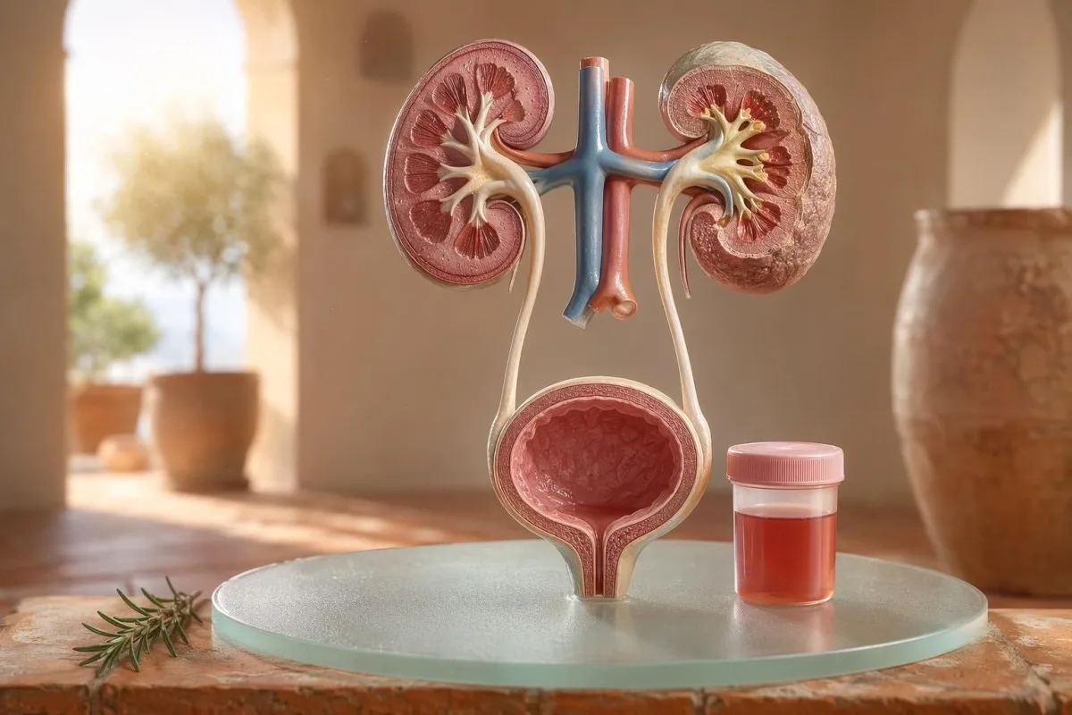
Sjúklingamiðaður leiðarvísir fyrir sýnilega og míkrósópíska blóðmigu, þar á meðal hvers vegna blóð á prófunarstrimli er ekki það sama og rauð blóðkorn í smásjá.


- Blóð í þvagi þýðir sýnilegt rautt, tebrúnt eða kóladökkt þvag, eða míkrósópíska blóðmigu sem finnst við rannsókn; sýnilegt blóð ávallt skilið eftir læknisfræðilegt eftirfylgni.
- Míkróhematuría er venjulega skilgreint sem 3 eða fleiri rauð blóðkorn á hverju sjónsviði með mikilli stækkun (high-power field) í rétt söfnuðu sýni fyrir þvag-míkrósjá.
- Blóð á prófunarstrimli greinir heme-virkni, þannig að það getur verið jákvætt vegna óskaddaðra rauðra blóðkorna, frjáls blóðrauða eða myoglobins eftir vöðvameiðsli.
- Ábendingar um UTI felur í sér sviða, tíðni, leukocyte esterase, nitrit og hvít blóðkorn; blóðmigu ætti að endurmeta þegar sýkingin er gengin yfir.
- Þvagsöðull niðurstöður eins og óeðlileg rauð blóðkorn (dysmorphic red cells), rauðkornabundnar þvagútfellingar (red cell casts) eða verulegt prótein benda til sjúkdóms í síueiningu nýrans.
- Þvagútfellingar (casts) eru mótað efni úr nýrnapíplum; rauðkornasteinar (red cell casts) eru merki um nýrnalækningavanda innan sömu viku, sérstaklega ef um er að ræða háan blóðþrýsting eða lækkandi eGFR.
- Viðvörunarmerki um krabbamein fela í sér sársaukalausa sjáanlega blóðmigu, blóðtappa, aldur yfir 45, reykingasögu, útsetningu fyrir efnum í starfi og endurtekin óútskýrð tilfelli.
- áreynslublóðmigu lagast venjulega innan 48-72 klukkustunda; viðvarandi blóð eftir hvíld ætti ekki að rekja til hlaupanna eða hjólreiða.
- Bráðamats er þörf er nauðsynlegt við blóði í þvagi með hita, verk í hlið (flank pain), blóðtappa sem valda stíflu í þvagfærum, þungun, áverka eða einkenni um alvarlega blóðleysi.
Hvað blóð í þvagi þýðir áður en þú lætur óttann taka yfir
Blóð í þvagi getur komið frá sýkingu, steinum, sjúkdómum sem hafa áhrif á síun í nýrum, sjúkdómum í blöðruhálskirtli eða þvagblöðru, lyfjum, áreynslu eða mengun frá tíðablæðingum. Hagnýta reglan er einföld: sjáanlegt blóð, endurtekin smásjárskoðunarbundin blóðmiga eða blóðmiga með verkjum, hita, blóðtappa, háum blóðþrýstingi, próteini eða skerta nýrnastarfsemi krefst tafarlauss mats frekar en að bíða og fylgjast með.
Ein bleikur litur í salernisskál eftir rauðrófur eða nýtt fæðubótarefni er ekki sama klíníska vandamálið og kólasvört (cola-lituð) þvag eftir hálsbólgu. Í minni vinnu segir litasagan mér oft fyrst hvert ég á að líta: skærrautt með blóðtöppum bendir til neðri þvagfæra, en brúnt eða te-litað þvag getur þýtt eldri heme-litarefni frá nýrnasíum.
Blóðmiga er ekki greining; hún er merki. Sjáanleg blóðmiga þýðir að þú sérð litabreytinguna, en smásjárskoðunarbundin blóðmiga þýðir að þvagið lítur eðlilega út en smásjáin finnur rauðkorn, venjulega við 3 eða fleiri rauðkorn á hverja high-power field.
Kantesti er AI blóðprufugreiningartæki sem hjálpar sjúklingum að tengja niðurstöður úr þvagi við nýrnamerki eins og kreatínín, eGFR, CRP, blóðrauða og blóðflögur í einni tímalínu. Ef liturinn er ruglingslegur, okkar leiðarvísir um lit þvags útskýrir hvers vegna rautt, brúnt, appelsínugult og dökkgult þvag þýðir ekki allt það sama.
Blóð á prófunarstrimli er ekki það sama og RBCs í smásjá
Þvagprófstrimill merktur blóð jákvætt greinir heme-líka efnafræðilega virkni, ekki endilega heilu rauð blóðkornin. Smásjáin svarar næstu spurningu: eru raunveruleg RBC í þvagsedimentinu og ef svo er, hvernig líta þau út?
Þvagprófstrimlaprófun er fljótleg vegna þess að hvarfefnissvæðið bregst við peroxídasa-virkni heme. Það þýðir að jákvæð niðurstaða getur komið fram með heilu RBC, frjálsu blóðrauða eftir niðurbrot rauðra blóðkorna, eða myóglóbíni eftir verulega vöðvameiðsli.
Smásjárskoðun er hægari en nákvæmari. Rannsóknarstofa sem vinnur af kostgæfni mun skilvinda þvag, skoða þvagsedimentið, og skrá rauðkorn á hverja high-power field; margir klínískir læknar nota 0-2 RBC/hpf sem eðlilegt og 3 eða fleiri RBC/hpf sem óeðlilegt hjá fullorðnum.
Mismunurinn skiptir máli. Þvagprófstrimill með 3+ blóði og 0-2 RBC/hpf fær mig til að spyrja um vöðvarýrnun (rhabdomyolysis), blóðlýsu (hemolysis), oxandi mengunarefni og meðhöndlun sýnis frekar en að stökkva beint í þvagblöðrusjúkdóm; munurinn er svipaður rökstuðningnum í okkar samanburður á þvagprófi og ræktun.
Þegar vísbendingar um UTI skýra blóðmigu og hvenær þær gera það ekki
Þvagfærasýking getur valdið blóði í þvagi, sérstaklega með sviða, bráðleika, tíðni, óþægindum í grindarholi, leukocyte esterase, nítrítum og hvítum blóðkornum. En blóðmigu ætti að hverfa eftir meðferð; viðvarandi rauð blóðkorn eftir þvagfærasýkingu þarf að skoða aftur.
Í einfaldri neðri þvagfærasýkingu býst ég við klasa: sviðaverk við þvaglát, tíðni, leukocyte esterase jákvætt, hvítfrumubólga (pyuria) oft yfir 5-10 hvítum blóðkornum/hpf og stundum nítrít. Hefðbundin ræktun jákvæðni er 100.000 CFU/mL, en hjá einkennakonum geta lægri gildi eins og 1.000-10.000 CFU/mL samt verið klínískt marktæk.
Leukocyte esterase er ensím úr hvítum blóðkornum, ekki sönnun um bakteríur. Jákvætt svar með blóðmigu getur komið fram vegna sýkingar, steina, mengunar eða bólgu í nýrum, þess vegna aðgreinir leukocyte esterase leiðarvísirinn okkar gagnlegar vísbendingar frá algengum fölskum jákvæðum.
Nítrít eru sértækari en minna næm vegna þess að ekki allar bakteríur breyta nítrati í nítrít. Ef einkennin eru dæmigerð en nítrít eru neikvæð getur ræktun samt verið jákvæð; hagnýtu smáatriðin eru fjallað í skýringunni okkar um nítrítniðurstöðu.
Sýnileg blóðmiga: litur, blóðtappa og tímasetningarvísbendingar
Sjónanleg blóðmigu er aldrei eitthvað sem ég hunsar, jafnvel þótt hún komi aðeins einu sinni. Bjart rautt þvag, blóðtappa eða blæðing án verkja þarf tímanlega mat, því að orsakir frá neðri þvagfærum verða líklegri eftir því sem aldur og reykingaáhrif aukast.
Tímasetningin getur verið óvænt gagnleg. Blóð aðeins í byrjun þvagláts getur bent til þvagrásar, blóð í gegnum allan þvagstrauminn getur komið frá þvagblöðru eða nýrnagjöf, og blóð í lokin nálægt lokum getur stundum bent til svæðis við háls þvagblöðru.
Blóðtappa þýðir venjulega að blæðingaruppspretta sé neðan við síur nýrna, því blóðtappar myndast ekki auðveldlega eftir að hafa farið í gegnum glomeruli og píplur. Blóðtappi sem hindrar þvagflæði er bráðamálefni: þensla þvagblöðru getur orðið sársaukafull innan nokkurra klukkustunda og gæti þurft leguþjónustu.
Karlar gera stundum ráð fyrir að sjáanleg blóðmiga eftir þvagseinkenni sé bara erting í blöðruhálskirtli. Það getur verið satt, en ef PSA var mælt meðan á sýkingu stóð eða stuttu eftir hana getur túlkun orðið flókin; okkar PSA eftir UTI leiðarvísir útskýrir hvers vegna margir klínískir læknar bíða 6–8 vikur áður en þeir endurtaka PSA.
Viðmiðunarmörk fyrir míkrósópíska blóðmigu sem læknar nota
Ósýnileg (ör)blóðmiga er almennt skilgreind sem 3 eða fleiri RBCs á hverju hástyrksstigi (high-power field) í réttilega safnaðri þvagsýni. AUA/SUFU leiðbeiningin notar þetta viðmið og mælir með áhættumiðaðri úrvinnslu frekar en að meðhöndla alla eins (Barocas o.fl., 2020).
Ekki ætti að nota stix (dipstick) einn og sér til að greina örblóðmigu. Í Barocas o.fl. 2020 AUA/SUFU leiðbeiningunni fer skilgreiningin eftir smásjá því stix getur verið jákvætt vegna heme-litarefna án þess að rauð blóðkorn séu til staðar.
Endurtekt er ekki veikleiki; það er góð læknisfræði. Ef einhver er með 3–10 RBC/hpf eftir mikla hreyfingu, hita, kynlíf, tíðablæðingar eða mengað safn, þá vil ég venjulega fá hreint endurtekið sýni áður en ég panta skannanir, alveg eins og við ráðleggjum fyrir leiðarvísir um endurteknar óeðlilegar rannsóknarniðurstöður.
Áhættubreyting breytir ferlinu. 24 ára einstaklingur með 4 RBC/hpf eftir hálfmaraþon er ekki það sama og 68 ára fyrrverandi reykingamaður með 25 RBC/hpf í tveimur sýnum, jafnvel þótt báðir uppfylli tæknilega skilgreininguna um blóðmigu.
Þvagsöðull, prótein og þvagútfellingar (casts) sem breyta rannsóknarferlinu
Þvagsöðull getur leitt í ljós hvort líklegt sé að blóðmiga komi frá síum nýrna. Óeðlileg rauð blóðkorn, rauðkornaþekjur (red cell casts) og marktæk próteinbreyting færa áhyggjur yfir í glomerular sjúkdóm frekar en einfalda sýkingu í þvagblöðru.
Rauð blóðkorn sem líta ójafn út, eru með blöðru-/blebmyndun eða hringlaga eru kölluð dysmorphic RBCs og benda til þess að þau hafi farið í gegnum bólgna glomerular síu. Acanthocytes yfir um það bil 5% af rauðum blóðkornum í þvagi eru oft meðhöndluð sem sterk glomerular vísbending, þó rannsóknarstofur séu mismunandi í því hversu örugglega þær greina þau.
Prótein er önnur vísbending sem ég hunsar ekki. Þvag-albúmín/kreatínín hlutfall yfir 30 mg/g, eða um 3 mg/mmol, er óeðlilegt og sjáanleg blóðmiga ásamt próteini á skilið hraðari yfirferð með áherslu á nýru; okkar prótein í þvagi leiðarvísir gefur viðmið á stigi sjúklings.
Þvagútfellingar (casts) eru örsmáir myglusveppir sem myndast inni í nýrnapíplum. Hyaline casts geta komið fram eftir ofþornun eða hreyfingu, en rauðkornaþekjur (red cell casts) eru óeðlilegar og benda venjulega til glomerulonephritis; fyrir ítarlegri viðmið í almennri þvaggreiningu, sjá okkar heildarleiðbeiningar um þvagrannsókn.
Steinar, hreyfing og tímabundnar orsakir blóðmigu
Steinar og erfið hreyfing geta valdið blóðmigu, en þær hafa mismunandi tímalínur. Blóðmiga vegna steina fylgir oft verk í hlið (flank pain) eða kristöllum, en blóðmiga vegna hreyfingar ætti venjulega að lagast innan 48–72 klukkustunda frá hvíld.
Nýrnassteinar valda klassískt bylgjum af verk í hlið, ógleði og ör- eða sjáanlegri blóðmigu. Kalsíumoxalatkristallar sanna ekki stein, en þeir auka grun þegar þeir fylgja kólíkuverkjum; okkar kalsíumoxalatkristalla leiðarvísir útskýrir smásjárvísbendingu í þvagi.
Blóðmiga vegna hreyfingar er raunveruleg. Ég hef séð maraþonhlaupara með 10–20 RBC/hpf morguninn eftir keppni, eðlilega ræktun, ekkert prótein og alveg eðlilega endurtekna þvaggreiningu 3 dögum síðar.
Hættan er að kenna öllu um þjálfun. Ef blóðmiga heldur áfram lengur en 72 klukkustundir, kemur fram með dökku kólasvipi þvagi, eða fylgir háu CK, hækkandi kreatíníni eða vöðvaverkjum, leitaðu lengra en í þvagblöðru; okkar æfingar leiðarvísir um kreatínín skarast á milli nýrna og vöðva.
Rauðir flaggar frá nýrum sem læknar hunsa ekki
Blóð í þvagi með próteini, hár blóðþrýstingur, bólga, skert eGFR, rauðkornaþekjur (red cell casts) eða hratt hækkandi kreatínín eru rauð flagg fyrir nýru. Þessar niðurstöður benda til þess að síueiningarnar geti verið bólgnar og bið í mánuði getur kostað nýrnastarfsemi.
KDIGO 2024 skilgreinir langvinna nýrnasjúkdóma út frá vísbendingum um skemmdir á nýrum, svo sem albúmínmigu, eða eGFR undir 60 mL/mín/1,73 m² í að minnsta kosti 3 mánuði. Í bráðum aðstæðum dugar hins vegar blóð í þvagi ásamt kreatínínhækkun á dögum eða vikum til að auka forgang áður en 3 mánaða skilgreiningin er uppfyllt.
Albúmínmiga er oft snemma viðvörunarmerki. ACR 30-300 mg/g er í meðallagi aukið, yfir 300 mg/g er alvarlega aukið og að para annaðhvort með blóði í þvagi breytir samtalinu; okkar leiðarvísir um nýrna-ACR útskýrir hvers vegna albúmín í þvagi finnur skemmdir áður en einkenni koma fram.
Blóðþrýstingur skiptir máli hér. Nýr BP 160/100 mmHg með kólasvörtu þvagi og bólgu í ökklum eftir hálsýkingu lætur mig hugsa um glomerulonephritis, ekki blöðrubólgu; ef eGFR er óvíst, okkar GFR endurathugunarleiðarvísir útskýrir hvenær cystatin C getur skýrt nýrnastarfsemi.
Ábendingar um krabbameinsáhættu án læti
Sársaukalaust sýnilegt blóð í þvagi, sérstaklega eftir 45 ára aldur, á skilið bráða úttekt vegna þess að krabbamein í þvagblöðru og efri þvagfærum getur komið fram á þennan hátt. NICE NG12 mælir með tilvísun vegna gruns um krabbamein við óútskýrðu sýnilegu blóði í þvagi hjá fullorðnum 45 ára og eldri, eða sýnilegu blóði í þvagi sem heldur áfram eftir meðferð við UTI.
Flest blóð í þvagi er ekki krabbamein. Samt eru tilvikin sem ég man rólegu: enginn verkur, enginn hiti, engin sviðatilfinning, bara tvö skipti af rauðu þvagi hjá 56 ára reykingamanni sem hefði nánast látið þetta fram hjá sér fara.
Áhættan safnast saman, ekki tvíund. Aldur yfir 45-50, reykingasaga, útsetning fyrir cyclophosphamide, grindargeislameðferð, endurtekið sýnilegt blóð í þvagi, atvinnuleg útsetning fyrir arómatískum amínum og blóðtappa allt eykur þörf fyrir blöðruspeglun eða myndgreiningu.
Blöðruhálskirtilsmerki útiloka ekki orsakir frá þvagblöðru. Ef þvagseinkenni, breytingar á PSA og blóð í þvagi skarast, metið tímasetningu og stöðu sýkingar vandlega; okkar leiðarvísir um blöðruhálskirtilsmarkara útskýrir hvers vegna PSA er aðeins einn hluti af mati á þvagfærum hjá körlum.
Lyf, blóðþynningarlyf og falskar viðvaranir
Blóðþynningarlyf geta sýnt blæðingu, en ætti ekki sjálfkrafa að kenna þeim um blóð í þvagi (hematuríu). Hematuría sem tengist segavarnarlyfjum þarf samt mat, því lyfið getur leitt í ljós steina, sýkingu, nýrnasjúkdóm eða vöxt í þvagfærum.
Warfarin, apixaban, rivaroxaban, aspirín og clopidogrel geta gert blæðingu augljósari. Ef INR er yfir markmiðsviðmiði skiptir máli að leiðrétta það, en eðlilegt INR gerir sýnilega hematuríu ekki skaðlausa.
Nokkrar orsakar sem ekki valda blæðingu geta líkt eftir hematuríu. Rifampicin getur breytt þvagi í appelsínugult-rauðan lit, phenazopyridine getur gert það skær appelsínugult, rauðrófur geta litað þvag hjá næmum einstaklingum og ofþornun getur dökkt litarefni nógu mikið til að hræða sjúklinga; blóðþynningareftirlit er fjallað um í öryggisrannsóknum vegna blóðþynningarlyfja.
Þvagprufan með stiku getur líka villt eftir erfiða hreyfingu eða vöðvameiðsli. Jákvætt heme-próf með fáum eða engum RBC ýtir hugsun minni í átt að myóglóbíni, sérstaklega ef CK er yfir 1.000 IU/L og þvagið er dökkt.
Börn, meðganga og mengun frá blæðingum
Hematuría hjá börnum, á meðgöngu eða í kringum tíðablæðingar krefst hreinna sýnatöku og lægri þröskulds fyrir endurskoðun. Sama þvagniðurstaða getur þýtt mismunandi hluti eftir aldri, stöðu á meðgöngu, hita, blóðþrýstingi og gæðum sýnatöku.
Tíðamengun er algeng og enginn ætti að skammast sín fyrir hana. Ef niðurstaðan er óvænt skaltu endurtaka miðstraums hreint-sýni að minnsta kosti 48 klukkustundum eftir að blæðingar hætta; viðvarandi 3 eða fleiri RBC/hpf verður þá mikilvægara.
Börn þurfa oft aðra nálgun. Hematuría eftir veirusjúkdóm getur verið tímabundin, en hematuría með bólgu, háum blóðþrýstingi, próteini eða minnkaðri þvagútskilnaði krefst tafarlausrar skoðunar hjá barnalækni.
Meðganga eykur veðmálin vegna þess að UTI, steinar, nýrnastreita tengd preeclampsíu og mengun geta skarast. Fyrir viðvörunarmynstur sama dags varðandi meðgöngu sem tengjast nýrum, lifur, blóðflögum og niðurstöðum úr þvagi, sjáðu meðgöngulábendingum í rannsóknarniðurstöðum.
Rannsóknir sem læknar venjulega panta eftir blóðmigu
Venjulegar næstu rannsóknir eftir hematuríu eru endurtekin þvaggreining með smásjá, þvagrækt ef sýking er möguleg, blóðpróf til að meta nýrnastarfsemi, þvagprótein eða ACR, mæling á blóðþrýstingi og myndgreining eða blöðruspeglun eftir áhættu. Nákvæm röð fer eftir einkennum og áhættu.
Fyrir óflókna blöðrubólgu geta ræktun og meðferð komið fyrst, og síðan endurtekin þvagsmásjá eftir að einkennum lýkur. Fyrir sársaukalausa sýnilega hematuríu fara margir læknar fyrr í myndgreiningu og blöðruspeglun, því bið eftir endurteknum þáttum getur seinkað greiningu.
Blóðpróf bæta samhengi sem þvag eitt og sér getur ekki veitt. Kreatínín, eGFR, þvagefni eða BUN, raflausnir, CBC, CRP, komplement C3/C4 og sjálfsofnæmismerki geta verið viðeigandi þegar grunur er um bólgu í nýrum; okkar leiðbeiningum um BUN/creatinine hlutfall útskýrir eitt algengt nýrnamynstur.
Kantesti AI getur skipulagt þessar niðurstöður í þróunarsýn, en læknisrýni er áfram nauðsynleg þegar rauðir fánar eru til staðar. Klínísk öryggisnálgun okkar er lýst í Læknisfræðileg staðfesting, þar á meðal hvernig óeðlileg þyrping er meðhöndluð frekar en stakar tölur.
Hvernig Kantesti hjálpar til við að skipuleggja samhengi blóðmigu
Kantesti er þjónustu fyrir túlkun á rannsóknarprófum með gervigreind sem les blóðmerki sem tengjast þvagi í samhengi, svo sem kreatínín, eGFR, CRP, blóðrauða, blóðflögur og glúkósa. Það greinir ekki hematuríu, en hjálpar sjúklingum að undirbúa hreiðri og gagnlegri samantekt fyrir lækni sinn.
Þegar ég met hematuríu lít ég sjaldan á eina niðurstöðu einvörðungu. Eðlilegt kreatínín 0,82 mg/dL, stöðugt eGFR yfir 90, engin prótein og neikvæð ræktun líður mjög öðruvísi en hematuría með eGFR 48 og CRP 65 mg/L.
Kantesti er AI lífmerkjatúlkunarvettvangur sem getur unnið úr innsendum PDF-skjölum eða myndum af blóðprófum og tengt utanviðmiðunarmörk nýrnamerkja, bólgumerkja og blóðleysismerkja við tímasetningu einkenna. Aðferðirnar á bak við þetta mynstralestur eru lýstar í okkar Tæknileiðarvísir, og ítarlegri umfjöllun um fleiri mælikvarða er útfærð í handbók um lífmerki.
Ein hagnýt ábending: áður en þú sendir inn skaltu athuga að einingar, dagsetningar og auðkenni sjúklings séu skráð rétt. OCR-villur geta breytt 0,9 í 9,0, sem er algjörlega önnur saga um nýru; okkar PDF-innhleðsluskoðunarlisti sýnir mistökin sem ég bið sjúklinga að ná fyrst.
Rannsóknarnótur, stjórnsýsla og endanlegur ávísunarlisti
Frá og með 11. júlí 2026 er öruggasta áætlun sjúklingsins að staðfesta hvort blóðmigu sé raunveruleg, greina vísbendingar um sýkingu, leita að rauðum merkjum í nýrum og efla viðbrögð við sýnilegri eða viðvarandi blóðmigu. Ekki treysta eingöngu á lit þvags.
Ég heiti Thomas Klein, MD, og ráðleggingar mínar eru vísvitandi íhaldssamar vegna þess að blóðmiga sem gleymist getur skipt máli. Kantesti er læknisfræðilegt efni yfirfarið með klínísku eftirliti frá okkar Læknisfræðileg ráðgjafarnefnd, og hlutverk okkar er að hjálpa sjúklingum að spyrja betri spurninga, ekki að koma í stað læknis þeirra.
Kantesti er AI-knúið blóðprófunargreiningartól er notað af milljónum manna í 127 löndum, þannig að persónuvernd og klínísk skýrleiki þarf að byggja inn í ferlið frekar en að bæta við síðar. Þú getur lesið meira um Kantesti LTD, breska fyrirtækjaskipulagið okkar og klíníska verkefni á Um okkur.
Kantesti AI Clinical Research Group. (2026). AI Blood Test Analyzer: 2,5M Tests Analyzed | Global Health Report 2026. Zenodo. DOI: https://doi.org/10.5281/zenodo.18175532. ResearchGate: https://www.researchgate.net/. Academia.edu: https://www.academia.edu/.
Kantesti AI Clinical Research Group. (2026). RDW Blood Test: Complete Guide to RDW-CV, MCV & MCHC. Zenodo. DOI: https://doi.org/10.5281/zenodo.18202598. ResearchGate: https://www.researchgate.net/. Academia.edu: https://www.academia.edu/.
Algengar spurningar
Er blóð í þvagi alltaf alvarlegt?
Blóð í þvagi er ekki alltaf alvarlegt, en það þarf að staðfesta og skýra. Einangrað smásjárskoðunarsvar með 3–5 RBC/hpf eftir æfingu eða tíðablæðingar getur gengið til baka við endurprófun, en sýnilegt blóð, blóðtappa, hiti, verkur í hlið (flank pain), prótein, hár blóðþrýstingur eða skert eGFR krefst tafarlausrar endurskoðunar. Hjá fullorðnum eldri en 45 ára á óútskýrð sýnileg blóðmyndun (hematuria) skilið brýnt mat jafnvel þótt enginn verkur sé til staðar.
Hver er munurinn á blóði á prófunarstrimli og RBC í þvagi?
Þvagprófstrimill (dipstick) greinir heme-líka efnafræðilega virkni, en þvagfrumumæling (urine microscopy) telur raunverulegar rauð blóðkorn. Þvagprófstrimill getur verið jákvæður vegna óskaddaðra RBC, frjáls blóðrauða eða myoglobíns eftir vöðvameiðsli, þannig að hann er ekki eins og staðfest blóðmigu. Smásæ blóðmigu er almennt skilgreind sem 3 eða fleiri RBC á hverju sjónsviði með mikilli stækkun (high-power field) í réttilega safnaðri sýni.
Getur þvagfærasýking valdið blóði í þvagi?
Já, þvagfærasýking (UTI) getur valdið blóði í þvagi, sérstaklega þegar sviði er við þvaglát, ásamt tíðni, bráðþörf, hvítfrumuesterasa (leukocyte esterase), nítrítum og hvítum blóðkornum. Hefðbundin jákvæðni þvagræktunar er oft 100.000 CFU/ml, en lægri mælingar geta skipt máli hjá einkennasjúklingum. Blóð í þvagi ætti venjulega að endurmeta eftir meðferð, því að viðvarandi blóð getur bent til steina, nýrnasjúkdóms eða annarrar orsakar í þvagfærum.
Hvenær ætti ég að fara á bráðamóttöku vegna blóðs í þvagi?
Leitaðu bráðrar læknishjálpar vegna blóðs í þvagi með hita, miklum verkjum í hlið, uppköstum, meðgöngu, nýlegu áverka, blóðtappa, vanhæfni til að þvagast, yfirliðs eða einkenna um alvarlega blóðleysi. Einnig er viðeigandi að fara í læknisskoðun sama viku vegna blóðmigu með próteini, rauðkornaþekjum (red cell casts), nýrri háþrýstingi eða hækkandi kreatíníni. Þvagþröng vegna blóðtappa getur orðið sársaukafull og óörugg innan nokkurra klukkustunda.
Getur hreyfing valdið örsmáu blóði í þvagi?
Kröftug hreyfing getur valdið örsmáu blóðmigu, sérstaklega eftir langhlaup, ákafa hjólreiðar eða snertiíþróttir. Blóðmiga sem tengist hreyfingu gengur venjulega til baka innan 48–72 klukkustunda frá hvíld og ætti ekki að fylgja viðvarandi próteinúría, lækkun á eGFR eða alvarlegur vöðvaverkur. Ef blóð á þvagprófunarstrimli er jákvætt en smásjárskoðun sýnir fáar RBC, geta læknar athugað CK þar sem myóglóbín frá vöðvaskaða getur valdið jákvæðri niðurstöðu á strimlinum.
Hvað þýðir þvagútfellingar þegar blóð finnst?
Þvagútfellingar eru mótað efni sem myndast inni í nýrnapíplum og tegund þeirra breytir merkingu blóðs í þvagi. Glærar útfellingar geta komið fram við ofþornun eða áreynslu, en rauðkornaútfellingar eru óeðlilegar og benda til bólgu í glomerulum í nýrum. Blóð í þvagi með rauðkornaútfellingum, próteini og háum blóðþrýstingi ætti að meðhöndla sem nýrnaviðvörun (kidney red flag) frekar en sem einfalt þvagfærasýkingu (UTI).
Getur ofþornun gert að þvag líkist blóði?
Ofþornun gerir þvag oft dökkgult eða gulleitt, en það skapar venjulega ekki rauðkorn í þvagi. Drekkið vatn og pantið þvagrannsókn ef liturinn er bleikur, rauður, brúnn eða líkjast kóka-kóla, sérstaklega ef hann er viðvarandi eftir vökvun eða fylgir honum verkur.
Fáðu AI-knúna greiningu á blóðprufum í dag
Vertu með yfir 2 milljónir notenda um allan heim sem treysta Kantesti fyrir tafarlausa og nákvæma greiningu á blóðprufum. Hladdu upp niðurstöðum blóðrannsókna þinna og fáðu yfirgripsmikla túlkun á 15,000+ lífmerkjum á sekúndum.
📚 Tilvísuð rannsóknarútgáfa
Klein, T., Mitchell, S., & Weber, H. (2026). AI blóðrannsóknargreining: 2,5M greindar rannsóknir | Heimsheilbrigðisskýrsla 2026. Kantesti AI Medical Research.
Klein, T., Mitchell, S., & Weber, H. (2026). Blóðpróf fyrir RDW: Heildarleiðbeiningar um RDW-CV, MCV og MCHC. Kantesti AI Medical Research.
📖 Ytri læknisfræðilegar heimildir
National Institute for Health and Care Excellence (2015). Grunur um krabbamein: greining og tilvísun. NICE-leiðbeining NG12. NICE leiðbeining.
Nýrnasjúkdómur: Bættar alþjóðlegar niðurstöður (KDIGO) CKD-vinnuhópurinn (2024). KDIGO 2024 klínískar leiðbeiningar um mat og meðferð langvinnrar nýrnasjúkdóms. Kidney International.
⚕️ Fyrirvari vegna læknisfræðilegra mála
Þessi grein er eingöngu til fræðslu og felur ekki í sér læknisráðgjöf. Leitaðu alltaf til hæfs heilbrigðisstarfsmanns vegna ákvarðana um greiningu og meðferð.
E-E-A-T traustmerki
Reynsla
Læknastýrð klínísk yfirferð á vinnuferlum við túlkun rannsóknarniðurstaðna.
Sérþekking
Áhersla á rannsóknarstofulækningar: hvernig lífmarkarar hegða sér í klínísku samhengi.
Yfirvald
Skrifað af Dr. Thomas Klein með yfirferð Dr. Sarah Mitchell og próf. Dr. Hans Weber.
Traustleiki
Rökstudd túlkun byggð á gögnum með skýrum eftirfylgnileiðum til að draga úr ávörun.