Hematuriya üzrə bələdçiSidik analizi2026 yenilənməsiXəstəyə uyğun
Görünən və mikroskopik hematuriya üçün xəstə yönümlü bələdçi; həmçinin niyə dipstikdəki qan mikroskop altında olan eritrositlərlə eyni deyil.
📖 ~11 dəqiqə📅
📝 Dərc edilib: 🩺 Tibbi baxış: ✅ Sübutlara əsaslanıb
🔄 Son yenilənmə:
Bu bələdçi Doktor Tomas Klein, MD ilə əməkdaşlıq çərçivəsində Kantesti AI Tibbi Məsləhət Şurası, o cümlədən Prof. Dr. Hans Weberin töhfələri və MD, PhD Dr. Sarah Mitchellin tibbi rəyi.
Baş Müəllif
Tomas Klein, MD
Baş Tibbi Məmur, Kantesti AI
Dr. Thomas Klein 15 ildən çox laboratoriya təbabəti və AI ilə dəstəklənən klinik analiz sahəsində təcrübəyə malik, şura tərəfindən təsdiqlənmiş klinik hematoloq və internistdir. Kantesti AI şirkətində Baş Tibb Məmuru kimi o, mülkiyyətçi neyron şəbəkənin tibbi dəqiqliyinə dair klinik nəzarəti təmin edir. Dr. Klein biomarkerlərin şərhi və laborator diaqnostika üzrə məqalələr dərc edib.
Baş Tibbi Məsləhətçi - Klinik Patologiya və Daxili Xəstəliklər
Dr. Sarah Mitchell laboratoriya tibbində və diaqnostik analizdə 18 ildən çox təcrübəyə malik, şura tərəfindən təsdiqlənmiş klinik patoloqdur. O, klinik kimya üzrə ixtisas sertifikatlarına malikdir və klinik praktikada biomarker panelləri və laborator analiz barədə geniş şəkildə nəşrlər edib.
Laboratoriya Təbabəti və Klinik Biokimya Professoru
Prof. Dr. Hans Weber klinik biokimya, laboratoriya tibb və biomarker tədqiqatı sahələrində 30+ illik təcrübə gətirir. Alman Klinik Kimya Cəmiyyətinin keçmiş prezidenti olaraq o, diaqnostik panel analizi, biomarkerlərin standartlaşdırılması və AI ilə dəstəklənən laboratoriya tibb üzrə ixtisaslaşır.
Sidikdə qan görünən qırmızı, çay rəngli və ya kola rəngli sidik, yaxud test zamanı aşkar olunan mikroskopik hematuriya deməkdir; görünən qan həmişə tibbi təqib tələb edir.
Mikroskopik hematuriya düzgün toplanmış sidik mikroskopiyası nümunəsində yüksək güclü sahədə (high-power field) 3 və daha çox eritrosit kimi adətən müəyyən edilir.
Dipstikdəki qan heme aktivliyini aşkarlayır, buna görə də əzələ zədələnməsindən sonra bütöv eritrositlərdən, sərbəst hemoglobindən və ya mioglobindən müsbət çıxa bilər.
UTI ipucları yanma, tez-tez sidiyə getmə, leykosit esterazası, nitritlər və ağ hüceyrələri; infeksiya təmizləndikdən sonra hematuriya yenidən yoxlanmalıdır.
Sidik çöküntüsü dismorf eritrositlər, eritrosit silindrləri və ya əhəmiyyətli zülal kimi tapıntılar böyrəyin filtrləmə vahidi xəstəliyinə işarə edir.
Sidik silindrləri böyrək borucuqlarından əmələ gələn qəliblənmiş materiallardır; eritrosit silindrləri eyni həftə içində görülən nefrologiya üçün qırmızı bayraqdır, xüsusən də yüksək qan təzyiqi və ya düşən eGFR fonunda.
Xərçəng üçün qırmızı bayraqlar ağrısız görünən hematuriya, laxtalar, 45 yaşdan yuxarı yaş, siqaret çəkmə tarixi, peşə kimyəvi maddə məruz qalması və təkrarlayan, izah olunmayan epizodları əhatə edir.
məşq zamanı hematuriya adətən 48-72 saat ərzində sakitləşir; istirahətdən sonra davam edən qanı qaçış və ya velosiped sürmə ilə bağlamaq olmaz.
Təcili qiymətləndirmə qızdırma, qabırğaaltı/yan ağrı, sidikdə tıxanıqlıq yaradan laxtalar, hamiləlik, travma və ya ağır anemiya əlamətləri olduqda sidikdə qan üçün lazımdır.
Panik etməzdən əvvəl sidikdə qan nə deməkdir
Sidikdə qan infeksiyadan, daşlardan, böyrəyin filtrləmə xəstəliyindən, prostat və ya sidik kisəsi vəziyyətlərindən, dərmanlardan, məşqdən və ya menstruasiya dövründən çirklənmədən qaynaqlana bilər. Praktik qayda sadədir: görünən qan, təkrarlanan mikroskopik hematuriya və ya ağrı, qızdırma, laxtalar, yüksək qan təzyiqi, protein və ya böyrək funksiyasının azalması ilə müşayiət olunan hematuriya diqqətli gözləmə yox, təcili qiymətləndirmə tələb edir.
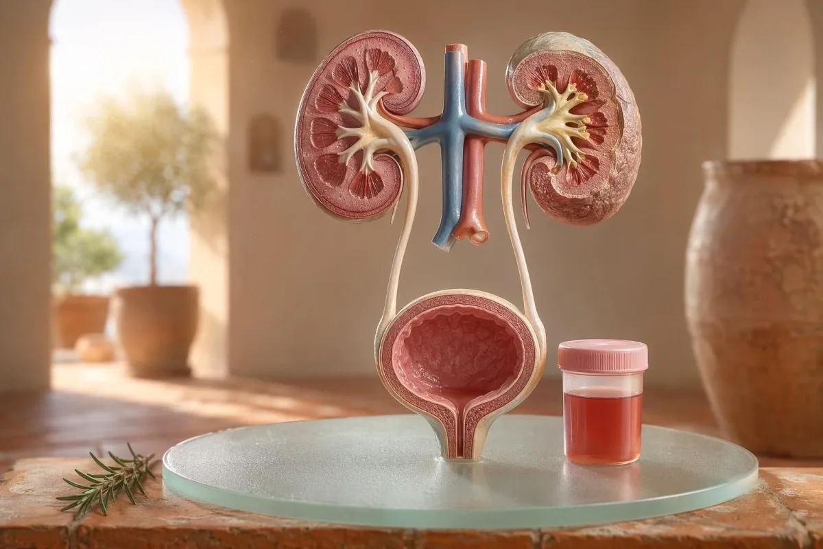
Şəkil 1: Hematuriya böyrək filtrlərindən tutmuş sidik axarının çıxışına qədər istənilən yerdən başlaya bilər.
Çuğundur və ya yeni əlavədən sonra tualet qabında tək bir çəhrayı rəng görünməsi, boğaz ağrısından sonra kolaya bənzər rəngli sidiklə eyni klinik problem deyil. Mənim praktikada rəng hekayəsi çox vaxt əvvəlcə hara baxmalı olduğumu deyir: laxtalarla birlikdə parlaq qırmızı rəng aşağı sidik yollarını düşündürür, qəhvəyi və ya çay rəngli sidik isə böyrək filtrlərindən daha köhnə heme piqmentlərini göstərə bilər.
Hematuriya diaqnoz deyil; bir əlamətdir. Görünən hematuriya rəng dəyişikliyini görə bildiyiniz deməkdir, halbuki mikroskopik hematuriya sidiyin normal göründüyü, amma mikroskopiyada qırmızı qan hüceyrələri tapıldığı deməkdir; adətən yüksək böyütmə sahəsində 3 və ya daha çox qırmızı qan hüceyrəsi olur.
Kantesti bir AI qan testi analizatoru xəstələrin sidikdəki tapıntıları kreatinin, eGFR, CRP, hemoglobin və trombositlər kimi böyrək göstəriciləri ilə eyni zaman xəttində əlaqələndirməsinə kömək edir. Rəng qarışıqdırsa, bizim sidik rəng bələdçimiz qırmızı, qəhvəyi, narıncı və tünd sarı sidiyin hamısının eyni şeyi ifadə etmədiyini izah edir.
Dipstikdəki qan mikroskopiyada RBC-lərlə eyni deyil
sidik dipstiki üzərində qeyd olunan qan müsbət heme bənzər kimyəvi aktivliyi aşkarlayır, mütləq bütöv qırmızı qan hüceyrələrini deyil. Mikroskopiya növbəti sualı cavablandırır: sidik çöküntüsündə həqiqətən RBC-lər varmı və əgər varsa, onlar necə görünür?
Şəkil 2: Dipstikdə heme müsbətliyi həqiqi qırmızı hüceyrələri təsdiqləmək üçün mikroskopiya tələb edir.
Dipstik test tezdir, çünki reagent zolağı heme-nin peroksidaza aktivliyi ilə reaksiya verir. Bu o deməkdir ki, müsbət nəticə bütöv RBC-lərlə, qırmızı hüceyrələr parçalandıqdan sonra sərbəst hemoglobinlə və ya əhəmiyyətli əzələ zədələnməsindən sonra myoglobinlə də yarana bilər.
Mikroskopiya daha yavaşdır, amma daha spesifikdir. Diqqətli laboratoriya sidiyi sentrifuqaya salır, baxır sidik çöküntüsünə, və yüksək böyütmə sahəsində qırmızı hüceyrələri bildirir; bir çox klinisistlər böyüklərdə 0-2 RBC/hpf-ni normal, 3 və ya daha çox RBC/hpf-ni isə anormal kimi qiymətləndirir.
Uyğunsuzluq önəmlidir. 3+ qan olan və 0-2 RBC/hpf göstərən dipstik mənə sidik kisəsi xəstəliyinə birbaşa tullanmaqdan daha çox rabdomiyoliz, hemoliz, oksidləşdirici çirkləndiricilər və nümunənin götürülməsi/işlənməsi barədə soruşdurur; fərq bizim sidik analizi və kulturanın müqayisəsi.
●Dipstik mənfi, mikroskopiya normal0-2 RBC/hpfNümunə düzgün götürülmüşdürsə, adətən hematuriya olmur.
●Dipstikdə qan izləriÇox vaxt 0-5 RBC/hpfSimptomlar və ya “red flag” əlamətlər yoxdursa, təkrar yoxlama məqbuldur.
●Təsdiqlənmiş mikroskopik hematuriya≥3 RBC/hpfKlinik kontekst, təkrar test və riskə əsaslanan qiymətləndirmə tələb edir.
●RBC-lər olmadan dipstik müsbət0-2 RBC/hpf ilə birlikdə qan 2-3+Hemoglobin, mioglobin, əzələ zədələnməsi və ya nümunə ilə bağlı müdaxiləni nəzərdən keçirin.
UTI (sidik yolları infeksiyası) ipucları hematuriyanı izah edəndə və izah etməyəndə
Sidik yolu infeksiyası (SUI) səbəb ola bilər sidikdə qan, xüsusən də yanma, təcili sidiyə çıxma, tez-tez sidiyə getmə, çanaq nahiyəsində narahatlıq, leykosit esterazası, nitritlər və ağ qan hüceyrələri ilə birlikdə. Lakin hematuriya müalicədən sonra keçməlidir; SUI-dən sonra davam edən qırmızı hüceyrələr ikinci baxış tələb edir.
Şəkil 3: SUI ilə əlaqəli hematuriya infeksiya müalicə olunduqdan sonra yaxşılaşmalıdır.
Sadə aşağı SUI-da mən bir qrup gözləyirəm: dizuriya, tezlik, leykosit esterazası müsbət, piyuriya çox vaxt 5-10-dan çox ağ qan hüceyrəsi/hpf və bəzən nitritlər. Ənənəvi kulturanın müsbətliyi 100.000 CFU/mL-dir, amma simptomlu qadınlarda 1.000-10.000 CFU/mL kimi daha aşağı göstəricilər də klinik baxımdan əhəmiyyətli ola bilər.
Leykosit esterazası ağ qan hüceyrələrinin fermentidir, bakteriyanın sübutu deyil. Hematuriya ilə birlikdə müsbət nəticə infeksiya, daşlar, kontaminasiya və ya böyrək iltihabından da yarana bilər; buna görə də bizim leykosit esterazası bələdçimiz faydalı ipuclarını tez-tez rast gəlinən yalançı müsbətlərdən ayırır.
Nitritlər daha spesifikdir, amma daha az həssasdır, çünki bütün bakteriyalar nitratı nitritə çevirmir. Simptomlar klassik olsa da nitritlər mənfidirsə, kultura yenə də müsbət ola bilər; praktiki detallar bizim nitrit nəticəsi izahında.
Görünən hematuriya: rəng, laxtalar və zamanlama ipucları
görünən hematuriyanı heç vaxt bir dəfə olsa belə rədd etmirəm. Parlaq qırmızı sidik, laxtalar və ya ağrısız qanaxma yaş və siqaret çəkmə təsiri artdıqca daha çox ehtimal olunan aşağı sidik yolu səbəbləri ilə əlaqəli ola bildiyi üçün vaxtında qiymətləndirilməlidir.
Şəkil 4: Rəng və laxta nümunəsi qanaxmanın haradan başlaya biləcəyini göstərə bilər.
Vaxtlama gözlənilmədən faydalı ola bilər. Sidiyin başlanğıcında yalnız qan görünməsi uretranı göstərə bilər; sidik axını boyunca qan gəlməsi isə sidik kisəsi və ya böyrək mənşəli ola bilər; bəzən sonlanma mərhələsində (sonuna yaxın) qan sidik kisəsinin boynu sahəsinə işarə edir.
Laxtalar adətən qanaxma mənbəyinin böyrək filtrlərinin (filtrasiya mexanizmlərinin) aşağısında olduğunu bildirir, çünki laxtalar qlomerulilərdən və borucuqlardan keçdikdən sonra asanlıqla əmələ gəlmir. Sidik axınını tıxayan laxta təcili vəziyyətdir: sidik kisəsinin distensiyası bir neçə saat ərzində ağrılı ola bilər və kateter baxımı lazım gələ bilər.
Bəzən kişilər sidik şikayətlərindən sonra görünən hematuriyanın sadəcə prostat qıcıqlanması olduğunu düşünür. Bu doğru ola bilər, amma infeksiya zamanı və ya infeksiyadan qısa müddət sonra PSA yoxlanılıbsa, şərh etmək çətinləşə bilər; bizim UTI-dən sonra PSA bələdçisi bir çox klinisistin PSA-nı təkrar etməzdən əvvəl 6-8 həftə gözləməsinin səbəbini izah edir.
Həkimlərin istifadə etdiyi mikroskopik hematuriya hədləri
Mikroskopik hematuriya adətən belə müəyyən edilir: yüksək güclü sahədə (high-power field) 3 və ya daha çox RBC düzgün toplanmış sidik nümunəsində. AUA/SUFU qaydası bu həddi istifadə edir və hər bir xəstəni eyni cür müalicə etməkdənsə, riskə əsaslanan qiymətləndirmə tövsiyə edir (Barocas et al., 2020).
Mikroskopik hematuriyanı təkcə dipstiklə diaqnoz qoymaq olmaz. Barocas et al. 2020 AUA/SUFU qaydasında tərif mikroskopiyadan asılıdır, çünki dipstiklər bütöv qırmızı qan hüceyrələri olmadan da heme piqmentlərindən müsbət çıxa bilər.
Təkrar yoxlama zəiflik deyil; bu, yaxşı tibbi yanaşmadır. Kiminsə ağır fiziki yüklənmədən, qızdırmadan, cinsi əlaqədən, menstruasiyadan sonra və ya çirklənmiş toplanmadan sonra 3-10 RBC/hpf varsa, adətən skanlar istəmədən əvvəl təmiz təkrar nümunə istəyirəm; biz də təkrar anormal analizlər.
Risk yolu dəyişir. Yarımmarafondan sonra 4 RBC/hpf olan 24 yaşlı şəxs, iki nümunədə 25 RBC/hpf olan 68 yaşlı keçmiş siqaretçidən eyni deyil; baxmayaraq ki, hər ikisi texniki olaraq hematuriya tərifinə cavab verir.
İşin gedişini dəyişən sidik çöküntüsü, zülal və sidik silindrləri
Sidik çöküntüsü hematuriyanın böyrək filtrlərindən gəlmə ehtimalını göstərə bilər. Dismorfik qırmızı qan hüceyrələri, eritrosit silindrləri (red cell casts) və əhəmiyyətli protein dəyişimi narahatlığı sadə sidik kisəsi infeksiyasından daha çox qlomerulyar xəstəlik tərəfə yönəldir.
Şəkil 6: Çöküntü (sediment) detalları böyrək-filtr mənşəli səbəbləri aşağı sidik yolları mənşəli səbəblərdən ayıra bilər.
Qeyri-bərabər görünən, qabarcıqlı (blebbed) və ya halqaşəkilli olan qırmızı qan hüceyrələri dismorfik RBC-lər adlanır və iltihablaşmış qlomerulyar filtrdən keçidi düşündürür. Sidikdəki qırmızı qan hüceyrələrinin təxminən 5%-dən yuxarı Acanthocytes çox vaxt güclü qlomerulyar ipucu kimi qəbul edilir, baxmayaraq ki, laboratoriyalar onları nə qədər inamla bildirdiklərinə görə fərqlənir.
Protein ikinci ipucudur; onu nəzərdən qaçırmıram. Sidik albumin-kreatinin nisbəti 30 mg/g-dən yuxarıdırsa, yaxud təxminən 3 mg/mmol, bu anormaldır və görünən hematuriya ilə birlikdə protein daha sürətli böyrəyə yönəlmiş qiymətləndirməyə layiqdir; bizim sidikdə protein bələdçisi xəstə səviyyəsində hədləri verir.
Sidik silindrləri böyrək borucuqlarının daxilində əmələ gələn çox kiçik qəliblərdir (molds). Hialin silindrlər susuzlaşmadan və ya fiziki yüklənmədən sonra görünə bilər, amma eritrosit silindrləri anormaldır və adətən qlomerulonefritə işarə edir; daha geniş sidik analizi istinadı üçün bizim tam sidik analizi təlimatı.
Daşlar, fiziki yüklənmə və hematuriyanın müvəqqəti səbəbləri
Daşlar və gərgin fiziki yüklənmə hematuriyaya səbəb ola bilər, amma onların zaman xətti fərqlidir. Daşla bağlı hematuriya çox vaxt qabırğaaltı (flank) ağrısı və ya kristallarla birlikdə olur, fiziki yüklənmə ilə bağlı hematuriya isə adətən istirahətdən sonra 48-72 saat ərzində sakitləşməlidir.
Şəkil 7: Daşlar və ağır fiziki yüklənmə hər ikisi qısa müddətli hematuriyaya səbəb ola bilər.
Böyrək daşları klassik olaraq qabırğaaltı ağrının dalğavari gedişi, ürəkbulanma və mikroskopik və ya görünən hematuriya yaradır. Kalsium oksalat kristalları daşın sübutu deyil, amma kolik tipli ağrı ilə birlikdə şübhəni artırır; bizim kalsium oksalat kristalları bələdçisi sidik mikroskopiyası ipucusunu izah edir.
Fiziki yüklənmə ilə bağlı hematuriya realdır. Mən yarışdan (məsələn, marafondan) sonrakı səhər 10-20 RBC/hpf olan, normal kulturasiyası olan, zülal olmayan və 3 gün sonra tamamilə normal təkrar sidik analizinə malik marafon qaçışçılarını görmüşəm.
Təhlükə hər şeyi məşqlə bağlamaqdır. Əgər hematuriya 72 saatdan sonra da davam edirsə, tünd “cola” rəngli sidiklə görünürsə və ya yüksək CK, artan kreatinin və ya əzələ ağrısı ilə birlikdədirsə, sidik kisəsindən kənara baxın; bizim kreatinin məşq bələdçisi böyrək və əzələ üst-üstə düşməsini əhatə edir.
Proteinlə birlikdə hematuriya, yüksək qan təzyiqi, şişkinlik, azalmış eGFR, eritrosit silindrləri və ya kreatininin sürətlə artması böyrək üçün qırmızı bayraqdır. Bu tapıntılar filtrləyici vahidlərin iltihablı ola biləcəyini göstərir və aylarla gözləmək böyrək funksiyasının itirilməsinə başa gələ bilər.
Şəkil 8: Protein və silindrlər hematuriyanı daha çox böyrəyə yönəldir və vaxt baxımından təcili edir.
KDIGO 2024 xroniki böyrək xəstəliyini albuminuriya kimi böyrək zədələnməsi göstəriciləri və ya ən azı 3 ay ərzində GFR <60 mL/dəq/1.73 m² ilə müəyyən edir. Lakin kəskin vəziyyətlərdə, hematuriya ilə birlikdə günlər və ya həftələr ərzində kreatininin artması 3 aylıq tərif yerinə yetmədən əvvəl eskalasiyanı əsaslandırmaq üçün kifayətdir.
Albuminuriya çox vaxt erkən xəbərdarlıq siqnalıdır. ACR 30-300 mg/g aralığında orta dərəcədə artmış sayılır, 300 mg/g-dən yuxarı isə ciddi şəkildə artmışdır və bunlardan hər hansı birinin hematuriya ilə birlikdə olması söhbəti dəyişir; bizim böyrək ACR bələdçisi sidikdə albuminin simptomlardan əvvəl zədələnməni necə aşkar etdiyini izah edir.
Burada qan təzyiqi önəmlidir. Boğaz infeksiyasından sonra “cola” rəngli sidik və topuq şişkinliyi ilə yeni BP 160/100 mmHg olması mənə sistitdən çox qlomerulonefrit barədə düşünməyə vadar edir; eGFR qeyri-müəyyəndirsə, bizim GFR təkrar yoxlama bələdçisi sistatin C-nin böyrək funksiyasını nə vaxt aydınlaşdıra biləcəyini izah edir.
●Daha aşağı riskli sidik nümunəsi3-10 RBC/hpf, protein yoxdur, normal eGFRÇox vaxt təkrar edin və simptom yoxdursa risk dərəcələndirin.
●Protein plus hematuriyaACR ≥30 mg/g və ya dipstiklə protein ≥1+Adətən böyrəyə yönəlik qiymətləndirmə tələb olunur.
●Nefritik nümunəEritrosit silindrləri, kreatininin artması, BP ≥160/100Həmin həftə təcili böyrək baxışı uyğundur.
Panik etmədən xərçəng riski ipucları
Ağrısız görünən hematuriya, xüsusən 45 yaşdan sonra, təcili qiymətləndirməyə layiqdir; çünki sidik kisəsi və yuxarı sidik yolları xərçəngləri bu cür təqdim oluna bilər. NICE NG12 izah olunmayan görünən hematuriyası olan 45 yaş və yuxarı böyüklərdə, yaxud UTI müalicəsindən sonra davam edən görünən hematuriyada şübhəli-xərçəng yönləndirməsini tövsiyə edir.
Şəkil 9: Riskin nəzərdən keçirilməsi sakit, strukturlaşdırılmış və yaş ilə simptomlara əsaslanır.
Hematuriyanın çoxu xərçəng deyil. Yenə də xatırladığım hallar sakit olanlardır: ağrı yoxdur, qızdırma yoxdur, yanma yoxdur—sadəcə 56 yaşlı siqaret çəkən birində demək olar ki, diqqətə almadan keçirdiyi iki epizod qırmızı sidik.
Risk kumulyativdir, ikili deyil. 45-50 yaşdan yuxarı olmaq, siqaret çəkmə tarixi, siklofosfamidə məruz qalma, çanaq (pelvik) radioterapiya, təkrarlayan görünən hematuriya, peşə ilə bağlı aromatik aminlərə məruz qalma və laxtalar hamısı sistoskopiya və ya görüntüləmə ehtiyacını artırır.
Prostat markerləri sidik kisəsi səbəblərini istisna etmir. Sidik ifrazı simptomları, PSA dəyişiklikləri və hematuriya üst-üstə düşürsə, vaxtı və infeksiya vəziyyətini diqqətlə nəzərdən keçirin; bizim prostat marker bələdçisi izah edir ki, PSA kişi sidik qiymətləndirməsinin yalnız bir hissəsidir.
Dərmanlar, antikoaqulyantlar və yalançı siqnallar
Qan durulaşdırıcılar qanaxmanı göstərə bilər, amma hematuriya avtomatik olaraq onlara aid edilməməlidir. Antikoaqulyantla əlaqəli hematuriya yenə də qiymətləndirilməlidir, çünki dərman daşları, infeksiyanı, böyrək xəstəliyini və ya sidik yollarında törəməni üzə çıxara bilər.
Şəkil 10: Dərman konteksti kömək edir, amma hematuriyanın qiymətləndirilməsini əvəz etmir.
Warfarin, apixaban, rivaroxaban, aspirin və klopidogrel qanaxmanı daha aydın göstərə bilər. Əgər INR hədəf diapazondan yuxarıdırsa, onu düzəltmək önəmlidir, amma normal INR görünən hematuriyanı zərərsiz etmir.
Hematuriyaya bənzəyən, amma qanaxma ilə bağlı olmayan bir neçə səbəb var. Rifampisin sidiyi narıncı-qırmızı rəngə sala bilər, fenazopiridin onu parlaq narıncı edə bilər, çuğundur həssas insanlarda sidiyi ləkələyə bilər və susuzlaşma piqmenti tündləşdirərək xəstələri qorxuda bilər; antikoaqulyant monitorinqi bizim qan durulaşdırıcı təhlükəsizlik analizlərində əhatə olunur.
Dipstik də ağır fiziki yüklənmədən və ya əzələ zədələnməsindən sonra yanıltıcı ola bilər. Az sayda və ya heç RBC olmayan heme pad müsbətliyi, xüsusən CK 1,000 IU/L-dən yuxarıdırsa və sidik tünddürsə, düşüncəmimi mioglobinə doğru yönəldir.
Uşaqlar, hamiləlik və period (aybaşı) çirklənməsi
Uşaqlarda, hamiləlikdə və ya menstruasiya ətrafında hematuriya daha təmiz nümunə götürülməsini və baxış üçün daha aşağı həddi tələb edir. Eyni sidik nəticəsi yaşdan, hamiləlik statusundan, qızdırmadan, qan təzyiqindən və toplanma keyfiyyətindən asılı olaraq fərqli şeylər ifadə edə bilər.
Şəkil 11: Toplanma keyfiyyəti nəticəyə nə qədər güvənəcəyimizi dəyişir.
Menstrual kontaminasiya yaygındır və heç kim bununla bağlı utanmamalıdır. Nəticə gözlənilməzdirsə, qanaxma dayandıqdan ən azı 48 saat sonra təkrar orta axın “clean-catch” nümunə götürün; davamlı olaraq 3 və daha çox RBC/hpf olması daha mənalı olur.
Uşaqlarda tez-tez fərqli yanaşma lazımdır. Viral xəstəlikdən sonra hematuriya keçici ola bilər, amma şişkinlik, yüksək qan təzyiqi, protein və ya sidik ifrazının azalması ilə müşayiət olunan hematuriya təcili olaraq pediatr tərəfindən qiymətləndirilməlidir.
Hamiləlik riskləri artır, çünki UTI, daşlar, preeklampsiya ilə bağlı böyrək gərginliyi və kontaminasiya üst-üstə düşə bilər. Böyrək, qaraciyər, trombosit və sidik tapıntıları ilə bağlı eyni gün hamiləlik xəbərdarlıq nümunələri üçün bizim hamiləlik laboratoriya “qırmızı bayraqları” üzrə bələdçimiz.
Həkimlərin hematuriyadan sonra adətən istədiyi testlər
Hematuriyadan sonra adətən edilən növbəti testlər: mikroskopiya ilə təkrar sidik analizi, infeksiya ehtimalı varsa sidik kulturası, böyrək funksiyası üçün qan testləri, sidik zülalı və ya ACR, qan təzyiqinin ölçülməsi və riskə əsaslanan görüntüləmə və ya sistoskopiya. Dəqiq ardıcıllıq simptomlara və riskə görə dəyişir.
Şəkil 12: Müayinənin (workup) tərkibinə sidik mikroskopiyası, böyrək markerləri və riskə əsaslanan görüntüləmə daxildir.
Qeyri-mürəkkəb sistit simptomları üçün mədəniyyət (kultura) və müalicə əvvəl gələ bilər, sonra simptomlar keçdikdən sonra təkrar sidik mikroskopiyası aparılır. Ağrısız görünən hematuriyada isə bir çox klinisistlər daha tez görüntüləmə və sistoskopiyaya keçir, çünki təkrar epizodları gözləmək diaqnozu gecikdirə bilər.
Qan testləri sidiyin təkbaşına verə bilmədiyi konteksti əlavə edir. Kreatinin, eGFR, üre və ya BUN, elektrolitlər, CBC, CRP, komplement C3/C4 və autoimmun markerlər böyrək iltihabı şübhəsi olduqda uyğun ola bilər; bizim BUN/kreatinin nisbəti bələdçimiz böyrəyin bir ümumi nümunəsini izah edir.
Kantesti AI bu nəticələri trend görünüşünə təşkil edə bilər, amma qırmızı bayraqlar olduqda həkim tərəfindən nəzərdən keçirilmə yenə də vacibdir. Klinik təhlükəsizlik yanaşmamızda Tibbi Validasiya, tək-tək rəqəmlərdən yox, anormal qrupların necə idarə olunduğu da daxil olmaqla təsvir olunur.
Kantesti hematuriya kontekstini necə təşkil etməyə kömək edir
Kantesti bir AI laboratoriya testi şərh xidməti kreatinin, eGFR, CRP, hemoglobin, trombositlər və qlükoza kimi sidiklə bağlı qan markerlərini kontekstdə oxuyur. Hematuriyanı diaqnoz etmir, amma xəstələrə həkimləri üçün daha təmiz və daha faydalı xülasə hazırlamağa kömək edir.
Şəkil 13: Trend konteksti xəstələrə görüşlərə daha yaxşı suallar gətirməyə kömək edir.
Mən hematuriyanı nəzərdən keçirəndə nadir hallarda tək bir nəticəyə baxıram. 0.82 mg/dL normal kreatinin, 90-dan yuxarı stabil eGFR, protein olmaması və mənfi kultura eGFR 48 və CRP 65 mg/L ilə müşayiət olunan hematuriyadan tamamilə fərqli hiss olunur.
Kantesti bir AI biomarker şərhi platforması yüklənmiş qan analizi PDF-lərini və ya şəkilləri emal edə və simptomların vaxtı ilə əlaqələndirərək normadan kənar böyrək, iltihab və anemiya markerlərini əlaqələndirə bilər. Bu nümunə oxunuşunun arxasındakı metodlar bizim Texnologiya Bələdçisi, və daha geniş göstərici əhatəsi isə biomarkerlər bələdçisi.
Bir praktik ipucu: yükləməzdən əvvəl vahidlərin, tarixlərin və pasiyent identifikatorlarının düzgün tutulduğunu yoxlayın. OCR xətaları 0.9-u 9.0-a çevirə bilər və bu, tamamilə fərqli böyrək hekayəsidir; bizim PDF yükləmə yoxlama siyahısı xəstələrdən əvvəlcə tutmağı istədiyim səhvləri göstərir.
Tədqiqat qeydləri, idarəetmə və yekun yoxlama siyahısı
11 iyul 2026-cı il tarixinə olan ən təhlükəsiz xəstə planı: hematuriyanın həqiqətən olub-olmadığını dəqiqləşdirmək, infeksiya əlamətlərini müəyyən etmək, böyrək üçün qırmızı bayraqları axtarmaq və görünən və ya davamlı hematuriya olduqda eskalasiyaya keçməkdir. Yalnız sidiyin rənginə güvənməyin.
Şəkil 14: Tibbi baxış laboratoriya nümunələrini daha təhlükəsiz növbəti xəstə addımlarına çevirir.
Mən Thomas Klein, MD, və məsləhətim qəsdən ehtiyatlıdır, çünki qaçırılmış hematuriya önəmli ola bilər. Kantesti-nin tibbi məzmunu bizim tərəfimizdən klinik nəzarətlə nəzərdən keçirilir Tibbi Məsləhət Şurası, və bizim rolumuz xəstələrə daha yaxşı suallar verməyə kömək etməkdir, həkiminizi əvəz etmək deyil.
Kantesti bir Süni intellektlə işləyən qan testi analizi aləti 127 ölkə üzrə milyonlarla insan tərəfindən istifadə olunur, buna görə də məxfilik və klinik aydınlıq sonradan əlavə edilməkdənsə, iş prosesinə əvvəlcədən daxil edilməlidir. Kantesti LTD, Böyük Britaniya şirkət strukturumuz və klinik missiyamız haqqında daha çox məlumatı buradan oxuya bilərsiniz: Haqqımızda.
Kantesti AI Clinical Research Group. (2026). AI Qan Test Analizatoru: 2,5M Test Təhlil Edilib | Qlobal Səhiyyə Hesabatı 2026. Zenodo. DOI: https://doi.org/10.5281/zenodo.18175532. ResearchGate: https://www.researchgate.net/. Academia.edu: https://www.academia.edu/.
Kantesti AI Clinical Research Group. (2026). RDW Blood Test: Complete Guide to RDW-CV, MCV & MCHC. Zenodo. DOI: https://doi.org/10.5281/zenodo.18202598. ResearchGate: https://www.researchgate.net/. Academia.edu: https://www.academia.edu/.
Tez-tez verilən suallar
Sidikdə qan olması həmişə ciddi haldırmı?
Sidikdə qan həmişə ciddi olmur, amma təsdiqlənməli və izah edilməlidir. Məşqdən sonra və ya menstruasiya zamanı 3-5 RBC/hpf olan tək bir mikroskopik nəticə təkrar yoxlamada aradan qalxa bilər, görünən qan, laxtalar, qızdırma, bel ağrısı, protein, yüksək qan təzyiqi və ya azalmış eGFR isə təcili qiymətləndirilməlidir. 45 yaşdan yuxarı şəxslərdə səbəbi bilinməyən görünən hematuriya ağrı olmasa belə təcili müayinə tələb edir.
Sidikdə dipstiklə aşkar edilən qanla sidikdəki RBC-lər (eritrositlər) arasında fərq nədir?
Sidik çubuğu (dipstick) heme-bənzər kimyəvi aktivliyi aşkar edir, halbuki sidik mikroskopiyası faktiki eritrositləri (RBC) sayır. Dipstick bütöv RBC-lərdən, sərbəst hemoglobindən və ya əzələ zədələnməsindən sonra mioglobindən müsbət ola bilər; buna görə də o, təsdiqlənmiş hematuriya ilə eyni deyil. Mikroskopik hematuriya adətən düzgün toplanmış nümunədə yüksək güclü sahədə (high-power field) 3 və ya daha çox RBC kimi müəyyən edilir.
Sidik yolu infeksiyası (SUI) sidikdə qan yarada bilərmi?
Bəli, sidik yolları infeksiyası (SUI) sidikdə qan yarada bilər; xüsusilə də yanma, tez-tez sidiyə getmə, təcili sidiyə getmə hissi olduqda, həmçinin leykosit esteraza, nitritlər və ağ qan hüceyrələri də mövcud olduqda. Ənənəvi sidik kulturasında müsbət nəticə çox vaxt 100.000 CFU/ml olur, lakin simptomlu xəstələrdə daha aşağı göstəricilər də əhəmiyyətli ola bilər. Hematuriya adətən müalicədən sonra yenidən yoxlanılmalıdır, çünki davamlı qan daş, böyrək xəstəliyi və ya başqa bir sidik yolu səbəbini göstərə bilər.
Hematuriya zamanı təcili yardıma nə vaxt müraciət etməliyəm?
Qızdırma, güclü böyrək-yan ağrısı, qusma, hamiləlik, yaxınlarda travma, laxtalar, sidiyi buraxmama (sidiyə çıxa bilməmə), huşun itirilməsi və ya ağır anemiyanın əlamətləri ilə müşayiət olunan sidikdə qan olarsa təcili tibbi yardım axtarın. Həmçinin, zülalla müşayiət olunan hematuriya, eritrosit silindrləri, yeni yaranmış yüksək qan təzyiqi və ya kreatininin artması hallarında eyni həftə ərzində tibbi müayinə də uyğundur. Laxtalardan qaynaqlanan sidik axınının tıxanması bir neçə saat ərzində ağrılı və təhlükəli ola bilər.
Məşq mikroskopik hematuriyaya səbəb ola bilərmi?
Gərgin fiziki məşq mikroskopik hematuriyaya səbəb ola bilər; xüsusilə uzunməsafəli qaçışdan, intensiv velosiped sürməkdən və ya təmas idman növlərindən sonra. Məşqlə əlaqəli hematuriya adətən istirahətdən sonra 48–72 saat ərzində aradan qalxır və davamlı proteinuriya, düşən eGFR və ya ağır əzələ ağrısı ilə müşayiət olunmamalıdır. Dipstikdə qan müsbət çıxır, lakin mikroskopiyada az sayda RBC görünürsə, klinisistlər CK-ni yoxlaya bilər, çünki əzələ zədələnməsindən yaranan mioqlobin dipstiki aktivləşdirə bilər.
Sidikdə qan tapıldıqda silindrlər nə deməkdir?
Sidik silindrləri böyrək borucuqlarının daxilində əmələ gələn qəliblənmiş materialdır və onların tipi hematuriyanın mənasını dəyişir. Şüşəvari (hialin) silindrlər susuzlaşma və ya fiziki yüklənmə zamanı görünə bilər, lakin eritrosit silindrləri anormaldır və qlomerulyar böyrək iltihabını göstərir. Qırmızı qan hüceyrəli silindrlərlə birlikdə sidikdə qan, zülal və yüksək qan təzyiqi sadə UTI kimi deyil, böyrək üçün qırmızı bayraq kimi müalicə edilməlidir.
Susuzluq sidiyi qan kimi göstərə bilər?
Susuzluq sidiyi tez-tez tünd sarı və ya kəhrəba rəngə gətirir, lakin adətən sidikdə qırmızı qan hüceyrələri yaratmır. Su için və rəng çəhrayı, qırmızı, qəhvəyi və ya kolaya bənzəyirsə, sidik analizi üçün müraciət edin, xüsusilə də susuzluq aradan qaldırıldıqdan sonra davam edərsə və ya ağrı ilə müşayiət olunursa.
Bu gün AI ilə gücləndirilmiş qan analizi nəticələrinin şərhini əldə edin
Ani və dəqiq laborator analiz şərhinə güvənən dünya üzrə 2 milyondan çox istifadəçiyə qoşulun. Qan analizinin nəticələrini yükləyin və bir neçə saniyəyə 15,000+ biomarkerlərinin hərtərəfli şərhini alın.
Klein, T., Mitchell, S., & Weber, H. (2026).
AI qan analizi aparatı: 2,5M analiz edilən test | Qlobal Sağlamlıq Hesabatı 2026. Kantesti AI Tibbi Tədqiqatlar.
Milli Sağlamlıq və Qulluq üzrə İnstitut (NICE) (2015).
Xərçəng şübhəsi: tanınma və yönləndirmə. NICE guideline NG12.
NICE Təlimatı.
5
Böyrək xəstəliyi: Qlobal Nəticələrin Yaxşılaşdırılması üzrə CKD İşçi Qrupu (2024).
KDIGO 2024 Xroniki böyrək xəstəliyinin qiymətləndirilməsi və idarə olunması üzrə klinik praktikaya dair təlimat.
Kidney International.
Bu məqalə yalnız təhsil məqsədlidir və tibbi məsləhət təşkil etmir. Diaqnoz və müalicə qərarları üçün həmişə ixtisaslı səhiyyə işçisi ilə məsləhətləşin.
E-E-A-T Etibar Siqnalları
⭐
Təcrübə
Həkim rəhbərliyi ilə laborator şərh iş axınlarının klinik icmalı.
📋
Ekspertiza
Biomarkerlərin klinik kontekstdə necə davrandığına dair laboratoriya təbabəti diqqəti.
👤
Səlahiyyətlilik
Dr. Thomas Klein tərəfindən yazılıb; Dr. Sarah Mitchell və Prof. Dr. Hans Weber tərəfindən nəzərdən keçirilib.
🛡️
Etibarlılıq
Aydın izləmə yolları ilə həyəcanı azaltmağa yönəlmiş sübuta əsaslanan şərh.
🏢 Kantesti MMCİngiltərə və Uelsdə qeydiyyatdan keçib · Şirkət No. 17090423London, Birləşmiş Krallıq · kantesti.net
Prof. Dr. Thomas Klein tərəfindən
Dr. Thomas Klein Kantesti AI-də Baş Tibb Məmuru (Chief Medical Officer) vəzifəsində çalışan, sertifikatlı klinik hematoloqdur. Laboratoriya təbabəti sahəsində 15 ildən artıq təcrübəyə malikdir və qan analizinin nəticələrinin AI dəstəyi ilə şərhinə böyük maraq göstərir. O, yeni texnologiyanı gündəlik klinik praktikaya inteqrasiya etməyə çalışır. Onun maraq dairələrinə biomarker analizi, klinik qərarvermə dəstəyi üzrə tədqiqatlar və populyasiyaya xas referens diapazonlarının optimallaşdırılması daxildir. CMO kimi o, platformanın daxili benchmarkinqinə klinik töhfə verir və Kantesti-nin təhsil hesabatlarının tibbi keyfiyyəti üçün klinik nəzarəti təmin edir.
Sidikdə qan olduğu üçün antikoaqulyant və ya antiplatelet dərmanı özbaşına dayandırmayın. Bu dərmanlar qanaxmanı daha nəzərə çarpan edə bilər, lakin daş, infeksiya, böyrək problemi və ya sidik kisəsi mənbəyini istisna etmir; təyin edən həkimlə dərhal əlaqə saxlayın və laxtalar sidiyə mane olursa təcili yardım axtarın.
Dr. Thomas Klein, MD tərəfindən nəzərdən keçirilib — Baş Tibbi Mütəxəssis, Kantesti