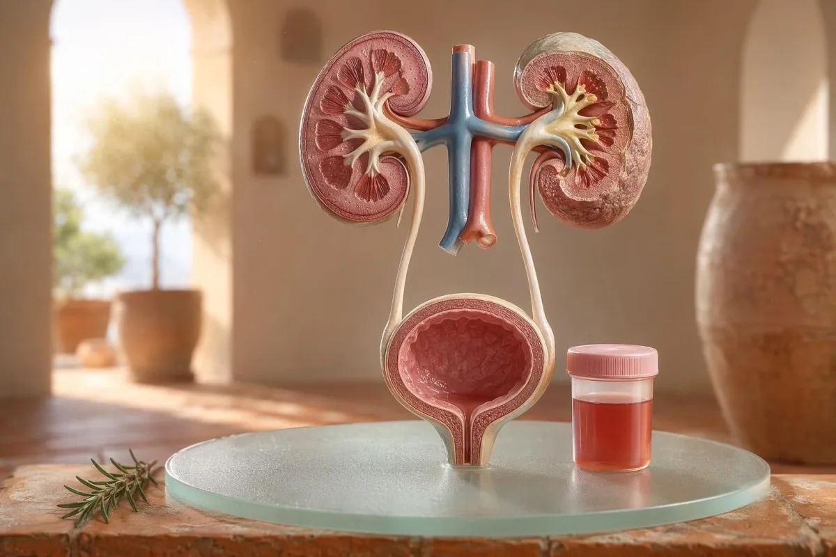
Памятка для пациента о видимой и микроскопической гематурии, включая то, почему кровь по тест-полоске не то же самое, что эритроциты под микроскопом.


- Кровь в моче означает видимую красную, цвета чая или цвета колы мочу либо микроскопическую гематурию, выявленную при анализе; видимая кровь всегда требует медицинского наблюдения.
- Microscopic hematuria обычно определяется как 3 или более эритроцитов на поле зрения при большом увеличении в образце мочи, правильно собранном для микроскопии.
- Кровь по тест-полоске выявляет активность гема, поэтому тест может быть положительным при наличии интактных эритроцитов, свободного гемоглобина или миоглобина после травмы мышц.
- Подсказки о UTI включают жжение, частое мочеиспускание, лейкоцитарную эстеразу, нитриты и лейкоциты; гематурию следует перепроверить после того, как инфекция пройдет.
- Мочевой осадок находки, такие как дисморфные эритроциты, эритроцитарные цилиндры или значимая протеинурия, указывают на заболевание фильтрационного аппарата почек.
- Цилиндры мочи представляют собой слепки из канальцев почек; эритроцитарные цилиндры — это красный флаг со стороны нефрологии в пределах одной недели, особенно при высоком артериальном давлении или снижающемся eGFR.
- Онкологические «красные флаги» включают безболезненную видимую гематурию, сгустки, возраст старше 45 лет, курение в анамнезе, профессиональное воздействие химических веществ и повторяющиеся необъяснимые эпизоды.
- Гематурия при физической нагрузке обычно проходит в течение 48–72 часов; сохраняющаяся кровь после отдыха не должна списываться на бег или езду на велосипеде.
- Срочная оценка необходимо при наличии крови в моче на фоне лихорадки, боли в боку, сгустков, вызывающих обструкцию мочеиспускания, беременности, травмы или симптомов тяжелой анемии.
Что означает кровь в моче, прежде чем вы начнете паниковать
Кровь в моче может возникать при инфекции, камнях, заболеваниях, связанных с фильтрацией в почках, состояниях предстательной железы или мочевого пузыря, при приеме лекарств, после физической нагрузки или из‑за контаминации во время менструаций. Практическое правило простое: видимая кровь, повторяющаяся микроскопическая гематурия или гематурия с болью, лихорадкой, сгустками, высоким артериальным давлением, протеинурией или сниженной функцией почек требуют срочной оценки, а не наблюдения без действий.
Один разовый розовый цвет унитаза после свеклы или нового БАД — это не та же клиническая проблема, что и моча цвета колы после ангины. В моей практике «история цвета» часто подсказывает, куда смотреть в первую очередь: ярко‑красная с сгустками обычно указывает на нижние мочевые пути, тогда как коричневая или цвета чая моча может означать более старые гемовые пигменты из почечных фильтров.
Гематурия — это не диагноз; это признак. Видимая гематурия означает, что вы видите изменение цвета, тогда как микроскопическая гематурия означает, что моча выглядит нормально, но микроскопия выявляет эритроциты, обычно при 3 или более эритроцитах на поле зрения при большом увеличении.
Кантести — это Анализатор анализа крови на основе искусственного интеллекта это помогает пациентам связать находки в моче с маркерами почек, такими как креатинин, eGFR, CRP, гемоглобин и тромбоциты, в одной временной шкале. Если цвет вызывает сомнения, наш справочник по цвету мочи объясняет, почему красная, коричневая, оранжевая и темно‑желтая моча — это не одно и то же.
Кровь по тест-полоске — не то же самое, что RBC при микроскопии
Тест-полоска для мочи с отметкой кровь положительная выявляет химическую активность, похожую на гем, а не обязательно неповрежденные эритроциты. Микроскопия отвечает на следующий вопрос: есть ли в мочевом осадке действительно эритроциты и, если да, как они выглядят?
Тестирование тест-полоской быстрое, потому что реагентная подушечка реагирует с пероксидазной активностью гема. Это означает, что положительный результат может быть при неповрежденных RBC, свободном гемоглобине после распада эритроцитов или миоглобине после значительного повреждения мышц.
Микроскопия медленнее, но более специфична. Тщательная лаборатория центрифугирует мочу, исследует мочевой осадок, и сообщает количество эритроцитов на поле зрения при большом увеличении; многие клиницисты считают нормой 0–2 RBC/HPF, а 3 или более RBC/HPF — отклонением у взрослых.
Несоответствие важно. Тест-полоска с 3+ по крови и 0–2 RBC/HPF заставляет меня расспрашивать о рабдомиолизе, гемолизе, окисляющих контаминантах и обращении с образцом, а не сразу переходить к заболеваниям мочевого пузыря; разница похожа на логику в нашем сравнение общего анализа мочи и посева.
Когда подсказки, указывающие на ИМП, объясняют гематурию, а когда — нет
Инфекция мочевых путей может вызывать кровь в моче, особенно при жжении, императивных позывах, учащенном мочеиспускании, дискомфорте в области таза, лейкоцитарной эстеразе, нитритах и лейкоцитах. Но гематурия должна исчезнуть после лечения; сохраняющиеся эритроциты после ИМП требуют повторного осмотра.
При типичной нижней ИМП я ожидаю «пакет»: дизурия, учащенное мочеиспускание, положительная лейкоцитарная эстераза, пиурия часто выше 5–10 лейкоцитов/HPF и иногда нитриты. Традиционная культуральная позитивность — 100 000 КОЕ/мл, но у симптомных женщин более низкие показатели, например 1 000–10 000 КОЕ/мл, все еще могут иметь клиническое значение.
Лейкоцитарная эстераза — это фермент лейкоцитов, а не доказательство бактерий. Положительный результат при гематурии может встречаться при инфекции, камнях, контаминации или воспалении почек, поэтому наш гид по лейкоцитарной эстеразе отделяет полезные подсказки от частых ложноположительных результатов.
Нитриты более специфичны, но менее чувствительны, потому что не все бактерии превращают нитраты в нитриты. Если симптомы типичные, но нитриты отрицательные, посев все равно может быть положительным; практические детали описаны в нашем разборе результата по нитритам.
Видимая гематурия: подсказки по цвету, сгусткам и времени появления
Видимая гематурия — это никогда не то, что я игнорирую, даже если она случилась один раз. Ярко-красная моча, сгустки или безболезненное кровотечение требуют своевременной оценки, потому что причины со стороны нижних мочевых путей становятся более вероятными по мере роста возраста и воздействия курения.
Время может быть удивительно полезным. Кровь только в начале мочеиспускания может указывать на уретру, кровь на протяжении всей струи может поступать из мочевого пузыря или почечного источника, а терминальная кровь ближе к концу иногда указывает на область шейки мочевого пузыря.
Сгустки обычно означают, что источник кровотечения находится ниже почечных фильтров, потому что сгустки не образуются легко после прохождения через клубочки и канальцы. Сгусток, который блокирует отток мочи, — неотложное состояние: растяжение мочевого пузыря может стать болезненным в течение нескольких часов, и может потребоваться уход за катетером.
Мужчины иногда предполагают, что видимая гематурия после симптомов со стороны мочевыводящих путей — это просто раздражение простаты. Это может быть правдой, но если PSA был проверен во время инфекции или вскоре после нее, интерпретация может быть затруднительной; наше Руководство по PSA после ИМП объясняет, почему многие клиницисты ждут 6–8 недель перед повторным определением PSA.
Пороговые значения микроскопической гематурии, которые используют врачи
Микроскопическая гематурия обычно определяется как 3 или более RBC в поле зрения при большом увеличении в правильно собранном образце мочи. Руководство AUA/SUFU использует этот порог и рекомендует оценку по уровню риска, а не лечение каждого пациента одинаково (Barocas et al., 2020).
Одного тест-полоски недостаточно, чтобы диагностировать микроскопическую гематурию. В руководстве AUA/SUFU Barocas et al., 2020 определение зависит от микроскопии, потому что тест-полоски могут быть положительными из‑за гемовых пигментов без сохраненных эритроцитов.
Повторное тестирование — не признак слабости; это правильная медицина. Если у кого-то 3–10 RBC/hpf после интенсивной физической нагрузки, лихорадки, секса, менструации или при загрязненном сборе, я обычно хочу получить чистый повторный образец перед назначением сканирований, как мы советуем для руководство по повторению аномальных анализов.
Изменение риска меняет путь. 24-летний человек с 4 RBC/hpf после полумарафона — это не то же самое, что 68-летний бывший курильщик с 25 RBC/hpf в двух образцах, даже если технически оба соответствуют определению гематурии.
Мочевой осадок, белок и цилиндры мочи, которые меняют тактику обследования
Мочевой осадок может показать, вероятнее ли гематурия исходит из почечных фильтров. Дисморфные эритроциты, цилиндры из эритроцитов и значимый сдвиг по белку смещают настороженность в сторону гломерулярного заболевания, а не простой инфекции мочевого пузыря.
Эритроциты, которые выглядят неровными, с «пузырьками» (blebbed) или кольцевидными, называются дисморфными RBC, и они предполагают прохождение через воспаленный гломерулярный фильтр. Акантоциты при уровне примерно выше 5% эритроцитов мочи часто рассматривают как сильную гломерулярную подсказку, хотя лаборатории различаются по тому, насколько уверенно они сообщают об этом.
Белок — вторая подсказка, которую я не игнорирую. Отношение альбумин/креатинин в моче выше 30 mg/g, или примерно 3 mg/mmol, является патологическим, и видимая гематурия плюс белок заслуживают более быстрого пересмотра с фокусом на почки; наше гид по белку в моче дает пороги на уровне пациента.
Цилиндры мочи — это крошечные «плесени», образующиеся внутри почечных канальцев. Гиалиновые цилиндры могут появляться после обезвоживания или физической нагрузки, но цилиндры из эритроцитов — патологические и обычно указывают на гломерулонефрит; для более широкого справочника по анализу мочи см. наше полное руководство по анализу мочи.
Камни, физическая нагрузка и временные причины гематурии
Камни и интенсивная физическая нагрузка могут вызывать гематурию, но у них разная временная динамика. Гематурия, связанная с камнем, часто сопровождается болью в боку или кристаллами, тогда как гематурия после нагрузки обычно должна стихнуть в течение 48–72 часов отдыха.
Почечные камни классически вызывают волны боли в боку, тошноту и микроскопическую или видимую гематурию. Кристаллы оксалата кальция не доказывают наличие камня, но повышают подозрение при сочетании с коликообразной болью; наше руководство по кристаллам оксалата кальция объясняет подсказку по микроскопии мочи.
Гематурия после физической нагрузки — реальна. Я видел у бегунов на марафоне 10–20 RBC/hpf утром на следующий день после забега, нормальную посевную культуру, отсутствие белка и полностью нормальный повторный анализ мочи через 3 дня.
Опасность — списывать все на тренировки. Если гематурия сохраняется дольше 72 часов, появляется при моче темного цвета «кола» или сопровождается высоким CK, растущим креатинином либо мышечной болью, ищите причины вне мочевого пузыря; наше руководство по креатинину при физической нагрузке описывает перекрытие между почками и мышцами.
«Красные флаги» со стороны почек, которые врачи не игнорируют
Гематурия с протеинурией, высокое артериальное давление, отеки, снижение eGFR, цилиндры из эритроцитов или быстро нарастающий креатинин — это красный флаг для почек. Эти находки указывают на воспаление фильтрующих единиц, и ожидание месяцев может стоить функции почек.
KDIGO 2024 определяет хроническую болезнь почек по маркерам повреждения почек, таким как альбуминурия, или eGFR ниже 60 мл/мин/1,73 м² как минимум в течение 3 месяцев. Однако в острой ситуации гематурия плюс повышение креатинина за дни или недели достаточно, чтобы эскалировать до того, как будет выполнено 3-месячное определение.
Альбуминурия часто является ранним сигналом тревоги. ACR 30–300 мг/г — умеренно повышен, выше 300 мг/г — резко повышен, а сочетание любого из этих показателей с гематурией меняет подход; наше руководство по ACR для почек объясняет, почему альбумин в моче выявляет повреждение раньше симптомов.
Здесь важны показатели артериального давления. Новое АД 160/100 мм рт. ст. с «колой» в моче и отеками лодыжек после инфекции горла заставляет меня думать о гломерулонефрите, а не о цистите; если eGFR неясен, наше руководство по повторной проверке GFR объясняет, когда цистатин C может уточнить функцию почек.
Подсказки о риске рака без паники
Безболезненная видимая гематурия, особенно после 45 лет, требует срочной оценки, потому что рак мочевого пузыря и верхних мочевых путей может проявляться так. NICE NG12 рекомендует направление к специалисту по подозрению на рак при необъяснимой видимой гематурии у взрослых 45 лет и старше или при видимой гематурии, которая сохраняется после лечения ИМП.
Большинство случаев гематурии — не рак. Но те случаи, которые я помню, — тихие: нет боли, нет температуры, нет жжения — просто два эпизода красной мочи у 56-летнего курильщика, который почти не обратил на это внимания.
Риск накапливается, а не является бинарным. Возраст старше 45–50, стаж курения, воздействие циклофосфамида, лучевая терапия таза, рецидивирующая видимая гематурия, профессиональное воздействие ароматических аминов и сгустки — все это повышает необходимость цистоскопии или визуализации.
Простатические маркеры не исключают причины со стороны мочевого пузыря. Если есть мочевые симптомы, изменения PSA и гематурия пересекаются, тщательно оцените сроки и статус инфекции; наше руководство по маркерам простаты объясняет, почему PSA — лишь одна часть оценки состояния мочевыводящих путей у мужчин.
Лекарства, антикоагулянты и ложные тревоги
Антикоагулянты могут выявлять кровотечение, но автоматически винить их в гематурии нельзя. Гематурия, связанная с антикоагулянтами, всё равно требует обследования, потому что лекарство может выявить камни, инфекцию, заболевание почек или новообразование мочевыводящих путей.
Варфарин, апиксабан, ривароксабан, аспирин и клопидогрел могут сделать кровотечение более заметным. Если INR выше целевого диапазона, его коррекция важна, но нормальный INR не делает видимую гематурию безвредной.
Несколько причин, не связанных с кровотечением, могут имитировать гематурию. Рифампицин может окрашивать мочу в оранжево-красный цвет, феназопиридин — делать её ярко-оранжевой, свёкла может окрашивать мочу у восприимчивых людей, а обезвоживание может затемнять пигмент настолько, что пугает пациентов; мониторинг антикоагулянтов описан в нашем лабораторном контроле безопасности антикоагулянтов.
Тест-полоска может также вводить в заблуждение после интенсивной физической нагрузки или травмы мышц. Положительная реакция на кровь при небольшом количестве или отсутствии RBC подталкивает мои мысли к миоглобину, особенно если CK выше 1 000 IU/L и моча тёмная.
Дети, беременность и контаминация менструальной кровью
Гематурия у детей, при беременности или в период, связанный с менструацией, требует более чистого сбора и более низкого порога для повторного рассмотрения. Один и тот же результат мочи может означать разное в зависимости от возраста, статуса беременности, температуры, артериального давления и качества сбора.
Менструальное загрязнение встречается часто, и никто не должен стесняться этого. Если результат неожидан, повторите пробу средней порции «чистый улов» минимум через 48 часов после прекращения кровотечения; стойкое наличие 3 или более RBC/hpf становится более значимым.
Детям часто нужен другой подход. Гематурия после вирусного заболевания может быть транзиторной, но гематурия с отёком, высоким артериальным давлением, протеинурией или снижением объёма мочи требует срочного осмотра педиатром.
При беременности ставки выше, потому что ИМП, камни, почечный стресс, связанный с преэклампсией, и загрязнение могут пересекаться. О предупреждающих паттернах при беременности в тот же день, связанных с почками, печенью, тромбоцитами и данными по моче, см. в нашем беременности: лабораторные «красные флаги».
Какие анализы врачи обычно назначают после выявления гематурии
Обычно следующие обследования после гематурии — повторный анализ мочи с микроскопией, посев мочи, если возможна инфекция, анализы крови для оценки функции почек, анализ белка в моче или ACR, измерение артериального давления и визуализация или цистоскопия по оценке риска. Точная последовательность зависит от симптомов и риска.
При симптомах неосложнённого цистита первыми могут быть посев и лечение, а затем — повторная микроскопия мочи после разрешения симптомов. При безболезненной видимой гематурии многие врачи переходят раньше к визуализации и цистоскопии, потому что ожидание повторных эпизодов может отсрочить диагностику.
Анализы крови добавляют контекст, который одна только моча дать не может. Креатинин, eGFR, мочевина или BUN, электролиты, CBC, CRP, комплемент C3/C4 и маркеры аутоиммунных заболеваний могут быть уместны, когда подозревается воспаление почек; наш руководстве по соотношению BUN/креатинин объясняет один распространённый почечный паттерн.
Kantesti AI может организовать эти результаты в виде тренда, но врачебная оценка остаётся критически важной при наличии «красных флагов». Наш подход к клинической безопасности описан в Медицинская валидация, включая то, как обрабатываются аномальные кластеры, а не отдельные числа.
Как Kantesti помогает организовать контекст гематурии
Кантести — это сервисе интерпретации тестов ИИ который читает маркеры крови, близкие к мочевым, в контексте, такие как креатинин, eGFR, CRP, гемоглобин, тромбоциты и глюкоза. Он не диагностирует гематурию, но помогает пациентам подготовить более чистое и полезное резюме для их врача.
Когда я оцениваю гематурию, я редко смотрю только на один результат. Нормальный креатинин 0,82 мг/дл, стабильный eGFR выше 90, отсутствие протеинурии и отрицательный посев ощущаются совершенно иначе, чем гематурия с eGFR 48 и CRP 65 мг/л.
Кантести — это платформа для интерпретации биомаркеров с помощью ИИ который может обрабатывать загруженные PDF с анализами крови или фотографии и связывать маркеры почек, воспаления и анемии, выходящие за пределы нормы, с временем появления симптомов. Методы, лежащие в основе чтения этого паттерна, описаны в нашем Руководство по технологиям, а более широкий охват маркеров изложен в руководство по биомаркерам.
Один практический совет: перед загрузкой проверьте, что единицы измерения, даты и идентификаторы пациента указаны корректно. Ошибки OCR могут превратить 0,9 в 9,0, что полностью меняет историю по почкам; наш чек-лист загрузки PDF показывает ошибки, которые я прошу пациентов сначала заметить.
Примечания к исследованиям, управление и окончательный контрольный список
По состоянию на 11 июля 2026 года самый безопасный план для пациента — подтвердить, является ли гематурия реальной, выявить признаки инфекции, искать настораживающие признаки со стороны почек и эскалировать видимую или сохраняющуюся гематурию. Не полагайтесь только на цвет мочи.
Я Томас Кляйн, MD, и мой совет намеренно консервативный, потому что пропущенная гематурия может иметь значение. Медицинское содержание Kantesti проходит проверку с клиническим надзором со стороны наших Медицинский консультативный совет, и наша роль — помогать пациентам задавать более хорошие вопросы, а не заменять их врача.
Кантести — это Инструмент анализа результатов анализа крови с ИИ используется миллионами людей в 127 странах, поэтому конфиденциальность и клиническая ясность должны быть встроены в рабочий процесс, а не добавлены позже. Вы можете узнать больше о Kantesti LTD, нашей структуре компании в Великобритании и клинической миссии на О нас.
Группа клинических исследований Kantesti AI. (2026). Анализатор AI для анализа крови: 2,5 млн тестов проанализировано | Global Health Report 2026. Zenodo. DOI: https://doi.org/10.5281/zenodo.18175532. ResearchGate: https://www.researchgate.net/. Academia.edu: https://www.academia.edu/.
Группа клинических исследований Kantesti AI. (2026). Тест крови RDW: Полное руководство по RDW-CV, MCV и MCHC. Zenodo. DOI: https://doi.org/10.5281/zenodo.18202598. ResearchGate: https://www.researchgate.net/. Academia.edu: https://www.academia.edu/.
Часто задаваемые вопросы
Кровь в моче всегда является серьезным признаком?
Кровь в моче не всегда является признаком серьезного заболевания, но ее следует подтвердить и объяснить причину. Однократный микроскопический результат 3–5 RBC/hpf после физической нагрузки или менструации может исчезнуть при повторном тестировании, тогда как видимая кровь, сгустки, лихорадка, боль в боку, протеинурия, высокое артериальное давление или сниженный eGFR требуют срочного осмотра. У взрослых старше 45 лет необъяснимая видимая гематурия заслуживает неотложной оценки даже при отсутствии боли.
В чем разница между кровью по тест-полоске и эритроцитами (RBC) в моче?
Тест-полоска выявляет активность, сходную с активностью гема, тогда как микроскопия мочи подсчитывает фактические эритроциты. Тест-полоска может быть положительной при наличии интактных RBC, свободного гемоглобина или миоглобина после травмы мышц, поэтому она не тождественна подтвержденной гематурии. Микроскопическая гематурия обычно определяется как 3 или более RBC на поле зрения при большом увеличении в надлежащим образом собранном образце.
Может ли инфекция мочевыводящих путей (ИМП) вызывать кровь в моче?
Да, ИМП может вызывать кровь в моче, особенно при жжении, учащенном мочеиспускании, императивных позывах; также присутствуют лейкоцитарная эстераза, нитриты и лейкоциты. Традиционная положительность посева мочи часто составляет 100 000 КОЕ/мл, но более низкие показатели могут иметь значение у пациентов с симптомами. Гематурию обычно следует повторно проверить после лечения, потому что сохраняющаяся кровь может указывать на камни, заболевание почек или другую причину со стороны мочевыводящих путей.
Когда мне следует обратиться в неотложную помощь при гематурии?
Обратитесь за неотложной медицинской помощью при наличии крови в моче с лихорадкой, сильной болью в боку (в области поясницы), рвотой, беременностью, недавней травмой, сгустками, невозможностью помочиться, обмороком или симптомами тяжелой анемии. Медицинский осмотр в течение той же недели также уместен при гематурии с протеинурией, цилиндрами из эритроцитов, новой высокой артериальной гипертензией или повышением креатинина. Закупорка мочевыводящих путей сгустками может стать болезненной и небезопасной в течение нескольких часов.
Может ли физическая нагрузка вызывать микрогематурию?
Интенсивные физические нагрузки могут вызывать микрогематурию, особенно после бега на длинные дистанции, интенсивной езды на велосипеде или контактных видов спорта. Гематурия, связанная с физической нагрузкой, обычно проходит в течение 48–72 часов отдыха и не должна сопровождаться персистирующей протеинурией, снижением eGFR или выраженной мышечной болью. Если по тест-полоске кровь положительная, но микроскопия выявляет немного RBC, клиницисты могут проверить CK, поскольку миоглобин при повреждении мышц может вызывать положительную реакцию тест-полоски.
Что означают цилиндры в моче, когда обнаруживается кровь?
Цилиндры в моче представляют собой формованные материалы, образующиеся внутри канальцев почек, и их тип меняет значение гематурии. Гиалиновые цилиндры могут появляться при обезвоживании или физической нагрузке, но эритроцитарные цилиндры являются патологическими и указывают на воспаление клубочков почек. Кровь в моче с эритроцитарными цилиндрами, белком и высоким артериальным давлением следует рассматривать как почечный красный флаг, а не как простой UTI.
Может ли обезвоживание сделать мочу похожей на кровь?
Обезвоживание часто делает мочу темно-желтой или янтарной, но обычно не вызывает появление эритроцитов в моче. Пейте воду и запланируйте анализ мочи, если цвет мочи розовый, красный, коричневый или похожий на колу, особенно если он сохраняется после регидратации или сопровождается болью.
Получите анализ крови с помощью ИИ уже сегодня
Присоединяйтесь к более чем 2 миллионам пользователей по всему миру, которые доверяют Kantesti для мгновенного и точного анализа лабораторных тестов. Загрузите результаты анализа крови и получите комплексную интерпретацию биомаркеров 15,000+ за считанные секунды.
📚 Ссылки на научные публикации
Кляйн, Т., Митчелл, С., и Вебер, Х. (2026). Анализатор анализа крови с помощью ИИ: проанализировано 2,5 млн тестов | Глобальный медицинский отчёт 2026. Kantesti AI Medical Research.
Кляйн, Т., Митчелл, С., и Вебер, Х. (2026). Анализ крови RDW: полное руководство по RDW-CV, MCV и MCHC. Kantesti AI Medical Research.
📖 Внешние медицинские источники
Национальный институт здравоохранения и передового медицинского ухода (NICE) (2015). Подозрение на рак: распознавание и направление. Рекомендация NICE NG12. Рекомендации NICE.
Группа по хронической болезни почек (ХБП) «Improving Global Outcomes CKD Work Group» (2024). Клиническое практическое руководство KDIGO 2024 по оценке и ведению хронической болезни почек. Kidney International.
⚕️ Медицинское предупреждение
Эта статья предназначена только для образовательных целей и не является медицинской консультацией. Всегда консультируйтесь с квалифицированным специалистом здравоохранения по вопросам диагностики и решений по лечению.
Сигналы доверия E-E-A-T
Опыт
Клинический обзор рабочих процессов интерпретации лабораторных данных под руководством врача.
Экспертиза
Акцент на лабораторной медицине: как биомаркеры ведут себя в клиническом контексте.
Авторитетность
Написано доктором Томасом Кляйном при рецензировании доктором Сарой Митчелл и профессором доктором Хансом Вебером.
Доверие
Интерпретация на основе доказательств с понятными маршрутами дальнейших действий, чтобы снизить тревожность.