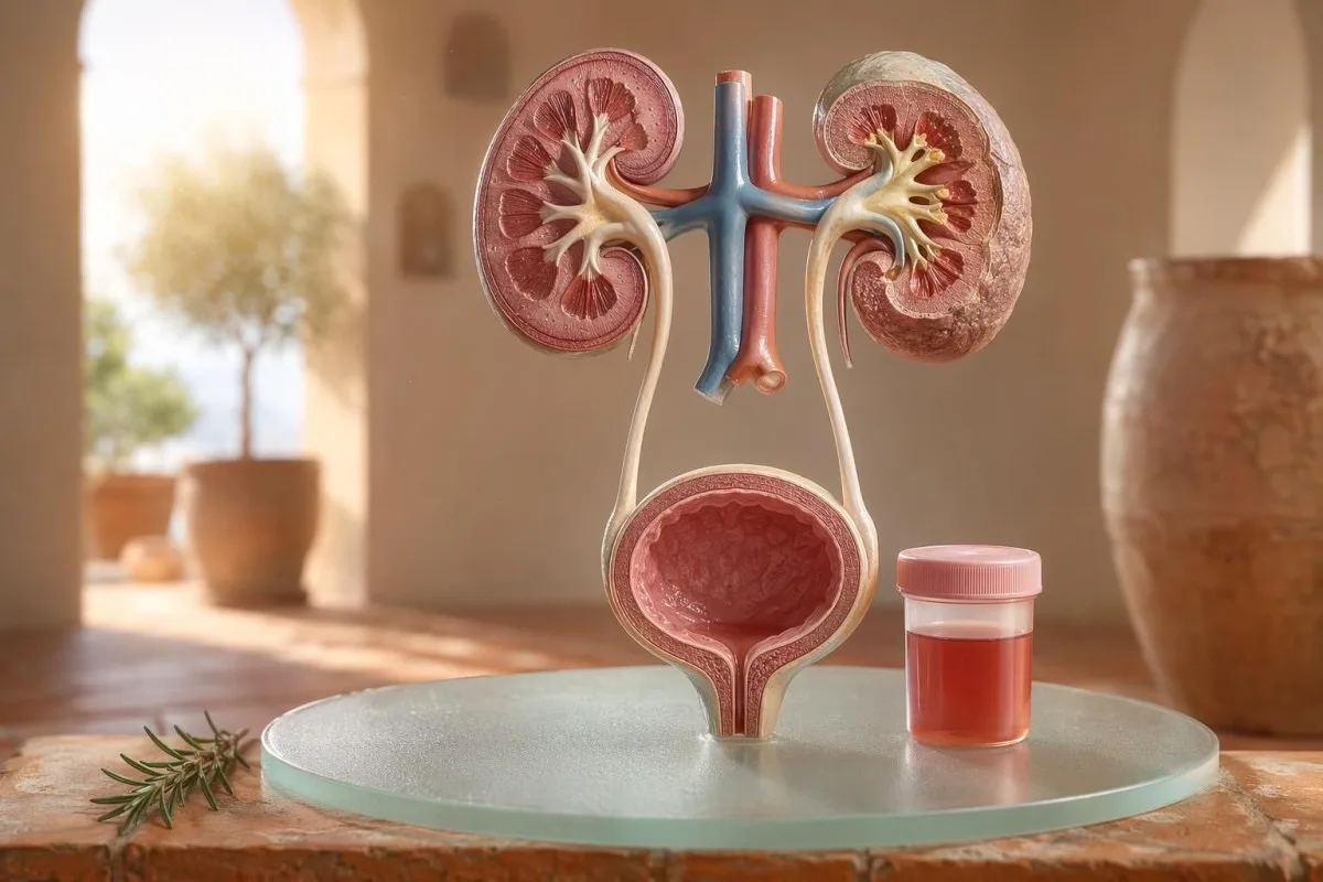
راهنمای «بیمار-محور» برای هیماتوریِ قابلمشاهده و میکروسکوپی، شامل اینکه چرا خونِ دیپاستیک با گلبولهای قرمز زیر میکروسکوپ یکسان نیست.


- خون در ادرار یعنی ادرارِ قرمزِ قابلمشاهده، رنگِ چای یا رنگِ کولا، یا هیماتوریِ میکروسکوپی که در آزمایش یافت میشود؛ خونِ قابلمشاهده همیشه نیاز به پیگیری پزشکی دارد.
- هماتوری میکروسکوپی معمولاً بهعنوان 3 یا بیشتر RBC در هر میدانِ پُرقدرت در نمونهٔ میکروسکوپیِ ادرار که بهدرستی جمعآوری شده تعریف میشود.
- خونِ دیپاستیک فعالیتِ هِم را تشخیص میدهد، بنابراین ممکن است از گلبولهای قرمزِ سالم، هموگلوبینِ آزاد یا میوگلوبین پس از آسیب عضلانی مثبت شود.
- نیشانەکانی UTI شامل سوزش، تکرر، استراز لکوسیت، نیتریتها و گلبولهای سفید است؛ باید هیماتوری پس از برطرف شدن عفونت دوباره بررسی شود.
- رسوب ادرار یافتههایی مانند RBCهای دیسمورفیک، استوانههای RBC یا پروتئینِ قابلتوجه به بیماریِ واحدهای فیلترکنندهٔ کلیه اشاره میکنند.
- استوانههای ادرار مادەی قالبگیریشوە لە لایەکانی تیووبەکانی کلیە؛ کاسـتـی سلولەی سوور (red cell casts) هەمان هەفتە ڕەنگی ئاگاداربوونەوەی نێوانەوەی نێوانەوەی کلیەیە (nephrology) ــ بە تایبەتی لەگەڵ بەرزبوونی فشاری خوێن یان کەمبوونی eGFR.
- ئاگاداربوونەوەکان لەسەر سەرتاسەری (Cancer) دەستپێدەکات بە هەڵسوکەوتی خوێن لە پیشاب بەدون نەخۆشی (painless visible hematuria)، لەخۆڵەکان (clots)، تەمەنی زیاتر لە ٤٥ ساڵ، مێژووی سیگارکێشان، بەکارهێنانی مادە کیمیاوی لە کاری، و هەڵسوکەوتە تێکچووەی هەستیار کە بەهۆی نەناسراو تکرار دەبێت.
- هەیماتۆریا لەگەڵ ڕاهێنان (Exercise hematuria) زۆرجار لە ماوەی ٤٨-٧٢ کاتژمێردا کەم دەبێتەوە؛ خوێن بەردەوام لەدوای ئارامگرتن نابێت بە ڕانین یان دوورکەوتن بە دوچرخه (cycling) بەهێڵ بکرێت.
- پێداچوونی هەراسان پێویستە بۆ خوێن لە پیشاب لەگەڵ تێکچوونی هەستیار (fever)، نەخۆشی لە بەرەوەی پهلو (flank pain)، کڵۆت کە دەبێتە هۆی بەستنی پیشاب، لەبارداری (pregnancy)، تروما (trauma) یان نەخۆشی/نیشانەکانی نەخۆشی سووربوونی خوێنی زۆر (severe anemia).
وجود خون در ادرار چه معنایی دارد پیش از اینکه وحشت کنید
خون در ادرار دەتوانێت لە هەڵسوکەوتی هەڵچوون (infection)، سنگ (stones)، نەخۆشییەکی فیلترکردنی کلیە (kidney filtering disease)، شێوەی پڕۆستات یان کێشەکانی پەردەی پیشاب (bladder conditions)، دارو (medicines)، ڕاهێنان (exercise)، یان ککړبوون لەگەڵ ماوەی قەدەغەکان (periods) هاتوو بێت. قاعدەی کارییەکە سادەیە: خوێنی دیار (visible blood)، هەیماتۆریای میکروسکوپی تکراربوو (repeated microscopic hematuria)، یان هەیماتۆریا لەگەڵ نەخۆشی (pain)، تێکچوون (fever)، کڵۆت، فشاری خوێنی بەرز، پڕۆتێن (protein)، یان کارکردنی کەمبووی کلیە (reduced kidney function) پێویستی بە بەدوای هەڵسەنگاندنی خێرا هەیە، نەک چارەسەری بەبێ تێکچوون (watchful waiting).
یەک جار خوێنی سوور-ڕەنگ (pink) لە کاسەی توالێت لەدوای beetroot یان سەرپێچی نوێ (new supplement) هەمان کێشەی کلینیکی نییە بەوەی پیشابی ڕەنگی کۆلا (cola-colored urine) لەدوای نەخۆشی گەلو (sore throat). لە پڕاکتیکمدا، ڕەنگەکە زۆرجار دەڵێت کوا لە سەرەتا بپرسین: سوور-ڕەنگی ڕوون بە کڵۆت دەگەڕێتەوە بۆ لایەکانی کێشەی ناوەوەی ڕێگای پیشاب (lower urinary tract)، بەڵام پیشابی قاوەیی (brown) یان ڕەنگی چای (tea-colored) دەتوانێت مانگەی هێمەی کۆنتر (older heme pigments) لە فیلترەکانی کلیە بگەیەنێت.
هەیماتۆریا نەک دیانۆزە (diagnosis) ــ بەڵکو نیشانەیە (sign). هەیماتۆریای دیار (Visible hematuria) واتە دەتوانیت ڕەنگەکە ببینیت لە گۆڕانەوە، بەڵام هەیماتۆریای میکروسکوپی (microscopic hematuria) واتە پیشابەکە دەبێت ڕەنگی ڕاستەقینە/عادی بنووسێت، بەڵام لە میکروسکوپدا سلولەی سوور دەبینرێت، زۆرجار لە ٣ یان زیاتر سلولەی سوور لە هەر high-power field.
Kantestî yek e Analyzerê testa xwînê ya AI کە یارمەتیدەدات بە نەخۆشەکان ببەستنەوەی ئەنجامەکانی پیشاب بە نیشانەکانی کلیە وەک creatinine، eGFR، CRP، hemoglobin و platelets لە یەک کاتنامەدا. ئەگەر ڕەنگەکە ڕێک نەکەوتوو بێت، our ڕێنمای ڕەنگی پیشاب (urine color guide) دەڵێت بۆچی هەموو ڕەنگەکانی سوور، قاوەیی، نارنجی و زەردی تۆخی (dark yellow) هەمان واتای نییە.
خونِ دیپاستیک با RBCها در میکروسکوپی یکسان نیست
دیپستیک (urine dipstick) ـەکە نیشان دەکات کە خوێن بەهێزە (blood positive) کاریگەرێکی کیمیاوی شێوە-هێمە (heme-like) دەدۆزێتەوە، نەک هەروەها سلولەی سووری ڕاستەقینەی بەهێز (intact red blood cells). میکروسکوپ وەڵامی پرسیاری دواتر دەدات: ئایا RBC ـی ڕاستەقینە هەیە لە کۆمەڵەی خاکی پیشاب (urine sediment)، و ئەگەر هەیە، چۆن دەبینرێن؟
تاقیکردنەوەی دیپستیک خێرایە چونکە پەدێ ڕەکێش (reagent pad) لەگەڵ کاریگەرێتی peroxidase ـی هێمە ڕێکدەکەوێت. ئەمەش واتە ئەنجامی بەهێز دەتوانێت لەگەڵ RBC ـی بەهێز (intact RBCs) ڕوو بدات، یان hemoglobin ـی ئازاد لەدوای شیکردنی سلولەی سوور، یان myoglobin لەدوای تروما/لەخۆچوونی گرنگی ماسی (significant muscle injury).
میکروسکوپ کندترە بەڵام تایبەتمەندی زیاتر هەیە. لابراتۆرییەکی بەدقت دەکاتەوە: کۆمەڵەی خاکی پیشاب (urine sediment), ، و RBC ـەکان دەنووسێت لە هەر high-power field؛ زۆر پزیشک بە ٠-٢ RBC/hpf وەک عادی دەزانن، و ٣ یان زیاتر RBC/hpf وەک نادیار/غیرعادی لە نێرەکان (adults).
گرنگی ناسازگاری (mismatch) هەیە. دیپستیک کە ٣+ خوێن دەنووسێت و ٠-٢ RBC/hpf دەبینێت، من وادەکەم بپرسین لەسەر rhabdomyolysis، hemolysis، کۆنتامێنێنتی (oxidizing contaminants) ـی هەڵهێنەر/خراپکار، و ڕێکخستنی نموونە (specimen handling) ــ نەک بەهێزەوە بڕۆین بۆ نەخۆشییەکی پەردەی پیشاب (bladder disease)؛ جیاوازییەکە شێوەی هەمان بیرکردنەوەیە لەسەر our بەراوردی ئازمایشەکانی ئێدرار و کەلتەر.
چه زمانی سرنخهای UTI هیماتوری را توضیح میدهند و چه زمانی توضیح نمیدهند
نەخۆشی ڕێگای ئێدرار (UTI) دەتوانێت هۆکار بێت بۆ خوێن لە ئێدرار, ، بە تایبەتی لەگەڵ سوتاندن، خێرایی/فراوانی (urgency)، زۆربوونی هەست بە پێویستی (frequency)، ناراحەتی لە ناوچەی لگن (pelvic discomfort)، leukocyte esterase، nitrites و سلولە سپییەکان. بەڵام هەیماتۆریا دەبێت لەدوای چارەسەری UTI ڕوون بێت؛ RBC ـی بەردەوام لەدوای UTI پێویستی بە دووبارە سەیرکردن هەیە.
لە UTI ـی سادەی سەرەوەی ڕێگای ئێدرار (lower UTI) ـدا، من دەستم بە کۆمەڵێک نیشانە دەکەم: سوتاندنی ئێدرار (dysuria)، فراوانی (frequency)، leukocyte esterase بەڵێنە، pyuria زۆرجار لەسەر 5-10 سلولە سپی/hpf زیاترە، و هەندێک جار nitritesیش دەبینرێت. بەڵگەی کەلتەری ڕەوتی (traditional culture positivity) 100,000 CFU/mL ـە، بەڵام لە ژنانە هەستیارەکان (symptomatic women)، کەمترەکان وەک 1,000-10,000 CFU/mL هێشتا دەتوانێت لە ڕووی کلینیکی گرنگ بێت.
Leukocyte esterase ئەزیمێکی سلولی سپییە، بەڵگەی سەختی بۆ باکتریا نییە. ئەنجامی بەڵێن لەگەڵ هەیماتۆریا دەتوانێت لە ڕێکەوتی نەخۆشی، سنگ، تێکەڵبوون (contamination)، یان هەڵکەوتنی هەڵسوکەوتی کلیە (kidney inflammation) ڕوو بدات؛ بۆیە our راهنمای leukocyte esterase جیا دەکاتەوە لە نیشانەی بەکارهێنراوەکان لەگەڵ هەڵەی زۆر هەڵەیی (common false positives).
Nitrites زۆر تایبەتمەندن بەڵام کەمتر دەستەبەر دەکات (less sensitive) چونکە هەموو باکتریا ناترات بۆ nitrite نایگۆڕێن. ئەگەر نیشانەکان ڕەوتی/کلاسیک بن بەڵام nitrites منفی بێت، کەلتەر هێشتا دەتوانێت بەڵێن بێت؛ زانیاری ڕووناکی ئەمە لە our ڕوونکردنەوەی ئەنجامی nitrite.
هیماتوریِ قابلمشاهده: سرنخهای رنگ، لختهها و زمانبندی
هەیماتۆریای دیار (visible hematuria) هەرگیز شتێک نییە کە من بەسەرچاوەی سادەوە بێت، تەنانەت ئەگەر یەکجار ڕوو بدات. ئێدراری سوور-ڕەش (bright red urine)، کڵۆت (clots)، یان خوێنڕێژی بەبێ سوتاندن (painless bleeding) پێویستی بە هەڵسەنگاندنی بەهێز هەیە، چونکە لەگەڵ زیادبوونی تەمەنی و کەشەی سیگار (smoking exposure)، هۆکارەکانی ڕێگای ئێدراری سەرەوە (lower urinary tract causes) زیاتر دەبنە هەمل.
ئەو کاتەبەندییە بە شێوەیەکی هێمن دەکرێت بەکارهێنانی سوودبەخش بێت. تەنها خوێن لە سەرەتای ڕۆیشتنی پێشەوە دەکرێت ڕوون بکاتەوە کە لە یورێتراوە هاتووە، خوێن لە هەموو کاتێکی ڕوونەکەدا دەکرێت لە سەرچاوەی وەشانی یان یان لە کلیەوە بێت، و خوێنی کۆتایی لە نزیک کۆتایی هەڵکەوتنەکەدا هەندێک جار دەلالەت دەکات بە ناوچەی دەریاچەی وەشانی (بلاډر نێک).
کڵۆتس (لختهخوێن) زۆرجار مانای ئەوەیە کە خوێڕێکە لە خوارەوەی فیلترەکانی کلیەوە دەهاتووە، چونکە لختهخوێن بە ئاسانی لە دوای تێپەڕبوون لە گلۆمەڕۆڵییەکان و تیووبولەکان دروست نابێت. لختهخوێنێک کە ڕێگای ڕۆیشتنی پێشەوە دەبەستێت فوریتە: پڕبوونەوەی وەشانی دەکرێت لە ماوەی چەند کاتژمێرێکدا دەردناک بێت، و پێویستی بە ڕێکخستنی کاتێتەر هەیە.
پیاوان هەندێک جار پندار دەکەن کە هیماتۆریای دیار (visible hematuria) دوای نەخۆشی/نیشانەکانی پێشەوە تەنها هەڵسوکەوتی ئازاری پڕۆستاتە. ئەمە دەکرێت ڕاست بێت، بەڵام ئەگەر PSA لە کاتی یان بە نزیکەی دوای هەڵچوونی هەڵچوون (UTI) چەک کرابێت، ڕوونکردنەوەکە دەبێت بە شێوەیەکی ئاڵۆز؛ PSA دوای UTI ڕوون دەکات کە بۆچی زۆربەی کلینیسینەکان 6-8 هەفتە دەوەستێن بۆ دووبارە چەککردنی PSA.
آستانههای هیماتوریِ میکروسکوپی که پزشکان استفاده میکنند
هیماتۆریای میکروسکوپی زۆرجار بە شێوەیەکی وەک 3 یان زیاتر RBC لە هەر فیلدێکی بەهێزی زۆر (high-power field) لە نموونەی پێشەوەیەکی بە شێوەیەکی تەواو کۆکراو. ڕێنمایی AUA/SUFU ئەم حدە بەکار دەهێنێت و پێشنیار دەکات بە بەستنی خەتەر (risk-based evaluation) بڕوانین، نەک چارەسەری هەموو نەخۆشێک بە یەک شێوە (Barocas et al., 2020).
تەنها دایپستیک (dipstick) نابێت بەکار بێت بۆ دۆزینەوەی هیماتۆریای میکروسکوپی. لە ڕێنمایی AUA/SUFU لە Barocas et al. 2020، ناسنامەکە پەیوەستە بە میکروسکوپی، چونکە دایپستیکەکان دەتوانن بەهۆی هێم پیگمنتەکان (heme pigments) بەبێ ئەوەی ڕەنگەڵە سوورەکان (red cells) بە تەواوی هەبن، بەردەوام/مثبت بن.
دووبارە چەککردن نەبێت بە مانای نەهێزی؛ ئەوە پزیشکی باشە. ئەگەر کەسێک لە دوای وەرزشێکی زۆر، هەستەوەی هەردەمی (fever)، سێکس، منداڵبوون/خۆڵەوە (menstruation)، یان کۆکردنێکی کەڵکەوتوو (contaminated collection) 3-10 RBC/hpf پیشان بدات، من زۆرجار دەمەوێت نموونەی پاکی دووبارە پێشکەش بکرێت پێش ئەوەی سکان (scans) داوا بکەم، وەک ئەوەی کە پێمان دەڵێت بۆ لابراتۆرییەکانی غیرعادی دووبارە.
خەتەر ڕێگاکە دەگۆڕێت. یەک 24 ساڵە کە لە دوای نیمەماراثۆن 4 RBC/hpf دەبینێت، لەگەڵ یەک 68 ساڵەی پێشتر سیگاری سەرکەوتوو (former smoker) کە لە دوو نموونەدا 25 RBC/hpf دەبینێت یەکسان نییە، هەرچەند هەردووکیان تەکنیکی لە هەمان ناسنامەی هیماتۆریا دەچن.
رسوب ادرار، پروتئین و استوانههای ادراری که روند بررسی را تغییر میدهند
رسوب ادرار دەتوانێت ڕوون بکاتەوە کە هیماتۆریا زۆرتر لە فیلترەکانی کلیەوە دەهاتووە. ڕەنگەڵە سوورەکان (red cells)ی ناسازگار (dysmorphic)، لختهکەستەکان/کستەکانی ڕەنگەڵە سوور (red cell casts)، و گۆڕانی گرنگ لە سەرەوەی پروتێن، نیگەرانی دەکاتەوە بۆ نەخۆشی گلۆمەڕۆڵی (glomerular disease) نەک تەنها نەخۆشی سادەی وەشانی.
ڕەنگەڵە سوورەکان کە وەک یەکسان نەبن، blebbed بن یان وەک شێوەی ڕینگ (ring-shaped) بن، دەوترێن dysmorphic RBCs، و دەلالەت دەکەن بە تێپەڕبوون لە فیلترێکی گلۆمەڕۆڵی هەڵسوکەوت/سووتاو. Acanthocytes لە سەرەوەی نزیکەی 5% لە ڕەنگەڵە سوورەکانی پێشەوە زۆرجار وەک ڕێنماییەکی قووڵ بۆ گلۆمەڕۆڵی چارەسەری پێدەدرێت، بەڵام لابراتۆرییەکان جیاوازن لە چۆنیەتی ڕاپۆرتکردنی بە چەند ڕێک و باوەڕپێکراو.
پروتێن دووەمین ڕێنماییە کە من پێی دەکەمەوە و پشتگوێی ناکەم. نێسبەتی ئالبومینی پێشەوە بە کرێاتینین (urine albumin-creatinine ratio) لە سەر 30 mg/g، یان نزیکەی 3 mg/mmol، ناسازگارە، و هیماتۆریای دیار لەگەڵ پروتێن دەبێت زووتر ڕەوی پشکنینی کلیە-محور بکرێت؛ ئەو ڕێنمایی پڕۆتێن لە ڕێژەدا حدەکان لە سەطحی نەخۆش (patient-level thresholds) پێشکەش دەکات.
استوانههای ادرار ئەمانە قالبی بچوکن (tiny molds)ن کە لە ناو تیووبولەکانی کلیە دروست دەبن. Hyaline casts دەتوانن دوای کەمبوونەوەی مایعات (dehydration) یان وەرزش پەیدا بن، بەڵام red cell casts ناسازگارە و زۆرجار دەلالەت دەکات بۆ glomerulonephritis؛ بۆ ڕێبەریەکی گەورەتر بۆ urinalysis، سەیری ئەو rêbernameya tevahî ya analîza mîzê.
سنگها، ورزش و علل موقتیِ هیماتوری
سنگەکان و وەرزشی زۆر دەتوانن هیماتۆریا دروست بکەن، بەڵام کاتەکان/تایملاینەکان جیاوازن. هیماتۆریای پەیوەندیدار بە سنگ زۆرجار لەگەڵ دەردی بەرەوپێش (flank pain) یان بلور/کریستالەکاندا دەبینرێت، بەڵام هیماتۆریای وەرزش زۆرجار دەبێت لە ماوەی 48-72 کاتژمێر لە دوای ڕامان (rest) کەم بێت و کۆتایی پێ بێت.
سنگەکانی کلیە بە شێوەی کلاسیک دەردی موجی بەرەوپێش (waves of flank pain)، نەخۆشی/تهقە (nausea)، و هیماتۆریای میکروسکوپی یان دیار دروست دەکەن. کریستالەکانی calcium oxalate شتێکی سنگ ڕاستەوخۆ نیشان ناکەن، بەڵام کاتێک لەگەڵ دەردی colicky جفت بن، نیگەرانی زیاتر دەکەن؛ ئەو ڕێنمایی کریستالەکانی calcium oxalate ڕوون دەکاتەوە کە ڕێنمایی میکروسکوپی پێشەوە چییە.
هیماتۆریای وەرزش ڕاستە. من بینیومە دوندەماراثۆنڕانەرەکان (marathon runners) لە سەرەتا/بەیانی ڕۆژی دوای ڕاگەیشتن (race) 10-20 RBC/hpf دەبینن، کەلتورێکی ڕاست/نرمال (normal culture) هەیە، پروتێن نییە، و لە 3 ڕۆژ دوای ئەوە urinalysis دووبارە بە تەواوی نرمالە.
مەترس ئەوەیە هەموو شتێک بەسەر ڕاهێنان (training) ببهستین. ئەگەر هیماتۆریا لە دوای 72 کاتژمێر هەبوو بمێنێت، لەگەڵ پێشەوەی تارەوە وەک cola دەردەکەوێت، یان لەگەڵ CK بەرز، کرێاتینین لە خوارەوە/دەبەرزبوون (rising creatinine)، یان دەردی ماسی (muscle pain) هاتووە، سەیری لای وەشانی بکەوە؛ ئەو راهنمای کراتینینِ ورزش همپوشانیِ کلیه و عضله را پوشش میدهد.
«پرچمهای قرمز» کلیه که پزشکان نادیده نمیگیرند
هیماتوریا همراه با پروتئین، فشارخون بالا، ورم، eGFR کاهشیافته، استوانههای گلبول قرمز یا بالا رفتن سریعِ کراتینین یک علامت هشدارِ کلیه است. این یافتهها نشان میدهند که واحدهای فیلتراسیون ممکن است ملتهب باشند، و منتظر ماندن چند ماه میتواند کارکرد کلیه را از دست بدهد.
KDIGO 2024 بیماری مزمن کلیه را با نشانگرهای آسیب کلیه تعریف میکند، مانند آلبومینوری، یا eGFR کمتر از 60 میلیلیتر/دقیقه/1.73 مترمربع به مدت حداقل 3 ماه. با این حال، در وضعیتهای حاد، هیماتوریا همراه با افزایش کراتینین طی روزها یا هفتهها بهتنهایی کافی است تا پیش از برآورده شدن تعریفِ 3 ماهه، اقدامِ تشدیدی انجام شود.
ئەڵبوومینیورییا زۆرجار هۆشداری سەرەتایییە. ACRی 30-300 mg/g بە زیادبوونی مامناوەندی دادەنرێت، زیاتر لە 300 mg/g بە زیادبوونی توند دادەنرێت، و هاوتایکردنی هەر یەکێکیان لەگەڵ هێماتوریا، قسەکان دەگۆڕێت؛ ئێمە ڕێبەری ACRی گورچیلە ڕووندەکاتەوە کە بۆچی ئەڵبوومینی میز پێش نیشانەکان زیان دەدۆزێتەوە.
پەستانی خوێن گرنگە لێرەدا. پەستانی خوێنی نوێی 160/100 mmHg لەگەڵ میزێکی وەک کۆلا و ئاوسانی قاچ پاش هەوکردنی قوڕگ وام لێدەکات بیر لە گڵۆمێرولۆنیفریت بکەمەوە، نەک هەوکردنی میزڵدان؛ ئەگەر eGFR ناڕوون بێت، ڕێبەری دووبارە پشکنینی GFR توضیح میدهد چه زمانی سیستاتین C میتواند عملکرد کلیه را روشنتر کند.
سرنخهای خطر سرطان بدون وحشت
هیماتوریا قابل مشاهدهٔ بدون درد، بهویژه بعد از سن 45 سالگی، نیاز به ارزیابی فوری دارد، چون سرطانهای مثانه و مجاری ادراری فوقانی میتوانند به این شکل ظاهر شوند. NICE NG12 ارجاعِ مشکوک به سرطان را برای هیماتوریا قابل مشاهدهٔ بدون علت در بزرگسالان 45 سال و بالاتر، یا هیماتوریا قابل مشاهدهای که بعد از درمان UTI ادامه پیدا میکند، توصیه میکند.
بیشترِ هیماتوریا سرطان نیست. با این حال، مواردی که به یاد دارم همانهای بیصدا هستند: بدون درد، بدون تب، بدون سوزش؛ فقط دو بار ادرارِ قرمز در یک فرد 56 سالهٔ سیگاری که تقریباً آن را نادیده گرفته بود.
ریسک تجمعی است، نه دودویی. سن بالای 45-50، سابقهٔ سیگار کشیدن، مواجهه با سیکلوفسفامید، پرتودرمانی لگنی، هیماتوریا قابل مشاهدهٔ عودکننده، مواجههٔ شغلی با آمینهای آروماتیک، و لختهها همگی نیاز به سیستوسکوپی یا تصویربرداری را افزایش میدهند.
نیشانەکانی پڕۆستات هۆکارەکانی میزڵدان ناڕەوێننەوە. ئەگەر نیشانەکانی میزکردن، گۆڕانکارییەکانی PSA و هێماتوریا پێکەوە بون، کات و بارودۆخی هەوکردن بە وردی دووبارە بکەرەوە؛ ئێمە راهنمای نشانەی پروستات ڕوون دەکات بۆچی PSA تەنها یەک بەشە لە بەسەرداچوونی پشکنینی ڕێگای پیشاب لە پیاوان.
داروها، ضدانعقادها و هشدارهای کاذب
خوێنڕقیقکەرەکان دەتوانن ڕوونکردنەوەی خوێربوون پیشان بدەن، بەڵام نابێت بە هەموو شێوەیەک خۆکارەکەی هەمووەڕاستەی هەموچوون (hematuria) بکرێن. هەموچوونی هەڵبەستراو بە خوێنڕقیقکەر هنوز پێویستی بە پشکنین هەیە، چونکە ئەم داروە دەتوانێت سنگ، نەخۆشی/وێرانی (infection)، نەخۆشیی کەلیە (kidney disease) یان ڕووی ڕێگای پیشاب (urinary tract growth) ڕوون بکاتەوە.
وارفارین، ئاپیکسابان، ریڤاروکسابان، ئاسپرین و کلوپیدوگرێل دەتوانن خوێربوون ڕوونتر بکەنەوە. ئەگەر INR لە ڕێژەی مەودای هەدەف زیاتر بێت، ڕاستکردن گرنگە، بەڵام INRی ئاسایی هەموچوونی ڕوونبوو (visible hematuria) بیخطر ناکات.
هەندێک هۆکاری تر کە خوێربوون نییە دەتوانن شێوەی هەموچوون هەمانان بنوێنن. ڕیفامپیسین دەتوانێت پیشاب ڕەنگی نارنجی-سوور بکات، فێنازۆپیریدین دەتوانێت پیشاب ڕەنگی نارنجی-سووری ڕوون بکات، چەقەڵ/بێتڕوت دەتوانێت پیشاب لە کەسانی لەسەرەوە ڕەنگی لکاندن بدات، و نەهێشتنی مایە (dehydration) دەتوانێت ڕەنگدەرەکە بەقەدەر توند بکات کە بیماران ترسان بکات؛ پشکنینی ایمنی خوێنڕقیقکەرەکان لە لابراتوارەکانی ئاسایش/ئەمنیەتی خوێنڕقیقکەرەکاندا ڕوونکراوە.
دیپستیک (dipstick) هەروەها دەتوانێت دووربکەوێت/گمراه بکات لەدوای هەستەوەری سەخت یان ئاسیبێکی میوە. پەیەی هێم (heme pad) بە بەدەستهێنانی پۆزەتیڤ و کەم یان هیچ RBC نەبێت، فکرم دەکاتە سەر میوگلوبین، بە تایبەتی ئەگەر CK لە 1,000 IU/L زیاتر بێت و پیشاب توند/تاریک بێت.
کودکان، بارداری و آلودگیِ مربوط به پریود
هەموچوون لە منداڵان، لە ماوەی نەخۆشی/بارداری (pregnancy) یان لە نزیک قەبارەی مانگی (menstruation) پێویستی بە نموونەگیری پاکتر و ئاستێکی کەمتر بۆ سەردان/لەبەرچاوگرتن هەیە. هەمان ئەنجامی پیشاب دەتوانێت واتای جیاواز هەبێت بە پێی تەمەنی، دۆخی بارەداری، هەبوونی تێکچوون/تێکچوونی هەستەوە (fever)، فشاری خوێن (blood pressure) و ڕێکخستنی نموونە.
کەڵەکانی مانگی (menstrual contamination) زۆر گرنگە و هیچ کەس نابێت خەجلەدار بێت لەبەر ئەوە. ئەگەر ئەنجام ناڕەوا بێت، دووبارە نموونەی midstream پاک-گرتن (clean-catch) بکە لە کەمتر لە 48 کاتژمێر پاش کۆتایی هاتنی خوێربوون؛ هەروەها ئەگەر 3 یان زیاتر RBC/hpf بەردەوام بێت، ئەوا واتادارتر دەبێت.
منداڵان زۆرجار پێویستی بە ڕەخنە/چاوێکی جیاواز هەیە. هەموچوون پاش نەخۆشی وێرەسی دەتوانێت کاتێکی بێدەنگ بێت (transient)، بەڵام هەموچوون لەگەڵ پەفکردن/سووژان (swelling)، فشاری خوێنی بەرز، پڕۆتێن، یان کەمبوونی دەرهێنانی پیشاب پێویستی بە ڕەوێنەی زوو لە لای دکتۆری منداڵان (pediatric review) هەیە.
بارەداری (pregnancy) بەرزتر دەکاتەوە گرنگی کارەکان، چونکە UTI، سنگ، فشارخوێنی بەرز لەسەرەوەی بارەداری (preeclampsia) و ستریسی نەخۆشیی کەلیە (kidney stress) لەسەرەوەی بارەداری و کەڵەکانی مانگی (contamination) دەتوانن یەکدیگر پێکەوە بپێچێن. بۆ ڕێما/پەتەرنە ئاگادارکردنەوەی بارەداری لە یەک ڕۆژدا کە لەسەر نەخۆشیی کەلیە، کەبد (liver)، پلیتڵت (platelet) و ئەنجامەکانی پیشاب دەگرێت، سەیری پرچمهای قرمز آزمایشهای بارداری.
آزمایشهایی که پزشکان معمولاً بعد از هیماتوری درخواست میکنند
پشکنینە سەرەتایی/دواترین پشکنینەکانی ئاسایی پاش هەموچوون بریتییە لە دووبارەکردنی urinalysis بە میکروسکوپ (microscopy)، کشتکردن/کالچر (urine culture) ئەگەر هەملانەی نەخۆشی هەبێت، تاقیکردنەوەی کارکردی نەخۆسی لە خوێن (kidney function blood tests)، پڕۆتێنی پیشاب یان ACR، ڕێکخستنی فشاری خوێن، و وێنەبردن (imaging) یان سیستۆسکۆپی (cystoscopy) بە پێی مەترسی. توالی/ڕیزبەندی دیار بە پێی نیشانەکان و مەترسی دەگۆڕێت.
بۆ نیشانەکانی سست/ناڕەخنەدار (uncomplicated) لەسەر سیستیت (cystitis)، کشتکردن و چارەسەری دەتوانێت یەکەم بێت، دواتر دووبارە میکروسکوپی پیشاب پاش کەمبوونی نیشانەکان. بۆ هەموچوونی ڕوونبوو بەبێ نەدرد (painless visible hematuria)، زۆر پزیشک زووتر دەچنە سەر imaging و cystoscopy، چونکە منتظربوون بۆ هەموچوونی دووبارە دەتوانێت دۆزینەوە (diagnosis) دواکەوتوو بکات.
تاقیکردنەوەی خوێن کۆنتێکستی زیاد دەدات کە تەنها پیشاب ناتوانێت پێی بدات. Creatinine، eGFR، urea یان BUN، electrolytes، CBC، CRP، complement C3/C4 و نیشانەکانی خودکار-بەخشی (autoimmune markers) دەتوانن بەجێ بێت ئەگەر گومان لەسەر سووزاندنی نەخۆسی هەبێت؛ ڕێژەی BUN/creatinine ڕوون دەکات یەک ڕەنگ/پەتەرنی زۆر بەکارهاتووی نەخۆسی.
Kantesti AI دەتوانێت ئەم ئەنجامانە ڕێکبکات بۆ نمایشێکی کۆنتێکستی ڕێژەیی (trend view)، بەڵام پشکنینی پزیشک هێشتا گرنگە ئەگەر پرچم/نیشانەی ئاگاداربوون (red flags) هەبێت. ڕێوشوێنی ئاسایش/ئەمنیای کلینیکی ئێمە لە Pejirandina Bijîşkî, ڕوونکراوە، لەوانەش چۆن خوشە/کۆمەڵە-ئەنجامە ناڕەواکان دەگۆڕێن بەجای تەنها ژمارەیەکی جیاواز.
اینکه چگونه Kantesti به سازماندهی زمینهٔ هیماتوری کمک میکند
Kantestî yek e خزمەتگوزاری تێکست/تێگەیشتنی تاقیکردنی لابراتۆریی AI کە نیشانەکانی خوێن نزیک-پیشاب (urine-adjacent blood markers) لە کۆنتێکستدا دەخوێنێت، وەک creatinine، eGFR، CRP، hemoglobin، platelets و glucose. ئەوە hematuria دۆزینەوە ناکات، بەڵام یارمەتیدەری بیماران دەکات بۆ ئامادەبوون بۆ کورتە-ڕاپۆرتێکی پاکتر و بەکارهێنانی زیاتر بۆ پزیشکەکەیان.
کاتێک من hematuria دەبینم، زۆرجار تەنها یەک ئەنجام لەبەرچاو دەگرم. creatinineی ئاسایی 0.82 mg/dL، eGFRی بێگۆڕان لەسەر 90، نەبوونی پڕۆتێن و کشتکردنی منفی (negative culture) بەکلی جیاوازە لە hematuria لەگەڵ eGFR 48 و CRP 65 mg/L.
Kantestî yek e پلاتفۆرمی تێکڕوانینی بایۆمارکەری AI کە دەتوانێت PDF-ەکانی تاقیکردنەوەی خوێن یان وێنەکان کە بارکراون (uploaded) پڕۆسە بکات و نیشانەکانی نەخۆشیی کەلیە، سووزاندن و نەهێلی/ئانێمی (anemia) کە لە ماودای ڕێژەیی (out-of-range) دابنێت لەگەڵ کات/هەنگاوی نیشانەکان. ڕێبازەکانی پشت ئەو خوێندنە پەتەرنە لە ڕێنمایی تەکنەلۆژی, ، و کۆمەڵە مارکرە گەورەترەکان لە rêbernameya nîşankerên biyolojîk.
یەک نکتەی کاری: پێش بارکردن، سەیری بکە کە یەکایەکان (units)، ڕێکەوتەکان (dates) و ناسنامەکانی نەخۆش (patient identifiers) بە شێوەی ڕاست دەگیرێن. هەڵەکانی OCR دەتوانێت 0.9 بگۆڕێت بۆ 9.0، کە حکایەتی نەخۆشیی کەلیە بەکلی جیاوازە؛ ئێمە ڕێنمای بارکردنی PDF (PDF upload checklist) نیشان دەدات هەڵەکان کە من داوای لێکردن لە نەخۆشەکان بکەن یەکەم جار بزانن.
یادداشتهای پژوهشی، حاکمیت و چکلیست نهایی
لە 11ی تەمموزی 2026ەوە، بەهێزترین پلانی نەخۆش بۆ ئەوە دەبێت دڵنیابین لەوەی هەیماتۆریا (hematuria) ڕاستە یان نا، ڕەخنەی هەڵسوکەوتی هەڵبژاردن/نیشانەکانی هەڵوەشاندن (infection) بدۆزینەوە، ئاگادارییەکانی کلیە (kidney) بدۆزینەوە، و هەیماتۆریای دیار یان بەردەوام کە بەرز دەبێت بەرز بکەینەوە (escalate). تەنها بە ڕەنگی ئاوەڵە (urine) مەبەستە.
من توماس کلاین، MDـم، و ڕێنماییەکەم بە شێوەی هەوڵدانەوەی خۆبەهێز (conservative) دەدەم، چونکە هەیماتۆریای لەبیرچووەکە گرنگ دەبێت. ناوەندی Kantesti ـی زانیاری پزیشکی لە ژێر چاودێریی کلینیکی لە Lijneya Şêwirmendiya Bijîşkî, ـەوە ڕەسەن دەکرێت، و ڕۆڵمان ئەوەیە یارمەتیدەین نەخۆشەکان پرسیارە باشتر بکەن، نەک ئەوەی پزیشکەکەیان جێگر بگرین.
Kantestî yek e ئامێری توێژینەوەی تاقیکردنەوەی خوێن بە پشتبەستن بە AI بە میلیۆنان کەس بەکاردەهێنن لە 127 واڵت، بۆیە نهێنی (privacy) و ڕوونی کلینیکی دەبێت لە ناوەڕاستی ڕێکخستنی کار (workflow) دروست بکرێت، نەک لە دواتر زیاد بکرێت. دەتوانیت زانیاری زیاتر لەسەر Kantesti LTD، ڕێکخستنی کۆمپانیایمان لە بریتانیا (UK) و مامەڵە/ماموریتی کلینیکی لە Çûna nava.
گرووپی توێژینەوەی کلینیکی AI ـی Kantesti. (2026). ئەنالیزەری تاقیکردنی خوێنی AI: 2.5M تاقیکردنەوە لێکۆڵکرا | ڕاپۆرتی تەندروستی گڵۆبال 2026. Zenodo. DOI: https://doi.org/10.5281/zenodo.18175532. ResearchGate: https://www.researchgate.net/. Academia.edu: https://www.academia.edu/.
گرووپی توێژینەوەی کلینیکی AI ـی Kantesti. (2026). تاقیکردنی خوێنی RDW: ڕێنمایی تەواو بۆ RDW-CV، MCV و MCHC. Zenodo. DOI: https://doi.org/10.5281/zenodo.18202598. ResearchGate: https://www.researchgate.net/. Academia.edu: https://www.academia.edu/.
Pirsên Pir tên Pirsîn
آیا وجود خون در ادرار همیشه جدی است؟
خون در ادرار همیشه جدی نیست، اما باید تأیید و علت آن توضیح داده شود. نتیجه میکروسکوپی یکبارهٔ ۳ تا ۵ RBC/hpf بعد از ورزش یا قاعدگی ممکن است در آزمایش تکراری برطرف شود، در حالی که خونِ قابلمشاهده، لختهها، تب، درد پهلو، پروتئین، فشار خون بالا یا کاهش eGFR نیاز به بررسی فوری دارد. در بزرگسالان بالای ۴۵ سال، هماتوریِ قابلمشاهدهٔ بدون علت حتی اگر درد وجود نداشته باشد، نیازمند ارزیابی فوری است.
تفاوت بین خون در نوار دیپاستیک و RBCها (گلبولهای قرمز) در ادرار چییە؟
کیت دیپاستیک خون فعالیت شیمیایی شبیه به هِم را تشخیص میدهد، در حالی که میکروسکوپی ادرار تعداد واقعی گلبولهای قرمز خون را میشمارد. دیپاستیک میتواند به دلیل RBCهای سالمِ دستنخورده، هموگلوبینِ آزاد، یا میوگلوبین پس از آسیب عضلانی مثبت شود، بنابراین با هماتوریاِ تأییدشده یکسان نیست. هماتوریا میکروسکوپی معمولاً بهصورت ۳ یا بیشتر RBC در هر میدانِ پُرقدرت در نمونهای که بهدرستی جمعآوری شده تعریف میشود.
آیا عفونت مجاری ادراری (UTI) میتواند باعث وجود خون در ادرار شود؟
بەلێ، عفونەتێکی ڕێگای پیشاب (UTI) دەتوانێت ببێتە هۆی خوێن لە ناو پیشاب، بە تایبەتی کاتێک سوتان هەیە، هەروەها کەمبوونەوەی زۆر، هەڵوەستبوون (urgency)، لەگەڵ هەبوونی leukocyte esterase، nitrites و پێکهاتەی سلولە سپییەکان (white cells). بەردەوامبوونی کەلتوری پیشاب بە شێوەی کۆنەوە زۆرجار 100,000 CFU/mL دەبێت، بەڵام کەمتر بوونیش دەتوانێت گرنگ بێت لە نەخۆشانی نەخۆشیهەبوو (symptomatic). دەبێت خوێن لە پیشاب (hematuria) دوای چارەسەردان دوبارە لێکۆڵینەوە بکرێت، چونکە ماندەبوونی خوێن دەتوانێت ڕوون بکاتەوە بۆ سنگەکان، نەخۆشییەکانی کلیە یان هۆکارێکی تر لە ڕێگای پیشاب.
چه زمانی باید برای هماتوری به اورژانس فوری مراجعه کنم؟
برای وجود خون در ادرار همراه با تب، درد شدید پهلو، ڕوودان/ڤۆمیت، بارداری، تروومای تازە، لختهها، ناتوانی لە بەردەوامکردنی ڕەوانەکردنی ادرار، هەڵکەوتن/غەشکردن، یان نەخۆشی/ئەلامەتەکانی نەخۆشی شدید لە ئانێمی، بە شێوەی هەورامی پێویستە بە پزیشکی فورسەیی سەردان بکەیت. لە هەمان هەفتەدا پێویستە پشکنینی پزیشکی بکرێت بۆ هیماتۆریا (خون لە ادرار) کە هاوکاتە بە پڕۆتئین، سیلندرەکانی سلولی سوور (red cell casts)، بەرزبوونەوەی نوێ لە خولەی خونی بەرز/فشارخونی، یان کەرتەی کرێاتینین کە لە دابەزینەوەدا دەبێت بەرز ببێت. ڕێگرتنەوەی ڕەوانەکردنی ادرار لە ڕێگەی لختهها دەتوانێت لە ماوەی چەند کاتژمێرێکدا ببێت بە دردناک و ناپارێزگار.
آیا ورزش میتواند هماتوری میکروسکوپی ایجاد کند؟
ورزش پڕفشار میتواند باعث هیماتۆریای میکروسکوپی بێت، بە تایبەتی دوای ڕێوڕەوی درێژخایەن، سایکڵی توند یان وەرزشە هاوکێشدارەکان. هیماتۆریای لەسەر هۆی وەرزش زۆرجار لە ماوەی 48-72 کاتژمێر دوای ئارامبوون چارەسەر دەبێت و دەبێت هاوکاری لەگەڵ پڕۆتئینی هەمیشەیی، کەمبوونی eGFR، یان توندی تێکچوونی سەختی (پەینەی سەخت) نەبێت. ئەگەر تاقیکردنەوەی دیپستیک بۆ خوێن بەردەوام/مثبت بێت بەڵام لە میکروسکوپدا ژمارەی کەم RBC دەبینرێت، پزیشکان دەتوانن CK پشکنین بکەن چونکە میۆگلوبین لە تێکچوونی سەخت میتواند دیپستیکەکە بە خوێن هەڵبگرێت.
ئایا ئەستێرەی پێستەوەی ڕەش (urine casts) چی مانای هەیە کاتێک خوێن دەبینرێت؟
قالبهای ادراری (Urine casts) مادەی شکلگرفتهای هستن کە داخل لولەکانی کلیە دروست دەبن، و جۆریان واتای هەڵوەشاندنەوەی خون لە ئادرار (hematuria) دەگۆڕێت. قالبهای شفاف (Hyaline casts) دەتوانن لە کاتێکدا لەبەر کمبوونەوەی مایعات (dehydration) یان وەرزش دەربکەون، بەڵام قالبە سوورەکان (red cell casts) ناهەنجارە و دەلالەت دەکەن لەسەر هەڵسوکەوتی هەڵکەوتی کلیە لە ناوچەی گلومێرول (glomerular kidney inflammation). خوێن لە ئادرار کە لەگەڵ قالبە سوورەکان، پڕۆتئین و بەرزی خوێن (high blood pressure) هاتووە، دەبێت وەک ئاگاداری کلیە (kidney red flag) چارەسەر بکرێت، نەک وەک تەنها UTI.
ئایا وشکبوونەوە دەتوانێت میز وەک خوێن دەربکەوێت؟
وشکبوونەوە زۆرجار میزەکە ڕەنگی زەردی تۆخ یان کەهروێڵەیی دەکات، بەڵام بە شێوەیەکی ئاسایی خانەی سووری خوێن لە میزدا دروست ناکات. ئاو بخۆرەوە و پشکنینی میز ڕێک بخە ئەگەر ڕەنگەکە پەمەیی، سوور، قاوەیی، یان وەک کۆلا بێت، بە تایبەتی ئەگەر دوای شیدابوونەوە بەردەوام بێت یان ئازاری لەگەڵ بێت.
ئەمڕۆ AI-پاوەرد لەسەر تاقیکردنەوەی خوێن بەدەست بهێنە
بە یارمەتی زیاتر لە 2 ملیۆن بەکارهێنەر لە هەموو جیهاندا کە Kantesti دەستپێدەکەن بۆ تاقیکردنەوەی لابراتۆری ڕاست و بەهێز لە کاتێکی کەم. ڕەخنەی تاقیکردنەوەی خوێنت بنێرە و تفسیرێکی تەواو لە 15,000+ نیشانەی زیستی (biomarkers) لە ماوەی چرکەکاندا وەرگرە.
📚 توێژینەوە سەرچاوە پەیوەندیدارەکان
کلاین، ت.، میچێڵ، س.، & وێبەر، ه. (2026). دۆزینەوەکەرێکی تاقیکردنەوەی خوێنی AI: 2.5M تاقیکردنەوە لێکۆڵکرا | ڕاپۆرتی تەندروستی گڵۆبال 2026. Kantesti توێژینەوەی پزیشکی AI.
کلاین، ت.، میچێڵ، س.، & وێبەر، ه. (2026). Testa Xwînê ya RDW: Rêbernameya Tevahî ji bo RDW-CV, MCV û MCHC. Kantesti توێژینەوەی پزیشکی AI.
📖 سەرچاوەی پزیشکی دەرەکی
موسسەی نیشتمانیدا بۆ تەندروستی و خزمەتگوزاری لە بریتانیا (National Institute for Health and Care Excellence) (2015). نەخۆشییەکە بەهێز/گومانکراو: ناساندن و راپێچکردن. ڕێنمایی NICE NG12. ڕێنمایی NICE.
کێشەی کێڵەیی: گەشەپێدانی ڕێکخستنی گشتی CKD Work Group (2024). KDIGO 2024 Clinical Practice Guideline بۆ هەڵسەنگاندن و چارەسەری نەخۆشی کلیەی مزمن. کیدنی ئینتەرناشناڵ.
⚕️ Daxuyaniya Bijîşkî
ئەم مادەیە تەنها بۆ. I think I must continue but user expects all items.
سەنگەری باوەڕپێکردن E-E-A-T
Tecribe
ڕەوی پشکنینی کلینیکی لەلایەن پزیشکەوە بۆ ڕێکخستنی ڕووداوەکانی لێکدانەوەی ئازمایش.
Pisporî
گرێدانی لابراتۆرییەی پزیشکی بەوەی چۆن بایۆمارکەرەکان لە کۆنتێکستی کلینیکی دەگۆڕن.
Desthilatdarî
نووسراوە لەلایەن د. توماس کلاین بە پشکنینی لەلایەن د. سارا میچێڵ و پڕۆف. د. هانس وێبەر.
Bawerî
لێکدانەوە بە بنەمای دڵنیابوون (Evidence-based) بە ڕێڕەوی دوایینەوەی ڕوون بۆ کەمکردنەوەی هەست بە ترس/هەڵوەشاندن.