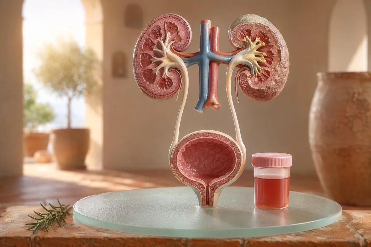
Guide destiné d’abord au patient pour l’hématurie visible et microscopique, y compris pourquoi le sang à la bandelette n’est pas la même chose que les globules rouges au microscope.


- Sang dans les urines signifie des urines rouges visibles, couleur thé ou couleur cola, ou une hématurie microscopique retrouvée lors des tests ; le sang visible mérite toujours un suivi médical.
- Hématurie microscopique est généralement défini comme 3 globules rouges ou plus par champ à fort grossissement sur un échantillon correctement recueilli pour une microscopie urinaire.
- Le sang à la bandelette détecte l’activité de l’hème ; il peut donc être positif à partir de globules rouges intacts, d’hémoglobine libre ou de myoglobine après une lésion musculaire.
- Indices d’IVU incluent la brûlure, la fréquence, la leucocyte estérase, les nitrites et les globules blancs ; l’hématurie doit être recontrôlée une fois l’infection résolue.
- Sédiment urinaire des constatations telles que des globules rouges dysmorphiques, des cylindres de globules rouges ou une protéinurie significative orientent vers une maladie de l’unité de filtration rénale.
- Cylindres urinaires sont des matériaux moulés issus des tubules rénaux ; les cylindres de globules rouges sont un signal d’alarme néphrologique de la même semaine, surtout en cas d’hypertension artérielle ou de baisse de l’eGFR.
- Signes d’alerte de cancer incluent une hématurie macroscopique indolore, des caillots, un âge supérieur à 45 ans, des antécédents de tabagisme, une exposition professionnelle à des produits chimiques et des épisodes récidivants inexpliqués.
- Hématurie d’effort se résorbe généralement en 48 à 72 heures ; un saignement persistant après le repos ne doit pas être attribué à la course ou au vélo.
- Une évaluation urgente est nécessaire en cas de sang dans les urines avec fièvre, douleur au flanc, caillots provoquant une obstruction urinaire, grossesse, traumatisme ou symptômes d’une anémie sévère.
Ce que signifie la présence de sang dans les urines avant de paniquer
Sang dans les urines peut provenir d’une infection, de calculs, d’une maladie de filtration rénale, de conditions de la prostate ou de la vessie, de médicaments, d’un effort, ou d’une contamination pendant les règles. La règle pratique est simple : un sang visible, une hématurie microscopique répétée, ou une hématurie avec douleur, fièvre, caillots, hypertension artérielle, protéinurie ou fonction rénale réduite nécessitent une évaluation rapide plutôt qu’une simple surveillance.
Un seul “rose” dans la cuvette après de la betterave ou un nouveau complément n’est pas le même problème clinique que des urines de couleur “cola” après un mal de gorge. Dans ma pratique, l’histoire de la couleur me dit souvent où chercher en premier : un rouge vif avec des caillots évoque plutôt une atteinte des voies urinaires basses, tandis qu’une urine brune ou couleur thé peut signifier des pigments hémoglobinés plus anciens provenant des filtres rénaux.
L’hématurie n’est pas un diagnostic ; c’est un signe. Hématurie macroscopique signifie que vous voyez le changement de couleur, tandis que l’hématurie microscopique signifie que les urines semblent normales mais que la microscopie trouve des globules rouges, généralement à 3 ou plus globules rouges par champ à fort grossissement.
Kantesti est un Analyseur de test sanguin AI qui aide les patients à relier les constatations urinaires à des marqueurs rénaux tels que la créatinine, l’eGFR, la CRP, l’hémoglobine et les plaquettes dans une seule chronologie. Si la couleur est déroutante, notre guide de couleur des urines explique pourquoi les urines rouges, brunes, orange et jaune foncé ne veulent pas toutes dire la même chose.
Le sang à la bandelette n’est pas la même chose que les RBC à la microscopie
Une bandelette urinaire marquée sang positif détecte une activité chimique de type hème, pas nécessairement des globules rouges intacts. La microscopie répond à la question suivante : y a-t-il de véritables RBC dans le sédiment urinaire, et si oui, à quoi ressemblent-ils ?
Le test par bandelette est rapide car le réactif du tampon réagit avec l’activité peroxydase du hème. Cela signifie qu’un résultat positif peut survenir avec des RBC intacts, une hémoglobine libre après la dégradation des globules rouges, ou de la myoglobine après une lésion musculaire importante.
La microscopie est plus lente mais plus spécifique. Un laboratoire soigneux centrifuge les urines, examine le sédiment urinaire, et rapporte le nombre de globules rouges par champ à fort grossissement ; de nombreux cliniciens utilisent 0 à 2 RBC/hpf comme normal et 3 ou plus RBC/hpf comme anormal chez l’adulte.
L’inadéquation compte. Une bandelette avec 3+ de sang et 0-2 RBC/hpf me fait rechercher une rhabdomyolyse, une hémolyse, des contaminants oxydants et la manipulation de l’échantillon plutôt que de sauter directement à une maladie de la vessie ; la différence est similaire au raisonnement de notre comparaison analyse d’urines et culture.
Quand les indices d’IVU expliquent l’hématurie, et quand ils ne l’expliquent pas
Une infection des voies urinaires peut provoquer du sang dans les urines, surtout en cas de brûlures, urgence, fréquence, gêne pelvienne, leucocyte estérase, nitrites et leucocytes. Mais l’hématurie devrait disparaître après le traitement ; des globules rouges persistants après une infection urinaire nécessitent un second examen.
Dans une infection urinaire basse simple, j’attends un ensemble : dysurie, fréquence, leucocyte estérase positive, pyurie souvent au-dessus de 5-10 leucocytes/hpf, et parfois des nitrites. La positivité classique de la culture est de 100 000 UFC/mL, mais chez les femmes symptomatiques, des taux plus faibles comme 1 000-10 000 UFC/mL peuvent rester significatifs sur le plan clinique.
La leucocyte estérase est une enzyme des leucocytes, pas une preuve de bactéries. Un résultat positif avec une hématurie peut survenir à cause d’une infection, de calculs, d’une contamination ou d’une inflammation rénale, c’est pourquoi notre guide de la leucocyte estérase sépare les indices utiles des faux positifs fréquents.
Les nitrites sont plus spécifiques mais moins sensibles, car toutes les bactéries ne convertissent pas le nitrate en nitrite. Si les symptômes sont typiques mais que les nitrites sont négatives, la culture peut quand même être positive ; les détails pratiques sont couverts dans notre explicateur du résultat des nitrites.
Hématurie visible : indices de couleur, de caillots et de chronologie
L’hématurie visible n’est jamais quelque chose que je balaie d’un revers de main, même si cela ne se produit qu’une fois. Des urines rouge vif, des caillots ou un saignement indolore nécessitent une évaluation rapide, car les causes des voies urinaires basses deviennent plus probables à mesure que l’âge et l’exposition au tabac augmentent.
Le moment peut être étonnamment utile. Du sang uniquement au début de la miction peut évoquer l’urètre, du sang tout au long du jet peut provenir de la vessie ou du rein, et un sang terminal près de la fin oriente parfois vers la région du col vésical.
Les caillots signifient généralement que la source du saignement se situe en dessous des filtres rénaux, car des caillots ne se forment pas facilement après le passage à travers les glomérules et les tubules. Un caillot qui bloque l’écoulement des urines est urgent : une distension vésicale peut devenir douloureuse en quelques heures, et des soins de sonde peuvent être nécessaires.
Les hommes supposent parfois que l’hématurie visible après des symptômes urinaires n’est qu’une irritation de la prostate. Cela peut être vrai, mais si une PSA a été contrôlée pendant ou peu après une infection, l’interprétation peut être délicate ; notre Guide PSA après infection urinaire (UTI) explique pourquoi de nombreux cliniciens attendent 6 à 8 semaines avant de répéter la PSA.
Seuils d’hématurie microscopique que les médecins utilisent
L’hématurie microscopique est généralement définie comme 3 globules rouges (RBC) ou plus par champ à fort grossissement sur un échantillon d’urine correctement recueilli. La recommandation AUA/SUFU utilise ce seuil et recommande une évaluation fondée sur le risque plutôt que de traiter chaque patient de la même façon (Barocas et al., 2020).
Une bandelette urinaire seule ne doit pas être utilisée pour diagnostiquer une hématurie microscopique. Dans la recommandation AUA/SUFU de Barocas et al., 2020, la définition dépend de la microscopie, car les bandelettes peuvent être positives pour des pigments hémés sans globules rouges intacts.
Répéter les tests n’est pas un signe de faiblesse ; c’est une bonne pratique médicale. Si quelqu’un a 3 à 10 RBC/hpf après un exercice intense, de la fièvre, des rapports sexuels, des menstruations, ou un recueil contaminé, je veux généralement un nouvel échantillon propre avant de prescrire des examens d’imagerie, un peu comme nous le conseillons pour guide des analyses anormales à répéter.
Le risque modifie la trajectoire. Un homme de 24 ans avec 4 RBC/hpf après un semi-marathon n’est pas la même situation qu’un homme de 68 ans, ancien fumeur, avec 25 RBC/hpf sur deux prélèvements, même si les deux répondent techniquement à la définition d’hématurie.
Sédiment urinaire, protéines et cylindres urinaires qui modifient l’exploration
Sédiment urinaire peut révéler si l’hématurie provient probablement des filtres rénaux. Des hématies dysmorphiques, des cylindres hématiques et un déplacement significatif des protéines orientent davantage vers une maladie glomérulaire qu’une simple infection de la vessie.
Les globules rouges qui ont un aspect irrégulier, bourgeonné (blebbed) ou en forme d’anneau sont appelés RBC dysmorphiques, et ils suggèrent le passage à travers un filtre glomérulaire enflammé. Des acanthocytes au-dessus d’environ 5% des globules rouges urinaires sont souvent traités comme un indice glomérulaire fort, bien que les laboratoires varient dans la confiance avec laquelle ils les rapportent.
La protéine est le deuxième indice que je n’ignore pas. Un rapport albumine/créatinine urinaire au-dessus de 30 mg/g, soit environ 3 mg/mmol, est anormal, et une hématurie visible plus une protéinurie mérite une réévaluation plus rapide centrée sur le rein ; notre guide protéine dans les urines fournit des seuils au niveau du patient.
Cylindres urinaires sont de minuscules moules formés à l’intérieur des tubules rénaux. Des cylindres hyalins peuvent apparaître après une déshydratation ou un exercice, mais des cylindres de globules rouges sont anormaux et orientent généralement vers une glomérulonéphrite ; pour une référence plus large d’analyse d’urines, voir notre guide complet d'analyse d'urine.
Calculs, exercice et causes temporaires d’hématurie
Les calculs et l’exercice intense peuvent provoquer une hématurie, mais ils ont des chronologies différentes. L’hématurie liée aux calculs s’accompagne souvent de douleurs lombaires ou de cristaux, tandis que l’hématurie d’effort devrait généralement se stabiliser dans les 48 à 72 heures de repos.
Les calculs rénaux provoquent classiquement des vagues de douleur lombaire, des nausées et une hématurie microscopique ou visible. Les cristaux d’oxalate de calcium ne prouvent pas un calcul, mais ils augmentent la suspicion lorsqu’ils sont associés à une douleur colique ; notre guide des cristaux d’oxalate de calcium explique l’indice de microscopie urinaire.
L’hématurie d’effort est réelle. J’ai vu des coureurs de marathon avec 10 à 20 RBC/hpf le matin après une course, une culture normale, pas de protéinurie, et une analyse d’urines de contrôle complètement normale 3 jours plus tard.
Le danger est d’attribuer tout à l’entraînement. Si l’hématurie persiste au-delà de 72 heures, apparaît avec une urine foncée type cola, ou s’accompagne d’une CK élevée, d’une créatinine qui augmente, ou de douleurs musculaires, regardez au-delà de la vessie ; notre guide sur la créatinine à l’effort couvre le chevauchement entre le rein et le muscle.
Signes d’alerte rénale que les médecins n’ignorent pas
Une hématurie avec protéinurie, une hypertension artérielle élevée, un gonflement, une baisse de eGFR, des cylindres de globules rouges ou une augmentation rapidement progressive de la créatinine sont un signal d’alerte rénal. Ces constatations suggèrent que les unités de filtration pourraient être enflammées, et attendre des mois peut coûter de la fonction rénale.
La KDIGO 2024 définit la maladie rénale chronique par des marqueurs de l’atteinte rénale, comme l’albuminurie, ou un eGFR inférieur à 60 mL/min/1,73 m² pendant au moins 3 mois. En revanche, en contexte aigu, une hématurie associée à une hausse de la créatinine sur quelques jours ou semaines suffit à intensifier la prise en charge avant que la définition des 3 mois ne soit atteinte.
L’albuminurie est souvent le signal d’alerte précoce. Un ACR de 30-300 mg/g est modérément augmenté, au-delà de 300 mg/g il est sévèrement augmenté, et associer l’un ou l’autre à une hématurie change la discussion ; notre guide ACR rénal explique pourquoi l’albumine urinaire détecte une atteinte avant l’apparition des symptômes.
La pression artérielle compte ici. Une nouvelle PA de 160/100 mmHg avec des urines couleur cola et un gonflement des chevilles après une infection de la gorge me fait penser à une glomérulonéphrite, pas à une cystite ; si l’eGFR est incertain, notre guide de recontrôle du GFR explique quand la cystatine C peut clarifier la fonction rénale.
Indices de risque de cancer sans panique
Hématurie visible indolore, surtout après 45 ans, mérite une évaluation urgente car les cancers de la vessie et des voies urinaires supérieures peuvent se présenter ainsi. NICE NG12 recommande une orientation pour suspicion de cancer en cas d’hématurie visible inexpliquée chez les adultes de 45 ans et plus, ou d’hématurie visible qui persiste après un traitement d’UTI.
La plupart des hématuries ne sont pas un cancer. Pourtant, les cas dont je me souviens sont ceux qui restent silencieux : pas de douleur, pas de fièvre, pas de brûlures, juste deux épisodes d’urines rouges chez un fumeur de 56 ans qui l’a presque ignoré.
Le risque est cumulatif, pas binaire. L’âge au-delà de 45-50 ans, l’historique tabagique, l’exposition au cyclophosphamide, la radiothérapie pelvienne, l’hématurie visible récidivante, l’exposition professionnelle aux amines aromatiques et la présence de caillots augmentent tous la nécessité d’une cystoscopie ou d’une imagerie.
Les marqueurs de la prostate n’excluent pas les causes vésicales. Si des symptômes urinaires, des variations de PSA et une hématurie se chevauchent, évaluez soigneusement le moment et le statut infectieux ; notre guide des marqueurs de la prostate explique pourquoi le PSA n’est qu’une partie de l’évaluation urinaire masculine.
Médicaments, anticoagulants et fausses alertes
Les anticoagulants peuvent révéler un saignement, mais ils ne doivent pas être accusés automatiquement en cas d’hématurie. Une hématurie associée aux anticoagulants nécessite toujours une évaluation, car le médicament peut mettre en évidence des calculs, une infection, une maladie rénale ou une tumeur des voies urinaires.
La warfarine, l’apixaban, le rivaroxaban, l’aspirine et le clopidogrel peuvent rendre le saignement plus évident. Si l’INR est au-dessus de la plage cible, le corriger est important, mais un INR normal ne rend pas une hématurie visible inoffensive.
Plusieurs causes non hémorragiques imitent une hématurie. La rifampicine peut rendre les urines orange-rouge, la phénazopyridine peut les rendre orange vif, la betterave peut tacher les urines chez les personnes sensibles, et la déshydratation peut assombrir suffisamment le pigment pour effrayer les patients ; le suivi des anticoagulants est couvert dans notre bilans de sécurité des anticoagulants.
La bandelette peut aussi induire en erreur après un exercice intense ou une lésion musculaire. Une bandelette hématine positive avec peu ou pas de RBC oriente plutôt ma réflexion vers la myoglobine, surtout si la CK est supérieure à 1 000 UI/L et si les urines sont foncées.
Enfants, grossesse et contamination par les règles
L’hématurie chez l’enfant, pendant la grossesse ou autour des menstruations nécessite un recueil plus rigoureux et un seuil plus bas pour réévaluation. Le même résultat urinaire peut signifier des choses différentes selon l’âge, le statut de grossesse, la fièvre, la pression artérielle et la qualité du recueil.
La contamination menstruelle est fréquente et personne ne devrait en avoir honte. Si le résultat est inattendu, répétez un prélèvement à mi-jet propre au moins 48 heures après l’arrêt des saignements ; une persistance de 3 RBC/hpf ou plus devient alors plus significative.
Les enfants ont souvent besoin d’un regard différent. Une hématurie après une maladie virale peut être transitoire, mais une hématurie avec gonflement, hypertension, protéinurie ou diminution de la diurèse nécessite une évaluation pédiatrique rapide.
La grossesse augmente les enjeux car une infection urinaire, des calculs, le stress rénal lié à la prééclampsie et la contamination peuvent se chevaucher. Pour les schémas d’alerte de grossesse le jour même impliquant les résultats rénaux, hépatiques, plaquettaires et urinaires, voir notre signaux d’alerte des analyses de grossesse.
Examens que les médecins prescrivent généralement après une hématurie
Les tests habituels suivants après une hématurie sont : une analyse d’urines répétée avec microscopie, une culture d’urines si une infection est possible, des analyses sanguines de la fonction rénale, une protéinurie urinaire ou un ACR, une mesure de la pression artérielle, et une imagerie ou une cystoscopie guidée par le risque. L’ordre exact dépend des symptômes et du risque.
Pour les symptômes de cystite non compliquée, la culture et le traitement peuvent venir en premier, suivis d’une nouvelle microscopie urinaire après la résolution des symptômes. Pour une hématurie visible indolore, de nombreux cliniciens passent plus tôt à l’imagerie et à la cystoscopie, car attendre des épisodes répétés peut retarder le diagnostic.
Les analyses sanguines apportent un contexte que l’urine seule ne peut pas fournir. La créatinine, l’eGFR, l’urée ou le BUN, les électrolytes, la CBC, la CRP, le complément C3/C4 et les marqueurs d’auto-immunité peuvent être appropriés lorsqu’une inflammation rénale est suspectée ; notre guide du rapport BUN/créatinine explique un schéma rénal fréquent.
L’IA Kantesti peut organiser ces résultats en vue de tendance, mais la revue par le médecin reste essentielle lorsque des signaux d’alerte sont présents. Notre approche de sécurité clinique est décrite dans Validation médicale, y compris la manière dont les regroupements anormaux sont gérés plutôt que des chiffres isolés.
Comment Kantesti aide à organiser le contexte de l’hématurie
Kantesti est un service d’interprétation des tests de laboratoire par l’IA qui lit des marqueurs sanguins proches de l’urine dans leur contexte, tels que la créatinine, l’eGFR, la CRP, l’hémoglobine, les plaquettes et la glucose. Elle ne diagnostique pas une hématurie, mais elle aide les patients à préparer un résumé plus propre et plus utile pour leur clinicien.
Quand j’évalue une hématurie, je ne regarde que rarement un seul résultat isolément. Une créatinine normale à 0,82 mg/dL, un eGFR stable au-dessus de 90, pas de protéinurie et une culture négative ne ressemblent pas du tout à une hématurie avec un eGFR à 48 et une CRP à 65 mg/L.
Kantesti est un Plateforme d’interprétation des biomarqueurs par IA qui peut traiter des PDF de bilans sanguins téléversés ou des photos et relier les marqueurs rénaux, inflammatoires et d’anémie hors normes à la chronologie des symptômes. Les méthodes à l’origine de cette lecture du schéma sont décrites dans notre Guide technologique, et une couverture plus large des marqueurs est détaillée dans le guide des biomarqueurs.
Un conseil pratique : avant de téléverser, vérifiez que les unités, les dates et les identifiants du patient sont correctement saisis. Les erreurs OCR peuvent transformer 0,9 en 9,0, ce qui correspond à une histoire rénale complètement différente ; notre checklist de téléversement PDF montre les erreurs que je demande aux patients de repérer en premier.
Notes de recherche, gouvernance et checklist finale
Au 11 juillet 2026, le plan patient le plus sûr consiste à confirmer si l’hématurie est réelle, à repérer des indices d’infection, à rechercher des signaux d’alerte rénaux et à faire remonter toute hématurie visible ou persistante. Ne vous fiez pas uniquement à la couleur des urines.
Je m’appelle Thomas Klein, MD, et mes conseils sont volontairement prudents, car une hématurie manquée peut avoir de l’importance. Le contenu médical de Kantesti est revu avec une supervision clinique par notre Conseil consultatif médical, et notre rôle est d’aider les patients à poser de meilleures questions, et non de remplacer leur médecin.
Kantesti est un Outil d’analyse de prise de sang alimenté par l’IA utilisé par des millions de personnes dans 127 pays, de sorte que la confidentialité et la clarté clinique doivent être intégrées au flux de travail plutôt qu’ajoutées plus tard. Vous pouvez en savoir plus sur Kantesti LTD, notre structure d’entreprise au Royaume-Uni et notre mission clinique sur À propos de nous.
Kantesti AI Clinical Research Group. (2026). AI Blood Test Analyzer : 2,5 M de tests analysés | Global Health Report 2026. Zenodo. DOI : https://doi.org/10.5281/zenodo.18175532. ResearchGate : https://www.researchgate.net/. Academia.edu : https://www.academia.edu/.
Kantesti AI Clinical Research Group. (2026). RDW Blood Test : Guide complet du RDW-CV, MCV et MCHC. Zenodo. DOI : https://doi.org/10.5281/zenodo.18202598. ResearchGate : https://www.researchgate.net/. Academia.edu : https://www.academia.edu/.
Questions fréquemment posées
Le sang dans les urines est-il toujours grave ?
La présence de sang dans les urines n’est pas toujours grave, mais elle doit être confirmée et expliquée. Un résultat microscopique unique de 3 à 5 RBC/hpf après un exercice ou des menstruations peut disparaître lors d’un nouveau test, tandis que du sang visible, des caillots, de la fièvre, une douleur au flanc, une protéinurie, une hypertension artérielle élevée ou une eGFR réduite nécessitent un examen rapide. Chez les adultes de plus de 45 ans, une hématurie visible inexpliquée mérite une évaluation urgente même en l’absence de douleur.
Quelle est la différence entre le sang à la bandelette urinaire et les GR dans les urines ?
La bandelette urinaire détecte une activité chimique de type hémique, tandis que la microscopie urinaire compte des globules rouges réels. Une bandelette peut être positive en présence de RBC intacts, d’hémoglobine libre ou de myoglobine après une lésion musculaire, de sorte qu’elle n’est pas identique à une hématurie confirmée. L’hématurie microscopique est couramment définie comme la présence de 3 ou plus RBC par champ à fort grossissement sur un échantillon correctement recueilli.
Une infection urinaire (IU) peut-elle provoquer du sang dans les urines ?
Oui, une infection urinaire (IU) peut provoquer du sang dans les urines, surtout en cas de brûlures, de fréquence et d’urgence mictionnelles, lorsque De plus, la présence d’esterase leucocytaire, de nitrites et de globules blancs est également observée. La positivité classique de la culture d’urines est souvent de 100 000 UFC/mL, mais des taux plus faibles peuvent être importants chez les patients symptomatiques. Une hématurie doit généralement être recontrôlée après le traitement, car la persistance de sang peut évoquer des calculs, une maladie rénale ou une autre cause des voies urinaires.
Quand dois-je consulter en soins urgents pour une hématurie ?
Consultez en urgence en cas de sang dans les urines avec fièvre, douleur lombaire intense, vomissements, grossesse, traumatisme récent, caillots, impossibilité d’uriner, malaise, ou symptômes d’une anémie sévère. Une réévaluation médicale dans la même semaine est également appropriée en cas d’hématurie avec protéinurie, cylindres de globules rouges, nouvelle hypertension élevée ou créatinine en augmentation. Un jet urinaire bloqué par des caillots peut devenir douloureux et dangereux en quelques heures.
L’exercice peut-il provoquer une hématurie microscopique ?
L’exercice intense peut provoquer une hématurie microscopique, en particulier après une course de longue distance, un cyclisme intense ou des sports de contact. L’hématurie liée à l’exercice disparaît généralement dans les 48 à 72 heures de repos et ne devrait pas s’accompagner d’une protéinurie persistante, d’une baisse de l’eGFR ou de douleurs musculaires sévères. Si le sang à la bandelette est positif mais que la microscopie montre peu de RBC, les cliniciens peuvent vérifier la CK, car la myoglobine provenant d’une lésion musculaire peut déclencher la réaction à la bandelette.
Que signifient les cylindres urinaires lorsqu’on trouve du sang ?
Les cylindres urinaires sont des matériaux moulés formés à l’intérieur des tubules rénaux, et leur type modifie la signification de l’hématurie. Les cylindres hyalins peuvent apparaître en cas de déshydratation ou d’exercice, mais les cylindres de globules rouges sont anormaux et suggèrent une inflammation glomérulaire du rein. Du sang dans les urines avec des cylindres de globules rouges, une protéinurie et une hypertension artérielle élevée doivent être traités comme un signal d’alarme rénal plutôt que comme une simple infection urinaire (UTI).
La déshydratation peut-elle donner l'apparence de sang dans l'urine ?
La déshydratation rend souvent l'urine jaune foncé ou ambrée, mais elle ne crée généralement pas de globules rouges dans l'urine. Buvez de l'eau et organisez un test d'urine si la couleur est rose, rouge, brune ou semblable à celle du cola, surtout si elle persiste après l'hydratation ou s'accompagne de douleur.
Obtenez dès aujourd’hui une analyse de sang par IA
Rejoignez plus de 2 millions d’utilisateurs dans le monde qui font confiance à Kantesti pour une analyse instantanée et précise des analyses de laboratoire. Téléversez vos résultats prise de sang et recevez une interprétation complète des biomarqueurs de 15,000+ en quelques secondes.
📚 Publications de recherche citées
Klein, T., Mitchell, S., & Weber, H. (2026). Analyseur de bilan sanguin par IA : 2,5 M de tests analysés | Rapport mondial sur la santé 2026. Recherche médicale par IA Kantesti.
Klein, T., Mitchell, S., & Weber, H. (2026). Analyse sanguine RDW : Guide complet du RDW-CV, du VGM et du CCMH. Recherche médicale par IA Kantesti.
📖 Références médicales externes
National Institute for Health and Care Excellence (2015). Cancer suspecté : reconnaissance et orientation. Recommandation NICE NG12. Recommandation NICE.
Maladie rénale : groupe de travail CKD (Improving Global Outcomes) (2024). Ligne directrice de pratique clinique KDIGO 2024 pour l’évaluation et la prise en charge de la maladie rénale chronique. Kidney International.
⚕️ Avertissement médical
Cet article est fourni à des fins éducatives uniquement et ne constitue pas un avis médical. Consultez toujours un professionnel de santé qualifié pour les décisions de diagnostic et de traitement.
Signaux de confiance E-E-A-T
Expérience
Revue clinique guidée par un médecin des flux d’interprétation des analyses.
Compétence
Orientation en médecine de laboratoire sur la façon dont les biomarqueurs se comportent dans un contexte clinique.
autorité
Rédigé par le Dr Thomas Klein, avec relecture par le Dr Sarah Mitchell et le Prof. Dr Hans Weber.
Fiabilité
Interprétation fondée sur des preuves, avec des parcours de suivi clairs pour réduire l’alarme.