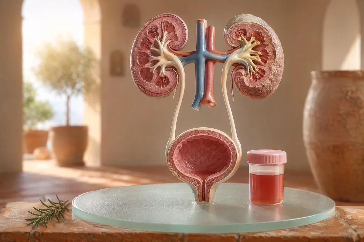
දෘශ්ය හා අන්වීක්ෂීය hematuria සඳහා රෝගියා-පළමු මාර්ගෝපදේශයක්; dipstick මගින් ලැබෙන රුධිරය අන්වීක්ෂය යටතේ ඇති රතු රුධිර සෛලවලට සමාන නොවන්නේ ඇයිද යන්න ඇතුළුව.


- මුත්රාවල රුධිරය යන්නෙන් අදහස් වන්නේ දෘශ්ය ලෙස රතු, තේ-වර්ණ හෝ කොලා-වර්ණ මුත්රාවක්, හෝ පරීක්ෂණයේදී හමු වූ අන්වීක්ෂීය hematuria; දෘශ්ය රුධිරය සැමවිටම වෛද්ය පසු විමර්ශනයක් ලැබිය යුතුය.
- ක්ෂුද්ර රුධිරමුත්රා (microscopic hematuria) නිසි ලෙස එකතු කරන ලද මුත්රා අන්වීක්ෂීය සාම්පලයක high-power field එකකට රතු රුධිර සෛල 3ක් හෝ ඊට වැඩි ලෙස සාමාන්යයෙන් අර්ථ දක්වයි.
- Dipstick රුධිරය heme ක්රියාකාරිත්වය හඳුනාගන්නා නිසා, මාංශ පේශි තුවාලයකින් පසු intact රතු රුධිර සෛලවලින්, නිදහස් hemoglobin වලින් හෝ myoglobin වලින් එය ධනාත්මක විය හැක.
- UTI සලකුණු දැවීම, නිතර මුත්රා කිරීම, leukocyte esterase, nitrites සහ සුදු රුධිර සෛල ඇතුළත් වේ; ආසාදනය ඉවත් වූ පසු hematuria නැවත පරීක්ෂා කළ යුතුය.
- මුත්රා අවසාදය dysmorphic රතු රුධිර සෛල, රතු රුධිර සෛල casts හෝ සැලකිය යුතු ප්රෝටීන් වැනි සොයාගැනීම් වකුගඩු පෙරහන-ඒකක රෝගයක් වෙත යොමු කරයි.
- Urine casts වකුගඩු නලිකා වලින් සෑදුණු ආකෘතිගත ද්රව්ය වේ; රතු සෛල කැස්ට් (red cell casts) එකම සතිය තුළ ඇතිවීම, විශේෂයෙන් රුධිර පීඩනය වැඩිවීමක් හෝ eGFR අඩුවීමක් සමඟ, නෙෆ්රොලොජි ක්ෂණික අවධානයට ලක්විය යුතු රතු අනතුරු ලකුණකි.
- පිළිකා අනතුරු සංඥා වේදනාවක් නැතිව පෙනෙන හීමැටූරියා (painless visible hematuria), ගැටිති (clots), වයස අවුරුදු 45ට වැඩි වීම, දුම්පානයේ ඉතිහාසය, වෘත්තීය රසායනික නිරාවරණය සහ නැවත නැවත පැහැදිලි නොවූ කථාංග.
- ව්යායාම මඟින් ඇතිවන හීමැටූරියා (Exercise hematuria) සාමාන්යයෙන් පැය 48-72 තුළ සන්සුන් වේ; විවේකයෙන් පසුවත් රුධිරය පවතින්නේ නම් එය දුවීම හෝ බයිසිකල් පැදීම නිසා යැයි දොස් නොදැමිය යුතුය.
- හදිසි ඇගයීම උණ, පැත්තේ වේදනාව (flank pain), මුත්රා අවහිර කරන ගැටිති (clots), ගර්භණීභාවය, කම්පනය (trauma) හෝ දරුණු රක්තහීනතාවයේ (severe anemia) ලක්ෂණ ඇති විට මුත්රාවේ රුධිරය සඳහා අවශ්ය වේ.
කලබල වීමට පෙර මුත්රාවල රුධිරය අදහස් කරන්නේ කුමක්ද
මුත්රාවල රුධිරය ආසාදනයෙන්, ගල් (stones) වලින්, වකුගඩු පෙරීමේ රෝගයෙන්, ප්රොස්ටේට් (prostate) හෝ මුත්රාශයේ (bladder) තත්ත්වයන්ගෙන්, ඖෂධවලින්, ව්යායාමයෙන්, හෝ මාසික කාලයෙන් (periods) ඇතිවිය හැකි අපවිත්රතාවයෙන් (contamination) පැමිණිය හැක. ප්රායෝගික නීතිය සරලයි: පෙනෙන රුධිරය (visible blood), නැවත නැවත ඇතිවන ක්ෂුද්ර හීමැටූරියා (repeated microscopic hematuria), හෝ වේදනාව, උණ, ගැටිති, රුධිර පීඩනය වැඩිවීම, ප්රෝටීන් (protein), හෝ වකුගඩු ක්රියාකාරිත්වය අඩුවීම සමඟ හීමැටූරියා ඇතිවීමක් නම්, බලා සිටීම (watchful waiting) වෙනුවට ඉක්මන් ඇගයීමක් අවශ්ය වේ.
බීට්රූට් (beetroot) හෝ නව අතිරේකයක් (new supplement) නිසා ඇති වූ පළමුවන වරට රෝස පැහැති වැසිකිළි බඳුනක් (pink toilet bowl) යනු, උගුරේ අමාරුවක් (sore throat) පසු කොලා වර්ණයට (cola-colored) සමාන මුත්රාවක් ඇතිවීම හා සමාන සායනික ගැටලුවක් නොවේ. මගේ පුරුද්දේදී, වර්ණ කතාව බොහෝ විට මට පළමුව බලන්න යන්නේ කොතැනද කියා කියා දෙයි: ගැටිති සමඟ දීප්තිමත් රතු පැහැය පහළ මුත්රා මාර්ගයට (lower urinary tract) සම්බන්ධ වීමට වැඩි ඉඩක් ඇති අතර, දුඹුරු හෝ තේ වර්ණයට (tea-colored) සමාන මුත්රාවක් වකුගඩු පෙරීමේ ස්ථානයෙන් පැරණි heme වර්ණක (heme pigments) නිසා විය හැක.
හීමැටූරියා (Hematuria) යනු රෝග නිර්ණායකයක් (diagnosis) නොවේ; එය ලක්ෂණයකි. පෙනෙන හීමැටූරියා (Visible hematuria) ඔබට වර්ණ වෙනස දැකගත හැකි බව අදහස් කරයි, එසේම ක්ෂුද්ර හීමැටූරියා (microscopic hematuria) යන්නෙන් අදහස් වන්නේ මුත්රාව සාමාන්ය ලෙස පෙනෙන නමුත් ක්ෂුද්රදර්ශනයෙන් රතු සෛල (red cells) හමු වීමයි; සාමාන්යයෙන් එය එක් high-power field එකකට රතු රුධිර සෛල 3ක් හෝ ඊට වැඩි වීමකි.
කන්ටෙස්ටි යනු AI රුධිර පරීක්ෂණ විශ්ලේෂකය එය රෝගීන්ට එක් කාලරේඛාවක (one timeline) creatinine, eGFR, CRP, hemoglobin සහ platelets වැනි වකුගඩු සලකුණු (kidney markers) සමඟ මුත්රා සොයාගැනීම් සම්බන්ධ කරගැනීමට උපකාරී වේ. වර්ණය ව්යාකූල නම්, අපේ මුත්රා වර්ණ මාර්ගෝපදේශය (urine color guide) රතු, දුඹුරු, තැඹිලි සහ අඳුරු කහ මුත්රා සියල්ලම එකම දෙයක් අදහස් නොකරන්නේ ඇයිද යන්න පැහැදිලි කරයි.
Dipstick රුධිරය අන්වීක්ෂය මත RBCs (රතු රුධිර සෛල) වලට සමාන නොවේ
මුත්රා ඩිප්ස්ටික් (urine dipstick) එකක සලකුණු කර ඇති රුධිරය ධනාත්මක (blood positive) යන්නෙන් අදහස් වන්නේ heme වැනි රසායනික ක්රියාකාරිත්වයක් හඳුනාගැනීම මිස අනිවාර්යයෙන්ම සම්පූර්ණ රතු රුධිර සෛල (intact red blood cells) නොවේ. ක්ෂුද්රදර්ශනය (microscopy) ඊළඟ ප්රශ්නයට පිළිතුරු දෙයි: මුත්රා අවසාදයේ (urine sediment) ඇත්තටම RBCs තිබේද, තිබේ නම් ඒවා පෙනෙන්නේ කෙසේද?
ඩිප්ස්ටික් පරීක්ෂාව වේගවත් වන්නේ reagent pad එක heme හි peroxidase ක්රියාකාරිත්වයට ප්රතික්රියා කරන නිසාය. එයින් අදහස් වන්නේ ධනාත්මක ප්රතිඵලයක් සම්පූර්ණ RBCs සමඟ, රතු සෛල බිඳවැටීමෙන් පසු නිදහස් hemoglobin සමඟ, හෝ සැලකිය යුතු මාංශ පේශි තුවාලයකින් පසු myoglobin සමඟ සිදුවිය හැකි බවයි.
ක්ෂුද්රදර්ශනය මන්දගාමී නමුත් වඩා නිශ්චිතය. ප්රවේශමෙන් කරන ලැබ් පරීක්ෂාවක් මුත්රාව centrifuge කර, මුත්රා අවසාදය (urine sediment), පරීක්ෂා කර, එක් high-power field එකකට රතු සෛල වාර්තා කරයි; බොහෝ වෛද්යවරු වැඩිහිටියන් තුළ 0-2 RBC/hpf සාමාන්ය ලෙසත් 3 හෝ ඊට වැඩි RBC/hpf අසාමාන්ය ලෙසත් සලකති.
මෙම නොගැලපීම වැදගත්ය. 3+ රුධිරය සහ 0-2 RBC/hpf ඇති ඩිප්ස්ටික් පරීක්ෂාවක් මට මුලින්ම bladder රෝගයට කෙලින්ම පැන නොයන්නට හේතු වේ; ඒ වෙනුවට rhabdomyolysis, hemolysis, oxidizing contaminants සහ සාම්පල හැසිරවීම (specimen handling) ගැන විමසීමට මට හේතු වේ; මෙම වෙනස අපේ තර්කනයට සමානයි. මුත්රා පරීක්ෂණය සහ සංස්කෘති සංසන්දනය.
UTI (මුත්රා මාර්ග ආසාදන) සලකුණු hematuria පැහැදිලි කරන විටත්, නොකරන විටත්
මුත්රා මාර්ග ආසාදනයක් (UTI) හේතු විය හැක මුත්රාව තුළ ලේ, විශේෂයෙන් දැවීම, හදිසි අවශ්යතාව, නිතර මුත්රා කිරීම, ශ්රෝණි අසහනය, ලියුකොසයිට් එස්ටරේස් (leukocyte esterase), නයිට්රයිට් (nitrites) සහ සුදු රුධිරාණු සමඟ. නමුත් UTI ප්රතිකාරයෙන් පසු හීමැටුරියා ඉවත් විය යුතුය; UTI එකෙන් පසුත් රතු රුධිරාණු පවතින්නේ නම් දෙවන වරට බැලීමක් අවශ්යයි.
සරල පහළ UTI එකකදී, මම එකතු වීමක් බලාපොරොත්තු වෙමි: ඩිස්යුරියා (dysuria), නිතර මුත්රා කිරීම, ලියුකොසයිට් එස්ටරේස් ධනාත්මක, පියුරියා (pyuria) බොහෝ විට 5-10කට වඩා වැඩි සුදු රුධිරාණු/hpf, සහ සමහර විට නයිට්රයිට්. සාම්ප්රදායික සංස්කෘති ධනාත්මකතාව 100,000 CFU/mL වේ, නමුත් ලක්ෂණ ඇති කාන්තාවන් තුළ 1,000-10,000 CFU/mL වැනි අඩු ගණන් ද සායනිකව වැදගත් විය හැක.
ලියුකොසයිට් එස්ටරේස් යනු සුදු රුධිරාණු එන්සයිමයක් වන අතර බැක්ටීරියා ඇති බවට සාක්ෂියක් නොවේ. හීමැටුරියා සමඟ ධනාත්මක ප්රතිඵලයක් ආසාදනයෙන්, ගල්වලින්, දූෂණයෙන්, හෝ වකුගඩු දැවිල්ලෙන් සිදුවිය හැක; ඒ නිසා අපේ ලියුකොසයිට් එස්ටරේස් මාර්ගෝපදේශය පොදු ව්යාජ ධනාත්මක ප්රතිඵලවලින් ප්රයෝජනවත් ඉඟි වෙන් කරයි.
නයිට්රයිට් වඩා විශේෂිත (specific) නමුත් අඩු සංවේදී (less sensitive) වේ, මන්ද සියලුම බැක්ටීරියා නයිට්රේට් නයිට්රයිට් බවට පරිවර්තනය නොකරයි. ලක්ෂණ සාමාන්ය ලෙස තිබුණත් නයිට්රයිට් නිෂේධ නම්, සංස්කෘතිය තවමත් ධනාත්මක විය හැක; ප්රායෝගික විස්තර අපේ නයිට්රයිට් ප්රතිඵල පැහැදිලි කිරීම.
දෘශ්ය hematuria: වර්ණය, ගැටිති සහ කාල සලකුණු
දෘශ්ය හීමැටුරියා (visible hematuria) මම කිසිවිටෙක නොසලකා හරින්නේ නැහැ—එය එක් වරක් සිදු වුණත්. දීප්තිමත් රතු මුත්රා, ගැටිති, හෝ වේදනාවක් නැති ලේ වහනයක් වහාම ඇගයීමක් අවශ්යයි, මන්ද වයස සහ දුම්පානයට නිරාවරණය වැඩි වන විට පහළ මුත්රා මාර්ග හේතු වඩාත් ඉඩකඩ ඇති වේ.
කාලය තීරණය කිරීම අතිශය ප්රයෝජනවත් විය හැක. මුත්රා කිරීම ආරම්භයේදී පමණක් රුධිරය තිබීම මුත්රා නාලය (urethra) වෙත යොමු විය හැකි අතර, ධාරාව පුරා රුධිරය තිබීම මුත්රාශය (bladder) හෝ වකුගඩු මූලාශ්රවලින් විය හැක. අවසානයට ආසන්නව රුධිරය තිබීම සමහර විට මුත්රාශය ගෙල ප්රදේශය (bladder neck area) වෙත යොමු කරයි.
ගැටිති (clots) සාමාන්යයෙන් අදහස් කරන්නේ රුධිර වහනය වකුගඩු පෙරහන් (kidney filters) වලට පහළින් ඇති බවයි, මන්ද glomeruli සහ tubules හරහා ගිය පසු ගැටිති පහසුවෙන් සෑදෙන්නේ නැත. මුත්රා ගලායාම අවහිර කරන ගැටිත්තක් හදිසි තත්ත්වයකි: පැය කිහිපයක් ඇතුළත මුත්රාශය පුම්බීම (bladder distension) වේදනාකාරී විය හැකි අතර, catheter රැකවරණය අවශ්ය විය හැක.
පුරුෂයන් සමහරවිට මුත්රා සම්බන්ධ රෝග ලක්ෂණවලින් පසුව දෘශ්ය hematuria (මුත්රාවේ රුධිරය) ඇතිවීම හුදෙක් ප්රොස්ටේට් (prostate) කෝපවීමක් ලෙස සිතයි. එය සත්ය විය හැකි නමුත්, ආසාදනයක් (infection) අතරතුර හෝ ඉක්මනින් පසුව PSA පරීක්ෂා කර තිබුණේ නම්, අර්ථකථනය අවුල් විය හැක; අපේ UTI පසුව PSA මාර්ගෝපදේශය බොහෝ වෛද්යවරු PSA නැවත පරීක්ෂා කිරීමට පෙර සති 6-8ක් බලා සිටින්නේ ඇයිද යන්න පැහැදිලි කරයි.
වෛද්යවරුන් භාවිතා කරන අන්වීක්ෂීය hematuria සීමා
Microscopic hematuria සාමාන්යයෙන් ලෙස අර්ථ දක්වන්නේ එක් high-power field එකකට RBC 3ක් හෝ ඊට වැඩි නිසි ලෙස එකතු කරන ලද මුත්රා සාම්පලයක. AUA/SUFU මාර්ගෝපදේශය මෙම සීමාව භාවිතා කරමින්, සෑම රෝගියෙකුටම එකම ලෙස ප්රතිකාර කිරීම වෙනුවට අවදානම් මත පදනම් වූ ඇගයීමක් නිර්දේශ කරයි (Barocas et al., 2020).
Microscopic hematuria හඳුනා ගැනීමට dipstick එක පමණක් භාවිතා නොකළ යුතුය. Barocas et al. 2020 AUA/SUFU මාර්ගෝපදේශයේ, අර්ථ දැක්වීම microscopy මත රඳා පවතී, මන්ද dipsticks මඟින් intact රතු රුධිර සෛල නොමැතිවද heme pigments නිසා ධනාත්මක විය හැක.
නැවත පරීක්ෂා කිරීම දුර්වලකමක් නොවේ; එය හොඳ වෛද්ය ක්රියාවකි. කෙනෙකුට දැඩි ව්යායාමයෙන් පසු, උණ (fever) ඇතිව, ලිංගික ක්රියාවෙන් පසු, මාසික (menstruation) අවස්ථාවේදී, හෝ අපිරිසිදු එකතු කිරීමක් (contaminated collection) නිසා RBC/hpf 3-10ක් තිබේ නම්, මම සාමාන්යයෙන් scans නියම කිරීමට පෙර පිරිසිදු නැවත සාම්පලයක් ලබා ගැනීමට කැමතියි; අපි උපදෙස් දෙන්නේ නැවත අසාමාන්ය රසායනාගාර ප්රතිඵල.
අවදානම මාර්ගය වෙනස් කරයි. අර්ධ මැරතන් එකකට පසු RBC/hpf 4ක් ඇති වයස අවුරුදු 24ක් වූ අයෙක්, තාක්ෂණිකව දෙදෙනාම hematuria අර්ථ දැක්වීමට ගැළපුණත්, specimen දෙකකදී RBC/hpf 25ක් ඇති වයස අවුරුදු 68ක් වූ හිටපු දුම් පානය කරන්නෙකුට සමාන නොවේ.
වැඩිදුර පරීක්ෂණය වෙනස් කරන මුත්රා අවසාදය, ප්රෝටීන් සහ urine casts
මුත්රා අවසාදය hematuria වකුගඩු පෙරහන් වලින් එන බවට ඇති ඉඩකඩ හෙළි කළ හැක. Dysmorphic රතු රුධිර සෛල (dysmorphic red cells), රතු සෛල casts (red cell casts), සහ සැලකිය යුතු ප්රෝටීන් වෙනස්වීම (significant protein shift) සරල මුත්රාශය ආසාදනයකට වඩා glomerular රෝගයක් වෙත ඇති සැකය වැඩි කරයි.
අසමාන ලෙස පෙනෙන, blebbed හෝ වළලු හැඩැති රතු රුධිර සෛල dysmorphic RBCs ලෙස හැඳින්වේ, සහ ඒවා දැවිල්ලට ලක් වූ glomerular පෙරහනක් හරහා ගමන් කිරීමක් යෝජනා කරයි. මුත්රාවේ රතු රුධිර සෛල වලින් ආසන්න වශයෙන් 5%කට ඉහළ acanthocytes බොහෝ විට ශක්තිමත් glomerular ඉඟියක් ලෙස සලකනු ලැබේ, නමුත් ඒවා කොතරම් විශ්වාසයෙන් වාර්තා කරන්නේද යන්න රසායනාගාර අනුව වෙනස් වේ.
ප්රෝටීන් දෙවන ඉඟියයි; මම එය නොසලකා නොහරිනවා. මුත්රා albumin-creatinine ratio 30 mg/gට ඉහළින්, හෝ ආසන්න වශයෙන් 3 mg/mmol, තිබීම අසාමාන්යයි. දෘශ්ය hematuria සමඟ ප්රෝටීන් තිබේ නම් වකුගඩු කේන්ද්ර කරගත් වේගවත් සමාලෝචනයක් ලැබිය යුතුය; අපේ මුත්රාවේ protein මාර්ගෝපදේශය රෝගියා මට්ටමේ සීමාවන් ලබා දෙයි.
Urine casts වකුගඩු tubules තුළ සෑදෙන ඉතා කුඩා අච්චු (tiny molds) වේ. Hyaline casts විජලනය (dehydration) හෝ ව්යායාමයෙන් පසුව පෙනී යා හැක, නමුත් red cell casts අසාමාන්ය වන අතර සාමාන්යයෙන් glomerulonephritis වෙත යොමු කරයි; වඩා පුළුල් urinalysis යොමු සඳහා අපේ සම්පූර්ණ මුත්රා විශ්ලේෂණ මාර්ගෝපදේශය.
ගල්, ව්යායාම සහ hematuria සඳහා තාවකාලික හේතු
ගල් (Stones) සහ දැඩි ව්යායාම hematuria ඇති කළ හැක, නමුත් ඒවාට වෙනස් කාල රටා ඇත. ගල් සම්බන්ධ hematuria බොහෝ විට පැත්තේ වේදනාව (flank pain) හෝ ස්ඵටික (crystals) සමඟ එයි. ව්යායාමයෙන් ඇතිවන hematuria සාමාන්යයෙන් විවේකයෙන් පැය 48-72ක් ඇතුළත නතර විය යුතුය.
වකුගඩු ගල් සාමාන්යයෙන් පැත්තේ වේදනාවේ (flank pain) තරංග, වමනය (nausea), සහ microscopic හෝ දෘශ්ය hematuria ඇති කරයි. Calcium oxalate ස්ඵටිකයක් ගලක් බව තහවුරු නොකරයි, නමුත් colicky වේදනාවක් සමඟ තිබේ නම් සැකය වැඩි කරයි; අපේ calcium oxalate ස්ඵටික මාර්ගෝපදේශය මුත්රා microscopy ඉඟිය පැහැදිලි කරයි.
ව්යායාමයෙන් ඇතිවන hematuria සැබෑ දෙයක්. මම මැරතන් ධාවකයන් දැකලා තියෙනවා—රේස් එකකට පසු උදෑසන RBC/hpf 10-20ක්, සාමාන්ය culture එකක්, ප්රෝටීන් නැතිව, සහ දින 3කට පසුව සම්පූර්ණයෙන්ම සාමාන්ය නැවත urinalysis එකක්.
අනතුර වන්නේ සියල්ලම පුහුණුවට (training) දොස් පැවරීමයි. hematuria පැය 72කට වඩා දිගටම පවතී නම්, අඳුරු cola වර්ගයේ මුත්රා සමඟ පෙනේ නම්, හෝ ඉහළ CK, ඉහළ යන creatinine, හෝ මාංශ පේශි වේදනාව සමඟ එන්නේ නම්, මුත්රාශය ඉක්මවා බලන්න; අපේ ව්යායාම ක්රියේටිනින් මාර්ගෝපදේශය වකුගඩු සහ මාංශ පේශි අතිච්ඡාදනය ආවරණය කරයි.
වෛද්යවරුන් නොසලකා නොහරින වකුගඩු “රතු කොඩි”
ප්රෝටීන් සමඟ හීමැටූරියා, අධි රුධිර පීඩනය, ඉදිමීම, අඩු eGFR, රතු සෛල සිලින්ඩර (red cell casts) හෝ වේගයෙන් ඉහළ යන ක්රියේටිනින් වකුගඩු පිළිබඳ රතු අනතුරු සංඥා වේ. මෙම සොයාගැනීම් පෙරහන් කරන ඒකක (filtering units) දැවිල්ලට ලක්විය හැකි බව පෙන්වයි, මාස ගණනක් බලා සිටීමෙන් වකුගඩු ක්රියාකාරිත්වය අහිමි විය හැක.
KDIGO 2024 වකුගඩු හානි සලකුණු, උදාහරණයක් ලෙස ඇල්බියුමිනූරියා (albuminuria), හෝ අවම වශයෙන් මාස 3ක් සඳහා 60 mL/min/1.73 m²ට අඩු eGFR මගින් නිදන්ගත වකුගඩු රෝගය (chronic kidney disease) නිර්වචනය කරයි. කෙසේ වෙතත්, හදිසි අවස්ථාවලදී දින හෝ සති තුළ ක්රියේටිනින් ඉහළ යාම සමඟ හීමැටූරියා 3-මාස නිර්වචනය සපුරා නොමැති වුවත් ඉක්මනින් ඉහළ මට්ටමට යොමු කිරීමට ප්රමාණවත් වේ.
ඇල්බියුමිනූරියා බොහෝ විට මුල් අනතුරු සංඥාවයි. ACR 30-300 mg/g මධ්යස්ථව වැඩිවීමක් වන අතර, 300 mg/gට වඩා වැඩිවීම දැඩි ලෙස වැඩිවීමක් වන අතර, හීමැටූරියා සමඟ ඒ දෙකක් එක්වීමෙන් සංවාදය වෙනස් වේ; අපි වකුගඩු ACR මාර්ගෝපදේශය මුත්රා ඇල්බියුමින් රෝග ලක්ෂණ ඇතිවීමට පෙර හානිය සොයාගන්නේ ඇයිද කියා පැහැදිලි කරයි.
මෙහිදී රුධිර පීඩනය වැදගත්ය. උගුරේ ආසාදනයකින් පසු කොලා වර්ගයේ මුත්රා සහ වළලුකර ඉදිමීම සමඟ නව BP 160/100 mmHg වීමෙන් මට සිතෙන්නේ සිස්ටිටිස් (cystitis) නොව ග්ලෝමෙරුලෝනෙෆ්රයිටිස් (glomerulonephritis) ගැනයි; eGFR අවිනිශ්චිත නම්, අපි GFR නැවත පරීක්ෂා කිරීමේ මාර්ගෝපදේශය සිස්ටැටින් C වකුගඩු ක්රියාකාරිත්වය පැහැදිලි කළ හැක්කේ කවදාද යන්න පැහැදිලි කරයි.
කලබල නොවී පිළිකා අවදානම් සලකුණු
වේදනාවක් නැතිව පෙනෙන හීමැටූරියා, විශේෂයෙන් වයස අවුරුදු 45ට පසු, හදිසි ඇගයීමක් ලැබිය යුතුය; මන්ද මුත්රාශය සහ ඉහළ මුත්රා මාර්ගයේ පිළිකා මේ ආකාරයෙන් පෙනී සිටිය හැක. NICE NG12 නිර්දේශ කරන්නේ, වැඩිහිටියන් 45 සහ ඊට වැඩි වයසේදී හේතුවක් නොදන්නා පෙනෙන හීමැටූරියා සඳහා හෝ UTI ප්රතිකාරයෙන් පසුත් පවතින පෙනෙන හීමැටූරියා සඳහා සැක සහිත-පිළිකා යොමු කිරීමක් (suspected-cancer referral) කිරීමයි.
බොහෝ හීමැටූරියා පිළිකාවක් නොවේ. එහෙත් මට මතක ඇති අවස්ථා සන්සුන් ඒවාය: වේදනාවක් නැත, උණක් නැත, දැවීමක් නැත—වයස අවුරුදු 56ක් වූ දුම් පානය කරන අයෙකු තුළ රතු මුත්රා කථා දෙකක් පමණක්; ඔහු එය බොහෝ දුරට නොසලකා හැරීමටත් ආසන්නව සිටියේය.
අවදානම එකතු වන්නේ, දෙකම/නැති දෙකම (binary) ලෙස නොවේ. වයස අවුරුදු 45-50ට වැඩිවීම, දුම් පානය කිරීමේ ඉතිහාසය, සයික්ලොෆොස්ෆමයිඩ් (cyclophosphamide) නිරාවරණය, ශ්රෝණි විකිරණ ප්රතිකාර (pelvic radiotherapy), නැවත නැවත පෙනෙන හීමැටූරියා, වෘත්තීය ඇරෝමැටික් ඇමයින් (occupational aromatic amine) නිරාවරණය, සහ ගැටිති (clots) සියල්ලම සිස්ටොස්කොපි (cystoscopy) හෝ රූපකරණය (imaging) අවශ්යතාව වැඩි කරයි.
ප්රොස්ටේට් සලකුණු (prostate markers) මුත්රාශය හේතු බැහැර නොකරයි. මුත්රා ලක්ෂණ තිබේ නම්, PSA වෙනස්වීම් සහ හීමැටූරියා එකට පවතින විට, වේලාව (timing) සහ ආසාදන තත්ත්වය (infection status) ගැන ඉතා සැලකිල්ලෙන් සමාලෝචනය කරන්න; අපි පුරස්ථ ග්රන්ථය සඳහා මාර්කර් මාර්ගෝපදේශය PSA පිරිමි මුත්රා ඇගයීමේ එක් කොටසක් පමණක් වන්නේ ඇයිද යන්න පැහැදිලි කරයි.
ඖෂධ, anticoagulants සහ බොරු අනතුරු
රුධිර තුනීකාරක (blood thinners) මගින් ලේ ගැලීම පෙන්විය හැක, නමුත් හීමැටුරියා (hematuria) සඳහා ඒවා ස්වයංක්රීයව දොස් පැවරිය යුතු නැත. ප්රතිදේහක (anticoagulant) ආශ්රිත හීමැටුරියා තවමත් ඇගයීමක් අවශ්යය, මන්ද එම ඖෂධය ගල්, ආසාදනය, වකුගඩු රෝගය හෝ මුත්රා මාර්ගයේ වර්ධනයක් හෙළිදරව් කර තිබිය හැක.
වෝෆරින් (Warfarin), ඇපික්සබෑන් (apixaban), රිවරුක්සබෑන් (rivaroxaban), ඇස්පිරින් (aspirin) සහ ක්ලෝපිඩොග්රෙල් (clopidogrel) ලේ ගැලීම වඩාත් පැහැදිලි කරයි. INR ඉලක්ක පරාසයට වඩා ඉහළ නම් එය නිවැරදි කිරීම වැදගත්ය, නමුත් සාමාන්ය INR එකක් පැහැදිලි හීමැටුරියා හානිකර නොවන බවට පත්කිරීමක් නොවේ.
ලේ ගැලීම නොවන හේතු කිහිපයක් හීමැටුරියා ලෙස පෙනෙන්නට පුළුවන. රිෆැම්පිසින් (Rifampicin) මුත්රාව තැඹිලි-රතු කරයි, ෆීනසොපිරයිඩීන් (phenazopyridine) එය දීප්තිමත් තැඹිලි කරයි, බීට්රූට් (beetroot) සමහර අයට මුත්රාව පැල්ලම් කරයි, සහ දියවීම (dehydration) වර්ණකය ප්රමාණවත් ලෙස අඳුරු කර රෝගීන් බිය ගන්විය හැක; ප්රතිදේහක නිරීක්ෂණය අපගේ රුධිර තුනීකාරක ආරක්ෂිතතා පරීක්ෂණවලින් ආවරණය කර ඇත.
ඩිප්ස්ටික් (dipstick) ද දැඩි ව්යායාමයකින් හෝ මාංශ පීඩාවකින් පසු වැරදි මඟ පෙන්විය හැක. RBC කිහිපයක් හෝ නොමැතිව heme pad එකක් ධනාත්මක වීම මගේ සිතුවිල්ල මයෝග්ලොබින් (myoglobin) වෙත යොමු කරයි, විශේෂයෙන් CK 1,000 IU/Lට වඩා ඉහළ නම් සහ මුත්රාව අඳුරු නම්.
ළමුන්, ගර්භණීභාවය සහ මාසික දූෂණය
ළමුන් තුළ, ගර්භණීභාවයේදී හෝ මාසික ඔසප් වීම ආසන්නයේ හීමැටුරියා සඳහා වඩා පිරිසිදු සාම්පල ලබාගැනීමක් සහ සමාලෝචනය සඳහා අඩු සීමාවක් අවශ්යය. එකම මුත්රා ප්රතිඵලය වයස, ගර්භණීභාවය, උණ, රුධිර පීඩනය සහ සාම්පල එකතු කිරීමේ ගුණාත්මකභාවය අනුව වෙනස් අර්ථයක් ගත හැක.
මාසික දූෂණය (menstrual contamination) සාමාන්ය දෙයක් වන අතර කිසිවෙකුට ඒ ගැන ලැජ්ජා විය යුතු නැත. ප්රතිඵලය අපේක්ෂිත නොවේ නම්, ලේ ගැලීම නතර වූ පසු අවම වශයෙන් පැය 48ක් ගතවූ විට නැවත midstream clean-catch සාම්පලයක් ගන්න; RBC/hpf 3ක් හෝ ඊට වැඩි ලෙස දිගටම පවතී නම් එය වඩාත් වැදගත් වේ.
ළමුන්ට බොහෝ විට වෙනස් දෘෂ්ටියක් අවශ්ය වේ. වෛරස් රෝගයකින් පසු ඇතිවන හීමැටුරියා තාවකාලික විය හැක, නමුත් ඉදිමීම (swelling), ඉහළ රුධිර පීඩනය, ප්රෝටීන් (protein), හෝ මුත්රා පිටවීම අඩුවීම සමඟ හීමැටුරියා සඳහා ඉක්මන් ළමා වෛද්ය සමාලෝචනයක් අවශ්යය.
ගර්භණීභාවය අවදානම වැඩි කරයි, මන්ද UTI, ගල්, preeclampsia ආශ්රිත වකුගඩු ආතතිය සහ දූෂණය එකිනෙකට අතිච්ඡාදනය විය හැක. වකුගඩු, අක්මාව, platelet සහ මුත්රා සොයාගැනීම් සම්බන්ධ එකම දින ගර්භණීභාව අනතුරු ඇඟවීම් රටා සඳහා, අපගේ ගර්භණීභාවය සඳහා රසායනාගාර අනතුරු සංඥා (lab red flags).
hematuria පසු වෛද්යවරුන් සාමාන්යයෙන් නියම කරන පරීක්ෂණ
හීමැටුරියා පසු සාමාන්යයෙන් කරන ඊළඟ පරීක්ෂණ වන්නේ: microscopy සමඟ නැවත මුත්රා පරීක්ෂාව, ආසාදනය හැකි නම් මුත්රා සංස්කෘතිය (urine culture), වකුගඩු ක්රියාකාරීත්වය සඳහා රුධිර පරීක්ෂණ, මුත්රා ප්රෝටීන් හෝ ACR, රුධිර පීඩනය මැනීම, සහ අවදානම මත පදනම් වූ රූපකරණ (imaging) හෝ cystoscopy. නිවැරදි අනුපිළිවෙල රෝග ලක්ෂණ සහ අවදානම මත රඳා පවතී.
සරල (uncomplicated) cystitis රෝග ලක්ෂණ සඳහා, සංස්කෘතිය (culture) සහ ප්රතිකාරය මුලින්ම විය හැකි අතර, රෝග ලක්ෂණ සුව වූ පසු නැවත මුත්රා microscopy සිදු කරයි. වේදනාවක් නැතිව පෙනෙන හීමැටුරියා සඳහා, බොහෝ වෛද්යවරු ඉක්මනින් රූපකරණ සහ cystoscopy වෙත යොමු වෙති, මන්ද නැවත නැවත සිදුවීම් බලා සිටීමෙන් රෝග නිદાનය ප්රමාද විය හැක.
රුධිර පරීක්ෂණ මගින් මුත්රා පමණක් ලබාදිය නොහැකි සන්දර්භය එකතු වේ. වකුගඩු දැවිල්ල (kidney inflammation) සැක කෙරේ නම් creatinine, eGFR, urea හෝ BUN, electrolytes, CBC, CRP, complement C3/C4 සහ autoimmune මාර්කර් සුදුසු විය හැක; අපගේ BUN/creatinine අනුපාත මාර්ගෝපදේශය වකුගඩු එක් පොදු රටාවක් (pattern) පැහැදිලි කරයි.
Kantesti AI මගින් එම ප්රතිඵල trend view එකකට සංවිධානය කළ හැක, නමුත් රතු කොඩි (red flags) තිබේ නම් වෛද්ය සමාලෝචනය තවමත් අත්යවශ්යය. අපගේ සායනික ආරක්ෂිතතා ප්රවේශය විස්තර කර ඇත්තේ වෛද්ය වලංගුකරණය, තුළය; එහිදී හුදකලා අංකවලට වඩා අසාමාන්ය පොකුරු (clusters) හැසිරවීම කෙසේද යන්න ඇතුළත් වේ.
Kantesti hematuria සන්දර්භය සංවිධානය කිරීමට උපකාර කරන්නේ කෙසේද
කන්ටෙස්ටි යනු AI රසායනාගාර පරීක්ෂණ අර්ථකථන සේවාව creatinine, eGFR, CRP, hemoglobin, platelets සහ glucose වැනි මුත්රාවට ආසන්න රුධිර මාර්කර් සන්දර්භය තුළ කියවීමට උපකාරී වේ. එය හීමැටුරියා නිદાન නොකරයි, නමුත් රෝගීන්ට තම වෛද්යවරයාට වඩා පිරිසිදු හා ප්රයෝජනවත් සාරාංශයක් සකස් කර ගැනීමට උපකාරී වේ.
මම හීමැටුරියා සමාලෝචනය කරන විට එක් ප්රතිඵලයක් පමණක් මම බොහෝ විට බලන්නේ නැහැ. 0.82 mg/dLක සාමාන්ය creatinine අගයක්, 90ට වඩා ඉහළ ස්ථාවර eGFR එකක්, ප්රෝටීන් නැතිවීම සහ ඍණාත්මක සංස්කෘතියක් තිබීම, eGFR 48 සහ CRP 65 mg/L සමඟ ඇති හීමැටුරියාට වඩා සම්පූර්ණයෙන්ම වෙනස් හැඟීමක් ලබා දෙයි.
කන්ටෙස්ටි යනු AI biomarker interpretation platform රෝග ලක්ෂණ කාලය (timing) සමඟින් උඩුගත කළ රුධිර පරීක්ෂණ PDF හෝ ඡායාරූප සැකසීමටත්, පරාසයෙන් ඉහළ/පහළ වකුගඩු, දැවිල්ල සහ රක්තහීනතාව (anemia) මාර්කර් සම්බන්ධ කිරීමටත් හැක. එම රටාව කියවීම පිටුපස ඇති ක්රම අපගේ තාක්ෂණික මාර්ගෝපදේශය, තුළ විස්තර කර ඇත, සහ පුළුල් සලකුණු ආවරණය (broader marker coverage) අපගේ ජෛව සලකුණු මාර්ගෝපදේශය.
එක් ප්රායෝගික උපදෙසක්: උඩුගත කිරීමට පෙර, ඒකක (units), දින (dates) සහ රෝගියා හඳුනාගැනීමේ (patient identifiers) තොරතුරු නිවැරදිව ග්රහණය වී ඇති බව පරීක්ෂා කරන්න. OCR දෝෂ 0.9ක් 9.0ක් බවට පත් කරයි—එය සම්පූර්ණයෙන්ම වෙනස් වකුගඩු කතාවක්; අපගේ PDF උඩුගත කිරීමේ චෙක්ලිස්ට් පළමුවෙන්ම රෝගීන්ට අල්ලාගන්න කියන වැරදි පෙන්වයි.
පර්යේෂණ සටහන්, පාලනය සහ අවසාන චෙක්ලිස්ට්
2026 ජූලි 11 වන විට, වඩාත් ආරක්ෂිත රෝගී සැලැස්ම වන්නේ හීමැටියුරියා (hematuria) සැබෑදැයි තහවුරු කිරීම, ආසාදන සලකුණු හඳුනාගැනීම, වකුගඩු පිළිබඳ අනතුරු සලකුණු සොයා බැලීම, සහ දෘශ්යමාන හෝ දිගටම පවතින හීමැටියුරියා (hematuria) සඳහා ඉක්මන් කිරීමයි. මුත්රා වර්ණය පමණක් මත විශ්වාස නොකරන්න.
මම තෝමස් ක්ලයින්, MD, සහ මගේ උපදෙස් හිතාමතාම සංරක්ෂණාත්මකයි, මන්ද මඟහැරුණු හීමැටියුරියා (hematuria) වැදගත් විය හැක. Kantesti හි වෛද්ය අන්තර්ගතය අපගේ වෛද්ය උපදේශක මණ්ඩලය, සහ අපගේ භූමිකාව වන්නේ රෝගීන්ට වඩා හොඳ ප්රශ්න ඇසීමට උදව් කිරීමයි; ඔවුන්ගේ වෛද්යවරයා වෙනුවට ආදේශ කිරීම නොවේ.
කන්ටෙස්ටි යනු AI මගින් බලගැන්වූ රුධිර පරීක්ෂණ විශ්ලේෂණ මෙවලම රටවල් 127ක් පුරා මිලියන ගණනක් භාවිතා කරන නිසා, පෞද්ගලිකත්වය සහ වෛද්යමය පැහැදිලි බව පසුව එකතු කරනවාට වඩා වැඩපිළිවෙලටම ගොඩනැගිය යුතුයි. Kantesti LTD, අපගේ UK සමාගම් ව්යුහය සහ වෛද්යමය මෙහෙවර ගැන වැඩිදුර කියවිය හැක්කේ මෙතැනින්: අපි ගැන.
Kantesti AI සායනික පර්යේෂණ කණ්ඩායම. (2026). AI රුධිර පරීක්ෂණ විශ්ලේෂකය: විශ්ලේෂණය කරන ලද පරීක්ෂණ 2.5M | ගෝලීය සෞඛ්ය වාර්තාව 2026. Zenodo. DOI: https://doi.org/10.5281/zenodo.18175532. ResearchGate: https://www.researchgate.net/. Academia.edu: https://www.academia.edu/.
Kantesti AI Clinical Research Group. (2026). RDW Blood Test: Complete Guide to RDW-CV, MCV & MCHC. Zenodo. DOI: https://doi.org/10.5281/zenodo.18202598. ResearchGate: https://www.researchgate.net/. Academia.edu: https://www.academia.edu/.
නිතර අසන ප්රශ්න
මුත්රාවල රුධිරය තිබීම සෑම විටම බරපතලද?
මුත්රාවල රුධිරය සෑම විටම බරපතල නොවිය හැකි නමුත් එය තහවුරු කර පැහැදිලි කළ යුතුය. ව්යායාම කිරීමෙන් හෝ ඔසප් වීමෙන් පසු එක් වරක් ලබාගත් අන්වීක්ෂීය ප්රතිඵලයක් ලෙස 3-5 RBC/hpf තිබේ නම් නැවත පරීක්ෂා කිරීමේදී එය ඉවත් විය හැකි අතර, දෘශ්ය රුධිරය, ගැටිති, උණ, පැත්තේ වේදනාව, ප්රෝටීන්, ඉහළ රුධිර පීඩනය හෝ අඩු eGFR වැනි දේවල් සඳහා ඉක්මන් පරීක්ෂාවක් අවශ්ය වේ. වයස අවුරුදු 45ට වැඩි වැඩිහිටියන් තුළ, වේදනාවක් නොමැති වුවද හේතුවක් නොදන්නා දෘශ්ය hematuria සඳහා හදිසි ඇගයීමක් අවශ්ය වේ.
尿中のディップスティック血液とRBCの違いは何ですか?
டிப்ஸ்டிக் இரத்தம் ஹீம் போன்ற வேதியியல் செயல்பாட்டை கண்டறிகிறது; அதே நேரத்தில் சிறுநீர் நுண்ணோக்கி பரிசோதனை உண்மையான சிவப்பு இரத்த அணுக்களை (RBC) எண்ணுகிறது. தசை காயத்திற்குப் பிறகு முழுமையான RBCகள், சுதந்திர ஹீமோகுளோபின், அல்லது மையோகுளோபின் காரணமாக டிப்ஸ்டிக் நேர்மறையாக இருக்கலாம்; எனவே அது உறுதிப்படுத்தப்பட்ட ஹீமாட்யூரியாவுடன் ஒரே மாதிரியல்ல. நுண்ணோக்கி ஹீமாட்யூரியா பொதுவாக சரியாக சேகரிக்கப்பட்ட மாதிரியில் ஒரு உயர் சக்தி களத்தில் (high-power field) 3 அல்லது அதற்கு மேற்பட்ட RBCகள் என வரையறுக்கப்படுகிறது.
UTI (மूத்திரमार्ग संक्रमण) එකක් මුත්රාවල ලේ ඇතිවීමට හේතු විය හැකිද?
ඔව්, UTI එකක් මුත්රාවල රුධිරය ඇති කළ හැක, විශේෂයෙන් දැවීම, නිතර මුත්රා කිරීම, හදිසි අවශ්යතාවය, leukocyte esterase, nitrites සහ සුදු රුධිරාණු ද තිබේ නම්. සාම්ප්රදායික මුත්රා සංස්කෘතියේ ධනාත්මක බව බොහෝ විට 100,000 CFU/mL ලෙස දක්නට ලැබේ, නමුත් රෝග ලක්ෂණ ඇති රෝගීන් තුළ අඩු ගණන් ද වැදගත් විය හැක. ප්රතිකාරයෙන් පසු Hematuria සාමාන්යයෙන් නැවත පරීක්ෂා කළ යුතුය, මන්ද දිගටම රුධිරය පැවතීම ගල්, වකුගඩු රෝගයක් හෝ වෙනත් මුත්රා මාර්ග හේතුවක් පෙන්නුම් කළ හැක.
හීමැටූරියා සඳහා මම හදිසි ප්රතිකාර මධ්යස්ථානයට යා යුත්තේ කවදාද?
උණ සමඟ මුත්රාවල රුධිරය තිබේ නම්, දැඩි පැත්තේ වේදනාව, වමනය, ගර්භණීභාවය, මෑතකාලීන තුවාල, ගැටිති, මුත්රා පිට කිරීමට නොහැකි වීම, ක්ලාන්ත වීම, හෝ දරුණු රක්තහීනතාවයේ රෝග ලක්ෂණ තිබේ නම් වහාම හදිසි ප්රතිකාර ලබාගන්න. ප්රෝටීන සමඟ හීමැටූරියා, රතු රුධිර සෛල කැස්ට්, නව ඉහළ රුධිර පීඩනය හෝ ක්රියේටිනින් අගය ඉහළ යාමක් ඇති විටද එම සතිය තුළ වෛද්ය පරීක්ෂාවක් සුදුසුය. ගැටිති නිසා මුත්රා ගලායාම අවහිර වීම පැය කිහිපයක් තුළ වේදනාකාරී හා අනාරක්ෂිත විය හැක.
ව්යායාම මගින් ක්ෂුද්ර රුධිරමුත්රා (microscopic hematuria) ඇති විය හැකිද?
දැඩි ව්යායාමය මගින් ක්ෂුද්ර රක්තමුත්රතාව (microscopic hematuria) ඇති විය හැක, විශේෂයෙන් දිගු දුර ධාවනය, දැඩි පාපැදි පැදීම හෝ සම්බන්ධතා ක්රීඩා වලින් පසුව. ව්යායාමයට සම්බන්ධ රක්තමුත්රතාව සාමාන්යයෙන් විවේකයෙන් පැය 48–72ක් ඇතුළත විසඳී යන අතර, එය පවතින ප්රෝටීන් (persistent protein), පහළ eGFR හෝ දැඩි මාංශ පේශි වේදනාව සමඟ නොවිය යුතුය. ඩිප්ස්ටික් පරීක්ෂාවේදී රුධිරය (blood) ධනාත්මක වුවද, ක්ෂුද්රදර්ශනයේදී RBC කිහිපයක් පමණක් දක්නට ලැබේ නම්, මාංශ පේශි තුවාලයෙන් ඇතිවන මයෝග්ලොබින් (myoglobin) ඩිප්ස්ටික් පරීක්ෂාවට ප්රතික්රියා කළ හැකි බැවින් වෛද්යවරු CK පරීක්ෂා කළ හැක.
රුධිරය සොයාගත් විට මුත්රා කාස්ට් (urine casts) යන්නෙන් අදහස් කරන්නේ කුමක්ද?
మూత్ర కాస్ట్లు మూత్రపిండాల ట్యూబ్యూల్స్ లోపల ఏర్పడే ఆకారంలో మలచబడిన పదార్థం, మరియు వాటి రకం హీమాటూరియாவின் అర్థాన్ని වෙනස් කරයි. హையలైన్ కాస్ట్లు నిర్జలీకరణం లేదా వ్యాయామం සමඟ కనిపించవచ్చు, కానీ රතු රුධිර కణ කాస్ట్లు අසාමාන්ය වන අතර ග්ලෝමරුලර් మూత్రපిండ දැවිල්ලක් సూచ කරයි. රතු රුධිර කణ කాస్ట్ల සමඟ මුත්රයේ රුධිරය, ප්රෝටීන් සහ ඉහළ රුධිර පීඩනය සරල UTI එකක් ලෙස නොව මුත්රපిండ රතු අනතුරු සංඥාවක් ලෙස ප්රතිකාර කළ යුතුය.
Ali lahko dehidracija povzroči, da urin izgleda kot kri?
Dehidracija pogosto povzroči, da je urin temno rumene ali jantarno obarvan, vendar običajno ne povzroči rdečih krvnih celic v urinu. Pijte vodo in se naročite na test urina, če je barva rožnata, rdeča, rjava ali podobna koli, še posebej, če vztraja po hidraciji ali jo spremlja bolečina.
අදම AI බලයෙන් රුධිර පරීක්ෂණ විශ්ලේෂණය ලබාගන්න
තත්පර කිහිපයකින් ක්ෂණික හා නිවැරදි රසායනාගාර පරීක්ෂණ විශ්ලේෂණය සඳහා Kantesti විශ්වාස කරන ලොව පුරා මිලියන 2කට වැඩි පරිශීලකයන්ට එක්වන්න. ඔබගේ රුධිර පරීක්ෂණ ප්රතිඵල උඩුගත කර, තත්පර කිහිපයකින් 15,000+ ජෛව සලකුණු පිළිබඳ සවිස්තර අර්ථකථනය ලබාගන්න.
📚 යොමු කර ඇති පර්යේෂණ ප්රකාශන
ක්ලයින්, ටී., මිචෙල්, එස්., සහ වෙබර්, එච්. (2026). AI රුධිර පරීක්ෂණ විශ්ලේෂකය: විශ්ලේෂණය කළ පරීක්ෂණ 2.5M | ගෝලීය සෞඛ්ය වාර්තාව 2026. Kantesti AI වෛද්ය පර්යේෂණ.
ක්ලයින්, ටී., මිචෙල්, එස්., සහ වෙබර්, එච්. (2026). RDW රුධිර පරීක්ෂණය: RDW-CV, MCV සහ MCHC සඳහා සම්පූර්ණ මාර්ගෝපදේශය. Kantesti AI වෛද්ය පර්යේෂණ.
📖 බාහිර වෛද්ය යොමු
National Institute for Health and Care Excellence (2015). පිළිකාව සැක: හඳුනාගැනීම සහ යොමු කිරීම. NICE මාර්ගෝපදේශය NG12.සාරවත්කරණ ගැටලු: ඇගයීම සහ ප්රතිකාර (NG23).
වකුගඩු රෝගය: ගෝලීය ප්රතිඵල වැඩිදියුණු කිරීම සඳහා වූ CKD කාර්ය සාධක කණ්ඩායම (2024). KDIGO 2024 වකුගඩු රෝග (Chronic Kidney Disease) ඇගයීම සහ කළමනාකරණය සඳහා වූ සායනික ප්රායෝගික මාර්ගෝපදේශය. Kidney International.
⚕️ වෛද්ය වියාචනය
මෙම ලිපිය අධ්යාපනික අරමුණු සඳහා පමණක් වන අතර වෛද්ය උපදෙස් ලෙස නොසැලකේ. රෝග නිદાન සහ ප්රතිකාර තීරණ සඳහා සෑම විටම සුදුසුකම් ලත් සෞඛ්ය සේවා සපයන්නෙකුගෙන් උපදෙස් ලබාගන්න.
E-E-A-T විශ්වාස සංඥා
අත්දැකීම්
වෛද්යවරයා විසින් මෙහෙයවන ලද රසායනාගාර අර්ථකථන ක්රියාවලි පිළිබඳ සමාලෝචනය.
ප්රවීණතාව
සායනික සන්දර්භය තුළ ජෛව සලකුණු (biomarkers) හැසිරෙන ආකාරය පිළිබඳ රසායනාගාර වෛද්ය විද්යා අවධානය.
අධිකාරීත්වය
ආචාර්ය තෝමස් ක්ලයින් විසින් ලියන ලද අතර ආචාර්ය සාරා මිචෙල් සහ මහාචාර්ය ආචාර්ය හෑන්ස් වෙබර් විසින් සමාලෝචනය කරන ලදී.
විශ්වසනීයත්වය
අනතුරු ඇඟවීම් අඩු කිරීමට පැහැදිලි පසුකැඳවීම් මාර්ග සහිත සාක්ෂි-පාදක අර්ථකථනය.