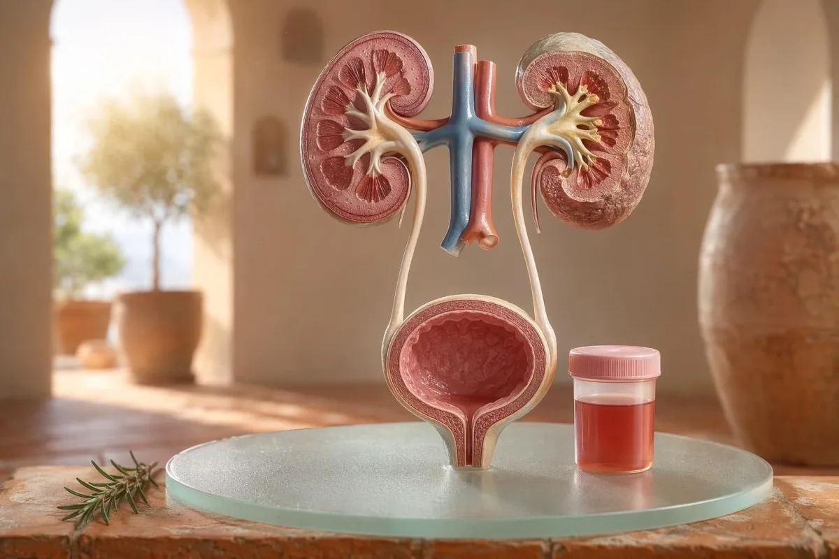
Görünür ve mikroskobik hematüri için hasta odaklı bir rehber; dipstick’teki kanın mikroskop altındaki eritrositlerle neden aynı şey olmadığını da içerir.


- İdrarda kan görünür şekilde kırmızı, çay rengi veya kola rengi idrar ya da testte saptanan mikroskobik hematüri anlamına gelir; görünür kanın her zaman tıbbi takip gerektirdiği unutulmamalıdır.
- Mikroskopik hematüri uygun şekilde toplanmış bir idrar mikroskopisi örneğinde, yüksek büyütme alanı başına 3 veya daha fazla eritrosit olarak tanımlanır.
- Dipstick’teki kan heme aktivitesini saptar; bu nedenle kas yaralanmasından sonra sağlam eritrositlerden, serbest hemoglobinden veya miyoglobinden pozitif çıkabilir.
- İYE ipuçları yanma, sık idrara çıkma, lökosit esteraz, nitritler ve beyaz hücreleri içerir; enfeksiyon temizlendikten sonra hematüri yeniden kontrol edilmelidir.
- İdrar sedimenti dismorfik eritrositler, eritrosit silendirleri veya anlamlı protein gibi bulgular, böbreğin süzme birimi hastalığına işaret eder.
- İdrar silendirleri böbrek tübüllerinden şekillendirilmiş materyaldir; eritrosit silendirleri aynı hafta içinde görülürse, özellikle yüksek kan basıncı veya düşen eGFR ile birlikte, acil bir nefroloji uyarı işaretidir.
- Kanser uyarı işaretleri ağrısız görülebilir hematüri, pıhtılar, 45 yaş üstü, sigara öyküsü, mesleki kimyasal maruziyet ve tekrarlayan, açıklanamayan atakları içerir.
- Egzersize bağlı hematüri genellikle 48-72 saat içinde düzelir; dinlenmeye rağmen devam eden kanama, koşu veya bisiklete bağlanmamalıdır.
- Acil değerlendirme ateş, yan ağrısı, idrar tıkanıklığına yol açan pıhtılar, gebelik, travma veya şiddetli anemi belirtileri olan idrarda kan için gereklidir.
Panik yapmadan önce idrarda kanın ne anlama geldiği
İdrarda kan enfeksiyondan, taşlardan, böbrek süzme hastalığından, prostat veya mesane durumlarından, ilaçlardan, egzersizden ya da adet döneminden kontaminasyondan kaynaklanabilir. Pratik kural basittir: görülebilir kan, tekrarlayan mikroskopik hematüri veya ağrı, ateş, pıhtı, yüksek kan basıncı, protein ya da azalmış böbrek fonksiyonu ile birlikte hematüri, izlemeyi beklemek yerine acil değerlendirme gerektirir.
Pancar veya yeni bir takviyeden sonra tuvalette tek bir pembe renk görülmesi, boğaz ağrısından sonra kola rengi idrar ile aynı klinik sorun değildir. Uygulamada, renk hikâyesi çoğu zaman önce nereye bakmam gerektiğini söyler: pıhtılarla birlikte parlak kırmızı genellikle alt üriner sistemle ilişkilidir; kahverengi veya çay rengi idrar ise böbrek filtrelerinden daha eski heme pigmentlerini düşündürebilir.
Hematüri bir tanı değildir; bir bulgudur. Görülebilir hematüri idrarın rengindeki değişikliği görebildiğiniz anlamına gelir; mikroskopik hematüri idrarın normal göründüğü, ancak mikroskopide kırmızı kan hücrelerinin görüldüğü anlamına gelir; genellikle yüksek büyütme alanı başına 3 veya daha fazla eritrosit.
Kantesti bir AI kan testi analizörü hastaların idrar bulgularını kreatinin, eGFR, CRP, hemoglobin ve trombositler gibi böbrek belirteçleriyle tek bir zaman çizelgesinde ilişkilendirmesine yardımcı olur. Renk kafa karıştırıcıysa, bizim idrar renk rehberimiz kırmızı, kahverengi, turuncu ve koyu sarı idrarın hepsinin aynı şeyi ifade etmediğini açıklar.
Dipstick’teki kan, mikroskopideki RBC’lerle aynı değildir
Üzerinde kan pozitif yazan bir idrar dipstiki, sağlam kırmızı kan hücreleri olmak zorunda olmayan heme-benzeri kimyasal aktiviteyi saptar. Mikroskopi bir sonraki soruyu yanıtlar: idrar sedimentinde gerçekten RBC var mı ve varsa nasıl görünür?
Dipstik test hızlıdır; çünkü reaktif ped, hemin peroksidaz aktivitesiyle reaksiyona girer. Bu da pozitif sonucun; sağlam RBC’lerde, eritrosit parçalanması sonrası serbest hemoglobinde veya anlamlı kas hasarı sonrası miyoglobinde görülebileceği anlamına gelir.
Mikroskopi daha yavaştır ama daha spesifiktir. Dikkatli bir laboratuvar idrarı santrifüje eder, idrar sedimentini, inceler ve yüksek büyütme alanı başına eritrosit sayısını raporlar; birçok klinisyen yetişkinlerde 0-2 RBC/hpf’yi normal ve 3 veya daha fazla RBC/hpf’yi anormal kabul eder.
Bu uyumsuzluk önemlidir. 3+ kan ve 0-2 RBC/hpf olan bir dipstik, mesane hastalığına doğrudan atlamaktan ziyade benden rabdomiyoliz, hemoliz, oksitleyici kontaminantlar ve örnek işleme hakkında düşünmemi ister; fark, bizim idrar tahlili ve kültür karşılaştırması.
İYE ipuçları hematürüyü açıkladığında ve açıklamadığında
Bir idrar yolu enfeksiyonu şunlara neden olabilir: idrarda kan, özellikle yanma, sık idrara çıkma, aciliyet, pelvik rahatsızlık, lökosit esteraz, nitritler ve beyaz hücreler ile birlikteyse. Ancak hematuri tedavi sonrası düzelmelidir; İYE (UTI) sonrası persistan eritrositler ikinci bir değerlendirme gerektirir.
Basit bir alt İYE’de, bir kümeyi beklerim: dizüri, sık idrara çıkma, lökosit esteraz pozitifliği, piyüri sıklıkla 5-10 beyaz hücre/hpf’nin üzerindedir ve bazen nitritler de görülür. Geleneksel kültür pozitifliği 100.000 CFU/mL’dir; ancak semptomatik kadınlarda 1.000-10.000 CFU/mL gibi daha düşük değerler de klinik olarak anlamlı olabilir.
Lökosit esteraz, bir beyaz hücre enzimidir; bakteri varlığının kanıtı değildir. Hematuri ile birlikte pozitif sonuç; enfeksiyon, taşlar, kontaminasyon veya böbrek iltihabından kaynaklanabilir; bu yüzden lökosit esteraz rehberimiz faydalı ipuçlarını yaygın yalancı pozitiflerden ayırır.
Nitritler daha özgüldür ancak daha az duyarlıdır; çünkü tüm bakteriler nitratı nitrite dönüştürmez. Belirtiler klasik olsa da nitritler negatifse kültür yine de pozitif çıkabilir; pratik ayrıntılar nitrit sonucu açıklayıcımızda.
Görünür hematüri: renk, pıhtılar ve zamanlama ipuçları
görünür hematuriyi asla küçümsemem; bir kez bile olsa. Parlak kırmızı idrar, pıhtılar veya ağrısız kanama, yaş ve sigara maruziyeti arttıkça alt üriner sistem nedenlerinin olasılığı yükseldiği için zamanında değerlendirme gerektirir.
Zamanlama şaşırtıcı derecede faydalı olabilir. İdrar yapmanın başlangıcında yalnızca kan görülmesi üretrayı düşündürebilir; akım boyunca kan gelmesi mesane veya böbrek kaynaklı olabilir ve bitişe yakın terminal kan bazen mesane boynu bölgesine işaret eder.
Pıhtılar genellikle kanamanın kaynağının böbrek filtrelerinin altında olduğunu gösterir; pıhtılar glomerüllerden ve tübüllerden geçtikten sonra kolay oluşmaz. İdrar akımını tıkayan bir pıhtı acildir: mesane distansiyonu birkaç saat içinde ağrılı hale gelebilir ve kateter bakımı gerekebilir.
Erkekler bazen idrar şikayetlerinden sonra görülen makroskopik hematürinin sadece prostat irritasyonu olduğunu varsayar. Bu doğru olabilir; ancak enfeksiyon sırasında veya hemen sonrasında bir PSA kontrol edildiyse yorum karmaşıklaşabilir; bizim UTI sonrası PSA rehberi birçok klinisyenin PSA’yı tekrar etmeden önce 6-8 hafta beklemesinin nedenini açıklar.
Doktorların kullandığı mikroskobik hematüri eşikleri
Mikroskopik hematüri genellikle yüksek güçlü alan başına 3 veya daha fazla RBC düzgün şekilde toplanmış bir idrar örneğinde tanımlanır. AUA/SUFU kılavuzu bu eşiği kullanır ve her hastayı aynı şekilde tedavi etmek yerine risk temelli değerlendirme önerir (Barocas ve ark., 2020).
Mikroskopik hematürüyü tek başına bir dipstick ile tanı koymak doğru değildir. Barocas ve ark. 2020 AUA/SUFU kılavuzunda tanım, mikroskopiye bağlıdır; çünkü dipstick’ler sağlam kırmızı hücreler olmadan da heme pigmentlerinden pozitif çıkabilir.
Tekrar test yaptırmak zayıflık değildir; iyi tıptır. Ağır egzersiz, ateş, cinsel ilişki, menstruasyon veya kontamine bir örnek sonrası birinde 3-10 RBC/hpf varsa, tarama istemeden önce genellikle temiz bir tekrar örneği isterim; tıpkı tekrarlı anormal testler.
Risk yolu değiştirir. Yarı maraton sonrası 4 RBC/hpf olan 24 yaşındaki bir kişi, iki örnekte 25 RBC/hpf olan 68 yaşındaki eski sigara içicisiyle aynı değildir; her ikisi teknik olarak hematüri tanımını karşılıyor olsa bile.
Değerlendirmeyi değiştiren idrar sedimenti, protein ve idrar silendirleri
İdrar sedimenti hematürinin böbrek filtrelerinden gelme olasılığını ortaya çıkarabilir. Disformik eritrositler, eritrosit silendirleri ve anlamlı protein değişimi, basit bir mesane enfeksiyonundan ziyade glomerüler hastalık endişesini öne çıkarır.
Düzensiz görünen, kabarcıklı (blebbed) veya halka şeklinde olan eritrositlere disformik RBC denir ve bu, iltihaplı bir glomerüler filtre üzerinden geçişi düşündürür. İdrardaki eritrositlerin yaklaşık 5% üzerindeki akantositler sıklıkla güçlü bir glomerüler ipucu olarak ele alınır; ancak laboratuvarlar bunları ne kadar güvenle raporladıklarında farklılık gösterebilir.
Protein ikinci ipucudur; bunu göz ardı etmem. İdrar albümin-kreatinin oranı 30 mg/g’nin üzerindeyse veya yaklaşık 3 mg/mmol ise anormaldir ve makroskopik hematüri ile birlikte protein daha hızlı böbreğe odaklı değerlendirmeyi hak eder; bizim idrarda protein rehberi hasta düzeyinde eşikleri verir.
İdrar silendirleri böbrek tübülleri içinde oluşan küçük kalıplardır. Hiyalin silendirler dehidratasyon veya egzersiz sonrası görülebilir; ancak eritrosit silendirleri anormaldir ve genellikle glomerülonefriti işaret eder; daha geniş bir idrar tahlili referansı için bizim idrar tahlili kılavuzunun tamamı.
Taşlar, egzersiz ve hematürünün geçici nedenleri
Taşlar ve şiddetli egzersiz hematüriye neden olabilir, ancak farklı zaman çizelgeleri vardır. Taşa bağlı hematüri çoğu zaman yan ağrısı veya kristallerle birlikte gelir; egzersize bağlı hematüri ise genellikle dinlenmeyle 48-72 saat içinde yatışmalıdır.
Böbrek taşları klasik olarak dalga dalga yan ağrısı, bulantı ve mikroskopik ya da makroskopik hematüri yapar. Kalsiyum oksalat kristalleri tek başına taş kanıtlamaz; ancak kolik tarz ağrı ile birlikte olduğunda şüpheyi artırır; bizim kalsiyum oksalat kristalleri rehberi idrar mikroskopisi ipucunu açıklar.
Egzersiz hematürisi gerçektir. Bir yarıştan sonraki sabah 10-20 RBC/hpf olan, normal kültürü bulunan, protein olmayan ve 3 gün sonra tamamen normal bir tekrar idrar tahlili yaptıran maraton koşucuları gördüm.
Tehlike, her şeyi antrenmana bağlamaktır. Hematüri 72 saati aşarak sürüyorsa, koyu kola renkli idrarla ortaya çıkıyorsa veya yüksek CK, yükselen kreatinin ya da kas ağrısıyla birlikte geliyorsa mesaneyi aşın; bizim kreatinin egzersiz kılavuzu böbrek ve kas örtüşmesini kapsar.
Doktorların göz ardı etmediği böbrek kırmızı bayrakları
Proteinle birlikte hematüri, yüksek kan basıncı, şişlik, azalmış eGFR, eritrosit silendirleri veya hızla yükselen kreatinin bir böbrek için kırmızı bayraktır. Bu bulgular, süzme birimlerinin iltihaplı olabileceğini düşündürür ve aylarca beklemek böbrek fonksiyonunu kaybettirebilir.
KDIGO 2024, kronik böbrek hastalığını; albüminüri gibi böbrek hasarı belirteçleriyle veya en az 3 ay boyunca 60 mL/dk/1,73 m²’nin altındaki eGFR ile tanımlar. Ancak akut durumlarda, günler ya da haftalar içinde kreatinin artışıyla birlikte hematüri, 3 aylık tanım sağlanmadan önce yükseltmek için yeterlidir.
Albüminüri çoğu zaman erken uyarı sinyalidir. ACR 30-300 mg/g orta düzeyde artmıştır, 300 mg/g’nin üzeri ciddi düzeyde artmıştır ve bunlardan herhangi birinin hematüriyle eşleşmesi konuşmayı değiştirir; bizim böbrek ACR rehberimiz idrar albümininin semptomlardan önce hasarı neden bulduğunu açıklar.
Burada kan basıncı önemlidir. Boğaz enfeksiyonundan sonra kola renkli idrar ve ayak bileği şişliğiyle birlikte yeni bir KB 160/100 mmHg olması, sistitten çok glomerülonefriti düşündürür; eGFR belirsizse, bizim GFR yeniden kontrol kılavuzu sistatin C’nin böbrek fonksiyonunu ne zaman netleştirebileceğini açıklar.
Panik olmadan kanser riski ipuçları
Ağrısız görülebilir hematüri, özellikle 45 yaş sonrası, mesane ve üst üriner sistem kanserleri bu şekilde ortaya çıkabildiği için acil değerlendirmeyi hak eder. NICE NG12, yetişkinlerde 45 yaş ve üzeri açıklanamayan görülebilir hematüri için veya İYE tedavisinden sonra devam eden görülebilir hematüri için şüpheli-kanser sevkini önerir.
Hematürünün çoğu kanser değildir. Yine de hatırladıklarım sessiz olanlar: ağrı yok, ateş yok, yanma yok; sadece 56 yaşında bir sigara içicisinde neredeyse önemsemeyecek kadar gözden kaçmış iki kez kırmızı idrar.
Risk kümülatiftir, ikili değildir. 45-50 yaş üstü, sigara öyküsü, siklofosfamid maruziyeti, pelvik radyoterapi, tekrarlayan görülebilir hematüri, mesleki aromatik amin maruziyeti ve pıhtılar; sistoskopi veya görüntüleme ihtiyacını artırır.
Prostat belirteçleri mesane kaynaklarını dışlamaz. İdrar şikayetleri, PSA değişiklikleri ve hematüri örtüşüyorsa; zamanlamayı ve enfeksiyon durumunu dikkatle gözden geçirin; bizim prostat belirteç rehberi PSA’nın erkeklerde idrar değerlendirmesinin yalnızca bir parçası olmasının nedenini açıklar.
İlaçlar, antikoagülanlar ve yanlış alarmlar
Kan sulandırıcılar kanamayı ortaya çıkarabilir, ancak hematüri için otomatik olarak suçlanmamalıdır. Antikoagülan ilişkili hematüri yine de değerlendirilmelidir; çünkü ilaç, taşları, enfeksiyonu, böbrek hastalığını veya üriner sistemde bir kitleyi ortaya çıkarıyor olabilir.
Varfarin, apiksaban, rivaroksaban, aspirin ve klopidogrel kanamayı daha belirgin hale getirebilir. INR hedef aralığın üzerindeyse düzeltmek önemlidir; ancak normal bir INR, görünür hematürinin zararsız olduğu anlamına gelmez.
Hematürüyü taklit eden, kanama ile ilişkili olmayan çeşitli nedenler vardır. Rifampisin idrarı turuncu-kırmızıya çevirebilir, fenazopiridin idrarı parlak turuncu yapabilir, pancar hassas kişilerde idrarı boyayabilir ve dehidratasyon pigmenti yeterince koyulaştırarak hastaları korkutabilir; antikoagülan izlemi bizim kan sulandırıcı güvenlik laboratuvarlarımızda ele alınır.
Dipsit de yoğun egzersiz veya kas yaralanması sonrası yanıltabilir. Az sayıda veya hiç RBC içermeyen pozitif heme pedi, özellikle CK 1.000 IU/L’nin üzerindeyse ve idrar koyu renkliyse, düşüncemi miyoglobine doğru iter.
Çocuklar, gebelik ve dönem (adet) kontaminasyonu
Çocuklarda, gebelikte veya menstruasyon çevresinde görülen hematüri daha temiz örnekleme ve inceleme için daha düşük bir eşik gerektirir. Aynı idrar sonucu; yaş, gebelik durumu, ateş, kan basıncı ve örnek toplama kalitesine göre farklı anlamlar taşıyabilir.
Menstrüel kontaminasyon yaygındır ve kimse bundan utanmamalıdır. Sonuç beklenmedikse, kanama durduktan en az 48 saat sonra tekrarlanan bir orta akım temiz yakalama örneği alın; ısrarcı olarak 3 veya daha fazla RBC/hpf bulunması daha anlamlı hale gelir.
Çocuklarda sıklıkla farklı bir bakış açısı gerekir. Viral bir hastalık sonrası hematüri geçici olabilir; ancak şişlik, yüksek kan basıncı, proteinüri veya idrar çıkışında azalma ile birlikte hematüri, acil pediatrik değerlendirme gerektirir.
Gebelik, UTI, taşlar, preeklampsiye bağlı böbrek stresi ve kontaminasyonun örtüşebilmesi nedeniyle riski artırır. Böbrek, karaciğer, trombosit ve idrar bulgularını içeren aynı gün gebelik uyarı paternleri için bizim gebelik laboratuvarı kırmızı bayrakları.
Doktorların hematüriden sonra genellikle istediği testler
Hematüriden sonra genellikle yapılan bir sonraki testler; mikroskopi ile tekrarlı idrar tahlili, enfeksiyon olasılığı varsa idrar kültürü, böbrek fonksiyonuna ilişkin kan testleri, idrar proteini veya ACR, kan basıncı ölçümü ve risk temelli görüntüleme veya sistoskopidir. Kesin sıra, semptomlara ve risklere bağlıdır.
Komplike olmayan sistit semptomlarında kültür ve tedavi önce gelebilir; semptomlar düzeldikten sonra tekrarlı idrar mikroskopisi yapılır. Ağrısız görünen hematüride ise birçok klinisyen, tekrarlayan atakları beklemek tanıyı geciktirebileceğinden daha erken görüntüleme ve sistoskopiye yönelir.
Kan testleri, yalnızca idrarın sağlayamadığı bağlam ekler. Kreatinin, eGFR, üre veya BUN, elektrolitler, CBC, CRP, kompleman C3/C4 ve otoimmün belirteçler; böbrek iltihabı şüphesi varsa uygun olabilir; bizim BUN/kreatinin oranı kılavuzumuz böbrek iltihabına ilişkin yaygın bir paterni açıklıyor.
Kantesti AI, bu sonuçları bir trend görünümüne dönüştürebilir; ancak kırmızı bayraklar varsa hekim incelemesi hâlâ esastır. Klinik güvenlik yaklaşımımızda Tıbbi Doğrulama, izole sayılar yerine anormal kümelerin nasıl ele alındığı da dahil olmak üzere açıklanır.
Kantesti, hematüri bağlamını organize etmeye nasıl yardımcı olur
Kantesti bir AI laboratuvar testi yorumlama hizmetinde yayımlanır. kreatinin, eGFR, CRP, hemoglobin, trombositler ve glukoz gibi idrarla ilişkili kan belirteçlerini bağlam içinde okur. Hematürüyü tanılamaz; ancak hastaların klinisyenleri için daha temiz ve daha kullanışlı bir özet hazırlamasına yardımcı olur.
Hematürüyü incelerken nadiren tek bir sonuca bakarım. 0.82 mg/dL normal kreatinin, 90’ın üzerindeki stabil eGFR, protein yokluğu ve negatif kültür; eGFR 48 ve CRP 65 mg/L ile birlikte hematüriden tamamen farklı bir şey gibi gelir.
Kantesti bir Yapay zekâ biyobelirteç yorumlama platformu yüklenen kan testi PDF’lerini veya fotoğraflarını işleyebilen ve semptomların zamanlamasıyla birlikte referans dışı böbrek, inflamasyon ve anemi belirteçlerini ilişkilendirebilen bir yapıdır. Bu patern okumanın arkasındaki yöntemler bizim Teknoloji Rehberi, ve daha geniş belirteç kapsamı ise biyobelirteç kılavuzu.
Bir pratik ipucu: Yüklemeden önce birimlerin, tarihlerinin ve hasta tanımlayıcılarının doğru yakalandığını kontrol edin. OCR hataları 0.9’u 9.0’a çevirebilir; bu da tamamen farklı bir böbrek hikâyesidir; bizim PDF yükleme kontrol listemiz hastaların önce yakalamasını istediğim hataları gösterir.
Araştırma notları, yönetişim ve son kontrol listesi
11 Temmuz 2026 itibarıyla en güvenli hasta planı, hematürinin gerçek olup olmadığını doğrulamak, enfeksiyon ipuçlarını belirlemek, böbrek için uyarı işaretlerini aramak ve görünen ya da kalıcı hematüride durumu acil olarak yükseltmektir. Sadece idrar rengine güvenmeyin.
Ben Thomas Klein, MD ve tavsiyem özellikle temkinlidir; çünkü atlanan hematürinin önemi olabilir. Kantesti'nin tıbbi içeriği, klinik gözetimle birlikte gözden geçirilmektedir. Tıbbi Danışma Kurulu, ve bizim rolümüz, hastaların daha iyi sorular sormasına yardımcı olmaktır; doktorun yerini almak değil.
Kantesti bir Yapay zekâ destekli kan testi analiz aracı 127 ülkede milyonlarca kişi tarafından kullanıldığı için, gizlilik ve klinik açıklık sonradan eklenmek yerine iş akışına yerleştirilmek zorundadır. Kantesti LTD, Birleşik Krallık şirket yapımız ve klinik misyonumuz hakkında daha fazlasını şurada okuyabilirsiniz: Hakkımızda.
Kantesti Yapay Zeka Klinik Araştırma Grubu. (2026). Yapay Zeka Kan Testi Analizörü: Analiz Edilen 2,5 Milyon Test | Küresel Sağlık Raporu 2026. Zenodo. DOI: https://doi.org/10.5281/zenodo.18175532. ResearchGate: https://www.researchgate.net/. Academia.edu: https://www.academia.edu/.
Kantesti AI Clinical Research Group. (2026). RDW Blood Test: Complete Guide to RDW-CV, MCV & MCHC. Zenodo. DOI: https://doi.org/10.5281/zenodo.18202598. ResearchGate: https://www.researchgate.net/. Academia.edu: https://www.academia.edu/.
Sıkça Sorulan Sorular
İdrarda kan her zaman ciddi midir?
İdrarda kan her zaman ciddi değildir; ancak doğrulanmalı ve açıklanmalıdır. Egzersiz sonrası veya menstruasyon sonrası 3-5 RBC/hpf düzeyinde tek seferlik mikroskobik bir sonuç tekrarlanan testte temizlenebilir; buna karşılık görülebilir kan, pıhtılar, ateş, yan ağrısı, protein, yüksek kan basıncı veya azalmış eGFR acil değerlendirme gerektirir. 45 yaş üzeri yetişkinlerde, ağrı olmasa bile açıklanamayan görülebilir hematüri acil değerlendirmeyi hak eder.
İdrarda dipstick kan ile idrardaki RBC’ler arasındaki fark nedir?
İdrar çubuğu (dipstick) kan, heme benzeri kimyasal aktiviteyi saptar; oysa idrar mikroskopisi gerçek eritrositleri (RBC) sayar. Bir dipstick, sağlam RBC’lerden, serbest hemoglobinden veya kas yaralanması sonrası miyoglobinden dolayı pozitif olabilir; bu nedenle doğrulanmış hematüri ile aynı şey değildir. Mikroskopik hematüri, uygun şekilde toplanmış bir örnekte yüksek büyütme alanı başına 3 veya daha fazla RBC olarak sıklıkla tanımlanır.
İdrar yolu enfeksiyonu (İYE) idrarda kana neden olabilir mi?
Evet, bir İYE idrarda kanamaya neden olabilir; özellikle yanma, sık idrara çıkma, idrar yapma isteği (aciliyet), lökosit esteraz, nitritler ve beyaz kan hücreleri de mevcutsa. Geleneksel idrar kültürü pozitifliği sıklıkla 100.000 CFU/mL’dir; ancak daha düşük sayımlar da semptomatik hastalarda anlamlı olabilir. Hematüri, tedavi sonrasında genellikle yeniden kontrol edilmelidir; çünkü devam eden kanama taşlara, böbrek hastalığına veya başka bir üriner sistem nedenine işaret edebilir.
Hematüri için ne zaman acil servise gitmeliyim?
Ateşle birlikte idrarda kan, şiddetli yan ağrısı, kusma, gebelik, yakın zamanda travma, pıhtılar, idrar yapamama, bayılma veya şiddetli anemi belirtileri varsa acil tıbbi yardım alın. Proteinüri ile birlikte hematüri, eritrosit silendirleri, yeni gelişen yüksek tansiyon veya kreatinin düzeyinde yükselme durumlarında da aynı hafta içinde tıbbi değerlendirme uygundur. Pıhtılardan kaynaklanan tıkalı bir idrar akımı birkaç saat içinde ağrılı ve güvenli olmayan hale gelebilir.
Egzersiz mikroskopik hematüriye neden olabilir mi?
Zorlu egzersiz mikroskopik hematüriye neden olabilir; özellikle uzun mesafe koşusu, yoğun bisiklet sürme veya temas sporlarından sonra. Egzersize bağlı hematüri genellikle dinlenmeyle 48-72 saat içinde düzelir ve kalıcı proteinüri, düşen eGFR veya şiddetli kas ağrısıyla birlikte olmamalıdır. Dipstick ile kan pozitif çıkıp mikroskopide az sayıda RBC görülürse, kas hasarından kaynaklanan miyoglobin dipstick’i tetikleyebileceğinden klinisyenler CK’ya bakabilir.
İdrar silendirleri kan bulunduğunda ne anlama gelir?
İdrar silendirleri, böbrek tübülleri içinde şekillenmiş kalıplanmış materyallerdir ve bunların tipi hematürinin anlamını değiştirir. Hiyalin silendirler dehidratasyon veya egzersizle ortaya çıkabilir; ancak eritrosit silendirleri anormaldir ve glomerüler böbrek iltihabını düşündürür. Eritrosit silendirleriyle birlikte idrarda kan, protein ve yüksek kan basıncı, basit bir İYE’den ziyade böbrek için bir kırmızı bayrak olarak ele alınmalıdır.
Dehidrasyon idrarın kana benzemesine neden olabilir mi?
Dehidrasyon genellikle idrarı koyu sarı veya kehribar rengine çevirir, ancak genellikle idrarda kırmızı hücre oluşturmaz. Sıvı alın ve renk pembe, kırmızı, kahverengi veya kola rengindeyse, özellikle de sıvı alımından sonra devam ediyorsa veya ağrı eşlik ediyorsa bir idrar testi ayarlayın.
Bugün Yapay Zekâ Destekli Kan Tahlili Analizini Alın
Anlık ve doğru laboratuvar testi analizi için Kantesti’ye güvenen dünya genelindeki 2 milyondan fazla kullanıcıya katılın. Kan testi sonuçlarınızı yükleyin ve saniyeler içinde 15,000+ biyobelirteçlerinin kapsamlı yorumunu alın.
📚 Kaynak Gösterilen Araştırma Yayınları
Klein, T., Mitchell, S., & Weber, H. (2026). Yapay Zeka Kan Testi Analizörü: 2,5M Analiz Edilen Test | Küresel Sağlık Raporu 2026. Kantesti Yapay Zeka Tıbbi Araştırma.
Klein, T., Mitchell, S., & Weber, H. (2026). RDW Kan Testi: RDW-CV, MCV ve MCHC için Tam Rehber. Kantesti Yapay Zeka Tıbbi Araştırma.
📖 Harici Tıbbi Kaynaklar
National Institute for Health and Care Excellence (2015). . EAU Kılavuz Ofisi.. NICE Kılavuzu.
Böbrek Hastalığı: İyileştirme İçin Küresel Sonuçlar (KDIGO) CKD Çalışma Grubu (2024). KDIGO 2024 Kronik Böbrek Hastalığının Değerlendirilmesi ve Yönetimi için Klinik Uygulama Kılavuzu. Kidney International.
⚕️ Tıbbi Uyarı
Bu makale yalnızca eğitim amaçlıdır ve tıbbi tavsiye niteliği taşımaz. Tanı ve tedavi kararları için her zaman yetkin bir sağlık hizmeti sağlayıcısına danışın.
E-E-A-T Güven Sinyalleri
Deneyim
Hekim liderliğinde laboratuvar yorumlama iş akışlarının klinik incelemesi.
Uzmanlık
Klinik bağlamda biyobelirteçlerin nasıl davrandığına odaklanan laboratuvar tıbbı.
Otorite
Dr. Thomas Klein tarafından yazılmış; Dr. Sarah Mitchell ve Prof. Dr. Hans Weber tarafından gözden geçirilmiştir.
Güvenilirlik
Alarmı azaltmaya yönelik net takip yollarıyla kanıta dayalı yorumlama.