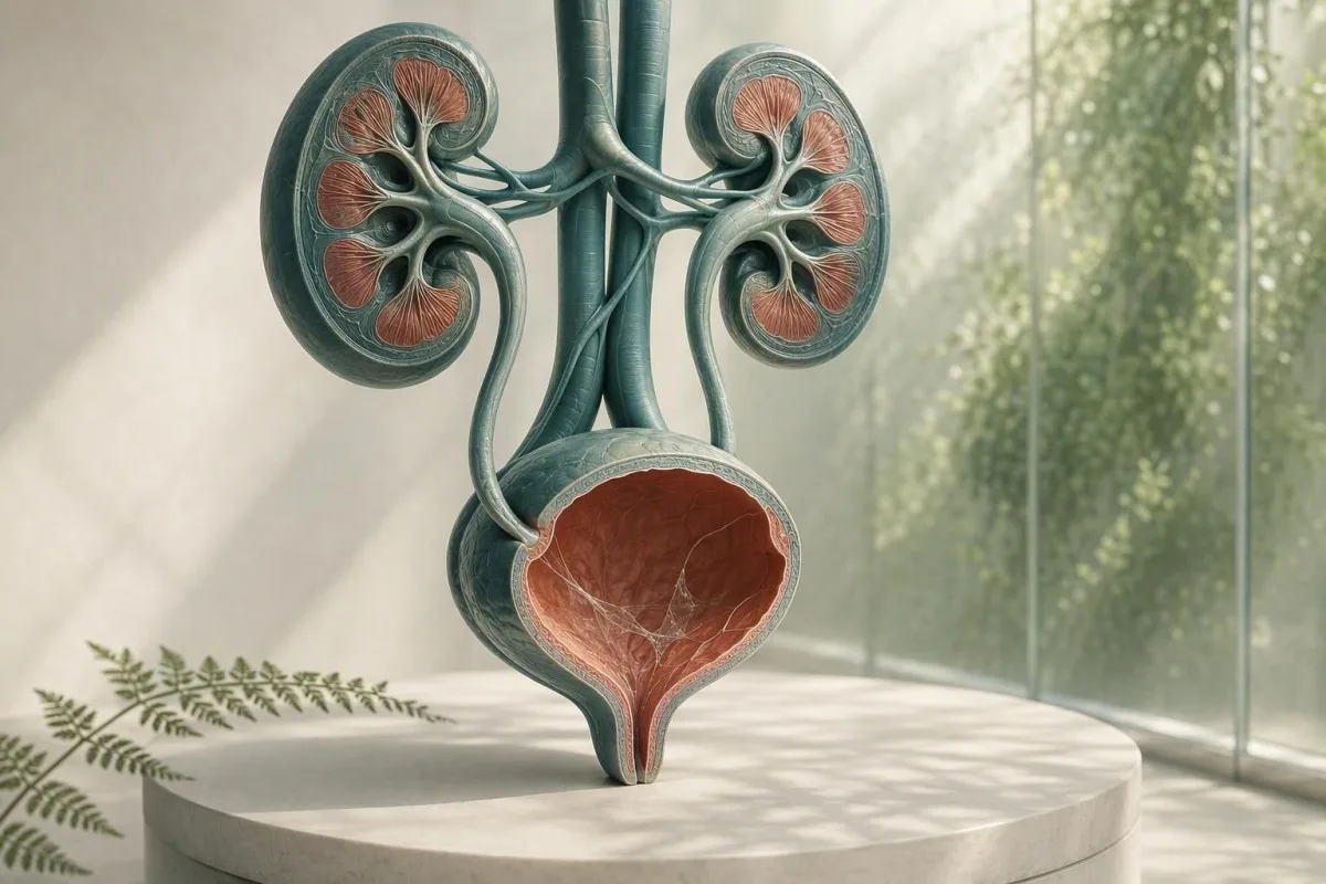
Les mucus dans un rapport de microscopie urinaire sont généralement un problème de prélèvement, pas un diagnostic. Les résultats d'analyse d'urine environnants, les symptômes et la qualité de l'échantillon déterminent s'ils nécessitent un suivi.


- Filaments de mucus seuls sont généralement inoffensifs, surtout dans un seul échantillon de jet propre sans symptômes urinaires.
- Seuil de pyurie de plus de 5 leucocytes par champ de haute puissance soutient l'inflammation des voies urinaires mais ne prouve pas une infection urinaire bactérienne.
- Déclencheur de culture est le mucus plus les brûlures, l'urgence, la fièvre, la douleur dans les flancs, les nitrites ou des leucocytes importants — pas le mucus seul.
- Cellules squameuses au-dessus d'environ 10 par champ de haute puissance indiquent souvent une contamination génitale-cutanée et rendent un résultat urinaire moins fiable.
- Sang visible avec douleur colique latérale nécessite une évaluation rapide pour un calcul, même si du mucus est également signalé.
- Grossesse modifie le seuil de suivi car la bactériurie asymptomatique est généralement dépistée par culture d'urine, et non par seule microscopie.
- Urine trouble peut refléter des cristaux, une urine concentrée, des pertes vaginales, du sperme ou des cellules ; il ne peut pas diagnostiquer une infection par son aspect.
- Répéter l’échantillon est judicieux lorsque le mucus est le seul résultat inhabituel et que le prélèvement n'a pas été soigneusement effectué par jet moyen après nettoyage.
Ce que les filaments de mucus dans une analyse d'urine signifient généralement
Les filaments de mucus dans l'urine sont généralement des brins de sécrétion protectrice normale ou une contamination génitale, et un résultat isolé signale rarement une maladie rénale. Le résultat devient significatif lorsqu'il apparaît avec des symptômes urinaires, des globules blancs, des bactéries, du sang ou des protéines.
Les laboratoires identifient le mucus comme des filaments fins et translucides observés au microscope ; de nombreux rapports le classent simplement comme rare, peu, modéré ou nombreux plutôt que d'attribuer une plage de référence numérique. L'urine contient naturellement une petite quantité de matière riche en glycoprotéines provenant des voies urinaires, et les sécrétions vaginales ou urétrales peuvent en ajouter davantage lors du prélèvement.
La formulation du rapport est moins alarmante qu'elle n'y paraît. À compter du 26 août 2026, il n'existe aucun décompte validé de filaments de mucus permettant de diagnostiquer une infection urinaire, une lithiase, un cancer ou une insuffisance rénale.; il s'agit d'une observation microscopique contextuelle. Pour une carte plus large des autres éléments d'un rapport de bandelette réactive et de microscope, consultez notre guide complet d'analyse d'urine.
Dans mon expérience clinique, un adulte asymptomatique avec du mucus marqué “modéré”, 0 à 2 globules blancs par champ de haute puissance, nitrite négatif et absence de sang est généralement mieux servi par une réassurance ou un échantillon répété correctement prélevé. Le Dr Thomas Klein constate souvent une anxiété antibiotique inutile due à un signalement de laboratoire confondu avec un diagnostic de maladie.
Kantesti est une plateforme d'interprétation de prises de sang par IA qui peut placer les marqueurs sanguins liés aux reins, tels que la créatinine et l'eGFR, à côté d'un résultat d'analyse d'urine — mais elle ne peut pas diagnostiquer une infection urinaire à partir du seul mucus. Un résultat de microscopie urinaire nécessite le contexte clinique qui l'accompagne.
Pourquoi un laboratoire le signale
Les systèmes d'information de laboratoire signalent le mucus car il est visuellement présent, et non parce que le laboratoire a établi un seuil dangereux. Un signal signifie donc “observé” plutôt que “marqueur de maladie anormale”, un peu comme un résultat isolé de traces peut ne nécessiter aucun traitement.
D'où provient le mucus dans un échantillon d'urine
Le mucus dans l'urine peut provenir de l'urètre, de la muqueuse vésicale, du col de l'utérus ou du vagin, du sperme, ou de la peau génitale externe lors du prélèvement. Sa source est souvent déduite du reste du schéma microscopique plutôt que des filaments eux-mêmes.
La vessie et l'urètre possèdent une couche de surface protectrice contenant des mucines, y compris du matériel associé à l'uroplakine, qui réduit la friction et l'adhésion microbienne. De petites quantités peuvent être éliminées dans l'urine après déshydratation, activité sexuelle récente, irritation mineure, ou simplement parce qu'un spécimen concentré du matin rend les filaments plus faciles à voir.
Chez les personnes qui ont leurs règles ou des pertes vaginales, les leucocytes et le mucus peuvent entrer dans le récipient sans provenir de la vessie. Plus de 10 cellules épithéliales squameuses par champ de haute puissance rend la contamination plus probable, bien que les laboratoires utilisent des seuils de déclaration différents. Notre guide sur les cellules épithéliales dans l'urine explique pourquoi cela change la confiance dans un résultat.
Le sperme peut également créer des matériaux filandreux ou une turbidité pendant plusieurs heures après l'éjaculation. Cela ne rend pas l'urine dangereuse, mais cela peut masquer la microscopie ; si une culture est envisagée, je conseille généralement de prélever un nouveau spécimen au moins 24 heures plus tard lorsque cela est possible.
Pourquoi le sexe et l'anatomie affectent le rapport
Une cupule d'urine n'isole pas la vessie des tissus avoisinants. C'est pourquoi “ mucus présent ” est rapporté plus souvent dans les échantillons avec des sécrétions externes, tandis qu'un spécimen obtenu par cathétérisme est parfois utilisé lorsque les cliniciens ont besoin d'une réponse plus propre.
Quand les filaments de mucus orientent vers une infection des voies urinaires
Les fils de mucus ne soutiennent une possible infection urinaire que lorsqu'ils accompagnent des symptômes et des signes inflammatoires objectifs, en particulier une pyurie, une positivité des nitrites ou une culture convaincante. Le mucus seul n'est pas un test d'infection urinaire.
Une infection urinaire basse symptomatique provoque couramment des brûlures, une impériosité, une pollakiurie, une gêne sus-pubienne et parfois une nouvelle odeur d'urine. Dans un échantillon correctement collecté, plus de 5 globules blancs par champ de haute puissance est souvent appelée pyurie ; elle soutient l'inflammation des voies urinaires mais peut également survenir avec des calculs, des infections sexuellement transmissibles et une contamination.
Le nitrite est très spécifique lorsqu'il est positif mais manque les infections causées par des organismes qui ne réduisent pas le nitrate, et un résultat négatif du nitrite n'exclut pas une infection urinaire. L'estérase leucocytaire détecte l'activité enzymatique des globules blancs et peut être faussement positive lorsque des sécrétions vaginales contaminent le spécimen ; notre explication de les résultats de la leucocyte estérase couvre ces pièges.
La directive IDSA déconseille le dépistage ou le traitement de la bacteriurie asymptomatique chez les adultes non enceintes en bonne santé car les antibiotiques nuisent sans bénéfice (Nicolle et al., 2019). Ce principe est particulièrement pertinent pour le mucus : absence de brûlure, absence de fièvre et absence de procédure urologique prévue signifient généralement pas d'antibiotique simplement parce qu'un champ de microscopie semblait désordonné.
Quand la culture est le meilleur test
La culture d'urine est plus utile qu'un test bandelette répété lorsque les symptômes persistent, les symptômes récidivent dans les 4 semaines, une grossesse est présente, une pyélonéphrite est suspectée, ou des antibiotiques antérieurs ont pu altérer le résultat. Une croissance bactérienne mixte indique couramment une contamination lors de la collecte plutôt qu'un seul pathogène urinaire ; voir interprétation de la culture d’urines.
Mucus, cristaux, calculs et irritation mécanique
Du mucus avec des douleurs lombaires intenses en forme d'onde ou du sang dans l'urine peut survenir avec un calcul urinaire, mais le mucus ne confirme ni n'exclut un calcul. Le sang, les cristaux, le schéma de douleur, l'imagerie et la fonction rénale sont plus importants.
Un calcul peut gratter ou obstruer la paroi urinaire, produisant des globules rouges, des globules blancs et du mucus supplémentaire. Le symptôme classique est une douleur colique abrupte irradiant vers l'aine, souvent accompagnée de nausées ; la présence de sang visible dans l'urine ou d'une urine de couleur thé augmente l'urgence. Lire la suite sur drapeaux rouges du sang dans l'urine plutôt que de supposer que chaque échantillon rose est une simple infection.
Les cristaux d'oxalate de calcium peuvent apparaître chez les personnes en bonne santé, en particulier dans l'urine concentrée et acide, donc un type de cristal ne prouve pas un calcul actif. Chez une personne souffrant de douleur, de calculs récidivants ou d'hématurie persistante, les cliniciens peuvent utiliser une échographie ou un scanner CT sans contraste à faible dose plutôt que de se fier uniquement au sédiment. Notre ressource sur les cristaux d’oxalate de calcium détaille les limites des rapports de cristaux.
Une exception importante est l'obstruction avec infection : fièvre de 38,0 °C ou plus, douleurs lombaires, vomissements et incapacité à uriner justifient une évaluation d'urgence. Le problème n'est pas le mucus ; c'est un système urinaire infecté et bloqué, qui peut se détériorer rapidement.
Irritation sans calcul
Une utilisation récente de cathéter, des procédures vésicales, du cyclisme intense et une radiothérapie pelvienne peuvent irriter les voies inférieures et augmenter la production de mucus ou de globules blancs. Un clinicien devrait interpréter ces résultats en fonction du moment, car un échantillon collecté dans les 48 heures suivant une instrumentation n'équivaut pas à un spécimen de dépistage de routine.
Signification de l'urine trouble : ce que l'apparence peut et ne peut pas vous dire
L'urine trouble peut être causée par des sels concentrés, des cristaux, du mucus, des cellules, des pertes génitales ou des bactéries, de sorte que l'apparence seule ne peut pas diagnostiquer une infection. Une urine trouble mais indolore et de courte durée est généralement non urgente.
Les cristaux de phosphate peuvent rendre l'urine alcaline trouble après refroidissement, tandis que les cristaux d'urate peuvent troubler l'urine acide concentrée. Un échantillon laissé à température ambiante pendant plus de 2 heures peut devenir de plus en plus trouble à mesure que les cellules se dégradent et que les bactéries se multiplient, c'est pourquoi un traitement rapide ou la réfrigération est important.
La turbidité associée à la dysurie, à l'urgence et à la pyurie mérite un examen ; la turbidité après l'exercice ou une mauvaise hydratation s'améliore souvent avec une hydratation ordinaire. Visez une urine jaune pâle plutôt que de forcer un apport excessif d'eau : une urine très claire ne signifie pas que les reins “éliminent” l'infection. Notre examen détaillé de les causes d'urine trouble sépare les indices visuels des tests fiables.
Kantesti est un Analyseur de test sanguin AI conçus pour interpréter les marqueurs sanguins dans un contexte clinique ; une créatinine élevée ou un eGFR réduit associé à des anomalies urinaires peut justifier un examen médical, tandis qu'une urine trouble seule n'indique pas une filtration rénale altérée. Un bilan métabolique de base peut apporter un contexte utile lorsque les symptômes suggèrent une déshydratation ou une obstruction.
L'odeur n'est pas une culture
Une urine à forte odeur reflète souvent la concentration, des métabolites d'asperges, des vitamines B ou un récipient resté trop longtemps. Une nouvelle odeur fétide accompagnée de fièvre ou de symptômes urinaires justifie une évaluation, mais l'odeur ne remplace pas la microscopie et la culture.
Pertes génitales et infections sexuellement transmissibles
Les pertes urétrales ou vaginales peuvent ressembler à du mucus dans l'urine, et une nouvelle perte avec une brûlure urinaire nécessite des tests de santé sexuelle ainsi qu'un examen urinaire. La culture d'urine de routine peut manquer la chlamydia et la gonorrhée.
La chlamydia et la gonorrhée peuvent provoquer une dysurie et une pyurie stérile – des globules blancs sans croissance bactérienne de routine – particulièrement après une nouvelle exposition sexuelle. Les tests d'amplification d'acides nucléiques, utilisant souvent la première miction plutôt qu'un échantillon du milieu du jet, sont le test approprié car la culture standard cible différents organismes.
Chez les personnes avec un vagin, la vaginose bactérienne, la candidose et l'inflammation cervicale peuvent ajouter des pertes à une tasse du milieu du jet ; un prélèvement vaginal ou un examen peut être plus informatif que de répéter la microscopie urinaire. Chez les personnes avec un pénis, les pertes urétrales visibles ne doivent pas être négligées comme des “filaments de mucus”. La distinction pratique est une perte remarquée en dehors de la miction par rapport à des filaments signalés uniquement par le laboratoire.
La ligne directrice de traitement des IST du CDC de 2021 recommande des tests basés sur l'AMNA sur les sites anatomiques pertinents en fonction de l'histoire d'exposition, pas des symptômes seuls (Workowski et al., 2021). Si une douleur pelvienne, une douleur testiculaire, de la fièvre, une grossesse ou une agression possible est impliquée, demandez un avis clinique le jour même plutôt que de vous auto-traiter avec des antibiotiques restants.
Pourquoi les antibiotiques peuvent brouiller les pistes
La prise de même 1 ou 2 doses d'antibiotiques avant une culture peut supprimer la croissance bactérienne normale tandis que les symptômes urinaires et les globules blancs persistent. Indiquez au clinicien exactement quel médicament, quelle dose et quelle heure de la dernière dose ont été utilisés ; ce détail modifie l'interprétation d'une culture négative.
Comment prélever un échantillon d'urine qui répond à la question
Un échantillon de milieu de jet propre réduit mieux le mucus, les cellules squameuses et la croissance bactérienne mixte que la collecte de la première ou de la dernière partie du jet. La technique est simple, mais les 2 premières secondes font une différence disproportionnée.
Lavez-vous les mains, écartez la peau génitale ou rétractez le prépuce si vous êtes à l'aise, nettoyez selon les instructions du kit, commencez à uriner dans les toilettes, puis collectez la partie médiane sans que le gobelet touche la peau. Un échantillon de 20 à 30 mL est généralement suffisant ; remplir un grand récipient à ras bord n'améliore pas le test.
Livrez le gobelet rapidement, idéalement dans l'heure ; si un retard est inévitable, réfrigérez-le selon les instructions du laboratoire et retournez-le dans les 24 heures. La réfrigération ralentit la croissance bactérienne mais ne restaure pas un échantillon déjà contaminé. Ceci est particulièrement important lorsque la demande comprend une culture plutôt qu'une simple bandelette réactive.
Kantesti est un Outil d’analyse de prise de sang alimenté par l’IA utilisés dans plus de 127+ pays, et nos protocoles examinés par des cliniciens traitent la qualité des spécimens comme une partie de l'interprétation plutôt qu'une note de bas de page. Le même principe s'applique à toute valeur de laboratoire : un échantillon techniquement médiocre peut créer un résultat médicalement persuasif mais trompeur. Voir notre liste de contrôle de la précision du laboratoire pour les questions à poser avant d'agir sur un drapeau isolé.
Ne pas collecter pendant ces situations si vous pouvez attendre
Évitez les tests de routine pendant les règles abondantes, immédiatement après un rapport sexuel, ou après l'utilisation de crèmes vaginales, à moins que le clinicien ne le demande spécifiquement. Lorsque les tests ne peuvent pas attendre, informez le laboratoire ou le clinicien, car ce contexte peut expliquer la présence de mucus, de globules rouges ou de cellules externes.
Comment les cliniciens interprètent le mucus dans le reste de l'analyse d'urine
Le schéma d'analyse d'urine le plus utile combine les symptômes avec les globules blancs, les globules rouges, le nitrite, l'estérase leucocytaire, les protéines, le glucose, la densité spécifique et les cellules épithéliales. Le mucus est une caractéristique mineure de soutien dans ce schéma.
Un échantillon propre avec du mucus, 0 à 2 leucocytes par champ d'immersion, nitrite négatif et sans sang ne nécessite généralement aucun traitement. Le mucus avec plus de 5 leucocytes par champ d'immersion et une estérase leucocytaire positive augmente la probabilité d'inflammation ; l'ajout de nitrite ou une culture à un seul organisme augmente la confiance que les bactéries en sont responsables.
Les protéines nécessitent leur propre voie. Les traces de protéines après fièvre, exercice ou urine concentrée peuvent être temporaires, mais les protéines persistantes doivent être quantifiées par un rapport albumine/créatinine plutôt qu'attribuées au mucus. Notre guide pour les protéines dans les urines explique pourquoi les protéines sur bandelette et le risque rénal ne sont pas interchangeables.
Une densité urinaire de 1,003 à 1,030 est courant chez les adultes, bien que les intervalles de référence varient selon le laboratoire. Une gravité spécifique élevée peut concentrer le mucus et créer un sédiment d'apparence plus spectaculaire, tandis qu'une gravité spécifique faible peut lyser les cellules et rendre la microscopie trompeusement fade ; notre guide de densité urinaire montre comment l'hydratation affecte l'interprétation.
La valeur des résultats négatifs
L'absence de sang, de protéines, de nitrites et d'estérase leucocytaire réduit significativement l'inquiétude chez une personne sans symptômes, même si le mucus est signalé comme modéré. Aucun test négatif unique n'est parfait, mais cet ensemble est plus rassurant qu'une gradation de mucus n'est préoccupante.
Quand répéter le test et quand demander une culture
Répéter un échantillon d'urine lorsque le mucus est isolé, les cellules épithéliales squameuses suggèrent une contamination ou la collecte a été précipitée ; demander une culture lorsque les symptômes sont persistants, récurrents, sévères ou à haut risque. Un test répété n'est pas “ ne rien faire ” — c'est souvent la prochaine étape la plus diagnostique.
Pour un adulte non enceinte sans symptômes et avec du mucus comme seul signal d'alerte, une nouvelle analyse d'urine par milieu de jet dans les 1 à 2 semaines est raisonnable si un réconfort est nécessaire ; de nombreux cliniciens ne la répéteraient pas du tout. Ne pas traiter un signal d'alerte de laboratoire avec des antibiotiques en attendant, sauf si un prescripteur identifie une indication clinique.
La culture avant les antibiotiques est particulièrement utile en cas de fièvre, de douleur au flanc, de grossesse, d'immunosuppression, de transplantation rénale, d'utilisation de cathéter urinaire, de symptômes urinaires chez l'homme ou de symptômes qui reviennent dans les 4 semaines. La directive de Gupta et al. (2011) soutient la culture en cas de suspicion de pyélonéphrite et dans les situations où une résistance ou un diagnostic alternatif est plus probable.
Les comptes de culture doivent être lus en tenant compte de la qualité de la collecte. Un seul organisme à 10^5 unités formant des colonies par mL a traditionnellement soutenu la bactériurie dans les cultures d'urine de milieu de jet propres, mais les patients symptomatiques peuvent présenter des taux plus bas cliniquement pertinents ; une “flore mixte” incite généralement à un nouvel échantillon plutôt qu'à un antibiotique à large spectre. Comparez les objectifs de l’analyse d’urines et de la culture avant de demander l'un ou l'autre.
La grossesse nécessite un seuil différent
La grossesse fait exception car la bactériurie asymptomatique peut augmenter le risque de pyélonéphrite et d'issues de grossesse défavorables. Les soins prénataux utilisent couramment une culture de dépistage tôt dans la grossesse ; la présence de mucus au microscope ne peut pas remplacer cette culture.
Quand les tests sanguins apportent un contexte utile pour les reins et l'infection
Les analyses de sang sont utiles lorsque du mucus dans l'urine s'accompagne de fièvre, de douleurs lombaires, d'infections récurrentes, de gonflement, d'une diminution du débit urinaire, ou de protéines ou de sang persistants. La créatinine et l'eGFR évaluent la filtration, tandis qu'une CBC et une protéine C-réactive peuvent aider à juger de la maladie systémique.
Un DFG (eGFR) inférieur à 60 mL/min/1,73 m² pendant au moins 3 mois remplit un critère d'insuffisance rénale chronique, mais un seul résultat bas lors d'une déshydratation ou d'une maladie aiguë n'est pas suffisant pour poser ce diagnostic. La créatinine est influencée par la masse musculaire, l'alimentation et certains médicaments, de sorte que les tendances et l'albuminurie sont souvent plus informatives qu'un seul chiffre.
Une augmentation du nombre de globules blancs ou de la CRP peut soutenir un processus inflammatoire mais ne peut pas identifier le tractus urinaire comme source. Chez une personne fébrile avec des douleurs lombaires, les cliniciens peuvent vérifier la créatinine avant de choisir l'imagerie ou les médicaments, surtout si des vomissements ou une obstruction pourraient altérer la fonction rénale. Consulter les stades de la maladie rénale chronique pour les catégories d'eGFR et d'albumine utilisées par les cliniciens.
Kantesti est un service d’interprétation des tests de laboratoire par l’IA qui met en évidence les tendances entre la créatinine, l'eGFR, les électrolytes, la CBC et les marqueurs inflammatoires plutôt que de traiter une seule notation de mucus comme un diagnostic rénal. Notre méthodologie est soumise à une surveillance de validation clinique, mais les symptômes urgents nécessitent toujours des soins médicaux directs, et non une interprétation via une application.
Une distinction pratique de la déshydratation
La déshydratation peut augmenter la densité urinaire et augmenter temporairement la créatinine, en particulier après une diarrhée, une exposition à la chaleur ou un exercice intense. Une production d'urine faible persistante, des vertiges ou une baisse de l'eGFR après réhydratation justifient un examen par un clinicien plutôt que des tests répétés à domicile.
Signes d'alerte urgents qui ne doivent pas attendre un nouveau test
Recherchez une évaluation urgente le jour même pour la présence de mucus dans l'urine avec une fièvre de 38,0°C ou plus, des douleurs lombaires, des vomissements, du sang visible, une incapacité à uriner, une confusion ou des symptômes urinaires liés à la grossesse. Ces combinaisons sont importantes car elles peuvent signaler une infection des voies supérieures, une obstruction ou une autre affection aiguë.
La fièvre associée à des douleurs dorsales ou lombaires unilatérales est plus préoccupante pour une pyélonéphrite que pour une simple cystite, en particulier avec des frissons intenses ou des vomissements. Un traitement tardif peut entraîner une déshydratation, une septicémie ou un stress rénal ; un échantillon d'urine d'aspect normal à domicile ne permet pas de l'exclure en toute sécurité.
La présence de sang visible doit être évaluée même lorsqu'une infection urinaire semble plausible, en particulier après 35 ans, chez les fumeurs, ou si le saignement persiste une fois les symptômes d'infection résolus. La recommandation de la directive de l'AUA sur la microhématurie recommande une évaluation basée sur les risques après que des explications bénignes ont été traitées (Barocas et al., 2020) ; le sang persistant n'est pas expliqué par la présence de mucus.
Les enfants, les adultes de plus de 65 ans, les personnes diabétiques, les personnes prenant des médicaments immunosuppresseurs et celles ayant un rein unique méritent un seuil d'évaluation plus bas. La règle pratique du Dr Thomas Klein est simple : si les symptômes s'aggravent sur 6 à 12 heures, n'attendez pas une deuxième tasse pour vous rassurer.
Appelez les services d'urgence maintenant en cas de maladie grave
Appelez les services d'urgence en cas de nouvelle confusion, de malaise, de faiblesse sévère, de lèvres bleues ou grises, de détresse respiratoire sévère ou d'incapacité à retenir les liquides avec des symptômes urinaires. Ce sont des signes de danger systémique, pas des symptômes d'infection urinaire de routine.
Mucus dans l'urine pendant la grossesse, l'enfance et plus tard dans la vie
La grossesse, les enfants et les personnes âgées nécessitent une interprétation plus prudente car la contamination est fréquente mais les conséquences d'une infection manquée peuvent être plus importantes. Les symptômes et la qualité de la culture restent plus précieux que le grade du mucus dans tous les groupes d'âge.
Pendant la grossesse, la fréquence urinaire peut être normale, ce qui rend les symptômes moins spécifiques ; la fièvre, la dysurie ou les douleurs dorsales doivent inciter à une évaluation obstétricale ou clinique rapide. Une culture de dépistage est généralement obtenue au début des soins prénatals, et une culture de contrôle peut être effectuée après le traitement en fonction du plan du clinicien.
Chez les enfants, l'urine collectée par sac est sujette à la contamination et ne doit généralement pas être utilisée seule pour diagnostiquer une infection urinaire avec culture. Un spécimen obtenu par cathétérisme ou par une méthode de prélèvement propre soigneusement réalisée peut être nécessaire lorsqu'un jeune enfant a de la fièvre sans source claire ; le mucus dans un échantillon de sac est particulièrement non spécifique.
Dans la vie plus avancée, la bactériurie et la pyurie deviennent plus courantes sans causer de symptômes. Un nouveau délire seul devrait déclencher une évaluation médicale large pour la déshydratation, les médicaments, la douleur, la constipation et les sources d'infection plutôt qu'un traitement automatique des infections urinaires ; la même retenue recommandée par Nicolle et al. (2019) s'applique.
Modifications liées à la menstruation et aux hormones
Le sang menstruel et le mucus cervical peuvent altérer l'analyse d'urine pendant plusieurs jours, et la sécheresse vaginale post-ménopausique peut provoquer une irritation locale qui ressemble à une brûlure urinaire. Si les résultats et les symptômes ne correspondent pas, un clinicien peut évaluer les causes génitales plutôt que de prescrire un nouveau traitement pour une infection urinaire.
Erreurs courantes après avoir constaté la présence de mucus sur un rapport de laboratoire
Ne commencez pas d'antibiotiques restants, ne nettoyez pas à l'intérieur et n'essayez pas de “chasser” le mucus en buvant une quantité extrême d'eau après une seule analyse d'urine d'apparence anormale. Ces actions peuvent masquer une culture, perturber la flore normale ou retarder le bon diagnostic.
Les antibiotiques pris sans culture peuvent rendre un test ultérieur faussement négatif et peuvent causer de la diarrhée, des éruptions cutanées, des symptômes de mycose ou une résistance. Si les symptômes sont légers mais persistants, obtenez l'échantillon en premier lieu lorsque cela est possible, puis suivez le plan de traitement d'un clinicien basé sur l'ensemble de la situation.
Les produits à base de canneberge peuvent réduire modestement le risque de récidive d'infections urinaires non compliquées chez certaines personnes, mais ils ne traitent pas la fièvre, la douleur lombaire ou une infection confirmée des voies supérieures. Évitez la vitamine C à haute dose en automédication : elle peut altérer certaines réactions des bandelettes réactives et peut augmenter la charge d'oxalate chez les personnes sujettes aux calculs d'oxalate de calcium.
N'inspectez pas à plusieurs reprises la cuvette des toilettes à la recherche de filaments. Les fibres de papier toilette, les résidus de nettoyage, les sécrétions génitales et la turbulence de l'eau peuvent imiter le mucus ; un échantillon de laboratoire collecté dans un récipient stérile est l'endroit approprié pour l'évaluer. Pour les changements de couleur associés, notre guide de couleur des urines offre un contexte visuel plus fiable.
La liste des médicaments est importante
La phénazopyridine peut rendre l'urine orange et interférer avec l'interprétation visuelle, tandis que les diurétiques peuvent concentrer ou diluer l'urine en fonction du moment. Apportez une liste des médicaments sur ordonnance, des produits en vente libre et des suppléments à votre rendez-vous, y compris les doses en mg.
Un plan de suivi pratique pour les filaments de mucus dans l'urine
La plupart des personnes présentant des filaments de mucus dans les urines et aucun symptôme n'ont besoin de traitement ; une répétition attentive de l'analyse d'urine est raisonnable si l'échantillon était discutable. Les symptômes ou les anomalies associées déterminent si une culture, un test d'IST, une imagerie ou des analyses de sang sont appropriés.
Étape 1 : vérifiez la présence de brûlures, d'urgence, de fièvre, de douleurs lombaires, de sang visible, d'écoulement, de grossesse, de procédures urinaires récentes et d'exposition sexuelle récente. Étape 2 : lisez le rapport pour les globules blancs, les nitrites, l'estérase leucocytaire, les globules rouges, les protéines, les bactéries et les cellules squameuses — pas seulement le mucus.
Étape 3 : si vous vous sentez bien et que le mucus est isolé, collectez un échantillon de mi-jet propre dans les 1 à 2 semaines uniquement si votre clinicien recommande une confirmation. Étape 4 : si des symptômes sont présents, demandez si une culture d'urine doit être collectée avant le traitement et si un test d'IST est pertinent ; les épisodes récurrents méritent un examen plus approfondi qu'un troisième antibiotique empirique.
Kantesti peut organiser les tendances des résultats sanguins pertinents et les questions pour un rendez-vous médical, tandis que notre Conseil consultatif médical soutient les normes de sécurité dirigées par le clinicien. Le bon résultat n'est pas un rapport de microscopie parfaitement “ propre ” — c'est une explication qui correspond à vos symptômes, à la qualité de l'échantillon et à vos facteurs de risque.
Questions à apporter à votre rendez-vous
Demandez si l'échantillon contenait des cellules squameuses, si la culture a produit un seul organisme ou une flore mixte, et si le sang ou les protéines persistaient après la résolution des symptômes. Ces 3 questions donnent souvent des réponses plus utiles que de demander la quantité de mucus observée.
Contexte de la recherche et limites du rapport de mucus
La classification du mucus n'est pas standardisée entre les laboratoires, c'est pourquoi les cliniciens devraient éviter d'utiliser “peu”, “modéré” ou “beaucoup” comme catégories de gravité de la maladie. La technique de microscopie, l'âge de l'échantillon et le logiciel de reporting peuvent tous modifier la formulation.
Certains laboratoires inspectent manuellement le sédiment après centrifugation d'environ 10 à 15 mL d'urine, tandis que d'autres utilisent une analyse automatisée des particules avec une revue manuelle des drapeaux sélectionnés. Cette variation signifie qu'un résultat de “mucus modéré” d'un laboratoire peut ne pas être reproduit exactement dans un autre laboratoire, même lorsque l'état de santé de la personne n'a pas changé.
Une approche utile en laboratoire consiste à se demander si un résultat est analytiquement réel, cliniquement significatif et reproductible. Cette approche est également familière dans les analyses de sang : nos guide des biomarqueurs sanguins expliquent pourquoi les intervalles de référence et les conditions pré-analytiques déterminent si un marqueur signalé mérite une action.
Kantesti AI utilise un contexte de laboratoire structuré pour expliquer les tendances des résultats dans les principaux panels, mais la microscopie urinaire nécessite toujours un jugement clinique spécifique à la source. Pour les lecteurs intéressés par la manière dont nous évaluons les performances techniques et les limites cliniques, notre Guide de technologie IA décrit les garanties qui sous-tendent notre approche interprétative.
Ce qu’un bon compte rendu doit rendre clair
Un rapport utile identifie le type d'échantillon, la date de collecte, les éléments microscopiques et tout organisme de culture et résultats de sensibilité lorsque la culture est effectuée. Si le rapport indique seulement “présence de filaments de mucus”, il est incomplet pour le diagnostic, mais souvent tout à fait adéquat pour une constatation fortuite à faible risque.
Questions fréquemment posées
Les fils de mucus dans l'urine sont-ils normaux ?
Les filaments de mucus dans les urines sont souvent normaux ou dus à une contamination par des sécrétions génitales, en particulier lorsqu'une personne n'a pas de symptômes urinaires. Une constatation isolée avec 0 à 2 globules blancs par champ de haute puissance, un nitrite négatif et l'absence de sang ou de protéines présente généralement un faible risque. Les laboratoires n'utilisent pas un nombre validé de filaments de mucus pour diagnostiquer une infection des voies urinaires (IVU) ou une maladie rénale. Un nouvel échantillon propre de milieu de jet est raisonnable si le prélèvement initial a été effectué à la hâte ou s'il était visiblement contaminé.
Les filaments de mucus dans l'urine signifient-ils que j'ai une infection urinaire ?
Les fils de mucus dans l'urine ne signifient pas à eux seuls une infection des voies urinaires (IVU). Une IVU devient plus probable lorsque des sensations de brûlure, une envie urgente, une miction fréquente, de la fièvre ou une douleur sus-pubienne surviennent avec plus de 5 globules blancs par champ de haute puissance, une estérase leucocytaire positive, un nitrite positif ou une culture positive. Un nitrite négatif n'exclut pas totalement une IVU car toutes les bactéries ne produisent pas de nitrite. La culture est généralement plus informative que la gradation du mucus lorsque les symptômes persistent ou récidivent.
La déshydratation peut-elle causer du mucus dans les urines ?
La déshydratation peut rendre le mucus plus facile à voir car une urine concentrée contient moins d'eau autour des sécrétions et des sédiments normaux. Une gravité spécifique urinaire élevée, généralement autour de 1,025–1,030, peut accompagner cet effet, bien qu'elle ne prouve pas la déshydratation en soi. Une réhydratation normale et la répétition d'un échantillon correctement recueilli peuvent clarifier la découverte. Un apport hydrique extrême est inutile et peut créer ses propres risques d'électrolytes.
Que signifient le mucus et les globules blancs dans les urines ?
Du mucus et des globules blancs dans les urines suggèrent une inflammation des voies urinaires ou génitales, mais ne prouvent pas une infection bactérienne. Plus de 5 globules blancs par champ à fort grossissement peuvent survenir en cas d'infection urinaire, de calculs, d'infections sexuellement transmissibles, de contamination vaginale ou d'instrumentation urinaire récente. Les symptômes, les nitrites, les cellules épithéliales et les résultats de culture déterminent la prochaine étape. Une fièvre de 38,0°C ou plus avec des douleurs au flanc nécessite une évaluation urgente.
Les pertes vaginales peuvent-elles provoquer la présence de mucus dans un test d'urine ?
Les pertes vaginales peuvent contaminer un échantillon d'urine à mi-jet et provoquent couramment la présence de mucus, de cellules épithéliales squameuses et parfois de leucocytes au microscope. Plus de 10 cellules squameuses par champ de grande puissance rendent souvent une contamination plus probable, bien que les laboratoires varient dans leurs rapports. Un échantillon soigneusement recueilli par méthode "catch clean" en dehors d'un flux menstruel abondant peut réduire ce problème. En cas de pertes, de démangeaisons, d'odeur ou de douleur pelvienne, un examen vaginal ou un prélèvement peut être plus utile que des cultures d'urine répétées.
Quand dois-je m'inquiéter de filaments de mucus et de sang dans les urines ?
Des filaments de mucus avec du sang visible dans les urines doivent être évalués rapidement, surtout si le sang persiste, en cas de douleur au flanc, de caillots, de fièvre ou de difficultés à uriner. Une calcul, une infection urinaire, une blessure, une maladie rénale ou, plus rarement, une tumeur des voies urinaires peuvent provoquer du sang ; le mucus n'identifie pas la cause présente. L'AUA recommande une évaluation basée sur les risques pour l'hématurie microscopique persistante après la prise en charge des causes temporaires. Une évaluation d'urgence est appropriée en cas de fièvre, de vomissements, de douleur intense ou de rétention urinaire.
Obtenez dès aujourd’hui une analyse de sang par IA
Rejoignez plus de 2 millions d’utilisateurs dans le monde qui font confiance à Kantesti pour une analyse instantanée et précise des analyses de laboratoire. Téléversez vos résultats prise de sang et recevez une interprétation complète des biomarqueurs de 15,000+ en quelques secondes.
📚 Publications de recherche citées
Klein, T., Mitchell, S., & Weber, H. (2026). Valeurs normales du TCA : D-dimères, protéine C – Guide de coagulation sanguine. Recherche médicale par IA Kantesti.
Klein, T., Mitchell, S., & Weber, H. (2026). Guide des protéines sériques : Analyse sanguine des globulines, de l’albumine et du rapport albumine/globuline (A/G).. Recherche médicale par IA Kantesti.
📖 Références médicales externes
⚕️ Avertissement médical
Cet article est fourni à des fins éducatives uniquement et ne constitue pas un avis médical. Consultez toujours un professionnel de santé qualifié pour les décisions de diagnostic et de traitement.
Signaux de confiance E-E-A-T
Expérience
Revue clinique guidée par un médecin des flux d’interprétation des analyses.
Compétence
Orientation en médecine de laboratoire sur la façon dont les biomarqueurs se comportent dans un contexte clinique.
autorité
Rédigé par le Dr Thomas Klein, avec relecture par le Dr Sarah Mitchell et le Prof. Dr Hans Weber.
Fiabilité
Interprétation fondée sur des preuves, avec des parcours de suivi clairs pour réduire l’alarme.