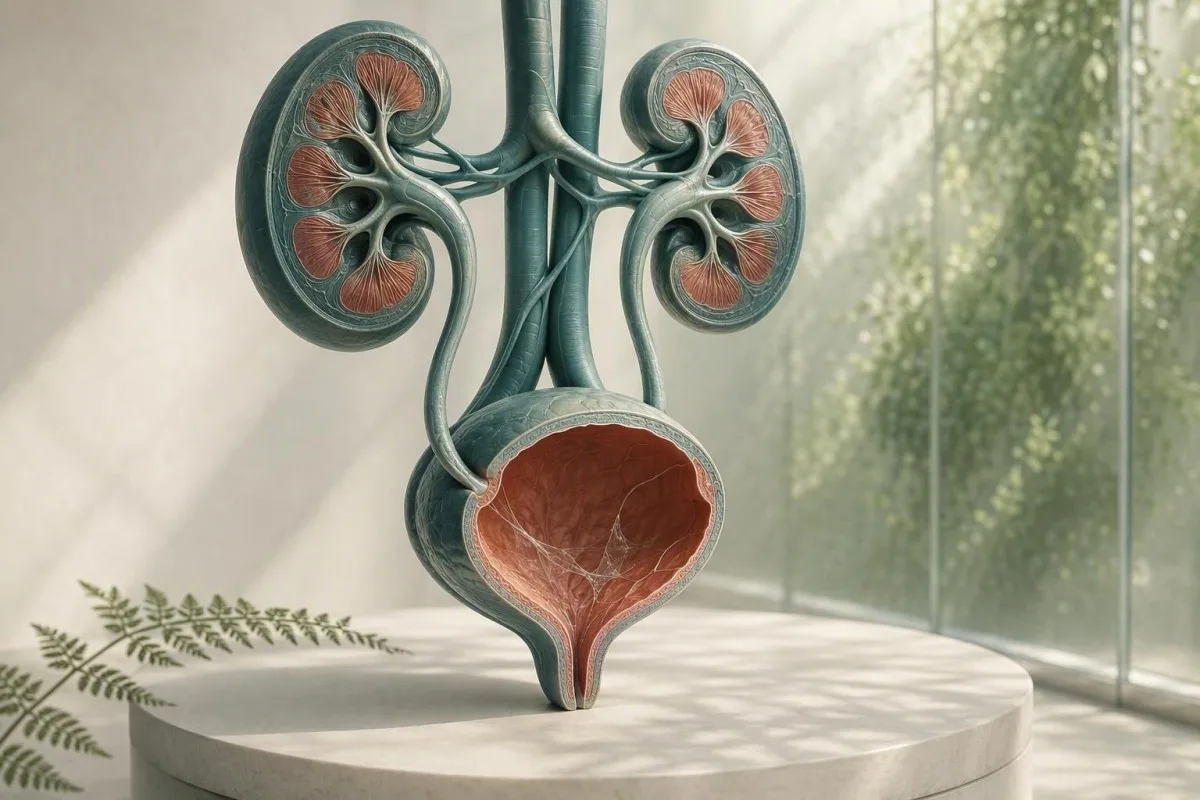
Schleim bei einer Urinmikroskopie ist normalerweise ein Entnahme-, kein Diagnoseproblem. Die umgebenden Urinanalyseergebnisse, Symptome und die Qualität der Probe bestimmen, ob eine Nachuntersuchung erforderlich ist.


- Schleimfäden allein sind häufig harmlos, besonders in einer einzelnen sauberen Auffangprobe ohne Harnwegssymptome.
- Pyurie-Schwellenwert von mehr als 5 Leukozyten pro Gesichtsfeld unterstützen eine Harnwegsentzündung, beweisen aber keine bakterielle HWI.
- Kulturauslöser ist Schleim plus Brennen, Harndrang, Fieber, Flankenschmerzen, Nitrite oder signifikante Leukozyten – nicht Schleim allein.
- Plattenepithelzellen über etwa 10 pro Gesichtsfeld weisen oft auf eine Kontamination der Genitalhaut hin und machen ein Urinergebnis weniger zuverlässig.
- Sichtbares Blut mit kolikartigen seitlichen Schmerzen bedarf einer sofortigen Abklärung auf einen Stein, auch wenn auch Schleim berichtet wird.
- Schwangerschaft ändert die Schwelle für die Nachuntersuchung, da asymptomatische Bakteriurie normalerweise mittels Urinkultur, nicht allein mittels Mikroskopie gescreent wird.
- Trüber Urin kann Kristalle, konzentrierten Urin, Vaginalsekret, Sperma oder Zellen widerspiegeln; eine Infektion kann anhand des Aussehens nicht diagnostiziert werden.
- Wiederholte Probe ist sinnvoll, wenn Schleim der einzige ungewöhnliche Befund ist und die Entnahme keine sorgfältige Mittelstrahl-Clean-Catch-Probenentnahme war.
Was Schleimfäden in der Urinanalyse normalerweise bedeuten
Schleimfäden im Urin sind normalerweise Stränge normaler schützender Sekrete oder Genitalverunreinigungen, und ein isoliertes Ergebnis deutet selten auf eine Nierenerkrankung hin. Der Befund wird aussagekräftig, wenn er zusammen mit Harnwegssymptomen, weißen Blutkörperchen, Bakterien, Blut oder Protein auftritt.
Labore identifizieren Schleim als dünne, durchscheinende Fäden, die unter dem Mikroskop sichtbar sind; viele Berichte stufen ihn einfach als selten, wenige, moderat oder viele ein, anstatt einen numerischen Referenzbereich anzugeben. Urin enthält von Natur aus eine geringe Menge an glykoproteinreichem Material aus den Harnwegen, und Vaginal- oder Urethralsekrete können während der Entnahme mehr hinzufügen.
Die Formulierung des Berichts ist weniger alarmierend, als sie klingt. Ab dem 26. August 2026, gibt es keine validierte Schleimfadenanzahl, die eine Harnwegsinfektion, einen Stein, Krebs oder Nierenversagen diagnostiziert; es handelt sich um eine kontextbezogene mikroskopische Beobachtung. Eine umfassendere Übersicht über die anderen Elemente eines Teststreifen- und Mikroskopieberichts finden Sie in unserer vollständiger Leitfaden zur Urinanalyse.
Nach meiner klinischen Erfahrung ist ein symptomfreier Erwachsener mit “moderat” markiertem Schleim, 0–2 Leukozyten pro Gesichtsfeld, negativem Nitrit und ohne Blut am besten mit Beruhigung oder einer ordnungsgemäß entnommenen Wiederholungsprobe bedient. Dr. Thomas Klein sieht oft unnötige Angst vor Antibiotika entstehen, weil eine Laborflagge mit einem Krankheitslabel verwechselt wird.
Kantesti ist eine KI-Plattform für die Blutbild Auswertung, die nierenbezogene Blutmarker wie Kreatinin und eGFR neben einem Urinanalysebefund platzieren kann – aber sie kann eine Harnwegsinfektion nicht allein anhand von Schleim diagnostizieren. Ein Ergebnis der Urinmikroskopie benötigt die klinische Geschichte, die damit einherging.
Warum ein Labor es markiert
Laborinformationssysteme markieren Schleim, weil er visuell vorhanden ist, nicht weil das Labor einen gefährlichen Grenzwert festgelegt hat. Eine Flagge bedeutet daher “beobachtet” und nicht “abnormaler Krankheitsmarker”, ähnlich wie ein isolierter Spurenbefund keine Behandlung erfordern kann.
Woher Schleim in einer Urinprobe stammt
Schleim im Urin kann aus der Harnröhre, der Blasenwand, dem Gebärmutterhals oder der Vagina, Sperma oder der äußeren Genitalhaut während der Entnahme stammen. Seine Quelle wird oft aus dem übrigen mikroskopischen Muster abgeleitet und nicht aus den Fäden selbst.
Die Blase und die Harnröhre haben eine schützende Oberflächenschicht, die Mucine enthält, einschließlich uroplakin-assoziiertem Material, das Reibung und mikrobielle Anhaftung reduziert. Kleine Mengen können nach Dehydrierung, kürzlich erfolgtem Geschlechtsverkehr, leichten Reizungen oder einfach dadurch, dass ein konzentrierte Morgenurinprobe Fäden leichter sichtbar macht, in den Urin abgeschilfert werden.
Bei Personen, die menstruieren oder vaginalen Ausfluss haben, können Leukozyten und Schleim in den Becher gelangen, ohne aus der Blase zu stammen. Mehr als 10 Plattenepithelzellen pro Gesichtsfeld machen eine Kontamination wahrscheinlicher, obwohl Labore unterschiedliche Reporting-Schwellenwerte verwenden. Unser Leitfaden zu Epithelzellen im Urin erklärt, warum dies die Zuverlässigkeit eines Ergebnisses verändert.
Sperma kann auch mehrere Stunden nach der Ejakulation fadenförmiges Material oder Trübungen verursachen. Das macht den Urin nicht unsicher, kann aber die Mikroskopie erschweren; wenn eine Kultur in Erwägung gezogen wird, rate ich im Allgemeinen dazu, eine neue Probe zu einem späteren Zeitpunkt, mindestens 24 Stunden später, zu sammeln, wenn dies möglich ist.
Warum Sex und Anatomie den Bericht beeinflussen
Ein Urinbecher trennt die Blase nicht vom umliegenden Gewebe. Deshalb wird “Schleim vorhanden” bei Proben mit externen Sekreten häufiger berichtet, während eine katheterisierte Probe manchmal verwendet wird, wenn Kliniker eine sauberere Antwort benötigen.
Wann Schleimfäden auf eine Harnwegsinfektion hinweisen
Schleimfäden unterstützen eine mögliche Harnwegsinfektion (UTI) nur, wenn sie von Symptomen und objektiven Entzündungsbefunden begleitet werden, insbesondere von Pyurie, Nitrit-Positivität oder einer überzeugenden Kultur. Schleim allein ist kein UTI-Test.
Eine symptomatische untere Harnwegsinfektion (UTI) verursacht häufig Brennen, Harndrang, häufigen Harndrang, Schmerzen im Unterbauch und manchmal einen neuen Urin-Geruch. Bei einer ordnungsgemäß gesammelten Probe, mehr als 5 weiße Blutkörperchen pro Hochleistungsfeld wird oft als Pyurie bezeichnet; sie unterstützt eine Entzündung des Harntrakts, kann aber auch bei Steinen, sexuell übertragbaren Infektionen und Kontaminationen auftreten.
Nitrit ist bei positivem Ergebnis hochspezifisch, übersieht aber Infektionen, die durch Organismen verursacht werden, die Nitrat nicht reduzieren, und ein negatives Nitritergebnis schließt eine UTI nicht aus. Leukozytenesterase weist auf die Enzymaktivität weißer Blutkörperchen hin und kann falsch positiv sein, wenn vaginale Sekrete die Probe kontaminieren; unsere Erklärung von Leukozytenesterase-Ergebnissen deckt diese Fallstricke ab.
Die IDSA-Richtlinie rät von der Untersuchung oder Behandlung von asymptomatischer Bakteriurie bei gesunden, nicht schwangeren Erwachsenen ab, da Antibiotika Schaden ohne Nutzen hinzufügen (Nicolle et al., 2019). Dieser Grundsatz ist besonders relevant für Schleim: kein Brennen, kein Fieber und keine geplante urologische Prozedur bedeuten in der Regel keine Antibiotika nur deshalb, weil ein Mikroskopiefeld unordentlich aussah.
Wann eine Kultur der bessere Test ist
Eine Urinkultur ist nützlicher als ein wiederholter Teststreifen, wenn Symptome anhalten, Symptome innerhalb von 4 Wochen wieder auftreten, eine Schwangerschaft besteht, eine Pyelonephritis vermutet wird oder frühere Antibiotika das Ergebnis möglicherweise beeinflusst haben. Ein gemischtes Bakterienwachstum deutet häufig auf eine Kontamination der Probe hin und nicht auf einen einzelnen Harnwegserreger; siehe Interpretation der Urinkultur.
Schleim, Kristalle, Steine und mechanische Reizung
Schleim mit starken wellenförmigen Flankenschmerzen oder Blut im Urin kann mit einem Harnstein auftreten, aber Schleim bestätigt oder schließt einen Stein weder ein noch aus. Blut, Kristalle, Schmerzmuster, Bildgebung und Nierenfunktion sind wichtiger.
Ein Stein kann die Harnwegsschleimhaut abschaben oder blockieren und rote Blutkörperchen, weiße Blutkörperchen und zusätzlichen Schleim produzieren. Das klassische Symptom sind abrupte kolikartige Schmerzen, die in die Leiste ausstrahlen, oft mit Übelkeit; sichtbarer roter oder teefarbener Urin erhöht den Harndrang. Lesen Sie mehr über rote Flaggen bei Blut im Urin anstatt anzunehmen, dass jede rosa Probe eine einfache Infektion ist.
Calciumoxalatkristalle können bei gesunden Menschen auftreten, insbesondere in konzentriertem saurem Urin, daher beweist ein Kristalltyp keinen aktiven Stein. Bei Personen mit Schmerzen, wiederkehrenden Steinen oder anhaltender Hämaturie können Ärzte Ultraschall oder eine niedrigdosierte Computertomographie ohne Kontrastmittel verwenden, anstatt sich nur auf den Sediment zu verlassen. Unsere Ressource über Calciumoxalatkristallen beschreibt die Grenzen von Kristallberichten.
Eine wichtige Ausnahme ist eine Obstruktion mit Infektion: Fieber von 38,0 °C oder höher, Flankenschmerzen, Erbrechen und Unfähigkeit, Urin auszuscheiden, erfordern eine Notfallbeurteilung. Die Sorge gilt nicht dem Schleim; es handelt sich um ein infiziertes blockiertes Harnsystem, das sich schnell verschlechtern kann.
Reizung ohne Stein
Kürzlicher Kathetergebrauch, Blasenoperationen, intensives Radfahren und Beckenbestrahlung können den unteren Harntrakt reizen und Schleim oder weiße Blutkörperchen erhöhen. Ein Arzt sollte diese Ergebnisse im zeitlichen Zusammenhang interpretieren, da eine Probe, die innerhalb von 48 Stunden nach der Instrumentierung entnommen wurde, nicht mit einer routinemäßigen Screening-Probe gleichzusetzen ist.
Trüber Urin Bedeutung: Was das Aussehen verraten kann und was nicht
Trüber Urin kann durch konzentrierte Salze, Kristalle, Schleim, Zellen, Genitalsekret oder Bakterien verursacht werden, daher kann das Aussehen allein keine Infektion diagnostizieren. Urin, der trüb, aber schmerzlos und kurzlebig ist, ist üblicherweise nicht dringlich.
Phosphatkristalle können alkalischen Urin nach dem Abkühlen trüben, während Uratkristalle sauren, konzentrierten Urin trüben können. Eine Probe, die länger als 2 Stunden bei Raumtemperatur steht, kann zunehmend trüb werden, da Zellen zerfallen und Bakterien sich vermehren. Deshalb ist die frische Verarbeitung oder Kühlung wichtig.
Trübungen in Verbindung mit Dysurie, Harndrang und Pyurie rechtfertigen eine Untersuchung; Trübungen nach körperlicher Anstrengung oder geringer Flüssigkeitszufuhr bessern sich oft durch normale Hydration. Streben Sie Urin an, der blassgelb ist, anstatt übermäßige Wasserzufuhr zu erzwingen – sehr klarer Urin bedeutet nicht, dass die Nieren eine Infektion “ausspülen”. Unsere detaillierte Übersicht von Ursachen für trüben Urin trennt visuelle Hinweise von zuverlässigen Tests.
Kantesti ist ein KI-Bluttestanalysator wurde entwickelt, um Blutmarker im klinischen Kontext zu interpretieren; ein hohes Kreatinin oder eine reduzierte eGFR zusammen mit Harnanomalien kann eine ärztliche Überprüfung rechtfertigen, während trüber Urin allein keine eingeschränkte Nierenfiltration begründet. Ein Basis-Metabolismus-Panel kann nützlichen Kontext liefern, wenn Symptome auf Dehydrierung oder Obstruktion hindeuten.
Geruch ist keine Kultur
Stark riechender Urin spiegelt oft die Konzentration, Stoffwechselprodukte von Spargel, B-Vitamine oder einen Behälter wider, der zu lange gestanden hat. Ein neuer, unangenehmer Geruch mit Fieber oder Harnwegssymptomen ist ein Grund, sich untersuchen zu lassen, aber der Geruch ist kein Ersatz für Mikroskopie und Kultur.
Genitaler Ausfluss und sexuell übertragbare Infektionen
Urethral- oder Vaginalsekret kann wie Schleim im Urin aussehen, und neues Sekret mit Brennen beim Wasserlassen erfordert neben einer Urinuntersuchung auch einen Test auf sexuell übertragbare Krankheiten. Routine-Uinkulturen können Chlamydien und Gonorrhö übersehen.
Chlamydien und Gonorrhö können Dysurie und sterile Pyurie – weiße Blutkörperchen ohne normale bakterielle Wachstums – verursachen, insbesondere nach einer neuen sexuellen Exposition. Nukleinsäureamplifikationstests, die häufig statt einer Mittelstrahlprobe den Erstfangurin verwenden, sind die geeigneten Tests, da die Standardkultur auf andere Organismen abzielt.
Bei Personen mit einer Vagina können bakterielle Vaginose, Candidiasis und Zervixentzündungen Sekret in einen Mittelstrahlbecher bringen; ein Vaginalabstrich oder eine Untersuchung kann aufschlussreicher sein als eine wiederholte Urinmikroskopie. Bei Personen mit einem Penis sollte sichtbares urethrales Sekret nicht als “Schleimfäden” abgetan werden. Der praktische Unterschied besteht darin, ob das Sekret außerhalb des Urinierens bemerkt wird oder ob Stränge nur vom Labor berichtet werden.
Die CDC STI-Behandlungsrichtlinie von 2021 empfiehlt NAAT-basierte Tests an relevanten anatomischen Stellen gemäß der Expositionshistorie, nicht nur anhand von Symptomen (Workowski et al., 2021). Wenn Beckenschmerzen, Hodenschmerzen, Fieber, Schwangerschaft oder ein möglicher Übergriff vorliegen, suchen Sie eine umgehende klinische Beratung auf, anstatt sich mit übrig gebliebenen Antibiotika selbst zu behandeln.
Warum Antibiotika das Bild verzerren können
Die Einnahme selbst von 1 oder 2 Dosen Antibiotika vor einer Kultur kann das normale Bakterienwachstum unterdrücken, während Harnwegssymptome und weiße Blutkörperchen bestehen bleiben. Teilen Sie dem Arzt genau mit, welches Medikament, welche Dosis und welche letzte Dosiszeit verwendet wurden; diese Details ändern die Interpretation eines negativen Ergebnisses.
Wie man eine Urinprobe entnimmt, die die Frage beantwortet
Eine Mittelstrahl-Clean-Catch-Probe reduziert Schleim, Plattenepithelzellen und gemischtes Bakterienwachstum besser als das Sammeln des ersten oder letzten Teils des Strahls. Die Technik ist einfach, aber die ersten 2 Sekunden machen einen unverhältnismäßig großen Unterschied.
Hände waschen, Genitalhaut trennen oder die Vorhaut zurückziehen, wenn es angenehm ist, gemäß den Anweisungen des Kits reinigen, mit dem Wasserlassen in die Toilette beginnen, dann den mittleren Teil auffangen, ohne dass der Becher die Haut berührt. Eine Probe von 20–30 ml ist in der Regel ausreichend; das Auffüllen eines großen Behälters bis zum Rand verbessert den Test nicht.
Liefern Sie den Becher umgehend, idealerweise innerhalb von 1 Stunde; falls eine Verzögerung unvermeidlich ist, kühlen Sie ihn gemäß den Anweisungen des Labors und geben Sie ihn innerhalb von 24 Stunden zurück. Die Kühlung verlangsamt das Bakterienwachstum, stellt aber keine bereits kontaminierte Probe wieder her. Dies ist besonders wichtig, wenn die Anforderung eine Kultur und nicht nur einen Teststreifen beinhaltet.
Kantesti ist ein KI-gestütztes Analyse-Tool für Bluttests werden in 127+ Ländern verwendet, und unsere klinisch geprüften Arbeitsabläufe behandeln die Probenqualität als Teil der Interpretation und nicht als Fußnote. Das gleiche Prinzip gilt für jeden Laborwert: Eine technisch schlechte Probe kann ein medizinisch überzeugendes, aber irreführendes Ergebnis liefern. Siehe unsere Checkliste für Laborgenauigkeit für Fragen, die Sie stellen sollten, bevor Sie auf eine isolierte Kennzeichnung reagieren.
Sammeln Sie nicht in diesen Situationen, wenn Sie warten können
Vermeiden Sie routinemäßige Tests während starker Menstruationsblutung, unmittelbar nach dem Geschlechtsverkehr oder nach der Anwendung von Vaginalcremes, es sei denn, der Arzt hat dies ausdrücklich angefordert. Wenn Tests nicht warten können, informieren Sie das Labor oder den Arzt, da dieser Kontext Schleim, rote Blutkörperchen oder externe Zellen erklären kann.
Wie Ärzte Schleim im Zusammenhang mit dem Rest der Urinanalyse interpretieren
Das nützlichste Muster der Urinanalyse kombiniert Symptome mit weißen Blutkörperchen, roten Blutkörperchen, Nitrit, Leukozytenesterase, Protein, Glukose, spezifischem Gewicht und Epithelzellen. Schleim ist ein untergeordnetes unterstützendes Merkmal in diesem Muster.
Eine saubere Probe mit Schleim, 0–2 weißen Blutkörperchen pro Gesichtsfeld, negativem Nitrit und ohne Blut erfordert normalerweise keine Behandlung. Schleim mit mehr als 5 weißen Blutkörperchen pro Gesichtsfeld und positiver Leukozytenesterase erhöht die Wahrscheinlichkeit einer Entzündung; die Zugabe von Nitrit oder eine Einzelkeimkultur erhöht die Sicherheit, dass Bakterien dafür verantwortlich sind.
Protein benötigt seinen eigenen Weg. Spuren von Protein nach Fieber, Anstrengung oder konzentriertem Urin können vorübergehend sein, aber persistierendes Protein sollte mit einem Albumin-Kreatinin-Verhältnis quantifiziert werden, anstatt Schleim dafür verantwortlich zu machen. Unsere Anleitung für Protein im Urin erklärt, warum Dipstick-Protein und Nierenrisiko nicht austauschbar sind.
Eine spezifische Dichte von 1,003 bis 1,030 ist bei Erwachsenen üblich, obwohl die Referenzintervalle je nach Labor variieren. Hohes spezifisches Gewicht kann Schleim konzentrieren und ein dramatischer aussehendes Sediment erzeugen, während niedriges spezifisches Gewicht Zellen lysieren und die Mikroskopie täuschend unscheinbar machen kann; unser Leitfaden zur spezifischen Dichte zeigt, wie die Hydratation die Interpretation beeinflusst.
Der Wert negativer Befunde
Negatives Blut, Protein, Nitrit und Leukozytenesterase verringern die Besorgnis bei einer Person ohne Symptome, auch wenn Schleim als moderat berichtet wird, erheblich. Kein einzelner negativer Test ist perfekt, aber diese Kombination ist beruhigender als eine Schleimbewertung besorgniserregend ist.
Wann der Test wiederholt und wann eine Kultur angefordert werden sollte
Wiederholen Sie eine Urinprobe, wenn Schleim isoliert ist, Plattenepithelzellen auf Kontamination hindeuten oder die Sammlung überstürzt erfolgte; fordern Sie eine Kultur an, wenn die Symptome anhaltend, wiederkehrend, schwerwiegend oder mit hohem Risiko verbunden sind. Ein wiederholter Test ist kein “Nichtstun” – er ist oft der diagnostisch wichtigste nächste Schritt.
Bei einer nicht schwangeren Erwachsenen ohne Symptome und mit Schleim als einzigem Auffälligkeit ist eine wiederholte Mittelstrahl-Urinanalyse innerhalb von 1–2 Wochen angemessen, wenn Beruhigung benötigt wird; viele Ärzte würden sie gar nicht wiederholen. Behandeln Sie ein Laborergebnis nicht mit Antibiotika, während Sie warten, es sei denn, ein verschreibender Arzt identifiziert eine klinische Indikation.
Die Kultur vor Antibiotika ist besonders nützlich bei Fieber, Flankenschmerzen, Schwangerschaft, Immunsuppression, Nierentransplantation, Harnkathetergebrauch, männlichen Harnwegssymptomen oder Symptomen, die innerhalb von 4 Wochen wiederkehren. Die Leitlinie von Gupta et al. (2011) unterstützt die Kultur bei Verdacht auf Pyelonephritis und Situationen, in denen Resistenzen oder eine alternative Diagnose wahrscheinlicher sind.
Kulturergebnisse müssen im Zusammenhang mit der Probenqualität abgelesen werden. Ein einzelner Organismus bei 10^5 koloniebildenden Einheiten pro ml hat traditionell eine Bakteriurie bei sauberer Mittelstrahlurinprobe unterstützt, aber symptomatische Patienten können klinisch relevante niedrigere Werte aufweisen; “gemischte Flora” veranlasst normalerweise die Entnahme einer neuen Probe statt einer breit angelegten Antibiotikagabe. Vergleichen Sie die Zwecke von Urinuntersuchung und Kultur vor der Anforderung einer der beiden.
Schwangerschaft erfordert einen anderen Grenzwert
Schwangerschaft ist eine Ausnahme, da asymptomatische Bakteriurie das Risiko einer Pyelonephritis und ungünstiger Schwangerschaftsausgänge erhöhen kann. Die Schwangerenvorsorge verwendet häufig eine Screening-Kultur früh in der Schwangerschaft; Schleim unter dem Mikroskop kann diese Kultur nicht ersetzen.
Wann Blutuntersuchungen nützlichen Kontext für Nieren und Infektionen liefern
Blutuntersuchungen sind nützlich, wenn Schleim im Urin mit Fieber, Flankenschmerzen, wiederkehrenden Infektionen, Schwellungen, reduzierter Urinausscheidung oder anhaltendem Protein oder Blut einhergeht. Kreatinin und eGFR beurteilen die Filtration, während ein CBC und C-reaktives Protein helfen können, systemische Erkrankungen einzuschätzen.
Eine eGFR unter 60 mL/min/1,73 m² für mindestens 3 Monate erfüllt ein Kriterium für chronische Nierenerkrankung, aber ein einzelner niedrigerer Wert während Dehydrierung oder akuter Erkrankung reicht nicht aus, um diese Diagnose zu stellen. Kreatinin wird durch Muskelmasse, Ernährung und einige Medikamente beeinflusst, daher sind Trends und Albumin im Urin oft aussagekräftiger als eine einzelne Zahl.
Ein erhöhter Leukozytenwert oder CRP kann einen Entzündungsprozess unterstützen, aber nicht die Harnwege als Quelle identifizieren. Bei fieberhaften Personen mit Flankenschmerzen können Ärzte Kreatinin überprüfen, bevor sie bildgebende Verfahren oder Medikamente wählen, insbesondere wenn Erbrechen oder eine Obstruktion die Nierenfunktion beeinträchtigen könnten. Siehe chronische Nierenerkrankungsstadien verfolgen für die eGFR- und Albumin-Kategorien, die Ärzte verwenden.
Kantesti ist ein KI-Labor-Befundinterpretationsservice die Muster über Kreatinin, eGFR, Elektrolyte, CBC und Entzündungsmarker hinweg hervorhebt, anstatt eine einzelne Schleimbeobachtung als Nierendiagnose zu behandeln. Unsere Methodik unterliegt einer klinischen Validierungsaufsicht, aber dringende Symptome erfordern immer noch eine direkte medizinische Versorgung, keine app-basierte Interpretation.
Eine praktische Unterscheidung von Dehydrierung
Dehydrierung kann die spezifische Urindichte erhöhen und vorübergehend das Kreatinin erhöhen, insbesondere nach Durchfall, Hitzeexposition oder intensiver Bewegung. Anhaltend geringe Urinausscheidung, Schwindel oder ein eGFR-Abfall nach Rehydrierung erfordern eine ärztliche Überprüfung und keine wiederholten Heimtests.
Dringende Warnsignale, die nicht auf einen Wiederholungstest warten sollten
Suchen Sie eine dringende Beurteilung am selben Tag bei Schleim im Urin mit Fieber von 38,0 °C oder höher, Flankenschmerzen, Erbrechen, sichtbarem Blut, Unfähigkeit zu urinieren, Verwirrung oder schwangerschaftsbedingten Harnwegssymptomen auf. Diese Kombinationen sind wichtig, da sie auf eine Infektion der oberen Harnwege, eine Obstruktion oder eine andere akute Erkrankung hindeuten können.
Fieber plus einseitige Rücken- oder Flankenschmerzen sind eher auf eine Pyelonephritis als auf eine einfache Zystitis zurückzuführen, insbesondere mit Schüttelfrost oder Erbrechen. Eine verzögerte Behandlung kann zu Dehydrierung, Sepsis oder Nierenstress führen; eine zu Hause unauffällige Urinprobe schließt dies nicht sicher aus.
Sichtbares Blut sollte auch dann beurteilt werden, wenn eine Harnwegsinfektion plausibel erscheint, insbesondere nach dem 35. Lebensjahr, bei Rauchern oder wenn die Blutung nachlässt, sobald die Infektionssymptome abklingen. Die AUA-Richtlinie für Mikrohämaturie empfiehlt eine risikobasierte Beurteilung, nachdem gutartige Erklärungen ausgeschlossen wurden (Barocas et al., 2020); anhaltendes Blut wird nicht durch Schleim erklärt.
Kinder, Erwachsene über 65, Menschen mit Diabetes, Menschen, die immunsuppressive Medikamente einnehmen, und Menschen mit einer einzelnen Niere verdienen einen niedrigeren Beurteilungsschwellenwert. Dr. Thomas Kleins Regel in der Praxis ist einfach: Wenn sich die Symptome über 6–12 Stunden verschlimmern, warten Sie nicht auf eine zweite Tasse, um Beruhigung zu geben.
Rufen Sie jetzt den Notdienst bei schwerer Krankheit an
Rufen Sie den Notdienst an bei neuer Verwirrung, Ohnmacht, starker Schwäche, blauen oder grauen Lippen, starker Atemnot oder Unfähigkeit, Flüssigkeiten bei Harnwegssymptomen bei sich zu behalten. Dies sind systemische Gefahrenzeichen, keine routinemäßigen Harnwegsinfektionssymptome.
Schleim im Urin während der Schwangerschaft, Kindheit und im späteren Leben
Schwangerschaft, Kinder und ältere Erwachsene erfordern eine sorgfältigere Interpretation, da Kontaminationen häufig vorkommen, aber die Folgen einer verpassten Infektion größer sein können. Symptome und Kulturergebnisse sind in jeder Altersgruppe weiterhin wertvoller als die Schleimqualität.
Während der Schwangerschaft kann häufiges Wasserlassen normal sein, was Symptome weniger spezifisch macht; Fieber, Dysurie oder Rückenschmerzen sollten eine umgehende geburtshilfliche oder klinische Beurteilung veranlassen. Eine Screening-Kultur wird typischerweise früh in der Schwangerenvorsorge entnommen, und eine Wiederholungskultur kann nach der Behandlung je nach Plan des Klinikers verwendet werden.
Bei Kindern ist Urin, der im Beutel gesammelt wurde, anfällig für Kontaminationen und sollte in der Regel nicht allein zur Diagnose einer HWI mit Kultur verwendet werden. Ein katheterisierter oder sorgfältig gewonnener Mittelstrahlurin kann bei einem jungen Kind mit Fieber ohne klare Ursache erforderlich sein; Schleim in einer Beutelprobe ist besonders unspezifisch.
Im höheren Alter werden Bakteriurie und Pyurie häufiger, ohne Symptome zu verursachen. Akute Verwirrtheit allein sollte eine breite medizinische Beurteilung auf Dehydrierung, Medikamente, Schmerzen, Verstopfung und Infektionsquellen auslösen, anstatt einer automatischen HWI-Behandlung; die gleiche Zurückhaltung wie von Nicolle et al. (2019) empfohlen, gilt.
Menstruation und hormonelle Veränderungen
Menstruationsblut und Zervixschleim können die Urinanalyse mehrere Tage lang verändern, und postmenopausale Vaginaltrockenheit kann lokale Reizungen verursachen, die einem brennenden Gefühl beim Wasserlassen ähneln. Wenn Ergebnisse und Symptome nicht übereinstimmen, kann ein Kliniker genitale Ursachen beurteilen, anstatt eine wiederholte HWI-Behandlung zu verschreiben.
Häufige Fehler nach dem Auftreten von Schleim auf einem Laborbericht
Beginnen Sie nicht mit übrig gebliebenen Antibiotika, reinigen Sie nicht innerlich und versuchen Sie nicht, Schleim mit extrem hoher Wasseraufnahme nach einer abnormal aussehenden Urinanalyse auszuscheiden. Diese Maßnahmen können eine Kultur verschleiern, die normale Flora stören oder die richtige Diagnose verzögern.
Antibiotika, die ohne Kultur eingenommen werden, können einen nachfolgenden Test fälschlicherweise negativ machen und Durchfall, Hautausschlag, Hefesymptome oder Resistenzen verursachen. Wenn die Symptome mild, aber hartnäckig sind, entnehmen Sie die Probe wann immer praktikabel zuerst und folgen Sie dann dem Behandlungsplan eines Klinikers, der auf dem Gesamtbild basiert.
Cranberry-Produkte können das Risiko von wiederkehrenden unkomplizierten Harnwegsinfektionen bei einigen Personen moderat reduzieren, aber sie behandeln kein Fieber, keine Flankenschmerzen oder eine bestätigte Infektion des oberen Harntrakts. Vermeiden Sie hochdosiertes Vitamin C als Selbstbehandlung: Es kann einige Teststreifenreaktionen verändern und die Oxalatbelastung bei Personen mit Neigung zu Kalziumoxalatsteinen erhöhen.
Untersuchen Sie die Toilettenschüssel nicht wiederholt auf Fäden. Toilettenpapierfasern, Reinigungsrückstände, Genitalsekrete und Wasserverwirbelungen können Schleim imitieren; eine im Labor in einem sterilen Behälter gesammelte Probe ist der richtige Ort, um ihn zu beurteilen. Für verwandte Farbveränderungen, unser Leitfaden zur Urinfarbe bietet einen zuverlässigeren visuellen Kontext.
Die Medikamentenliste ist wichtig
Phenazopyridin kann den Urin orange färben und die visuelle Interpretation beeinträchtigen, während Diuretika den Urin je nach Zeitpunkt konzentrieren oder verdünnen können. Bringen Sie eine Liste der verschreibungspflichtigen Medikamente, rezeptfreien Produkte und Nahrungsergänzungsmittel zur Untersuchung mit, einschließlich der Dosen in mg.
Ein praktischer Nachbehandlungsplan für Schleimfäden im Urin
Die meisten Menschen mit Schleimfäden im Urin und ohne Symptome benötigen keine Behandlung; eine sorgfältige wiederholte Urinanalyse ist sinnvoll, wenn die Probe fragwürdig war. Symptome oder begleitende Auffälligkeiten bestimmen, ob eine Kultur, STI-Tests, bildgebende Verfahren oder Blutuntersuchungen angebracht sind.
Schritt 1: Prüfen Sie auf Brennen, Harndrang, Fieber, Flankenschmerzen, sichtbares Blut, Ausfluss, Schwangerschaft, kürzliche Harnwegseingriffe und neue sexuelle Kontakte. Schritt 2: Lesen Sie den Bericht auf Leukozyten, Nitrit, Leukozytenesterase, Erythrozyten, Protein, Bakterien und Plattenepithelzellen – nicht nur auf Schleim.
Schritt 3: Wenn Sie sich gut fühlen und isolierter Schleim vorliegt, sammeln Sie nur eine Mittelstrahl-Clean-Catch-Probe innerhalb von 1–2 Wochen, wenn Ihr Kliniker eine Bestätigung empfiehlt. Schritt 4: Wenn Symptome vorliegen, fragen Sie, ob vor der Behandlung eine Urinkultur entnommen werden sollte und ob STI-Tests relevant sind; wiederkehrende Episoden verdienen eine sorgfältigere Überprüfung als ein drittes empirisches Antibiotikum.
Kantesti kann relevante Blutbefundtrends und Fragen für einen Arzttermin organisieren, während unser Medizinischer Beirat unterstützt klinikergeführte Sicherheitsstandards. Das richtige Endziel ist kein perfekt “sauberer” Mikroskopiebericht – es ist eine Erklärung, die zu Ihren Symptomen, der Probenqualität und den Risikofaktoren passt.
Fragen für Ihren Arzttermin
Fragen Sie, ob das Präparat Plattenepithelzellen enthielt, ob die Kultur einen oder gemischte Flora ergab und ob nach Abklingen der Symptome Blut oder Protein persistierte. Diese 3 Fragen liefern oft nützlichere Antworten als die Frage nach der Menge des beobachteten Schleims.
Forschungskontext und Grenzen der Schleimberichterstattung
Die Schleimbewertung ist in den Laboren nicht standardisiert, weshalb Ärzte “wenig”, “mäßig” oder “viel” als Kategorien für die Schwere der Erkrankung vermeiden sollten. Mikroskopietechnik, Alter des Präparats und Berichtssoftware können die Formulierungen ändern.
Einige Labore inspizieren manuell das Sediment nach dem Zentrifugieren von etwa 10–15 ml Urin, während andere automatisierte Partikelanalysen mit manueller Überprüfung ausgewählter Flags verwenden. Diese Variation bedeutet, dass ein “moderater Schleim”-Befund aus einem Labor in einem anderen Labor nicht exakt reproduzierbar ist, selbst wenn sich der Gesundheitszustand der Person nicht geändert hat.
Eine nützliche Denkweise im Labor ist die Frage, ob ein Ergebnis analytisch real, klinisch bedeutsam und reproduzierbar ist. Dieser Ansatz ist auch von Blutuntersuchungen vertraut: unsere Blutbiomarker-Leitfaden erklärt, warum Referenzintervalle und präanalytische Bedingungen darüber entscheiden, ob ein markierter Marker Handlungsbedarf besteht.
Kantesti AI nutzt einen strukturierten Labor-Kontext, um Ergebnis-Muster über große Panels hinweg zu erklären, aber die Urinmikroskopie erfordert weiterhin eine quellenspezifische klinische Beurteilung. Für Leser, die daran interessiert sind, wie wir technische Leistung und klinische Grenzen bewerten, beschreibt unser KI-Technologie-Leitfaden die Sicherheitsvorkehrungen hinter unserem Interpretationsansatz.
Was ein guter Bericht klar machen sollte
Ein nützlicher Bericht identifiziert den Probentyp, das Entnahmedatum, mikroskopische Elemente und jegliche Kultur-Organismen sowie Resistenztestergebnisse, wenn eine Kultur durchgeführt wird. Wenn der Bericht nur “Schleimfäden vorhanden” besagt, ist er für die Diagnose unvollständig, aber oft für einen zufälligen Befund mit geringem Risiko völlig ausreichend.
Häufig gestellte Fragen
Sind Schleimfäden im Urin normal?
Schleimfäden im Urin sind oft normal oder auf eine Kontamination durch Genitalsekrete zurückzuführen, insbesondere wenn eine Person keine Harnwegssymptome aufweist. Ein isolierter Befund mit 0–2 weißen Blutkörperchen pro Gesichtsfeld (high-power field), negativem Nitrit und ohne Blut oder Protein ist in der Regel mit einem geringen Risiko verbunden. Labore verwenden keine validierte Schleimfadenanzahl zur Diagnose von Harnwegsinfektionen (UTI) oder Nierenerkrankungen. Eine erneute Mittelstrahlprobe nach sauberer Gewinnung ist vertretbar, wenn die ursprüngliche Sammlung überstürzt oder sichtbar kontaminiert war.
Bedeuten Schleimfäden im Urin, dass ich eine Harnwegsinfektion habe?
Schleimfäden im Urin bedeuten für sich allein keine Harnwegsinfektion (HWI). Eine HWI wird wahrscheinlicher, wenn Brennen, Harndrang, häufiges Wasserlassen, Fieber oder suprapubische Schmerzen zusammen mit mehr als 5 Leukozyten pro Gesichtsfeld, positiver Leukozytenesterase, Nitrit oder einer positiven Kultur auftreten. Ein negatives Nitrit schließt eine HWI nicht vollständig aus, da nicht alle Bakterien Nitrit produzieren. Die Kultur ist bei anhaltenden oder wiederkehrenden Symptomen in der Regel aussagekräftiger als die Schleimbeurteilung.
Kann Dehydrierung Schleim im Urin verursachen?
Dehydratisierung kann den Schleim leichter sichtbar machen, da konzentrierter Urin weniger Wasser um normale Sekrete und Sedimente herum aufweist. Eine hohe Urinspezifische Gewicht, üblicherweise um 1,025–1,030, kann diesen Effekt begleiten, obwohl er für sich allein keine Dehydratisierung beweist. Eine normale Rehydrierung und die Wiederholung einer ordnungsgemäß entnommenen Probe können den Befund klären. Extremer Wasserkonsum ist unnötig und kann eigene Elektrolytrisiken bergen.
Was bedeutet Schleim und weiße Blutkörperchen im Urin?
Schleim plus weiße Blutkörperchen im Urin deuten auf eine Entzündung der Harnwege oder des Genitaltrakts hin, beweisen aber keine bakterielle Infektion. Mehr als 5 weiße Zellen pro Gesichtsfeld können bei Harnwegsinfektionen, Steinen, sexuell übertragbaren Infektionen, vaginaler Kontamination oder kürzlichen instrumentellen Eingriffen an den Harnwegen auftreten. Symptome, Nitrit, Epithelzellen und Kultur-Ergebnisse bestimmen das weitere Vorgehen. Fieber von 38,0°C oder höher mit Flankenschmerzen erfordert eine dringende Abklärung.
Kann vaginaler Ausfluss Schleim im Urintest verursachen?
Vaginalsekret kann in eine Mittelstrahlurinprobe gelangen und verursacht häufig Schleim, Plattenepithelzellen und manchmal Leukozyten im Urinsediment. Mehr als 10 Plattenepithelzellen pro Gesichtsfeld (HPF) deuten oft auf eine Kontamination hin, obwohl die Labore in ihrer Berichterstattung variieren. Eine sorgfältige Clean-Catch-Probe, die außerhalb des starken Menstruationsflusses entnommen wird, kann dieses Problem reduzieren. Bei Vorliegen von Ausfluss, Juckreiz, Geruch oder Unterleibsschmerzen kann eine vaginale Untersuchung oder ein Abstrich nützlicher sein als wiederholte Urinkulturen.
Wann sollte ich mir wegen Schleimfäden und Blut im Urin Sorgen machen?
Schleimfäden mit sichtbarem Blut im Urin sollten umgehend beurteilt werden, insbesondere wenn das Blut anhält, Flankenschmerzen, Gerinnsel, Fieber oder Schwierigkeiten beim Wasserlassen auftreten. Ein Stein, eine Harnwegsinfektion, eine Verletzung, eine Nierenerkrankung oder seltener ein Tumor im Harntrakt können Blut verursachen; Schleim identifiziert nicht, welche Ursache vorliegt. Die AUA empfiehlt eine risikobasierte Untersuchung von anhaltendem mikroskopischem Hämaturie, nachdem vorübergehende Ursachen behoben wurden. Eine Notfallbeurteilung ist bei Fieber, Erbrechen, starken Schmerzen oder Harnverhalt angebracht.
Hol dir heute eine KI-gestützte Bluttest-Analyse
Schließe dich über 2 Millionen Nutzern weltweit an, die Kantesti für sofortige, genaue Laboranalysen vertrauen. Lade deine Blutwerte Ergebnisse hoch und erhalte in Sekunden eine umfassende Interpretation von 15,000+-Biomarkern.
📚 Referenzierte Forschungsveröffentlichungen
Klein, T., Mitchell, S., & Weber, H. (2026). aPTT-Normalbereich: D-Dimer, Protein C Blutgerinnungsleitfaden. Kantesti KI-Medizinische Forschung.
Klein, T., Mitchell, S., & Weber, H. (2026). Leitfaden zu Serumproteinen: Globuline, Albumin und Albumin/Globulin-Quotient (A/G-Quotient) – Bluttest. Kantesti KI-Medizinische Forschung.
📖 Externe medizinische Referenzen
⚕️ Medizinischer Haftungsausschluss
Dieser Artikel dient nur zu Bildungszwecken und stellt keine medizinische Beratung dar. Wende dich für Diagnose- und Behandlungsentscheidungen immer an eine qualifizierte medizinische Fachkraft.
E-E-A-T Vertrauenssignale
Erfahrung
Ärztlich geleitete klinische Überprüfung von Labor-Interpretations-Workflows.
Sachverstand
Fokus der Labormedizin darauf, wie Biomarker sich im klinischen Kontext verhalten.
Autorität
Verfasst von Dr. Thomas Klein, überprüft von Dr. Sarah Mitchell und Prof. Dr. Hans Weber.
Vertrauenswürdigkeit
Evidenzbasierte Interpretation mit klaren nächsten Schritten zur Reduzierung von Alarm.