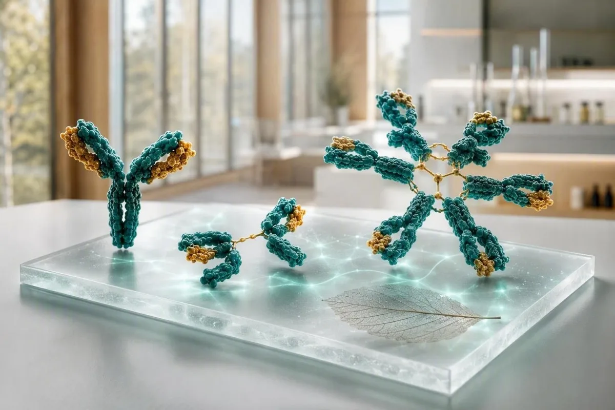
Xét nghiệm globulin miễn dịch hữu ích nhất khi IgG, IgA và IgM được đọc theo một mô hình, không phải ba dấu hiệu riêng lẻ. Giá trị cao có thể phản ánh sự hoạt hóa miễn dịch rộng hoặc một protein bất thường đơn lẻ; giá trị thấp có thể chỉ ra thuốc, mất protein hoặc suy giảm sản xuất kháng thể.


- IgG thường là 700-1.600 mg/dL ở người trưởng thành; sự gia tăng kéo dài ở nhiều lớp thường phản ánh tình trạng viêm mãn tính hoặc bệnh gan.
- IgA thường là 70-400 mg/dL ở người trưởng thành; tình trạng thiếu hụt chọn lọc có thể làm cho xét nghiệm celiac dựa trên IgA âm tính không đáng tin cậy.
- kháng thể IgM thường là 40-230 mg/dL ở người trưởng thành; sự gia tăng đơn lẻ, kéo dài cần điện di protein huyết thanh để loại trừ protein đơn dòng.
- Cả ba đều thấp đáng lo ngại hơn một kết quả thấp và cần xem xét tình trạng nhiễm trùng, thuốc, mất protein và phản ứng kháng thể với vắc xin.
- Tăng đa dòng có nghĩa là nhiều lớp globulin miễn dịch bị tăng cao; nó thường đi kèm với bệnh tự miễn, bệnh gan mãn tính hoặc nhiễm trùng kéo dài.
- Mô hình đơn dòng có nghĩa là một lớp bị tăng cao một cách không tương xứng hoặc một dải hẹp xuất hiện trên điện di; nó cần xét nghiệm miễn dịch cố định và xét nghiệm chuỗi nhẹ tự do để xác nhận.
- Cần xem xét khẩn cấp được chỉ định hợp lý đối với sốt kèm nhiễm trùng nghiêm trọng tái phát, lú lẫn mới, khó thở, vàng da, sụt cân, đau xương hoặc tổng protein tăng nhanh.
- Xu hướng quan trọng vì sự khác biệt 10-20% có thể phát sinh do tình trạng hydrat hóa, biến đổi xét nghiệm, bệnh gần đây hoặc phương pháp xét nghiệm khác.
Cách đọc xét nghiệm globulin miễn dịch như một mô hình
Kết quả IgG, IgA và IgM có ý nghĩa khác nhau khi kết hợp lại so với khi đứng riêng lẻ. Một bảng xét nghiệm tăng rộng thường báo hiệu sự kích thích miễn dịch đa dòng, trong khi một lớp chiếm ưu thế—đặc biệt là với tổng protein cao—đặt ra câu hỏi riêng về protein đơn dòng. Tính đến ngày 5 tháng 9 năm 2026, sự phân biệt đó vẫn là bước đầu tiên thực tế trước khi chẩn đoán được gắn với xét nghiệm máu immunoglobulin.
Khoảng tham chiếu của người trưởng thành thay đổi theo phương pháp, nhưng nhiều phòng thí nghiệm sử dụng IgG 700-1.600 mg/dL, IgA 70-400 mg/dL và IgM 40-230 mg/dL. Một giá trị 5% nằm ngoài khoảng không tự động là bệnh; Tôi kiểm tra trước khoảng tham chiếu của phòng thí nghiệm, tuổi, tình trạng mang thai, nhiễm trùng gần đây, mất qua thận và liệu mẫu có được lấy sau khi truyền immunoglobulin tĩnh mạch hay không.
Kantesti là một Máy phân tích xét nghiệm máu AI đọc IgG, IgA và IgM cùng với tổng protein, albumin, các chỉ số gan, creatinine và CBC thay vì coi cảnh báo đỏ là chẩn đoán. Việc kiểm tra chéo đó hữu ích vì albumin thấp với globulin tăng nói lên một câu chuyện rất khác với tình trạng mất nước khi cả hai giá trị đều cô đặc; bảng về chỉ dấu sinh học của chúng tôi của chúng tôi giải thích các phép đo liên quan.
Trong công việc lâm sàng của tôi, bảng xét nghiệm đáng tin cậy nhất thường không phải là mức trung bình hoàn hảo mà là ổn định: ví dụ, IgG 1.690 mg/dL trong ba lần lấy mẫu trong 18 tháng với điện di bình thường và không có triệu chứng. Bác sĩ Thomas Klein vẫn sẽ hỏi về khô mắt, sưng khớp, nhiễm trùng tái phát và thuốc, nhưng sự biến đổi nhẹ ổn định hiếm khi có trọng lượng như mức tăng 600 mg/dL trong sáu tháng.
Ba loại immunoglobulin bằng ngôn ngữ đơn giản
kháng thể IgM là phản ứng miễn dịch ban đầu, đáp ứng rộng, IgA bảo vệ bề mặt niêm mạc ở đường hô hấp và ruột, và IgG cung cấp hoạt tính kháng thể lưu thông bền vững nhất. Thời gian bán hủy sinh học của chúng khác nhau rõ rệt: IgG tồn tại khoảng 21 ngày, IgA khoảng 6 ngày trong huyết thanh và IgM khoảng 5 ngày, vì vậy những thay đổi liên tiếp không di chuyển với tốc độ như nhau.
Khoảng tham chiếu thông thường ở người trưởng thành và lý do phòng xét nghiệm của bạn có thể khác
Khoảng tham chiếu được in bên cạnh kết quả của bạn sẽ ưu tiên hơn bất kỳ khoảng trực tuyến nào. Phương pháp đo độ đục và các nhóm dân số phòng thí nghiệm hiệu chuẩn theo tuổi tạo ra các giới hạn khác biệt vừa phải, đặc biệt đối với IgA và IgM.
Khoảng thông thường ở người trưởng thành của IgG 700-1.600 mg/dL, IgA 70-400 mg/dL và IgM 40-230 mg/dL chuyển đổi thành khoảng 7-16 g/L, 0,7-4,0 g/L và 0,4-2,3 g/L. Trẻ em có các giá trị phụ thuộc nhiều vào tuổi, và không bao giờ nên sử dụng khoảng tham chiếu của người trưởng thành để đánh giá trẻ sơ sinh hoặc thanh thiếu niên.
Một kết quả gần ngưỡng đặc biệt dễ bị ảnh hưởng bởi sự biến đổi sinh học thông thường. Nếu IgM là 238 mg/dL so với giới hạn trên là 230 mg/dL sau khi bị bệnh đường hô hấp, việc lặp lại trong 8-12 tuần thường tiết lộ nhiều hơn là đặt một loạt xét nghiệm lớn ngay lập tức; xem hướng dẫn của chúng tôi về những thay đổi thực sự giữa các lần xét nghiệm máu.
Thông thường không cần nhịn ăn đối với định lượng immunoglobulin, nhưng tình trạng mất nước nặng có thể làm cô đặc protein và immunoglobulin IV có thể làm tăng IgG đo được trong nhiều tuần. Hỏi bác sĩ lâm sàng chỉ định xét nghiệm xem bạn có nhận được liệu pháp thay thế kháng thể, rituximab, corticosteroid, thuốc chống co giật hoặc liệu pháp kháng-CD20 trong 6 tháng trước đó hay không.
Khi cả ba loại globulin miễn dịch đều cao
Nồng độ IgG, IgA và IgM cao cùng lúc thường chỉ ra sự hoạt hóa miễn dịch đa dòng hơn là một dòng tế bào bất thường. Các tình trạng lâm sàng phổ biến bao gồm bệnh gan mạn tính, các bệnh tự miễn, nhiễm trùng kéo dài và một số rối loạn viêm ruột.
Sự tăng đa dòng thường làm mở rộng vùng gamma trên điện di protein huyết thanh thay vì tạo ra một đỉnh sắc nét hẹp. Về mặt thực tế, IgG 2.100 mg/dL, IgA 520 mg/dL và IgM 310 mg/dL với CRP tăng gợi ý một con đường khác với IgG 2.400 mg/dL với hai lớp còn lại bị ức chế.
Bệnh tự miễn có thể gây ra kiểu hình này, nhưng các triệu chứng hướng dẫn xét nghiệm tiếp theo. Khô mắt và miệng kéo dài, sưng khớp, phát ban, tiêu chảy hoặc bệnh thần kinh có thể biện minh cho việc thực hiện xét nghiệm kháng thể tự miễn và bổ thể đặc hiệu; phần của chúng tôi C3 và C4 giải thích lý do tại sao sự thay đổi bổ thể thấp lại làm thay đổi diễn giải đó.
Nhiễm trùng mạn tính cũng có thể làm tăng nhiều lớp trong nhiều tháng, mặc dù globulin miễn dịch không xác định được sinh vật hoặc chứng minh nhiễm trùng đang hoạt động. Tốc độ lắng hồng cầu (ESR) cao kèm theo các triệu chứng toàn thân cần được xem xét lâm sàng, và phần giải thích của chúng tôi về nguyên nhân ESR cao cho thấy tại sao một dấu hiệu viêm không đặc hiệu không thể xác định nguyên nhân.
Ý nghĩa của IgM cao đơn lẻ
Kết quả IgM tăng đơn độc có thể xảy ra sau nhiễm trùng gần đây, xảy ra với bệnh gan tự miễn tắc mật, hoặc đại diện cho protein đơn dòng IgM. Sự kéo dài, mức độ tăng, các triệu chứng và điện di quan trọng hơn một con số giới hạn duy nhất.
IgM là một kháng thể lớn dạng pentamer, và nồng độ chỉ trên 230 mg/dL thường ổn định sau một thách thức miễn dịch. Kết quả IgM lặp lại là 800 mg/dL hoặc 1.200 mg/dL, đặc biệt với IgG và IgA bình thường hoặc thấp, nên kích hoạt điện di protein huyết thanh, miễn dịch cố định và xét nghiệm chuỗi nhẹ tự do trong huyết thanh thay vì đoán mò.
Viêm đường mật nguyên phát cổ điển liên quan đến tăng IgM, đặc biệt khi phosphatase kiềm và gamma-glutamyl transferase cũng cao. Hướng dẫn của EASL khuyên nên đánh giá bệnh gan tắc mật khi phosphatase kiềm tăng dai dẳng và có kháng thể kháng ty thể (EASL, 2017); phần của chúng tôi về kiểu ứ mật đề cập đến các dấu hiệu đi kèm.
Protein IgM đơn dòng không giống như bệnh Waldenström macroglobulinemia, và hầu hết những người mắc bệnh gammopathy đơn dòng nhỏ không được chẩn đoán ung thư ngay lập tức. Tuy nhiên, các triệu chứng như nhìn mờ, đau đầu, chảy máu cam, bệnh thần kinh mới hoặc mệt mỏi không rõ nguyên nhân cần được đánh giá khẩn cấp vì IgM rất cao có thể làm thay đổi độ nhớt của huyết thanh; đọc thêm về IgM cao gây.
IgA thấp: cạm bẫy xét nghiệm celiac và gợi ý về nhiễm trùng
IgA thấp có thể làm cho xét nghiệm sàng lọc bệnh Celiac với kháng thể kháng tissue-transglutaminase IgA cho kết quả âm tính giả. Thiếu IgA chọn lọc thường được định nghĩa là nồng độ IgA trong huyết thanh dưới 7 mg/dL ở người trên 4 tuổi với IgG và IgM bình thường, mặc dù các định nghĩa địa phương và xét nghiệm lặp lại là quan trọng.
Nếu tổng IgA thấp, các bác sĩ lâm sàng thường sử dụng xét nghiệm peptide gliadin deamidated dựa trên IgG hoặc xét nghiệm tissue-transglutaminase IgG cùng với bệnh sử lâm sàng. Không bắt đầu chế độ ăn không chứa gluten trước khi xét nghiệm thích hợp trừ khi được khuyến cáo, vì nồng độ kháng thể có thể giảm và che khuất kết quả; hướng dẫn của chúng tôi về thử thách gluten mô tả vấn đề về thời gian.
Nhiều người bị thiếu hụt IgA một phần không gặp vấn đề sức khỏe lớn, trong khi những người khác báo cáo nhiễm trùng xoang, ngực hoặc đường tiêu hóa tái phát. Tần suất, mức độ nghiêm trọng và các nguyên nhân vi khuẩn được ghi nhận quan trọng hơn là bị hai lần cảm lạnh thông thường trong một mùa đông; lịch sử tiêm chủng và phản ứng kháng thể với phế cầu có thể cung cấp nhiều thông tin hơn là chỉ đo IgA toàn phần.
Phản ứng với chế phẩm máu không phổ biến nhưng có liên quan: những người bị thiếu hụt IgA nặng và có kháng thể kháng IgA có thể cần các thành phần được lựa chọn đặc biệt nếu cần truyền máu. Đây là lý do để thông báo cho đội ngũ y tế của bệnh viện về tình trạng thiếu hụt nặng đã được xác nhận, không phải là lý do để tránh chăm sóc y tế cần thiết; xem thảo luận chi tiết hơn của chúng tôi về kết quả IgA thấp.
IgG thấp và nhiễm trùng tái phát: khi kết quả quan trọng
IgG thấp trở nên có ý nghĩa lâm sàng khi nó đi kèm với các nhiễm trùng tái phát, bất thường nghiêm trọng hoặc khó khỏi. IgG ở người trưởng thành dưới 400 mg/dL thường đáng lo ngại hơn giá trị 650 mg/dL, nhưng phản ứng kháng thể sau tiêm chủng chức năng có thể làm thay đổi các giả định dựa trên số lượng đơn thuần.
Các bệnh nhiễm trùng khiến các nhà miễn dịch học phải chú ý là viêm phổi tái phát, giãn phế quản, nhiễm trùng xoang do vi khuẩn lặp đi lặp lại cần dùng nhiều loại kháng sinh, bệnh xâm lấn do vi khuẩn hoặc bệnh giardia mạn tính. Bốn lần cảm lạnh do virus nhẹ ở người làm việc tại trung tâm chăm sóc trẻ em không tương đương với ba lần viêm phổi được xác nhận bằng hình ảnh trong 24 tháng.
Các nguyên nhân thứ phát phổ biến: rituximab và các liệu pháp nhắm vào tế bào B khác, corticosteroid uống kéo dài, một số thuốc chống co giật, mất protein ở mức độ bệnh thận hư, bệnh ruột lây truyền protein và u lympho có thể làm giảm IgG. Protein niệu, albumin, số lượng bạch cầu lympho và tiền sử dùng thuốc thường mang lại nhiều thông tin hơn là một bảng xét nghiệm kháng thể ngẫu nhiên khác; hướng dẫn của chúng tôi về protein trong nước tiểu giải thích một con đường mất mát chính.
Bonilla và cộng sự (2015) khuyến cáo đánh giá định lượng globulin miễn dịch cùng với phản ứng kháng thể đặc hiệu khi nghi ngờ rối loạn miễn dịch nguyên phát. Theo kinh nghiệm của tôi, phản ứng sau tiêm chủng bình thường có thể làm yên tâm ngay cả khi IgG giảm nhẹ, trong khi phản ứng kém với IgG dưới 500 mg/dL cần được chuyên gia tư vấn.
Khi IgG, IgA và IgM đều thấp
Giảm IgG, IgA và IgM cùng lúc được gọi là giảm gammaglobulin máu và cần được điều tra có hệ thống, đặc biệt là khi có nhiễm trùng. Mô hình này có thể phản ánh suy giảm sản xuất kháng thể, tác dụng của thuốc, mất protein, bệnh lympho tiến triển hoặc ít phổ biến hơn là thay đổi tạm thời sau bệnh.
Một bảng kết quả IgG 360 mg/dL, IgA 28 mg/dL và IgM 18 mg/dL không phải là kết quả chờ đợi và theo dõi nếu người bệnh bị sốt, nhiễm trùng ngực tái phát hoặc sụt cân. Bác sĩ lâm sàng có thể lặp lại bảng xét nghiệm, kiểm tra điện di, CBC với công thức bạch cầu, xét nghiệm thận và gan, protein niệu và kháng thể đặc hiệu cho vắc-xin, sau đó giới thiệu đến chuyên khoa miễn dịch lâm sàng hoặc huyết học.
Suy giảm miễn dịch thể hỗn hợp phổ biến thường biểu hiện ở tuổi trưởng thành, nhưng chẩn đoán đòi hỏi nhiều hơn là chỉ số lượng thấp: các triệu chứng, loại trừ các nguyên nhân thứ phát và giảm sản xuất kháng thể chức năng là yếu tố trung tâm. Kantesti AI là một dịch vụ diễn giải kết quả xét nghiệm của AI có thể sắp xếp các phát hiện xung quanh này thành bản tóm tắt buổi khám bác sĩ, nhưng nó không thể chẩn đoán tình trạng suy giảm miễn dịch hoặc quyết định về việc thay thế globulin miễn dịch.
Liệu pháp thay thế globulin miễn dịch được kê đơn để ngăn ngừa các bệnh nhiễm trùng nặng ở những bệnh nhân được chọn, không chỉ đơn thuần để bình thường hóa kết quả xét nghiệm. Các phác đồ thay thế điển hình được cá nhân hóa và thường bắt đầu khoảng 400-600 mg/kg mỗi tháng bằng đường tiêm tĩnh mạch hoặc tiêm dưới da, với liều lượng được điều chỉnh để kiểm soát nhiễm trùng và nồng độ IgG đáy chứ không phải là một mục tiêu phổ quát; hướng dẫn của chúng tôi về các xét nghiệm máu hệ miễn dịch sẽ cung cấp thêm ngữ cảnh hữu ích.
Xét nghiệm globulin miễn dịch có thể chỉ ra protein đơn dòng như thế nào
Một protein đơn dòng được gợi ý khi một lớp globulin miễn dịch tăng quá mức, các lớp khác bị ức chế, hoặc điện di cho thấy một dải hẹp. Định lượng globulin miễn dịch đơn thuần không thể chứng minh tính clonality; điện di protein huyết thanh và miễn dịch cố định là các công cụ xác nhận.
Xem xét IgG 2.900 mg/dL với IgA 38 mg/dL và IgM 21 mg/dL: mô hình đó đáng ngờ hơn là cả ba đều tăng nhẹ. Tổng protein cao, tỷ lệ albumin/globulin thấp, thiếu máu, eGFR giảm, canxi cao, đau xương hoặc nhiễm trùng tái phát làm tăng tính cấp bách lâm sàng; xem hướng dẫn của chúng tôi về tổng protein cao.
Nhóm nghiên cứu quốc tế về đa u tủy phân biệt MGUS với đa u tủy đang hoạt động bằng cách sử dụng lượng protein, các phát hiện trong tủy xương và các ảnh hưởng đến cơ quan—không chỉ dựa vào kết quả immunoglobulin (Rajkumar và cộng sự, 2014). Mức protein M dưới 3 g/dL vẫn có thể cần theo dõi, trong khi mức thấp hơn kèm theo thiếu máu hoặc tổn thương thận có thể cần đánh giá huyết học nhanh hơn.
Không nên để bị nhầm lẫn bởi tổng lượng protein bình thường. Các rối loạn chỉ chuỗi nhẹ có thể không tạo ra sự gia tăng protein rõ rệt, đó là lý do tại sao các bác sĩ lâm sàng kết hợp chuỗi nhẹ tự do trong huyết thanh, điện di miễn dịch và các xét nghiệm nước tiểu trong một biểu hiện phù hợp; bài viết của chúng tôi lộ trình xét nghiệm ung thư máu phác thảo những gì xảy ra tiếp theo.
Mô hình bệnh gan: gợi ý IgA, IgG và IgM
Bệnh gan có thể làm tăng immunoglobulin, nhưng lớp chiếm ưu thế chỉ là một gợi ý, không phải là chẩn đoán. IgA thường tăng trong tổn thương gan liên quan đến rượu, IgG trong viêm gan tự miễn, và IgM trong viêm đường mật nguyên phát; sự trùng lặp là phổ biến.
Mức IgG tăng trên 1,1 lần giới hạn trên của bình thường ủng hộ viêm gan tự miễn khi men transaminase và kháng thể tự miễn phù hợp, nhưng nó không đủ nhạy hoặc đủ đặc hiệu để chẩn đoán một mình. Nhiễm virus cấp tính, tổn thương gan mãn tính và bệnh tự miễn hệ thống đều có thể tạo ra tín hiệu tương tự, vì vậy các bác sĩ lâm sàng thường sử dụng chẩn đoán hình ảnh và xét nghiệm theo chỉ định của chuyên gia.
Mức IgA tăng cùng với GGT, AST và hồng cầu to có thể phù hợp với tổn thương gan liên quan đến rượu, tuy nhiên không bao giờ được giả định sử dụng rượu từ một bảng xét nghiệm. Tác dụng của thuốc, bệnh gan nhiễm mỡ do chuyển hóa và các nguyên nhân khác là phổ biến; phần giải thích của chúng tôi về kết quả ALT giúp đặt các enzyme cạnh immunoglobulin.
Thông điệp an toàn hữu ích là vàng da, nước tiểu sẫm màu, phân nhạt màu, lú lẫn, bụng chướng hoặc nôn ra máu cần được chăm sóc y tế khẩn cấp bất kể giá trị immunoglobulin. Sự gia tăng IgA đơn độc ổn định ở mức 430 mg/dL không gây ra các triệu chứng đó và không nên làm xao nhãng việc đánh giá ngay lập tức.
Phân biệt nhiễm trùng, viêm và hoạt động tự miễn
Nồng độ immunoglobulin tăng quá chậm và không đặc hiệu để chẩn đoán nhiễm trùng cấp tính một mình. CRP, công thức máu toàn bộ, nuôi cấy hoặc xét nghiệm mầm bệnh đích trả lời các câu hỏi nhiễm trùng cấp tính tốt hơn, trong khi immunoglobulin mô tả bối cảnh miễn dịch rộng hơn.
IgM thường được mô tả là kháng thể nhiễm trùng sớm, nhưng tổng lượng IgM trong huyết thanh không giống với xét nghiệm IgM đặc hiệu với mầm bệnh. Tổng lượng IgM là 290 mg/dL không thể xác nhận cúm, virus Epstein-Barr hoặc nhiễm trùng đường tiết niệu; các triệu chứng và xét nghiệm trực tiếp xác định có cần điều trị hay không.
Viêm tự miễn có thể làm tăng IgG trong nhiều năm trước khi chẩn đoán rõ ràng, đặc biệt là với tăng gammaglobulin máu và mẫu điện di rộng. ANA, yếu tố dạng thấp và các bảng ANA nên được đặt hàng để trả lời câu hỏi lâm sàng, không phải là một cuộc thăm dò ý kiến; ví dụ, hướng dẫn anti-dsDNA của chúng tôi giải thích tại sao kết quả dương tính cần bối cảnh thận và bổ thể.
Bác sĩ Thomas Klein đã thấy những bệnh nhân sợ hãi vì IgG cao sau khi bị bệnh do virus mà đã bình thường hóa sau 3 tháng lặp lại. Ngược lại, IgG dai dẳng trên 2.000 mg/dL cộng với C3 thấp, protein niệu và các triệu chứng khớp không phải là thứ tôi sẽ hoãn lại một năm—những phát hiện kết hợp đó biện minh cho việc đánh giá lâm sàng kịp thời.
Thuốc, mất protein và ảnh hưởng của thời gian làm thay đổi kết quả
Thuốc và mất protein có thể làm giảm immunoglobulin mà không có rối loạn miễn dịch nguyên phát. Thuốc chống CD20 có thể làm giảm IgM trước và IgG sau, trong khi mất protein ở thận hoặc ruột có thể làm giảm nhiều lớp cùng với albumin.
Tình trạng giảm gammaglobulin máu liên quan đến Rituximab có thể xuất hiện nhiều tháng sau lần truyền cuối cùng vì sự phục hồi tế bào B và sự phục hồi kháng thể không giống nhau. Mang theo danh sách thuốc đầy đủ—bao gồm các loại thuốc sinh học, liều steroid, thuốc chống co giật và hóa trị—đến cuộc hẹn; hướng dẫn xu hướng an toàn thuốc của chúng tôi giải thích tại sao ngày tháng lại quan trọng.
Các tình trạng tiêu hao protein thường để lại thêm dấu vết: giảm albumin dưới khoảng 3,5 g/dL, phù nề, tiêu chảy, tỷ lệ albumin niệu/creatinine niệu tăng hoặc giảm tổng protein. Ngược lại, mất nước có thể làm tăng giả các globulin miễn dịch trong khi albumin và hematocrit tăng song song.
Việc xét nghiệm lặp lại thường tốt nhất khi bạn ổn định về mặt lâm sàng, sử dụng cùng một phòng xét nghiệm nếu có thể. Kantesti có thể so sánh tệp PDF hoặc ảnh đã có ngày tháng với các kết quả trước đó và đánh dấu sự thay đổi lớn, nhưng cần kiểm tra kỹ việc tải lên là hợp lý vì đơn vị và phạm vi tham chiếu có thể bị ghi nhận không chính xác.
Các xét nghiệm theo dõi làm rõ bảng kết quả bất thường
Xét nghiệm theo dõi phù hợp phụ thuộc vào mô hình: điện di cho các mức tăng không tương xứng, kháng thể vắc-xin cho tình trạng nghi ngờ thiếu hụt, và xét nghiệm nước tiểu hoặc gan cho tình trạng mất protein hoặc các dấu hiệu về gan. Lặp lại ba số liệu giống nhau mà không có câu hỏi thường ít có ý nghĩa.
Đối với các kết quả cao hoặc không đồng đều, bác sĩ lâm sàng thường yêu cầu điện di protein huyết thanh, miễn dịch cố định, chuỗi nhẹ tự do trong huyết thanh, xét nghiệm công thức máu, canxi, creatinine, albumin và các enzyme gan. Một dải hẹp trên điện di nên được định kiểu bằng miễn dịch cố định vì nhóm—IgG, IgA hoặc IgM—thay đổi chẩn đoán phân biệt và kế hoạch theo dõi.
Đối với các kết quả thấp kèm theo nhiễm trùng, xét nghiệm có thể bao gồm các phân nhóm IgG và hiệu giá kháng thể với uốn ván, *Haemophilus influenzae* loại b hoặc các type huyết thanh phế cầu trước và sau khi tiêm chủng. Không có một ngưỡng bảo vệ phế cầu duy nhất phù hợp với mọi phòng xét nghiệm và nhóm tuổi, đây là một lĩnh vực mà việc giải thích của chuyên gia quan trọng hơn giới hạn trực tuyến.
Kantesti là một Công cụ phân tích xét nghiệm máu dựa trên AI có thể tổng hợp các kết quả này thành một dòng thời gian và xác định bối cảnh còn thiếu, chẳng hạn như thiếu albumin hoặc điện di, để bác sĩ lâm sàng của bạn xem xét. hướng dẫn công nghệ giải thích cách các cờ bối cảnh được tạo ra mà không thay thế việc ra quyết định y tế.
Các triệu chứng không nên chờ đợi để lặp lại xét nghiệm định kỳ
Một kết quả globulin miễn dịch đơn lẻ hiếm khi gây ra tình trạng khẩn cấp, nhưng một số triệu chứng thì có. Tìm kiếm đánh giá khẩn cấp trong cùng ngày đối với sốt cao kèm bệnh nặng, khó thở, lú lẫn, yếu mới khởi phát, vàng da, giảm lượng nước tiểu rõ rệt, đau đầu dữ dội kèm thay đổi thị lực, hoặc chảy máu không kiểm soát.
Mức globulin miễn dịch thấp kèm sốt từ 38,0°C trở lên đáng lo ngại hơn ở người đang hóa trị, dùng steroid liều cao, điều trị nhắm đích tế bào B hoặc có tiền sử nhiễm trùng xâm lấn. Đừng đợi tin nhắn trên cổng thông tin nếu khó thở, nồng độ oxy thấp, hoặc người bệnh có vẻ lú lẫn hoặc buồn ngủ bất thường.
Một protein đơn dòng có thể cần được xem xét nhanh hơn khi xảy ra kèm theo đau xương mới, thiếu máu không rõ nguyên nhân, tăng creatinine, canxi cao hơn phạm vi trên của địa phương, bệnh thần kinh hoặc sụt cân không chủ ý. Đây không phải là bằng chứng của rối loạn tế bào plasma, nhưng chúng là những đặc điểm mà bác sĩ lâm sàng sử dụng để ưu tiên xét nghiệm; về ý kiến thứ hai có thể giúp chuẩn bị câu hỏi.
Đối với các bất thường nhẹ, ổn định mà không có các dấu hiệu cảnh báo, một cuộc hẹn theo kế hoạch trong vòng vài tuần thường là hợp lý. Mang theo các kết quả trước đó, ngày nhiễm trùng, thay đổi thuốc và tiền sử gia đình; biểu đồ 2 năm thường hữu ích hơn về mặt lâm sàng so với một con số được làm nổi bật.
Sử dụng diễn giải AI một cách an toàn cho kết quả globulin miễn dịch
AI có thể sắp xếp một mô hình xét nghiệm globulin miễn dịch, nhưng nó không thể xác định liệu một người có bị suy giảm miễn dịch, bệnh gan hay rối loạn đơn dòng hay không. Việc giải thích an toàn giữ cho phạm vi phòng xét nghiệm, triệu chứng, thuốc, xu hướng và các xét nghiệm xác nhận hiển thị.
AI của Kantesti xem xét IgG, IgA và IgM cùng với các protein và dấu ấn cơ quan thay đổi ý nghĩa của chúng, sau đó trình bày các câu hỏi theo dõi thay vì đưa ra chẩn đoán. Trong quy trình phân tích của chúng tôi, IgA thấp đơn độc gợi ý cảnh báo xét nghiệm celiac, trong khi cả ba giá trị thấp cộng với albumin thấp gợi ý xem xét tình trạng tiêu hao, tác dụng của thuốc và xem xét miễn dịch lâm sàng.
Quyền riêng tư rất quan trọng đối với các kết quả liên quan đến miễn dịch và ung thư. Kantesti Ltd sử dụng quy trình xử lý tuân thủ GDPR, và trang xác thực y khoa mô tả sự giám sát lâm sàng và các giới hạn đằng sau việc giải thích kết quả xét nghiệm tự động.
Một báo cáo tốt nên giúp bạn hỏi: “Đây là phản ứng miễn dịch lan rộng, tình trạng tiêu hao protein, giảm sản xuất, hay một protein đơn lẻ?” Đó là mức độ chắc chắn phù hợp trước khi điện di hoặc đánh giá của chuyên gia đưa ra câu trả lời.
Câu hỏi cần hỏi sau khi có bảng kết quả globulin miễn dịch bất thường
Câu hỏi tiếp theo hay nhất thường là “đây là mẫu gì?” thay vì “làm thế nào để tôi giảm hoặc tăng con số này?” Hỏi xem kết quả có dai dẳng, đa dòng hay đơn dòng không, liệu nhiễm trùng có gợi ý chức năng suy giảm không và những nguyên nhân thứ phát nào đã được kiểm tra.
Các câu hỏi hữu ích bao gồm: Có thực hiện điện di protein huyết thanh không? Albumin, protein nước tiểu và các enzyme gan có nhất quán với tình trạng mất hoặc bệnh gan không? Tôi có nên xét nghiệm kháng thể vắc xin không? Có cần xét nghiệm lại sau 8-12 tuần không? Những câu hỏi đó mang lại hiệu quả hơn là mua các chất bổ sung, vốn không có tác dụng đáng tin cậy để khắc phục tình trạng thiếu hụt kháng thể có ý nghĩa lâm sàng.
Nếu có kế hoạch giới thiệu đến chuyên khoa, hãy ghi lại số lần nhiễm trùng đã được điều trị bằng kháng sinh trong 12 tháng qua, các lần nhập viện, viêm phổi được xác nhận bằng hình ảnh và việc sử dụng thuốc. Một hồ sơ chính xác—“ba đợt điều trị kháng sinh và một lần viêm phổi”—sẽ hữu ích hơn là “tôi hay ốm vặt”; trình theo dõi kết quả xét nghiệm cung cấp một định dạng hồ sơ thực tế.
Của chúng tôi Hội đồng tư vấn y tế hỗ trợ các tiêu chuẩn đánh giá lâm sàng của Kantesti, nhưng bác sĩ lâm sàng của bạn vẫn là người có thể khám cho bạn, chỉ định các xét nghiệm xác nhận và hành động dựa trên các phát hiện khẩn cấp. Hầu hết bệnh nhân thấy rằng một cuộc trò chuyện bình tĩnh, dựa trên mẫu hình sẽ biến một dấu hiệu đáng lo ngại thành một bước tiếp theo có thể quản lý được.
Những câu hỏi thường gặp
Phạm vi xét nghiệm immunoglobulin bình thường cho người lớn là gì?
Khoảng immunoglobulin điển hình ở người lớn là IgG 700-1.600 mg/dL, IgA 70-400 mg/dL và IgM 40-230 mg/dL, mặc dù khoảng giá trị in trên phiếu kết quả của mỗi phòng thí nghiệm nên được sử dụng để diễn giải. Kết quả cũng được báo cáo dưới dạng g/L ở nhiều quốc gia: 7-16 g/L đối với IgG, 0,7-4,0 g/L đối với IgA và 0,4-2,3 g/L đối với IgM. Kết quả hơi nằm ngoài khoảng giá trị có thể phản ánh tình trạng bệnh gần đây, tình trạng hydrat hóa hoặc sự biến đổi của xét nghiệm. Các bất thường dai dẳng và các triệu chứng sẽ xác định xem có cần theo dõi thêm hay không.
Nồng độ cao IgG, IgA và IgM cùng lúc có nghĩa là gì?
Nồng độ IgG, IgA và IgM cao đồng thời thường chỉ ra sự hoạt hóa miễn dịch đa dòng do viêm mãn tính, bệnh tự miễn, nhiễm trùng kéo dài hoặc bệnh gan. Sự gia tăng trên diện rộng của cả ba loại khác với protein đơn dòng, thường tạo ra một loại có nồng độ cao bất thường hoặc một dải điện di hẹp. Các giá trị như IgG 2.000 mg/dL, IgA 500 mg/dL và IgM 300 mg/dL nên được diễn giải cùng với CRP, xét nghiệm chức năng gan, albumin và điện di protein huyết thanh. Bản thân kiểu hình này không chẩn đoán bệnh tự miễn hoặc nhiễm trùng.
Nồng độ immunoglobulin thấp có thể gây nhiễm trùng thường xuyên không?
Nồng độ immunoglobulin thấp có thể làm tăng nguy cơ nhiễm trùng đường hô hấp hoặc đường tiêu hóa do vi khuẩn tái phát, đặc biệt khi IgG dưới 400-500 mg/dL hoặc đáp ứng kháng thể vắc xin kém. Kiểu hình đáng lo ngại nhất là viêm phổi tái phát, giãn phế quản, nhiễm khuẩn xâm lấn hoặc một vài trường hợp nhiễm trùng xoang đã được điều trị bằng kháng sinh, thay vì cảm lạnh do virus thỉnh thoảng. Giảm IgG, IgA và IgM cùng lúc nên được xem xét để kiểm tra thuốc, mất protein ở thận hoặc ruột, các tình trạng về lympho và suy giảm miễn dịch nguyên phát. Xét nghiệm chức năng kháng thể thường làm rõ nguy cơ tốt hơn một giá trị IgG tổng.
Nồng độ IgA thấp có ảnh hưởng đến xét nghiệm bệnh celiac không?
Nồng độ IgA thấp có thể gây ra kết quả âm tính giả trong xét nghiệm kháng thể IgA chống enzyme transglutaminase mô (IgA tTG) ở bệnh nhân không dung nạp gluten (coeliac) vì xét nghiệm này phụ thuộc vào sự sản xuất kháng thể IgA. Thiếu hụt IgA chọn lọc thường được định nghĩa là nồng độ IgA dưới 7 mg/dL sau 4 tuổi, với IgG và IgM bình thường. Khi nồng độ IgA tổng thể thấp, các bác sĩ lâm sàng thường sử dụng xét nghiệm kháng thể IgG chống peptide gliadin biến tính (IgG DGP) hoặc xét nghiệm kháng thể IgG chống enzyme transglutaminase mô (IgG tTG) thay thế. Tiếp tục ăn gluten cho đến khi có kế hoạch xét nghiệm được thống nhất, vì việc tránh gluten có thể làm giảm nồng độ kháng thể và giảm độ nhạy của xét nghiệm.
Xét nghiệm nào xác nhận protein miễn dịch đơn dòng?
Điện di protein huyết thanh phát hiện ra một mô hình protein bất thường, và cố định miễn dịch xác nhận xem có immunoglobulin đơn dòng hay không và xác định lớp của nó. Xét nghiệm chuỗi nhẹ tự do trong huyết thanh thường được bổ sung vì một số rối loạn tế bào plasma chủ yếu sản xuất chuỗi nhẹ và có thể không làm tăng đáng kể tổng lượng protein. Kết quả đơn dòng đòi hỏi phải đánh giá nồng độ M-protein, công thức máu, canxi, chức năng thận và các triệu chứng; M-protein đơn thuần không tự động có nghĩa là ung thư. Nghiên cứu protein nước tiểu cũng có thể cần thiết trong các trường hợp được chọn.
Căng thẳng có thể làm tăng IgG, IgA hoặc IgM không?
Căng thẳng tâm lý hàng ngày thường không gây ra sự gia tăng cô lập lớn về IgG, IgA hoặc IgM trong xét nghiệm máu định lượng miễn dịch. Căng thẳng có thể ảnh hưởng đến giấc ngủ, khả năng nhiễm trùng và tín hiệu viêm, nhưng các giá trị dai dẳng trên giới hạn trên của phòng thí nghiệm không nên được quy cho căng thẳng mà không xem xét các dấu ấn gan, CRP, thuốc và điện di protein khi thích hợp. Sự thay đổi khiêm tốn từ 10-20% có thể xảy ra giữa các lần lấy máu do sự biến đổi sinh học và phân tích bình thường. Lặp lại xét nghiệm sau 8-12 tuần khi sức khỏe ổn định thường là bước đầu tiên hợp lý đối với sự gia tăng nhẹ không rõ nguyên nhân.
Nhận phân tích xét nghiệm máu được hỗ trợ bởi AI ngay hôm nay
Tham gia hơn 2 triệu người dùng trên toàn thế giới, những người tin tưởng Kantesti để phân tích xét nghiệm máu tức thì và chính xác. Tải lên kết quả xét nghiệm máu của bạn và nhận phần giải thích toàn diện về các chỉ dấu sinh học 15,000+ trong vài giây.
📚 Các ấn phẩm nghiên cứu được trích dẫn
Klein, T., Mitchell, S., & Weber, H. (2026). Kantesti LTD. (2026). AI Blood Test Analyzer: 2,5 Triệu Xét Nghiệm Đã Phân Tích | Báo Cáo Sức Khỏe Toàn Cầu 2026. Zenodo.. Nghiên cứu y khoa bằng AI của Kantesti.
Klein, T., Mitchell, S., & Weber, H. (2026). Kantesti LTD. (2026). Xét nghiệm máu RDW: Hướng dẫn đầy đủ về RDW-CV, MCV và MCHC. Zenodo.. Nghiên cứu y khoa bằng AI của Kantesti.
📖 Tài liệu tham khảo y khoa bên ngoài
⚕️ Tuyên bố miễn trừ trách nhiệm y tế
Bài viết này chỉ nhằm mục đích giáo dục và không cấu thành lời khuyên y tế. Luôn tham khảo ý kiến của nhà cung cấp dịch vụ chăm sóc sức khỏe đủ năng lực để đưa ra quyết định chẩn đoán và điều trị.
Tín hiệu tin cậy E-E-A-T
Kinh nghiệm
Đánh giá lâm sàng do bác sĩ phụ trách đối với quy trình diễn giải kết quả xét nghiệm.
Chuyên môn
Tập trung vào y học xét nghiệm: cách các chỉ dấu sinh học (biomarker) hoạt động trong bối cảnh lâm sàng.
Tính uy quyền
Được viết bởi Tiến sĩ Thomas Klein, có rà soát bởi Tiến sĩ Sarah Mitchell và Giáo sư Tiến sĩ Hans Weber.
Độ tin cậy
Diễn giải dựa trên bằng chứng, kèm các lộ trình theo dõi rõ ràng để giảm mức độ báo động.