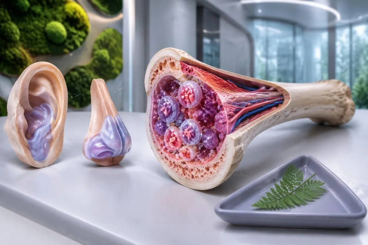
VEXAS syndrome is an acquired inflammatory condition caused by somatic UBA1 mutations in blood-forming cells. Recurrent fever, swollen ear or nose cartilage and unexplained macrocytic anemia should prompt specialist assessment; diagnosis requires appropriate genetic testing, not a routine autoimmune panel.

Temel çıkarımlar
- VEXAS syndrome usually begins after age 50 and combines systemic inflammation with abnormalities in blood-forming cells.
- Cartilage swelling can affect the ears, nose or airway; ear cartilage inflammation often spares the soft earlobe.
- Makrositoz means an MCV above approximately 100 fL in adults, but this finding alone does not diagnose VEXAS.
- Düşük trombosit below 150 × 10^9/L can strengthen concern when they accompany recurrent inflammation and unexplained anemia.
- CRP commonly has an upper reference limit of 5 mg/L, although some laboratories use 10 mg/L; no CRP cutoff confirms VEXAS.
- UBA1 mutation testing looks for an acquired pathogenic variant, usually using peripheral blood or bone marrow rather than a routine inherited-risk panel.
- Autoimmune panels such as ANA, rheumatoid factor and ANCA cannot confirm or exclude VEXAS syndrome.
- Acil belirtiler include new breathlessness, chest pain, airway difficulty or fever of 38°C or higher during significant immunosuppression.
What is VEXAS syndrome, and why do blood counts matter?
VEXAS syndrome is an acquired autoinflammatory disorder caused by a somatic UBA1 mutation in blood-forming cells. The combination of recurrent inflammation, swollen cartilage and unexplained abnormal blood counts—especially macrocytic anemia in someone over 50—should raise suspicion, while diagnosis depends on identifying an appropriate pathogenic UBA1 variant in clinical context.
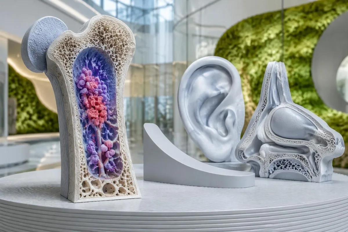
The name describes 5 features: vacuoles, E1 enzyme, X-linked, autoinflammatory and somatic. These are clues to the biology, not a checklist that every person must satisfy; visible marrow vacuoles, cartilage symptoms and marked anemia do not necessarily appear together at the first appointment.
Beck et al. first described VEXAS in the New England Journal of Medicine in 2020, linking adult-onset inflammation to acquired UBA1 variants. That discovery explains why a person may previously have received more than 1 diagnosis, such as relapsing polychondritis and a marrow disorder, before the shared cause was recognized.
A single elevated CRP result does not point specifically to VEXAS, and an MCV of 103 fL often has more common explanations. The concern rises when inflammatory attacks recur alongside persistent or worsening cytopenias, because that pattern suggests an inflammatory process connected to abnormal blood-cell production rather than an isolated laboratory fluctuation.
Kantesti bir AI kan testi analizörü that can explain how hemoglobin, MCV, platelets and CRP fit together, while leaving VEXAS diagnosis to specialists. Our organizasyonumuz ve amacımız are described separately; 1 uploaded CBC cannot reveal a UBA1 mutation or replace a clinical examination.
Who develops VEXAS, and is the UBA1 mutation inherited?
VEXAS syndrome most often affects older men, usually with symptoms beginning after age 50. Its disease-causing UBA1 mutation is generally acquired within a population of blood-forming cells, rather than inherited from a parent or present in every cell from birth.
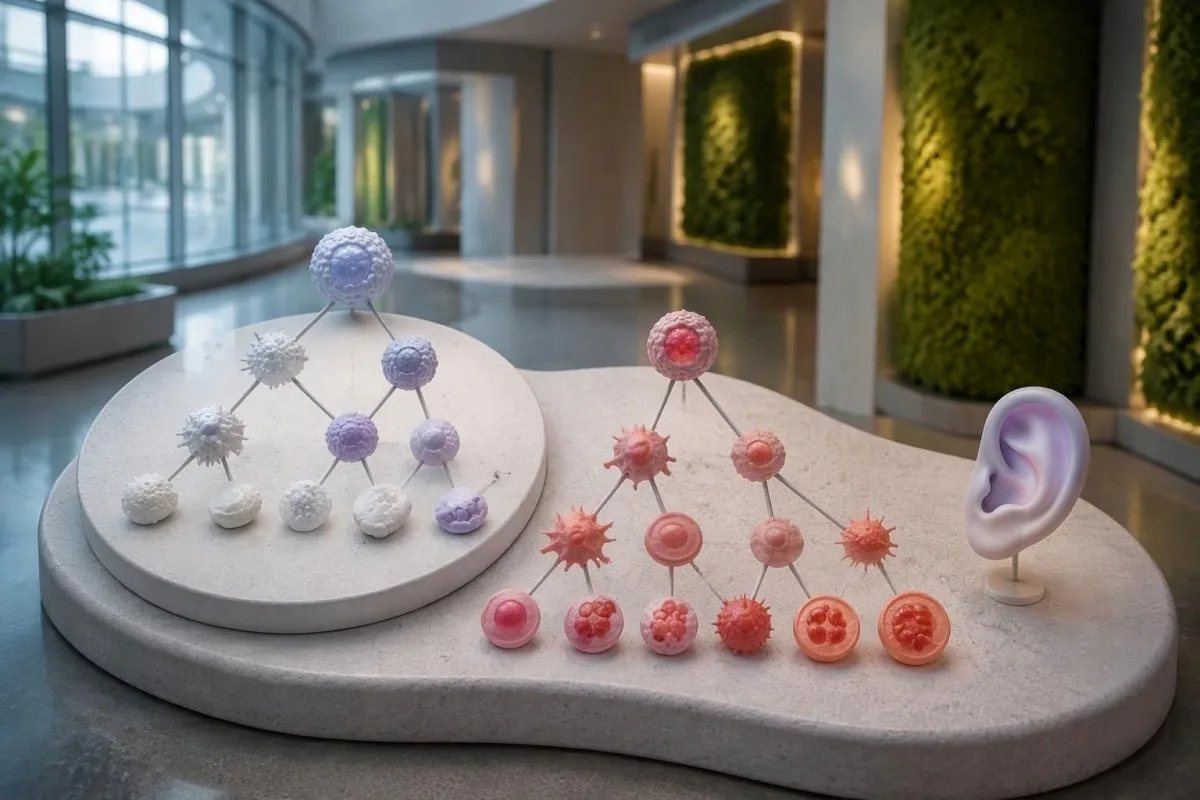
UBA1 lies on the X chromosome, and most affected men have 1 X chromosome. VEXAS also occurs in women, sometimes in association with loss of an X chromosome in affected cells, but sex alone should not stop assessment when the clinical pattern is strongly suggestive.
In a JAMA study of 163,096 participants in a US health-system cohort, Beck et al. identified 11 people with disease-associated UBA1 variants. The estimated frequency among men older than 50 was about 1 in 4,269; this was a particular, predominantly European-ancestry cohort, not a worldwide prevalence estimate.
A family history is usually absent because the relevant mutation emerges during a person's lifetime. This differs from inherited recurrent-fever disorders covered in our MEFV testi açıklamamız; testing 2 relatives for a familial variant is not the standard starting point for typical somatic VEXAS.
The word genetic does not automatically mean hereditary, and an X-linked gene does not automatically imply transmission to children. If a report raises a constitutional variant or an unusual presentation before age 50, a clinical genetics specialist can clarify whether additional tissue testing or family counseling is appropriate.
Which VEXAS syndrome symptoms should raise suspicion?
VEXAS syndrome symptoms commonly include recurrent fever, inflammatory skin changes, fatigue, lung involvement and cartilage inflammation. Swelling of ear or nasal cartilage becomes particularly informative when it occurs with unexplained anemia or other abnormal blood counts in an adult over 50.
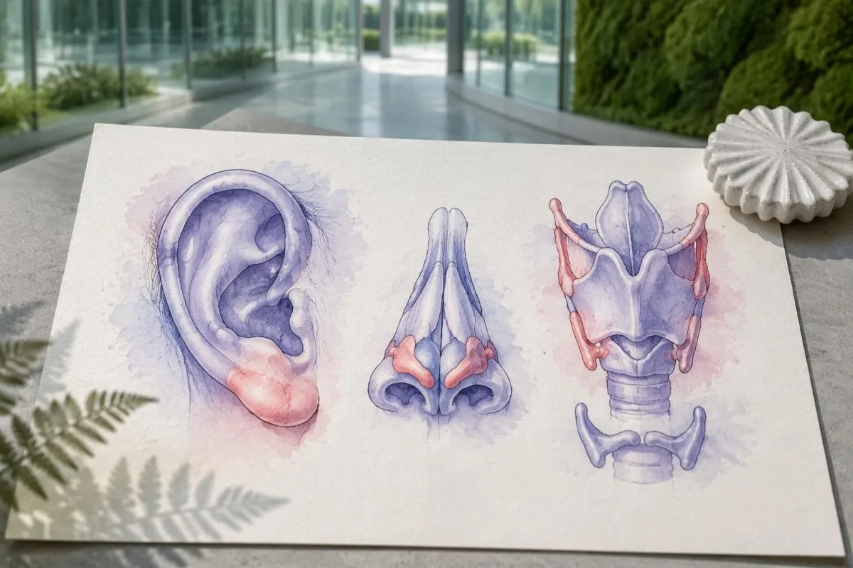
Ear chondritis often affects the firm upper ear while sparing the soft earlobe, which contains no cartilage. That anatomical distinction can help describe an episode accurately, but 1 red ear still requires assessment for more common causes such as local infection, injury or dermatitis.
Skin findings may include tender raised areas, plaques or purplish changes; appearance alone cannot identify the cause. A tissue examination can help distinguish neutrophilic dermatosis, vasculitis and other processes, while photographs of 2 separate untreated episodes may help a specialist recognize a recurrent pattern.
Inflammatory attacks can resemble established rheumatological conditions, including relapsing polychondritis or polymyalgia rheumatica. Our explanation of PMR laboratory limitations describes why inflammatory markers are not disease-specific; persistent macrocytosis above 100 fL or falling platelets should prompt a broader assessment rather than automatic acceptance of the first label.
New hoarseness, noisy breathing or difficulty breathing needs urgent assessment because airway cartilage can be involved. A useful symptom record includes 3 details for each episode: the affected site, its duration and whether fever or a medication change accompanied it; do not wait for another flare if breathing is affected.
What blood test pattern can suggest VEXAS syndrome?
makrositik anemi, sometimes accompanied by low platelets or other cytopenias, is a major laboratory clue to VEXAS. Adult MCV is commonly about 80–100 fL, but neither an MCV above 100 fL nor an elevated CRP result can confirm the condition.
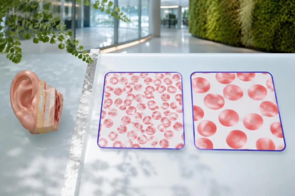
Hemoglobin measures anemia, while MCV measures average red-cell size; the 2 results answer different questions. An MCV of 108 fL with hemoglobin of 10.2 g/dL deserves a different assessment from an isolated MCV of 101 fL with stable hemoglobin, particularly when inflammatory symptoms are also present.
Many laboratories use a platelet reference interval of approximately 150–450 × 10^9/L. A falling platelet count can matter before it crosses the lower limit, and our borderline MCV explanation shows why persistent trends are often more useful than treating one near-boundary number as a diagnosis.
CRP is often markedly elevated during active VEXAS, but infection and many other inflammatory disorders can produce the same result. A hypothetical CRP of 80 mg/L is 16 times an upper limit of 5 mg/L; that comparison describes inflammatory intensity, not the probability of a UBA1 mutation.
RDW describes variation in red-cell size and can help explain a mixed CBC pattern, but it is not a VEXAS-specific biomarker. Our RDW ve MCV rehberimiz explains how 2 overlapping processes, such as iron deficiency and macrocytosis, can produce a deceptively ordinary average MCV.
| Düzey | Aralık | Anlamı |
|---|---|---|
| Typical adult MCV interval | 80–100 fL | Use the reporting laboratory's interval; an in-range result does not exclude early or masked macrocytosis. |
| Makrositoz | >100 fL | Investigate nutritional, medication, liver, thyroid and marrow causes; this is not a VEXAS diagnostic threshold. |
| Trombositopeni | <150 × 10^9/L | Low platelets can support concern when combined with persistent inflammation and unexplained anemia. |
| CRP above the laboratory limit | Often >5 mg/L; some laboratories use >10 mg/L | Indicates an inflammatory response without identifying its cause. |
Why can't routine autoimmune panels confirm VEXAS?
Routine autoimmune panels cannot confirm VEXAS syndrome because they measure antibodies or related immune markers, not acquired UBA1 mutations. ANA, rheumatoid factor, ANCA and complement results may help assess alternative or overlapping conditions, but no combination substitutes for appropriate molecular testing.
ANA testing detects antibodies directed against nuclear components; it does not read the UBA1 gene. A positive ANA at 1:160 may deserve interpretation in its own clinical context, while a negative ANA does not make unexplained fever, chondritis and macrocytic anemia safe to dismiss.
Repeating several antibody panels can create incidental positives without resolving the central question. Our ANA result explanation describes why 1 positive result is not equivalent to a disease diagnosis; the same caution applies when an antibody is found during a suspected VEXAS evaluation.
Complement testing can be useful when clinicians suspect an immune-complex disorder, but normal C3 and C4 cannot exclude VEXAS. The complement and antibody guide explains these 2 proteins; their biological role differs from the acquired enzyme dysfunction caused by a disease-associated UBA1 variant.
Autoinflammatory and autoimmune are related but not interchangeable descriptions: VEXAS is driven principally by abnormal innate immune activity associated with mutated blood-forming cells. Clinicians may still order 2 types of investigation in parallel—antibody testing for alternatives and molecular testing for VEXAS—because another diagnosis can coexist rather than disappear.
What other causes must be checked before blaming VEXAS?
Vitamin B12 or folate deficiency, medication effects, alcohol use, liver disease, hypothyroidism and marrow disorders can cause macrocytosis. An MCV above 100 fL should trigger evaluation of these possibilities, while recurrent chondritis or unexplained inflammatory attacks may justify investigating VEXAS at the same time.
Vitamin B12 and folate results need interpretation alongside symptoms, treatment history and the CBC. A borderline B12 result may require additional assessment rather than immediate attribution of an MCV of 107 fL to VEXAS; our B12 versus folate explanation also explains why folate treatment can obscure part of a B12 deficiency picture.
Copper deficiency can cause cytopenias and vacuolated marrow precursors, making it an especially relevant mimic. Reviewing high-dose zinc supplements is therefore useful, and our düşük bakır değerlendirme rehberimiz explains why 2 supplement ingredients—zinc and copper—may matter even when a product is marketed for general immune support.
Myelodysplastic syndrome can cause macrocytic anemia and may coexist with VEXAS rather than simply compete with it as an explanation. Our MDS confirmation guide distinguishes CBC clues from marrow evidence; 1 abnormal count cannot establish either diagnosis or determine whether both are present.
Medication and infection reviews are essential because treatment can change the very numbers being investigated. For example, glucocorticoids may increase neutrophils while lowering CRP, and a recent transfusion can alter MCV; bring the dates of these events, not just 2 laboratory reports that appear to disagree.
How does somatic UBA1 testing support VEXAS diagnosis?
VEXAS syndrome diagnosis is established by a disease-associated somatic UBA1 variant in a compatible clinical setting. Peripheral blood is commonly used for testing, while marrow or a more sensitive method may be appropriate after an inconclusive result; many recognized variants affect methionine at position 41.
The request should specify suspected VEXAS and testing for somatic UBA1 variants rather than a general hereditary disease panel. These are 2 different diagnostic tasks: an inherited-risk assay may not include the relevant gene regions, cell source or detection sensitivity needed to identify an acquired clone.
Beck et al. linked UBA1 variants to altered ubiquitin-activating enzyme function in their 2020 New England Journal of Medicine report. Position 41 is a major hotspot, but a hotspot-only test is not equivalent to comprehensive relevant UBA1 analysis, because disease-associated noncanonical variants have also been described.
A negative result needs the laboratory method attached to it before its meaning is clear. An assay validated to detect a 5% variant allele fraction may miss a smaller clone; ask which regions were covered, the reported detection limit and whether the sample was suitable for somatic testing.
Kantesti bir AI kan tahlili yorumlama platformu that can help explain accompanying CBC and inflammatory-marker results, but it cannot infer a UBA1 sequence from 1 routine report. Our Yapay zeka teknolojisi açıklaması describes report interpretation; molecular diagnosis still requires an accredited laboratory and specialist interpretation.
How should you read a UBA1 mutation report?
A UBA1 report should state the variant, classification, sample type and testing limitations, often including variant allele fraction. A pathogenic or likely pathogenic disease-associated variant can support VEXAS, but a variant of uncertain significance is not diagnostic, and a 10% allele fraction is not a 10% severity score.
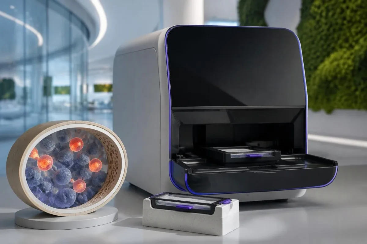
Variant allele fraction is the proportion of analyzed sequence reads carrying a particular change at that location. In a simple 1-X-chromosome model it may approximate the proportion of affected sampled cells, but cell mixtures, chromosome changes and laboratory methods complicate that estimate; it does not directly measure organ damage.
A hypothetical report showing a recognized disease-associated variant at 12% allele fraction may be meaningful even though most reads are unchanged. By contrast, a novel uncertain variant at 40% still requires expert assessment, because abundance cannot turn an unproven genetic finding into an established cause of symptoms.
Different clonal disorders require different molecular or cellular tests, even when anemia and thrombosis overlap. Our PNH clone testing guide explains a separate disorder assessed by flow cytometry; 1 negative PNH test does not evaluate UBA1, and a UBA1 result does not replace PNH testing when indicated.
Kantesti cannot determine whether an uncertain UBA1 finding is disease-causing simply by matching it to an elevated CRP of 70 mg/L. Ask the specialist whether additional sequencing, another specimen or formal variant review is needed, and keep the complete report rather than a screenshot containing only the word positive.
Are bone marrow vacuoles enough to diagnose VEXAS?
Bone marrow vacuoles are a clue, not proof of VEXAS syndrome. Vacuoles may appear in early myeloid and erythroid precursors, but they are neither completely specific nor universally present; an adult with an MCV above 100 fL still needs the broader clinical and molecular assessment.
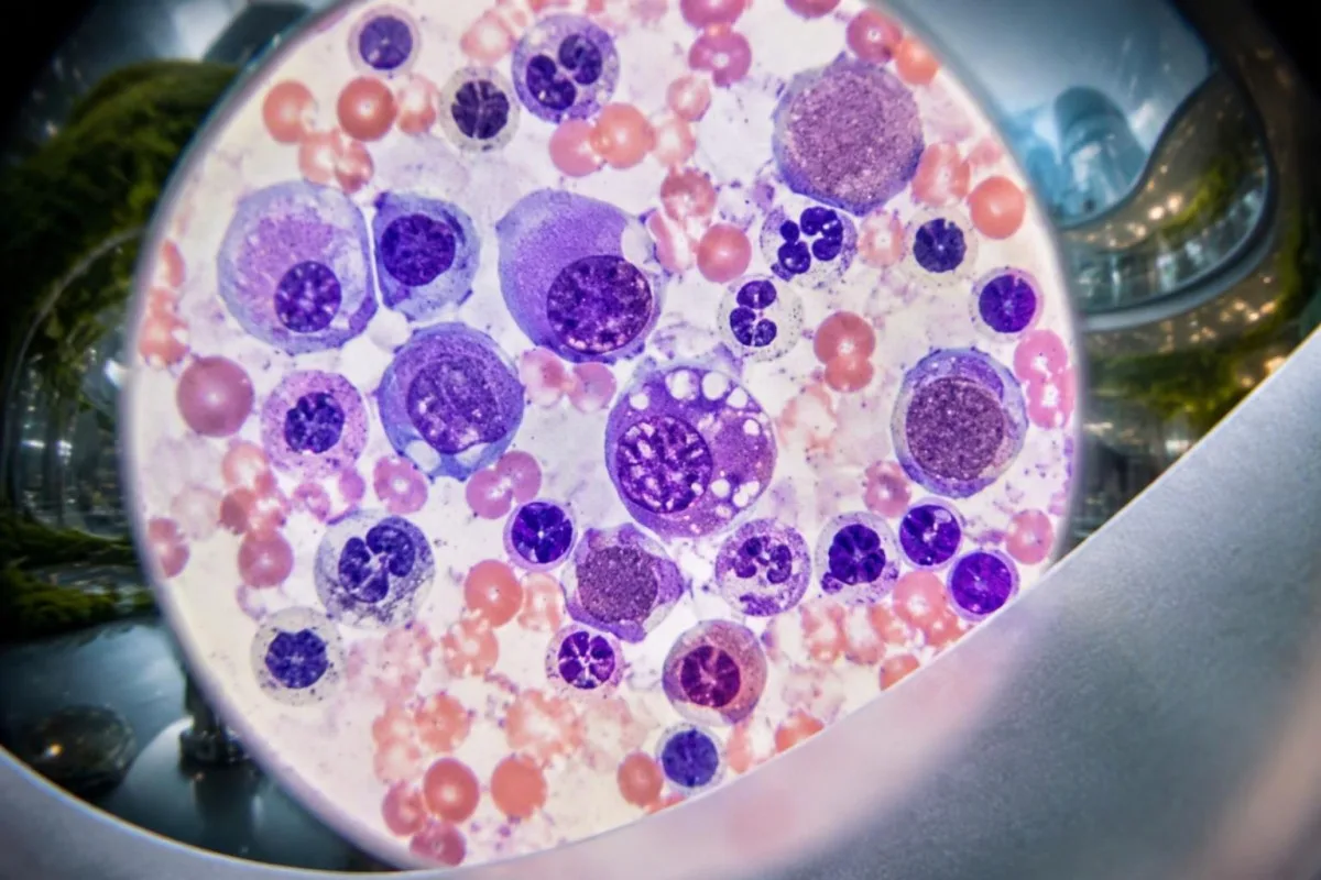
Vacuoles are small clear spaces visible within cells under microscopy, not holes that a patient can see on an ordinary CBC. Their absence on 1 marrow examination does not automatically exclude VEXAS, particularly when specimen quality, disease stage or the specific UBA1 variant affects the appearance.
Copper deficiency, alcohol-related effects and other conditions can produce vacuolated precursors. That overlap is why 2 complementary investigations may be needed: laboratory assessment for reversible mimics and somatic UBA1 testing, rather than treating a descriptive pathology finding as the final diagnosis.
Marrow examination also assesses cellularity, dysplasia and other abnormalities that may influence treatment decisions. Our explanation of teardrop cells and marrow clues describes a different microscopy finding; neither teardrop cells nor vacuoles can independently explain a platelet count of 95 × 10^9/L.
A marrow procedure is not automatically necessary for every isolated MCV of 102 fL, and a positive molecular result may already establish VEXAS in the right context. Hematology decides whether marrow assessment is needed to investigate persistent cytopenias, suspected MDS or another process that would change management.
When is rheumatology or hematology assessment appropriate?
Specialist assessment is appropriate when recurrent unexplained inflammation accompanies macrocytic anemia, falling platelets or other persistent cytopenias. Suspicion is higher after age 50, especially with chondritis, inflammatory skin findings or lung involvement; rheumatology and hematology often need to evaluate the pattern together.
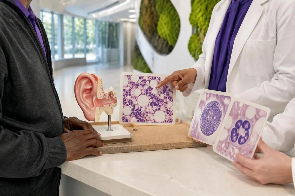
There is no universal CRP level, age boundary or number of attacks that automatically mandates UBA1 testing. A person aged 68 with recurrent ear chondritis and unexplained anemia has a different pretest probability from a person aged 28 with 1 viral fever and an otherwise stable CBC.
Steroid-responsive inflammation that repeatedly returns during dose reduction deserves reassessment, particularly when blood counts remain abnormal. Improvement over 2 days may show that symptoms are inflammatory, but it does not distinguish VEXAS from several other disorders and should not become the only justification for ongoing treatment.
Unexplained pain needs a broader workup when it accompanies constitutional symptoms or changing counts. Our açıklanamayan ağrı test rehberi explains why inflammatory markers are only part of that workup; an MCV above 100 fL adds a marrow-related question that a pain score cannot answer.
A referral should highlight 3 elements: documented inflammatory episodes, the CBC trend and the results of common-cause investigations. If symptoms are progressing rapidly, new organs are involved or transfusions are being considered, ask the referring clinician whether the appointment needs to be expedited rather than waiting for another routine repeat test.
A worked example: recurrent ear swelling and a changing CBC
A hypothetical combination of recurrent chondritis, macrocytic anemia and low platelets warrants specialist evaluation, not an AI-generated diagnosis. Consider a 68-year-old man with hemoglobin 10.2 g/dL, MCV 108 fL, platelets 112 × 10^9/L and CRP 86 mg/L during a third episode of upper-ear swelling.

The first reasoning step is to separate symptom severity from diagnostic specificity: CRP of 86 mg/L confirms a substantial inflammatory response but does not identify its cause. The MCV of 108 fL and platelet count of 112 × 10^9/L add evidence that blood-cell production also needs investigation.
Suppose the person's B12, folate and thyroid assessments do not reveal an explanation, and medication, alcohol and liver reviews are unrevealing. A negative ANA would not close the case; the combination of 3 inflammatory episodes and persistent unexplained cytopenias would justify discussing somatic UBA1 testing with hematology and rheumatology.
Now suppose CRP falls to 12 mg/L after prescribed glucocorticoids, but hemoglobin falls to 9.8 g/dL. Those 2 trends are not contradictory: inflammatory activity may improve while marrow dysfunction or another anemia mechanism persists, so treatment response cannot be used as a substitute for investigating the falling hemoglobin.
Kantesti can help explain why those 2 trends deserve separate attention, but cannot establish VEXAS or choose immunosuppressive treatment. Our ilaç trend analizi kılavuzumuz explains why dose changes, transfusions and intercurrent illness should be recorded beside each result; the example here is entirely hypothetical.
How do specialists treat confirmed VEXAS syndrome?
VEXAS treatment aims to control inflammation, manage cytopenias and reduce treatment-related harm. Glucocorticoids often suppress symptoms but may be difficult to taper; other options include selected immunomodulators, marrow-directed treatment and allogeneic stem-cell transplantation, with no single regimen appropriate for every adult over 50.
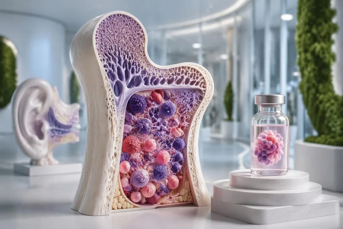
A falling CRP does not by itself establish treatment success if anemia, thrombosis or lung disease is worsening. Specialists follow at least 2 dimensions—clinical inflammatory activity and hematological status—and weigh these against cumulative glucocorticoid exposure, because prolonged treatment can cause infections, diabetes, osteoporosis and muscle weakness.
JAK inhibitors and IL-6-directed therapy are among approaches used by specialists, while azacitidine may be considered in selected patients, particularly with associated marrow disease. Evidence and access vary, and much published experience is observational; a response lasting 6 months in one cohort does not reliably predict an individual's longer-term benefit.
Allogeneic stem-cell transplantation can potentially eliminate the abnormal blood-forming clone and produce sustained remission in selected patients. It also carries substantial risks, including graft-versus-host disease and serious infection, so 2 people of the same age may receive different recommendations because fitness, organ involvement and donor availability differ.
Infection prevention deserves planning before immunosuppression, not only after fever develops. Depending on exposures and the proposed regimen, clinicians may consider screening or prevention measures described in our Strongyloides testing explanation; even 1 relevant past exposure can matter before corticosteroids, and prescribed steroids should never be stopped abruptly without guidance.
Hangi belirtiler acil veya derhâl tıbbi yardım gerektirir?
New breathing difficulty, chest pain, airway symptoms, neurological deficits or significant bleeding require urgent assessment in someone with suspected or confirmed VEXAS. Fever of 38°C or higher during substantial immunosuppression also needs prompt clinical advice, particularly when neutrophils are low.
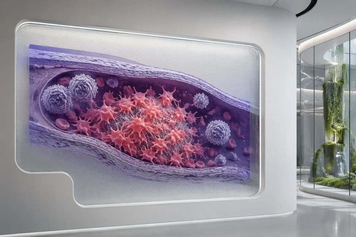
VEXAS is associated with venous thrombosis, so a painful swollen leg or sudden breathlessness should not be dismissed as another inflammatory flare. Normal routine clotting times do not exclude a clot; our pıhtılaşma panelinin kısıtlılıkları explain why 2 normal tests, PT and aPTT, cannot provide that reassurance.
An absolute neutrophil count below 0.5 × 10^9/L represents severe neutropenia and increases concern when fever occurs. Treatment can also blunt the temperature response, so feeling acutely unwell with shaking chills, confusion or rapidly worsening weakness can require urgent assessment even when a thermometer reads below 38°C.
Breathlessness can reflect lung inflammation, infection, pulmonary embolism or worsening anemia, and these require different treatments. Our breathlessness blood test guide explains why hemoglobin of 8 g/dL may contribute without proving the entire cause; new chest pain or sudden breathing difficulty should not wait for an uploaded report.
Persistent nose or gum bleeding, black stools or a severe new headache needs urgent advice, especially if platelets are very low. A count below 20 × 10^9/L generally warrants prompt clinical review, but the urgency depends on symptoms and context; significant bleeding is an emergency even above that number.
Follow-up, AI limits and research publications
Useful VEXAS follow-up combines symptoms, treatment history, CBC trends and inflammatory markers, with molecular and marrow findings interpreted by specialists. An AI explanation of 1 laboratory report can support preparation for care, but it cannot diagnose VEXAS, exclude infection or establish whether a UBA1 variant is pathogenic.

Bring original CBC reports, inflammatory markers and the complete UBA1 result, including specimen type and detection limit. Record 3 timeline features beside them: flare dates, medication changes and transfusions; this makes it easier to distinguish a true disease trend from a treatment effect or a change in testing conditions.
Kantesti bir Yapay zekâ destekli kan testi analiz aracı that can explain laboratory patterns without converting an MCV above 100 fL into a rare-disease diagnosis. Genetic reports can reveal sensitive information, so review our gizlilik ve veri bilgilerini before sharing records, and include only documents you are authorized to upload.
Kantesti's 2 DOI-linked publications below concern broader blood-test interpretation and RDW education, not clinical trials of VEXAS diagnosis or UBA1 sequencing. They should therefore be read separately from the disease-specific medical studies listed in the external references; neither publication establishes VEXAS-specific diagnostic sensitivity or treatment effectiveness.
Bizim metodoloji ve klinik standartlar explain the distinction between report interpretation and medical diagnosis. Before the next appointment, write 2 questions: whether the laboratory method adequately assessed somatic UBA1 variants, and whether persistent cytopenias need marrow evaluation; these are more actionable than asking an AI to calculate an unsupported VEXAS probability.
Sıkça sorulan sorular
VEXAS sendromunun ilk belirtileri nelerdir?
VEXAS sendromu genellikle 50 yaşından sonra tekrarlayan, nedeni açıklanamayan ateş, inflamatuvar cilt değişiklikleri, yorgunluk veya kulak ya da burun kıkırdağında şişlikle başlar. Makrositik anemi veya diğer anormal kan sayımı bulguları bu belirtilerden önce, bunlarla birlikte ya da sonrasında ortaya çıkabilir. Yetişkinlerde yaklaşık 100 fL'nin üzerindeki MCV bir ipucudur, tanı değildir. Tekrarlayan inflamatuvar belirtilerle birlikte nedeni açıklanamayan kalıcı sitopeniler, romatoloji ve hematoloji değerlendirmesinin görüşülmesini gerektirir.
VEXAS sendromunu hangi test doğrular?
VEXAS sendromu, uygun bir klinik tabloda hastalıkla ilişkili somatik bir UBA1 varyantının saptanmasıyla teşhis edilir. Testlerde genellikle periferik kan kullanılır; ancak seçilmiş vakalarda kemik iliği örneği veya daha duyarlı bir test gerekebilir. Tanımlanmış varyantların çoğu 41. pozisyondaki metiyonini etkiler; ancak sınırlı sayıda sıcak nokta varyantını inceleyen testler, hastalıkla ilişkili diğer varyantları saptamayabilir. Rapor; varyant sınıflandırması, gen kapsamı ve saptama sınırı göz önünde bulundurularak yorumlanmalıdır.
Normal bir ANA veya otoimmün panel VEXAS'ı dışlayabilir mi?
Normal bir ANA veya rutin otoimmün panel, bu testler somatik UBA1 mutasyonlarını saptamadığından VEXAS sendromunu dışlayamaz. 1:160 titrede pozitif bir ANA da VEXAS tanısını doğrulamaz veya mutlaka farklı bir otoimmün hastalığın varlığını göstermez. Antikor testleri alternatifleri araştırmaya yardımcı olabilirken CBC örüntüleri, belirtiler ve moleküler testler farklı sorulara yanıt verir. Kalıcı makrositik anemi ve tekrarlayan kıkırdak iltihabı, yalnızca antikor testleri negatif olduğu için göz ardı edilmemelidir.
Is VEXAS syndrome inherited, and should my children be tested?
Typical VEXAS syndrome is acquired rather than inherited, so routine testing of children is generally not indicated. The relevant UBA1 mutation usually develops within blood-forming cells during adult life, often with symptoms after age 50. Although UBA1 is on the X chromosome, that does not make a somatic VEXAS variant automatically transmissible. Unusual or possibly constitutional findings should be reviewed by a clinical genetics specialist.
Can women develop VEXAS syndrome?
Women can develop VEXAS syndrome, although most recognized cases occur in older men. UBA1 is located on the X chromosome, and loss of 1 X chromosome in affected cells is one mechanism reported in women. A 2023 JAMA health-system study identified 2 women among 11 participants with disease-associated UBA1 variants. Sex alone should not prevent specialist assessment when recurrent inflammation and unexplained blood-count abnormalities are strongly suggestive.
Does a negative UBA1 test exclude VEXAS syndrome?
A negative UBA1 test lowers the likelihood of VEXAS only to the extent that the specimen and assay were appropriate. A test with a 5% variant allele fraction detection limit may miss a smaller clone, and hotspot-only testing may miss variants outside the covered region. Strong clinical suspicion can justify specialist review of coverage, sensitivity and sample type. A properly performed negative result should also prompt careful reassessment of alternative diagnoses rather than repeated testing without a clear reason.
Is there a cure for VEXAS syndrome?
Allogeneic stem-cell transplantation can potentially produce sustained remission by replacing the abnormal blood-forming clone, but it is suitable only for selected patients and carries substantial risks. Other treatments aim to control inflammation, manage cytopenias and reduce glucocorticoid exposure rather than reliably eliminate the clone. For adults over 50, fitness, organ involvement and associated marrow disease can strongly influence treatment choices. No supplement, dietary plan or AI blood-test interpretation has been shown to cure VEXAS.
Kan tahlili sonuçları ne anlama gelir
Laboratuvar raporunuzu yükleyin; Kantesti'nin AI'ı her biyobelirteci saniyeler içinde sade bir dille açıklar. Dünya genelinde 2 milyondan fazla kişi tarafından kullanılıyor.
Referanslar
- Klein, T., Mitchell, S. & Weber, H. (2026). Yapay Zeka Kan Testi Analizörü: 2,5M Test Analiz Edildi | Global Health Report 2026. Kantesti AI Tıbbi Araştırma. DOI
- Klein, T., Mitchell, S. & Weber, H. (2026). RDW Kan Testi: RDW-CV, MCV ve MCHC için Eksiksiz Rehber. Kantesti AI Tıbbi Araştırma. DOI
- Beck DB et al. (2020). Somatic Mutations in UBA1 and Severe Adult-Onset Autoinflammatory Disease. New England Journal of Medicine. DOI
- Beck DB et al. (2023). Estimated Prevalence and Clinical Manifestations of VEXAS Syndrome in a Large US Health Care System. JAMA. DOI
- 2 milyondan fazlaAnaliz edilen testler
- 127+Ülkeler
- 100+Diller
Sağlık ekibi hakkında


E-E-A-T Güven Sinyalleri
Deneyim
Hekim liderliğinde laboratuvar yorumlama iş akışlarının klinik incelemesi.
Uzmanlık
Klinik bağlamda biyobelirteçlerin nasıl davrandığına odaklanan laboratuvar tıbbı.
Otorite
Dr. Thomas Klein tarafından tıbbi açıdan incelenmiş; Dr. Sarah Mitchell ve Prof. Dr. Hans Weber'in katkılarıyla hazırlanmıştır.
Güvenilirlik
Alarmı azaltmaya yönelik net takip yollarıyla kanıta dayalı yorumlama.

Tıbbi sorumluluk reddi
Bu makale yalnızca eğitim amaçlıdır ve tıbbi tavsiye niteliği taşımaz. Tanı ve tedavi kararları için her zaman yetkin bir sağlık hizmeti sağlayıcısına danışın.
Yayınlanmış · Yayın ekibiyle iletişime geçin
Kantesti Ltd tarafından yayımlanmıştır; İngiltere ve Galler'de kayıtlı, şirket no. 17090423, Londra, Birleşik Krallık.
