
การตรวจไข้ร็อกกีเมาน์เทนสปอตเต็ดอาจให้ผลเป็นลบในสัปดาห์แรก เนื่องจากยังไม่มีการสร้างแอนติบอดี การตรวจพบไตเตอร์ IgG เป็นบวกเพียงครั้งเดียวไม่สามารถยืนยันการติดเชื้อเฉียบพลันได้; ตัวอย่างที่เก็บเป็นคู่ซึ่งแสดงการเพิ่มขึ้นสี่เท่าสนับสนุนการติดเชื้อเมื่อไม่นานมานี้ แพทย์ควรเริ่มการรักษาโดยเร็วเมื่อสงสัย RMSF—ไม่ควรรอการยืนยันจากห้องปฏิบัติการ.

ประเด็นสำคัญ
- ผลลบลวงในระยะแรก: การตรวจแอนติบอดี RMSF มักให้ผลเป็นลบในช่วง 7 วันแรกของการเจ็บป่วย ซึ่งเป็นช่วงที่ต้องตัดสินใจรักษาอย่างเร่งด่วนที่สุด.
- วิธีตรวจแอนติบอดีที่แนะนำ: การตรวจแอนติบอดีด้วยวิธีอิมมูโนฟลูออเรสเซนต์ทางอ้อมสำหรับ IgG หรือ IFA ใช้เปรียบเทียบไตเตอร์แอนติบอดีกับแอนติเจนของริกเก็ตเซียกลุ่มไข้สปอตเต็ด.
- ตัวอย่างที่เก็บเป็นคู่: เก็บตัวอย่างระยะเฉียบพลันตั้งแต่เนิ่น ๆ และตัวอย่างระยะพักฟื้นในภายหลัง; แนวทางของ CDC ระบุช่วงห่างระหว่างการเก็บตัวอย่างไว้ที่ 2–10 สัปดาห์.
- การเพิ่มขึ้นสี่เท่า: การเปลี่ยนแปลงจาก 1:64 เป็น 1:256 ถือเป็นการเพิ่มขึ้นสี่เท่า และสนับสนุนว่ามีการติดเชื้อริกเก็ตเซียกลุ่มไข้สปอตเต็ดเมื่อไม่นานมานี้ในผู้ป่วยที่มีอาการสอดคล้องกัน.
- IgG เป็นบวกเพียงครั้งเดียว: แอนติบอดีอาจคงอยู่ได้นานหลายปี แม้ไตเตอร์ 1:256 ก็ไม่สามารถใช้ระบุได้โดยลำพังว่าได้รับเชื้อเมื่อใดหรือเป็นริกเก็ตเซียสายพันธุ์ใด.
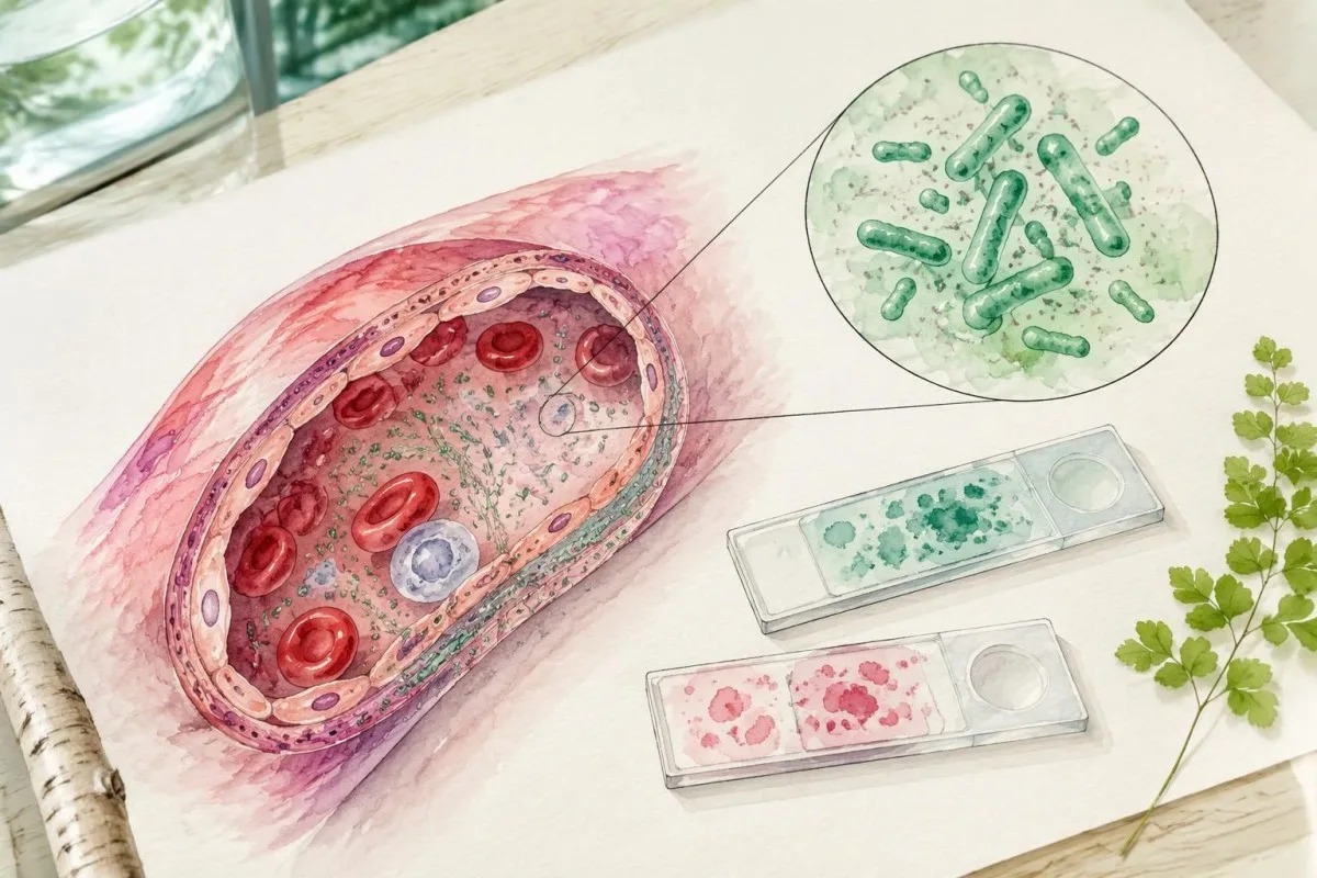
- PCR ให้ผลลบ: PCR จากเลือดครบส่วนอาจตรวจไม่พบ RMSF เนื่องจาก Rickettsia rickettsii ส่งผลต่อเซลล์บุผนังหลอดเลือดเป็นหลัก ไม่ใช่เม็ดเลือดขาวที่ไหลเวียนอยู่.
- ช่วงเวลาเริ่มการรักษา: ดอกซีไซคลินเป็นการรักษาลำดับแรกสำหรับผู้ใหญ่และเด็กทุกวัยที่สงสัยว่าเป็น RMSF แพทย์ไม่ควรรอผลตรวจ.
- แนวทางการใช้ยาโดยทั่วไปในผู้ใหญ่: แนวทางของ CDC แนะนำดอกซีไซคลิน 100 mg ทุก 12 ชั่วโมง โดยทั่วไปอย่างน้อย 5–7 วัน และจนกว่าไข้จะหายไปอย่างน้อย 3 วันพร้อมกับอาการทางคลินิกดีขึ้น.
การตรวจไข้ร็อกกีเมาน์เทนสปอตเต็ดแบบใดควรทำก่อน?
แนวทางมาตรฐานในการตรวจแอนติบอดีเพื่อวินิจฉัย RMSF คือการตรวจ IgG แบบจับคู่ด้วยวิธี indirect fluorescent antibody assay ซึ่งมักเรียกว่า IFA. อย่างไรก็ตาม แพทย์อาจเริ่มให้ดอกซีไซคลินก่อนมีผลตรวจครั้งแรก เนื่องจากตัวอย่างแอนติบอดีที่เก็บในช่วงวันที่ 1–7 มักไม่สามารถยืนยันหรือตัดโรคนี้ออกได้.

RMSF เกิดจากเชื้อ Rickettsia rickettsii, ซึ่งเป็นแบคทีเรียที่แพร่โดยเห็บและสามารถทำลายหลอดเลือดขนาดเล็กได้อย่างรวดเร็ว คู่มือปฏิบัติของ CDC แบ่งงานออกเป็น 2 ส่วน ได้แก่ รักษาโรคที่อาจเป็นอันตรายในขณะนี้ และตรวจหาหลักฐานทางห้องปฏิบัติการเพื่อยืนยันภายหลัง การสับสนระหว่างสองส่วนนี้ทำให้เกิดความล่าช้าที่หลีกเลี่ยงได้ (Biggs et al., 2016).
คำสั่งตรวจที่ระบุว่าเป็นแผงตรวจ RMSF อาจรวม IgG, IgM หรือทั้งสองชนิด และผลตรวจคัดกรองอาจตามด้วยการตรวจไตเตอร์ด้วย IFA ควรถามว่าแต่ละผลได้มาจากวิธีใด เพราะผลคัดกรองที่เป็นบวก 1 รายการกับไตเตอร์เชิงปริมาณ 1 รายการใช้แทนกันไม่ได้ คู่มือของเราเกี่ยวกับ ผลการตรวจแอนติบอดีเป็นบวก อธิบายความแตกต่างนั้นไว้.
คันเตสตีเป็น เครื่องวิเคราะห์ผลเลือด AI จัดทำขึ้นเพื่ออธิบายรายงานผลตรวจทางห้องปฏิบัติการ ไม่ใช่เพื่อใช้ตัด RMSF ออกด้วยผลลบเพียง 1 ครั้ง ของเรา ข้อมูลพื้นฐานของบริษัท ระบุองค์กรที่อยู่เบื้องหลังบริการดังกล่าว การตรวจร่างกาย การประเมินการสัมผัสเชื้อ การสั่งตรวจ และการตัดสินใจใช้ยาปฏิชีวนะยังคงเป็นความรับผิดชอบของแพทย์ผู้รักษา.
เหตุใดการตรวจแอนติบอดี RMSF ในระยะแรกจึงให้ผลเป็นลบ?
หนึ่ง ผลตรวจ RMSF ลบลวง มักเกิดจากช่วงเวลาที่ยังตรวจไม่พบแอนติบอดี กล่าวคือ ระบบภูมิคุ้มกันยังผลิตแอนติบอดีที่ตรวจพบได้ไม่เพียงพอ การตรวจ IgG IFA มักให้ผลลบในช่วง 7 วันแรกของอาการ ดังนั้นรายงานผลลบในระยะแรกจึงไม่ใช่เหตุผลให้ระงับการรักษาเมื่อภาพทางคลินิกบ่งชี้ว่าอาจเป็น RMSF.

การนับช่วงเวลาสำหรับการตรวจเริ่มจาก การเริ่มมีอาการ, ไม่ใช่วันที่ห้องปฏิบัติการได้รับตัวอย่าง ตัวอย่างเช่น ผู้ป่วยสมมติที่ได้รับการตรวจในวันที่ 3 ของการเจ็บป่วยอาจเป็น RMSF จริงแม้ผล IgG จะเป็นลบ ส่วนผลลบแบบเดียวกันที่ได้ในอีกหลายสัปดาห์ต่อมาจะมีความหมายในการแปลผลต่างออกไป โดยเฉพาะหากตรวจแบบจับคู่ครบแล้ว.
การสร้างแอนติบอดีไม่ได้เกิดขึ้นตรงกันตามวันปฏิทินที่แน่นอนในทุกคน แนวทางการวินิจฉัยทางคลินิกและห้องปฏิบัติการของ CDC อธิบายว่าการตรวจทางซีรัมวิทยาในสัปดาห์แรกมักให้ผลลบ ประสิทธิภาพสูงของห้องปฏิบัติการในการตรวจตัวอย่างที่มีแอนติบอดีเป็นบวกอยู่แล้ว ไม่ได้หมายความว่าจะมีความไวในการตรวจเทียบเท่ากันในช่วงวันที่ 1–7.
การตรวจ IgG ซ้ำเพียง 24–48 ชั่วโมงหลังผลลบในระยะแรกอาจให้ประโยชน์เพิ่มขึ้นเพียงเล็กน้อย เพราะช่วงเวลาดังกล่าวมักสั้นเกินกว่าจะแสดงการเปลี่ยนแปลงของแอนติบอดีตามที่คาดไว้ ส่วน Monospot testing window illustrates a similar timing principle, although its assay, illness, and recommended follow-up schedule differ from RMSF.
ความหมายที่แท้จริงของผล IgG ต่อ RMSF เป็นบวกครอบคลุมอะไรบ้าง?
RMSF IgG positive meaning includes previous exposure, recent infection, or antibodies reacting with another spotted fever group rickettsia. A single positive titer—even 1:256 or higher—cannot independently establish acute RMSF, because antibody persistence and cross-reactivity prevent the result from acting as a reliable date stamp.

Antibodies against R. rickettsii can remain elevated for months and sometimes up to 4 years after illness, according to CDC diagnostic guidance. Persistent positivity does not mean bacteria remain active, and the height of a single titer does not reliably distinguish a new infection from an older immune response.
Background antibodies also affect interpretation: CDC notes that 10% or more of healthy people in some areas may have elevated titers from past exposure to spotted fever group rickettsiae. That figure is geographically dependent, not a universal prevalence estimate; testing someone without compatible symptoms increases the chance of finding a clinically misleading positive.
Consider a hypothetical asymptomatic adult with IgG 1:128 on an incidental panel and no recent febrile illness. That 1 result is not a reason by itself to prescribe doxycycline; antibody persistence after exposure is another example of why serology must be matched to the organism, symptoms, and sampling dates.
ปฏิกิริยาข้ามหรือ IgM ทำให้ได้ผลตรวจที่ทำให้เข้าใจผิดได้หรือไม่?
การเกิดปฏิกิริยาข้าม can produce positive RMSF serology after exposure to another spotted fever group rickettsia, and อธิบายว่าห้องแล็บติดป้ายกำกับตัวบ่งชี้ภูมิคุ้มกันเหล่านี้อย่างไร is less specific than IgG for diagnosing recent illness. A positive IgM result on day 5 is therefore not a dependable shortcut around an early negative IgG result.
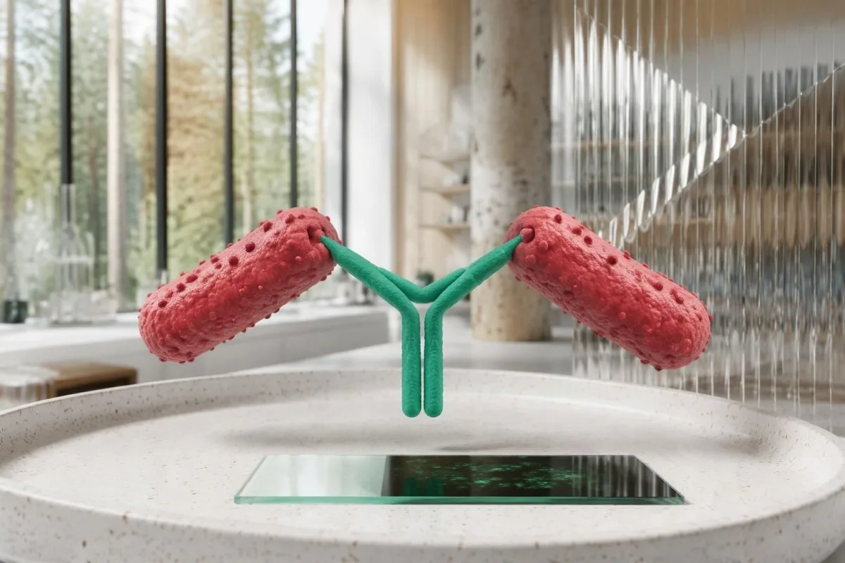
R. rickettsii, R. parkeri, and related organisms share antigenic features that routine commercial serology often cannot distinguish. A 4-fold IgG rise can establish recent spotted fever group rickettsial infection in a compatible illness, but identifying R. rickettsii specifically may require species-specific molecular testing or specialized reference-laboratory investigation.
IgM may appear without convincing evidence of recent rickettsial illness and should not be used as a stand-alone diagnostic marker. The CDC practical guide favors paired IgG IFA for confirmation rather than interpreting 1 isolated IgM positive as proof of acute RMSF (Biggs et al., 2016).
Not all antibody families behave alike: Q fever antibody patterns use phase-specific interpretation that should not be imported into RMSF. For a report showing IgM positive and IgG negative on day 4, ask about clinical suspicion, assay method, and the planned convalescent specimen rather than assuming the sequence proves infection.
ตัวอย่างที่เก็บเป็นคู่และการเพิ่มขึ้นสี่เท่าสนับสนุนการวินิจฉัยอย่างไร?
Paired IgG IFA samples support diagnosis by showing whether antibody titers rise substantially between acute illness and recovery. A rise from 1:64 to 1:256 is fourfold; with compatible symptoms, it supports recent spotted fever group rickettsial infection rather than merely documenting an antibody that was already present.
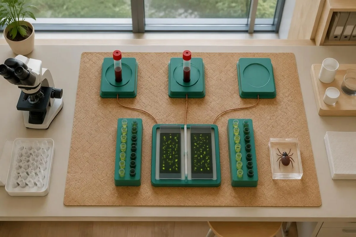
IFA titers describe the highest dilution at which antibody remains detectable; 1:256 means detection after greater dilution than 1:64. In a common 2-fold dilution series, 1:64 to 1:128 is one step, while 1:64 to 1:256 is two steps—the fourfold change used to reduce ambiguity from small fluctuations.
CDC RMSF guidance describes paired specimens collected 2–10 weeks apart, while the 2016 practical guide emphasizes an acute specimen and a convalescent specimen ideally 2–4 weeks later. These recommendations are not competing treatment deadlines; the clinician and laboratory should agree on follow-up timing that can capture the antibody response.
Use the same laboratory and assay when possible, and retain the original reports rather than comparing 2 abbreviated portal flags. Our explanations of ผลเชิงปริมาณเทียบกับผลเชิงคุณภาพ และ การเปลี่ยนแปลงระหว่างการเยี่ยมชมห้องปฏิบัติการ help distinguish a meaningful paired titer rise from unrelated reporting differences.
| ระดับ | ช่วง | ความหมาย |
|---|---|---|
| Early negative | Below laboratory cutoff on days 1–7 | Does not exclude RMSF; antibodies may not yet be detectable. |
| Single positive | Example: 1:128 on one specimen | Shows detectable antibody, not the date or species of infection. |
| Twofold rise | Example: 1:64 → 1:128 | Does not meet the standard fourfold-rise criterion. |
| Fourfold rise | ตัวอย่าง: 1:64 → 1:256 | Supports recent spotted fever group infection with compatible illness; does not measure severity. |
PCR ตรวจพบ RMSF ได้ก่อนที่แอนติบอดีจะปรากฏหรือไม่?
PCR can detect rickettsial DNA before antibodies develop, but a negative whole-blood PCR does not exclude RMSF. R. rickettsii mainly targets vascular endothelial cells, so an illness-day-3 blood specimen may contain too little bacterial DNA for detection even when the disease is clinically significant.
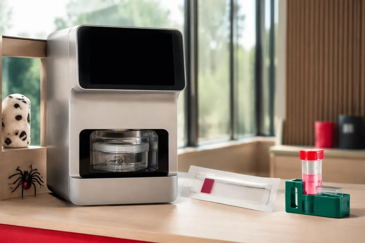
Unlike serology, PCR looks for the organism's genetic material rather than the host's immune response. Its usefulness depends on 3 practical variables: the specimen collected, illness severity and timing, and whether antibiotics have already reduced detectable organisms; there is no single sensitivity percentage applicable to every RMSF PCR order.
A clinician may arrange PCR or immunohistochemical testing of a rash skin biopsy through an appropriate laboratory, because affected tissue can provide evidence unavailable in circulating blood. Even when tissue testing is considered, collecting a specimen before the first doxycycline dose is desirable only if it does not delay urgent treatment.
Ask whether a panel actually includes R. rickettsii and whether it reports species-specific results or a broader rickettsial group. The PCR timing explanation for whooping cough demonstrates why molecular tests have specimen and treatment windows, but its respiratory sampling rules do not apply to RMSF.
เหตุใดผลตรวจ RMSF และเออร์ลิชิโอซิสจึงแตกต่างกัน?
RMSF and ehrlichiosis require different diagnostic reasoning even when the same tick-exposure evaluation includes both. Whole-blood PCR is generally more useful early in ehrlichiosis because Ehrlichia organisms infect white cells, whereas R. rickettsii primarily affects vessel-lining cells; 1 negative tick-borne panel is not a universal exclusion test.

Fever, headache, muscle aches, and abnormal liver enzymes can occur in more than 1 tick-borne illness. Exposure location, platelet count, white-cell count, rash, and illness timing guide test selection; clinicians should not assume that an order called a tick panel covers every relevant organism or uses the best specimen for each.
การ ehrlichiosis PCR and titer guide explains why acute PCR and later antibodies may disagree in that illness. For a hypothetical day-4 presentation, an Ehrlichia PCR positive and RMSF IgG negative need interpretation as separate findings—not evidence that all other tick-transmitted illnesses were conclusively excluded.
คันเตสตีเป็น แพลตฟอร์มการแปลผลตรวจเลือดของ AI that explains submitted laboratory reports, not a substitute for organism-specific diagnostic assessment. If a report contains 2 negative results from different methods, we emphasize the distinction between the method's target and the clinician's question rather than treating the word negative as a complete answer.
ผลตรวจเลือดทั่วไปใดบ้างที่ช่วยสนับสนุนข้อสงสัยว่าเป็น RMSF?
Low platelets, mildly elevated AST or ALT, and low sodium can support suspicion of RMSF, but none confirms the diagnosis and early values may be normal. A platelet count of 110 × 10⁹/L and sodium of 130 mmol/L are contextual clues—not organism-specific thresholds or prerequisites for starting treatment.
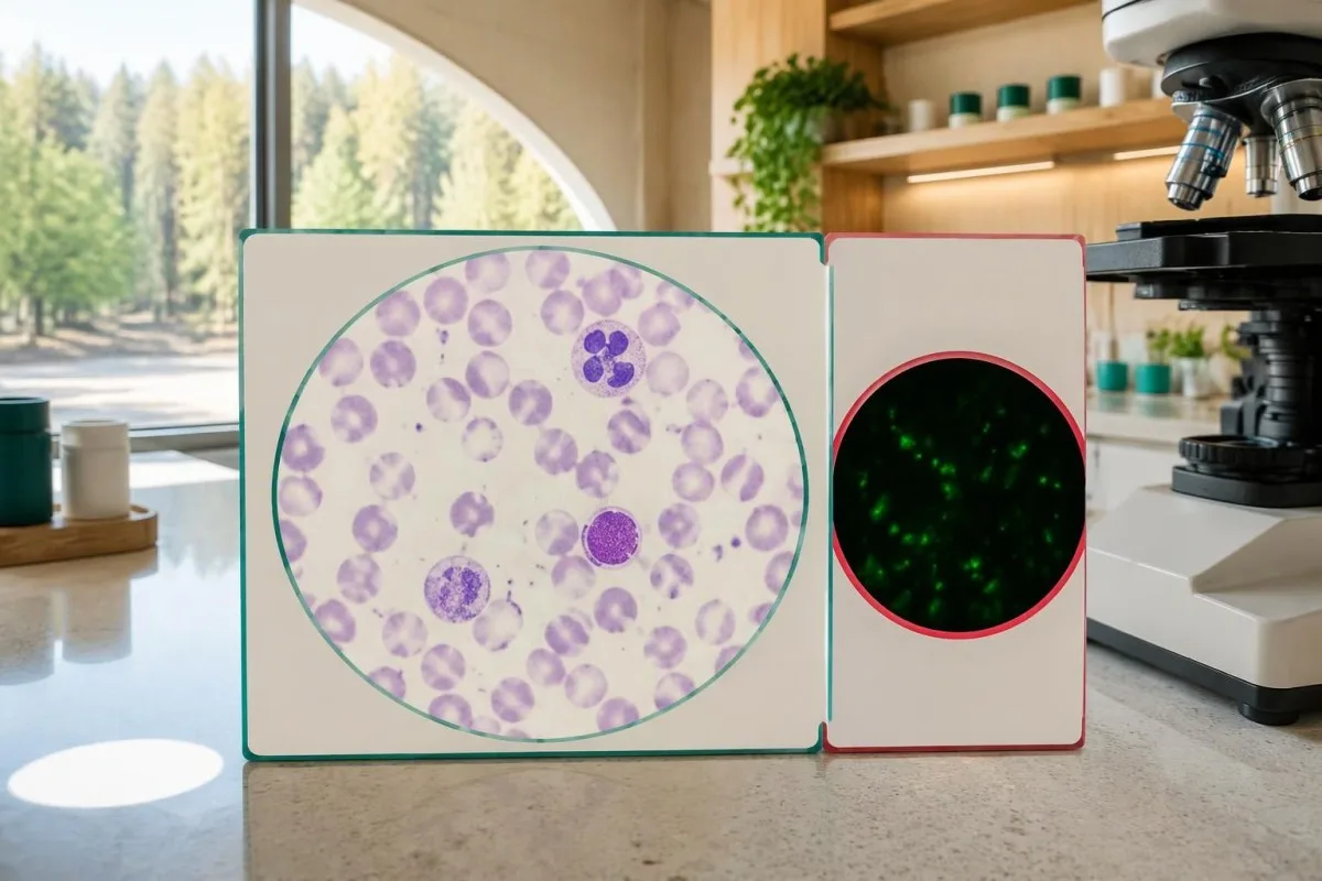
Typical adult platelet reference intervals are approximately 150–450 × 10⁹/L, and serum sodium is commonly 135–145 mmol/L, although local ranges vary. The reason clinicians consider falling platelets together with fever and low sodium is that the combination can suggest systemic illness; it does not distinguish RMSF from several infectious or noninfectious causes.
A hypothetical febrile traveler with platelets 95 × 10⁹/L, AST 82 U/L, and recent outdoor exposure deserves clinical assessment even if white cells are normal. Our การเปรียบเทียบ WBC และ CRP explains why normal inflammatory markers cannot independently clear a person with concerning symptoms.
Unexpectedly low platelets may also reflect sample-related clumping, which requires laboratory verification rather than automatic attribution to RMSF; เกล็ดเลือดจับกลุ่มจาก EDTA explains that artifact. If systemic illness is accompanied by abnormal clotting or organ dysfunction, the แนวทางการตรวจการแข็งตัวของเลือดของเรา helps explain additional tests, but a repeat specimen must not postpone urgent care.
เหตุใดจึงควรเริ่มรักษา RMSF ที่สงสัยก่อนทราบผลตรวจ?
Suspected RMSF requires prompt empiric doxycycline, because early tests can be negative and delayed treatment increases the risk of severe illness and death. Clinicians should not wait for IgG seroconversion, a PCR report, or a convalescent sample 2–10 weeks later before treating a compatible acute presentation.

Treatment timing is a safety issue, not simply a preference for faster symptom relief. Kirkland et al. reported higher mortality with treatment initiated after the first 5 days of illness in their retrospective study; its observational design limits precise causal estimates, but the finding supports the urgency emphasized in CDC guidance (Kirkland et al., 1995).
CDC recommends doxycycline 100 mg every 12 hours for adults and children weighing at least 45 kg; children under 45 kg receive 2.2 mg/kg every 12 hours, up to 100 mg per dose. These are clinician-prescribed regimens, not instructions for self-treatment; oral versus intravenous therapy depends on severity and the ability to retain medication.
Treatment generally lasts at least 5–7 days and continues until fever has resolved for at least 3 days with clinical improvement; complicated cases may need longer care. Pregnancy or suspected doxycycline allergy requires urgent individualized advice rather than automatic delay, and urgent infection assessment illustrates the same need to coordinate testing and treatment, although Listeria requires different antibiotics.
ควรแปลผลตรวจ RMSF ที่พบบ่อยอย่างไร?
RMSF test results must be interpreted using symptoms, illness dates, specimen types, and changes between samples. An early negative result may be uninformative, a single positive may reflect old exposure, and a fourfold paired IgG rise can support recent spotted fever group rickettsial infection without proving the species.

Example 1 is hypothetical: a person develops fever and severe headache after outdoor exposure, tests IgG negative on day 3, and begins clinician-directed doxycycline that day. An acute titer below 1:64 followed by a convalescent titer of 1:256 can provide supportive laboratory evidence, but the laboratory must verify the change against its starting dilution and reporting limits.
Example 2 is also hypothetical: an otherwise well person has IgG 1:256 on 2 specimens obtained several weeks apart, without a compatible intervening illness. Stable positivity does not prove new RMSF, and escalating antibiotics to make the titer disappear is not justified; Kantesti explains this as an interpretation limitation, not a declaration that every stable titer is harmless.
Example 3 involves fever, IgG 1:128, and no properly timed acute comparator, leaving recent versus previous exposure uncertain. The next step depends on clinical assessment and possible additional testing; แอนติบอดีต่อ Babesia เทียบกับการติดเชื้อ demonstrates a related distinction, but babesiosis needs its own organism-detection approach and is not treated with doxycycline alone.
เมื่อใดควรให้ความสำคัญกับอาการมากกว่าผล RMSF ที่เป็นลบ?
A negative RMSF result should not override a concerning acute illness with fever, severe headache, gastrointestinal symptoms, or rash after plausible tick exposure. Symptoms typically begin 3–12 days after an infected tick bite, and neither an unremembered bite nor the absence of an early rash reliably excludes RMSF.

The classic rash often develops 2–4 days after fever begins, so waiting for wrist, ankle, palm, or sole spots can delay care. RMSF can occur without a recognized rash, and its name is geographically misleading: clinicians assess exposure beyond the Rocky Mountain region, including travel and locally circulating ticks.
Confusion, breathing difficulty, fainting, rapidly worsening illness, or a spreading purple or pinpoint rash warrant emergency assessment—not a plan to repeat antibodies in 2 weeks. A fever and severe headache after possible tick exposure merit prompt clinical contact even if an online portal marks the first RMSF test negative.
After a tick bite without symptoms, routine antibody screening and preventive antibiotics are not recommended specifically to prevent RMSF; CDC guidance advises symptom monitoring for approximately 14 days. Tick exposure can also lead to conditions with entirely different testing logic, such as ภูมิแพ้แอลฟากัลหลังถูกเห็บกัด, which involves allergy assessment rather than RMSF IgG interpretation.
ควรตรวจแอนติบอดี RMSF ซ้ำหลังการรักษาหรือไม่?
Repeat RMSF antibodies are useful for diagnostic confirmation, not as a routine test of cure. A convalescent sample collected 2–10 weeks after the acute specimen can document an antibody rise, but persistent IgG after recovery does not by itself mean treatment failed or that more antibiotics are needed.

Fever often improves within 24–48 hours after early appropriate doxycycline treatment, although severe illness may respond more slowly. Failure to improve or clinical deterioration requires reassessment for complications, an alternative diagnosis, inadequate medication absorption, or another illness; it should not be managed by waiting for a titer to fall.
Early antibiotic treatment can sometimes delay or diminish antibody responses in rickettsial illness, so the absence of a fourfold rise is not an absolute retrospective exclusion in every treated case. Do not stop effective treatment to improve test yield; the clinician should interpret the 2 specimens alongside treatment dates, sampling quality, and the original clinical presentation.
Kantesti AI can help organize submitted results for trend review, but a trend graph cannot establish that RMSF is cured or still active. Our คู่มือไทม์ไลน์การตรวจเลือด explains why illness and medication dates matter; persistent symptoms after treatment need clinical reassessment, because lasting complications can occur without evidence of chronic ongoing RMSF infection.
ควรตรวจสอบอะไรบ้างก่อนขอให้ AI อธิบายรายงานผล RMSF?
A useful RMSF report explanation requires the assay method, exact titer, collection date, and symptom-onset date, not just the words positive or negative. Before seeking an AI explanation, identify whether 2 reports represent comparable acute and convalescent specimens; urgent symptoms still require clinical care before any upload or interpretation.

Record at least 5 details: symptom onset, sample collection, IgG versus IgM, IFA titer versus screening index, and the first antibiotic dose. Add recent travel and exposure information for the clinician; a numerical index of 2.1 from one assay cannot be mathematically converted into an IFA titer such as 1:128.
คันเตสตีเป็น เครื่องมือวิเคราะห์ผลตรวจเลือดที่ขับเคลื่อนด้วย AI that helps explain submitted laboratory information without replacing clinical diagnosis. Our คู่มือเทคโนโลยี AI describes the interpretation approach; for suspected RMSF, 1 negative early result must never become an AI-generated reassurance that postpones medical assessment.
An explanation should separate 3 categories: what the laboratory measured, what that finding can support, and what remains uncertain. Our การตรวจสอบ describe that safety boundary; useful questions include whether paired IgG is planned, whether PCR covered the suspected species, and whether treatment has already started.
งานวิจัยที่ตีพิมพ์และข้อจำกัดของหลักฐานการวินิจฉัย
RMSF diagnostic guidance and AI engineering publications answer different questions and should not be treated as interchangeable evidence. The 2016 CDC practical guide supports paired serology and immediate treatment when clinically indicated; 2 Figshare records listed below provide publication context, not proof that an AI system can diagnose or exclude RMSF.
The 1995 therapeutic-delay study and the 2016 CDC recommendations directly address RMSF care, whereas a laboratory's assay validation addresses the particular test it performs. These are 3 different evidence levels—clinical outcomes, diagnostic guidance, and analytical performance—and none makes a first-week negative antibody result a reliable reason to delay treatment.
Kantesti's 2 requested Figshare publication records concern women's health and AI-assisted hantavirus triage, respectively; neither should be cited as an RMSF diagnostic-validation study. A DOI identifies a publication record rather than guaranteeing peer review, clinical accuracy, or applicability to another disease; our มาตรฐานเนื้อหาทางการแพทย์ explain the need to distinguish directly relevant sources from contextual material.
For this topic, the practical evidence boundary is straightforward: a 4-fold paired IgG rise supports recent spotted fever group infection, while immediate care depends on clinical suspicion rather than a future titer. Our กรอบการตรวจยืนยันทางคลินิก provides broader methodology context, but suspected RMSF still requires a clinician who can examine, prescribe, and arrange appropriate confirmatory testing.
คำถามที่พบบ่อย
การตรวจไข้ด่างดำร็อกกีเมาน์เทนสามารถให้ผลเป็นลบในระยะแรกได้หรือไม่?
ใช่ การตรวจแอนติบอดีต่อไข้ด่างดำร็อกกีเมาน์เทนมักให้ผลลบในช่วง 7 วันแรกของการเจ็บป่วย เนื่องจากแอนติบอดีที่ตรวจพบได้ยังไม่พัฒนาขึ้น ดังนั้น ผลตรวจ IgG IFA ในระยะแรกที่เป็นลบจึงไม่สามารถตัดโรค RMSF ออกได้ แพทย์ควรเริ่มให้ด็อกซีไซคลินโดยเร็วเมื่ออาการเจ็บป่วยและประวัติการสัมผัสบ่งชี้ว่าอาจเป็น RMSF แทนที่จะรอการตรวจซ้ำ การเก็บตัวอย่างเลือดในระยะพักฟื้นภายหลังอาจช่วยยืนยันการวินิจฉัยได้.
ผลตรวจ RMSF IgG เป็นบวก หมายความว่าฉันกำลังติดเชื้ออยู่หรือไม่?
ผล RMSF IgG ที่เป็นบวกไม่ได้ยืนยันการติดเชื้อที่กำลังดำเนินอยู่โดยลำพัง แม้ค่าไตเตอร์จะอยู่ที่ 1:256 แอนติบอดีอาจคงอยู่เป็นเวลาหลายเดือนหรือหลายปี และอาจทำปฏิกิริยาข้ามกับริกเก็ตเซียกลุ่มไข้ผื่นจุดชนิดอื่น อาการและการสัมผัสเชื้อล่าสุด รวมถึงการเพิ่มขึ้นสี่เท่าของค่าไตเตอร์ระหว่างตัวอย่างที่เก็บเป็นคู่ซึ่งเปรียบเทียบกันได้ ทำให้มีความเป็นไปได้มากขึ้นว่าเป็นการติดเชื้อล่าสุด ผลบวกเพียงครั้งเดียวในผู้ที่ไม่มีอาการที่สอดคล้องกัน ไม่เพียงพอที่จะเป็นเหตุผลให้ใช้ยาปฏิชีวนะโดยลำพัง.
ควรเก็บตัวอย่างเลือดเพื่อตรวจแอนติบอดีต่อ RMSF ครั้งที่สองเมื่อใด?
แนวทางการวินิจฉัย RMSF ของ CDC ระบุถึงการเก็บตัวอย่าง IgG ระยะเฉียบพลันและระยะพักฟื้น โดยเว้นระยะห่างกัน 2–10 สัปดาห์ คู่มือเชิงปฏิบัติของ CDC ปี 2016 ระบุว่าควรเก็บตัวอย่างระยะพักฟื้นในช่วง 2–4 สัปดาห์หลังเก็บตัวอย่างระยะเฉียบพลัน ผู้รักษาและห้องปฏิบัติการควรเลือกระยะเวลาที่เหมาะสมและใช้วิธีตรวจที่เทียบเคียงกันได้ ห้ามรอผลตัวอย่างที่สองก่อนเริ่มการรักษา.
ระดับไตเตอร์แอนติบอดีต่อ RMSF ที่เพิ่มขึ้นเป็นสี่เท่าคืออะไร?
การเพิ่มขึ้นสี่เท่าหมายถึงค่าไตเตอร์ IgG ในภายหลังสูงกว่าค่าไตเตอร์ก่อนหน้าสี่เท่า เช่น เพิ่มจาก 1:64 เป็น 1:256 ในชุดการเจือจางแบบสองเท่าที่ใช้กันทั่วไป การเปลี่ยนแปลงนี้เท่ากับการเจือจางสองขั้น เมื่อมีอาการเจ็บป่วยที่สอดคล้องกัน การเปลี่ยนแปลงดังกล่าวสนับสนุนว่ามีการติดเชื้อริกเก็ตเซียในกลุ่มไข้ผื่นจุดเมื่อไม่นานมานี้ อย่างไรก็ตาม การตรวจทางซีรั่มวิทยาตามปกติอาจยังไม่สามารถระบุ Rickettsia rickettsii โดยเฉพาะ หรือบ่งชี้ความรุนแรงของการเจ็บป่วยได้.
ผล PCR หาเชื้อ RMSF เป็นลบ สามารถตัดโรคนี้ออกได้หรือไม่?
ผล PCR ของเลือดครบส่วนที่เป็นลบสำหรับ RMSF ไม่ได้ตัดโรคนี้ออก เนื่องจาก Rickettsia rickettsii ส่งผลต่อเซลล์บุผนังหลอดเลือดเป็นหลัก ไม่ใช่เม็ดเลือดขาวที่หมุนเวียนอยู่ ตัวอย่างที่เก็บในวันที่ 3 ของการป่วยอาจมี DNA ของแบคทีเรียไม่เพียงพอสำหรับการตรวจพบ และยาปฏิชีวนะอาจทำให้ประสิทธิภาพการตรวจลดลงอีก การตรวจเฉพาะทางจากเนื้อเยื่อผิวหนังบริเวณที่ได้รับผลกระทบอาจให้หลักฐานเพิ่มเติมได้เมื่อเหมาะสม แพทย์ไม่ควรชะลอการรักษาระหว่างเก็บตัวอย่างหรือรอผล PCR.
เด็กควรได้รับด็อกซีไซคลินก่อนที่ผลตรวจ RMSF จะออกหรือไม่?
Children of all ages should receive clinician-prescribed doxycycline promptly when RMSF is suspected, without waiting for laboratory confirmation. CDC guidance recommends 2.2 mg/kg every 12 hours for children under 45 kg, with a maximum of 100 mg per dose. Short courses used for rickettsial disease have not been shown to cause the permanent tooth staining associated with older tetracycline concerns. The child's clinician determines route, duration, and whether hospital care is needed.
ทำไมผลตรวจแอนติบอดี IgG ต่อโรคไข้ด่างดำร็อกกีเมาน์เทนของฉันยังคงเป็นบวกหลังการรักษา?
IgG ต่อเชื้อ RMSF อาจยังให้ผลบวกเป็นเวลาหลายเดือน และบางครั้งนานถึง 4 ปีหลังเจ็บป่วย ดังนั้นการมีผลบวกต่อเนื่องจึงไม่ได้บ่งชี้ถึงความล้มเหลวในการรักษาเพียงอย่างเดียว การตรวจแอนติบอดีไม่ใช่การตรวจยืนยันการหายเป็นปกติที่ทำเป็นประจำ ไม่ควรสั่งยาปฏิชีวนะเพิ่มเติมเพียงเพื่อทำให้ค่าไตเตอร์เป็นลบ อาการที่ยังคงอยู่หรือแย่ลงจำเป็นต้องได้รับการประเมินทางคลินิกซ้ำเพื่อค้นหาภาวะแทรกซ้อนหรือการวินิจฉัยอื่น แทนการแปลผลจากตัวเลขแอนติบอดีเพียงอย่างเดียว.
อ่านผลตรวจเลือดอย่างไร
อัปโหลดรายงานผลตรวจของคุณ แล้ว AI ของ Kantesti จะอธิบายตัวบ่งชี้ทางชีวภาพแต่ละรายการด้วยภาษาที่เข้าใจง่ายภายในไม่กี่วินาที มีผู้ใช้ทั่วโลกมากกว่า 2 ล้านคน.
เอกสารอ้างอิง
- ไคลน์, ที., มิตเชลล์, เอส., & เวเบอร์, เอช. (2026). คู่มือสุขภาพสตรี: การตกไข่ วัยหมดประจำเดือน และอาการที่เกี่ยวกับฮอร์โมน. Kantesti AI การวิจัยทางการแพทย์. โดอิ
- ไคลน์, ที., มิตเชลล์, เอส., & เวเบอร์, เอช. (2026). ระบบสนับสนุนการตัดสินใจทางคลินิกที่ใช้ AI หลายภาษาเพื่อคัดกรองการติดเชื้อฮันตาไวรัสในระยะเริ่มต้น: การออกแบบ การตรวจสอบความถูกต้องทางวิศวกรรม และการนำไปใช้จริงกับรายงานผลตรวจเลือดที่แปลผลแล้ว 50,000 ฉบับ. Kantesti AI การวิจัยทางการแพทย์. โดอิ
- Biggs HM et al. (2016). Diagnosis and Management of Tickborne Rickettsial Diseases: Rocky Mountain Spotted Fever and Other Spotted Fever Group Rickettsioses, Ehrlichioses, and Anaplasmosis — United States. MMWR Recommendations and Reports. โดอิ
- Kirkland KB et al. (1995). Therapeutic delay and mortality in cases of Rocky Mountain spotted fever. Clinical Infectious Diseases.
- ศูนย์ควบคุมและป้องกันโรค (ไม่ปรากฏปีที่เผยแพร่). Clinical and Laboratory Diagnosis for Rocky Mountain Spotted Fever. แนวทางทางคลินิกของ CDC.
- 2 ล้าน+การตรวจที่วิเคราะห์
- 127+ประเทศ
- 100+ภาษา
เกี่ยวกับทีมแพทย์


สัญญาณความน่าเชื่อถือ E-E-A-T
ประสบการณ์
การทบทวนเชิงคลินิกโดยแพทย์ที่นำโดยกระบวนการตีความผลตรวจในห้องแล็บ.
ความเชี่ยวชาญ
โฟกัสด้านเวชศาสตร์ห้องปฏิบัติการเกี่ยวกับพฤติกรรมของไบโอมาร์กเกอร์ในบริบททางคลินิก.
อำนาจ
ทบทวนทางการแพทย์โดย นพ. Thomas Klein พร้อมผลงานจาก นพ. Sarah Mitchell และศาสตราจารย์ ดร. Hans Weber.
ความน่าเชื่อถือ
การตีความที่อิงหลักฐาน พร้อมเส้นทางการติดตามที่ชัดเจนเพื่อลดความตื่นตระหนก.





ข้อจำกัดความรับผิดชอบทางการแพทย์
บทความนี้จัดทำเพื่อวัตถุประสงค์ด้านการศึกษาเท่านั้น และไม่ถือเป็นคำแนะนำทางการแพทย์ โปรดปรึกษาผู้ให้บริการด้านสุขภาพที่มีคุณสมบัติเหมาะสมเสมอสำหรับการตัดสินใจด้านการวินิจฉัยและการรักษา.
เผยแพร่แล้ว · ติดต่อทีมบรรณาธิการ
เผยแพร่โดย Kantesti Ltd จดทะเบียนในอังกฤษและเวลส์ เลขทะเบียนบริษัท. 17090423, ลอนดอน สหราชอาณาจักร.
