
Testi për ethet me njolla të Maleve Shkëmbore mund të dalë negativ gjatë javës së parë, sepse antitrupat ende nuk janë zhvilluar. Një titër i vetëm pozitiv i IgG nuk mund të vërtetojë infeksionin akut; mostrat e çiftuara që tregojnë një rritje katërfish mbështesin infeksionin e kohëve të fundit. Klinikët duhet ta fillojnë menjëherë trajtimin kur dyshohet për RMSF—pa pritur konfirmimin laboratorik.

Përfundimet kryesore
- Rezultate të rreme negative të hershme: Testet për antitrupat ndaj RMSF janë shpesh negative gjatë 7 ditëve të para të sëmundjes, kur vendimet për trajtimin janë më urgjente.
- Metoda e preferuar për antitrupat: Testimi indirekt i antitrupave fluoreshentë për IgG, ose IFA, krahason titrat e antitrupave kundrejt antigjeneve riketsiale të grupit të etheve me njolla.
- Mostra të çiftuara: Merrni një mostër në fazën akute herët dhe një mostër në fazën e konvaleshencës më vonë; udhëzimet e CDC-së përshkruajnë një interval prej 2–10 javësh ndërmjet mostrave.
- Rritje katërfish: Një ndryshim nga 1:64 në 1:256 përfaqëson një rritje katërfish dhe mbështet infeksionin e kohëve të fundit nga riketsiet e grupit të etheve me njolla në rastin e një sëmundjeje përputhëse.
- IgG e vetme pozitive: Antitrupat mund të qëndrojnë për vite; edhe një titër prej 1:256 nuk mund të përcaktojë i vetëm se kur ka ndodhur ekspozimi ose të identifikojë specien e rikecieve.
- PCR negative: PCR-ja në gjakun e plotë mund të mos e zbulojë RMSF-në, sepse Rickettsia rickettsii prek kryesisht qelizat endoteliale vaskulare, jo leukocitet qarkulluese.
- Koha e trajtimit: Doksiciklina është trajtimi i linjës së parë për RMSF-në e dyshuar te të rriturit dhe fëmijët e të gjitha moshave; mjekët nuk duhet të presin rezultatet e analizave.
- Skema e zakonshme për të rriturit: Udhëzimet e CDC-së rekomandojnë doksiciklinë 100 mg çdo 12 orë, përgjithësisht për të paktën 5–7 ditë dhe derisa ethet të jenë qetësuar për të paktën 3 ditë dhe gjendja klinike të jetë përmirësuar.
Cili test për ethet me njolla të Maleve Shkëmbore kryhet i pari?
Qasja standarde me antitrupa për një diagnozë të RMSF-së është testimi i çiftuar i IgG-së me anë të analizës indirekte me antitrupa fluoreshentë, që zakonisht quhet IFA. Megjithatë, mjeku mund ta fillojë doksiciklinën para se të dalë rezultati i parë, sepse një mostër antitrupash e marrë gjatë ditëve 1–7 shpesh nuk mund ta konfirmojë ose përjashtojë sëmundjen.

RMSF shkaktohet nga Rickettsia rickettsii, një bakter i transmetuar nga rriqrat që mund të dëmtojë shpejt enët e vogla të gjakut. Udhëzuesi praktik i CDC-së dallon 2 detyra: trajtimin e menjëhershëm të një sëmundjeje potencialisht të rrezikshme dhe marrjen më vonë të provave laboratorike për konfirmim; ngatërrimi i këtyre detyrave shkakton vonesa të shmangshme (Biggs et al., 2016).
Një kërkesë e përshkruar si panel për RMSF mund të përfshijë IgG, IgM ose të dyja, dhe një rezultat depistues mund të pasohet nga një titër IFA. Pyetni se cila metodë e dha secilin rezultat, sepse 1 rezultat depistues pozitiv dhe 1 titër sasior nuk janë të këmbyeshëm; udhëzuesi ynë për rezultateve pozitive të antitrupave e shpjegon këtë dallim.
Kantesti është një Analizues i testeve të gjakut AI është hartuar për të shpjeguar raportet laboratorike, jo për të përjashtuar RMSF-në mbi bazën e 1 rezultati negativ. Udhëzuesi ynë sfondin e kompanisë identifikon organizatën që qëndron pas këtij shërbimi; ekzaminimi, vlerësimi i ekspozimit, kërkesa për analiza dhe vendimet për antibiotikët mbeten përgjegjësi e mjekut kurues.
Pse rezulton negativ një test i hershëm për antitrupat ndaj RMSF?
Një Testi fals negativ për RMSF shpesh pasqyron periudhën dritare të antitrupave: përgjigjja imunitare ende nuk ka prodhuar mjaftueshëm antitrupa të zbulueshëm. IFA-ja për IgG është shpesh negative gjatë 7 ditëve të para të simptomave, ndaj një raport i hershëm negativ nuk e justifikon mosdhënien e trajtimit kur pamja klinike sugjeron RMSF.

Ora e testimit fillon me fillimi i simptomave, jo dita kur laboratori e merr mostrën. Për shembull, një person hipotetik i testuar në ditën e 3-të të sëmundjes mund të ketë vërtet RMSF pavarësisht IgG-së negative; i njëjti rezultat negativ disa javë më vonë do të interpretohej ndryshe, veçanërisht nëse është kryer testimi i çiftuar.
Prodhimi i antitrupave nuk ndodh në të njëjtën ditë të saktë te çdo person. Udhëzimet e CDC-së për diagnozën klinike dhe laboratorike shpjegojnë se serologjia gjatë javës së parë shpesh është negative; performanca e lartë e një laboratori me mostra të konfirmuara si pozitive për antitrupa nuk nënkupton ndjeshmëri të njëjtë gjatë ditëve 1–7.
Përsëritja e testit të IgG-së vetëm 24–48 orë pas një rezultati të hershëm negativ mund të sjellë pak dobi, sepse ky interval zakonisht është tepër i shkurtër për të treguar ndryshimin e pritshëm të antitrupave. Të Monospot testing window illustrates a similar timing principle, although its assay, illness, and recommended follow-up schedule differ from RMSF.
Çfarë përfshin në të vërtetë kuptimi i IgG pozitive për RMSF?
RMSF IgG positive meaning includes previous exposure, recent infection, or antibodies reacting with another spotted fever group rickettsia. A single positive titer—even 1:256 or higher—cannot independently establish acute RMSF, because antibody persistence and cross-reactivity prevent the result from acting as a reliable date stamp.

Antibodies against R. rickettsii can remain elevated for months and sometimes up to 4 years after illness, according to CDC diagnostic guidance. Persistent positivity does not mean bacteria remain active, and the height of a single titer does not reliably distinguish a new infection from an older immune response.
Background antibodies also affect interpretation: CDC notes that 10% or more of healthy people in some areas may have elevated titers from past exposure to spotted fever group rickettsiae. That figure is geographically dependent, not a universal prevalence estimate; testing someone without compatible symptoms increases the chance of finding a clinically misleading positive.
Consider a hypothetical asymptomatic adult with IgG 1:128 on an incidental panel and no recent febrile illness. That 1 result is not a reason by itself to prescribe doxycycline; antibody persistence after exposure is another example of why serology must be matched to the organism, symptoms, and sampling dates.
A mund të shkaktojnë reaktiviteti i kryqëzuar ose IgM një rezultat çorientues?
Reaktiviteti i kryqëzuar can produce positive RMSF serology after exposure to another spotted fever group rickettsia, and antitrupat IgM is less specific than IgG for diagnosing recent illness. A positive IgM result on day 5 is therefore not a dependable shortcut around an early negative IgG result.
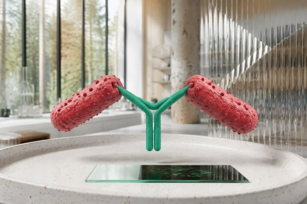
R. rickettsii, R. parkeri, and related organisms share antigenic features that routine commercial serology often cannot distinguish. A 4-fold IgG rise can establish recent spotted fever group rickettsial infection in a compatible illness, but identifying R. rickettsii specifically may require species-specific molecular testing or specialized reference-laboratory investigation.
IgM may appear without convincing evidence of recent rickettsial illness and should not be used as a stand-alone diagnostic marker. The CDC practical guide favors paired IgG IFA for confirmation rather than interpreting 1 isolated IgM positive as proof of acute RMSF (Biggs et al., 2016).
Not all antibody families behave alike: Q fever antibody patterns use phase-specific interpretation that should not be imported into RMSF. For a report showing IgM positive and IgG negative on day 4, ask about clinical suspicion, assay method, and the planned convalescent specimen rather than assuming the sequence proves infection.
Si e mbështesin diagnozën mostrat e çiftuara dhe një rritje katërfish?
Paired IgG IFA samples support diagnosis by showing whether antibody titers rise substantially between acute illness and recovery. A rise from 1:64 to 1:256 is fourfold; with compatible symptoms, it supports recent spotted fever group rickettsial infection rather than merely documenting an antibody that was already present.
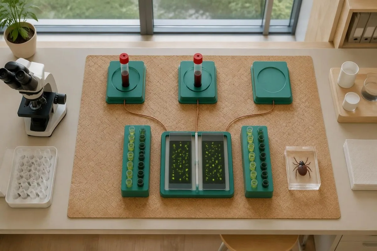
IFA titers describe the highest dilution at which antibody remains detectable; 1:256 means detection after greater dilution than 1:64. In a common 2-fold dilution series, 1:64 to 1:128 is one step, while 1:64 to 1:256 is two steps—the fourfold change used to reduce ambiguity from small fluctuations.
CDC RMSF guidance describes paired specimens collected 2–10 weeks apart, while the 2016 practical guide emphasizes an acute specimen and a convalescent specimen ideally 2–4 weeks later. These recommendations are not competing treatment deadlines; the clinician and laboratory should agree on follow-up timing that can capture the antibody response.
Use the same laboratory and assay when possible, and retain the original reports rather than comparing 2 abbreviated portal flags. Our explanations of rezultateve sasiore kundrejt cilësore dhe ndryshimet midis vizitave në laborator help distinguish a meaningful paired titer rise from unrelated reporting differences.
| Niveli | Intervali | Çfarë do të thotë |
|---|---|---|
| Early negative | Below laboratory cutoff on days 1–7 | Does not exclude RMSF; antibodies may not yet be detectable. |
| Single positive | Example: 1:128 on one specimen | Shows detectable antibody, not the date or species of infection. |
| Twofold rise | Example: 1:64 → 1:128 | Does not meet the standard fourfold-rise criterion. |
| Rritje katër herë | Shembull: 1:64 → 1:256 | Supports recent spotted fever group infection with compatible illness; does not measure severity. |
A mund ta zbulojë PCR RMSF para se të shfaqen antitrupat?
PCR-ja can detect rickettsial DNA before antibodies develop, but a negative whole-blood PCR does not exclude RMSF. R. rickettsii mainly targets vascular endothelial cells, so an illness-day-3 blood specimen may contain too little bacterial DNA for detection even when the disease is clinically significant.
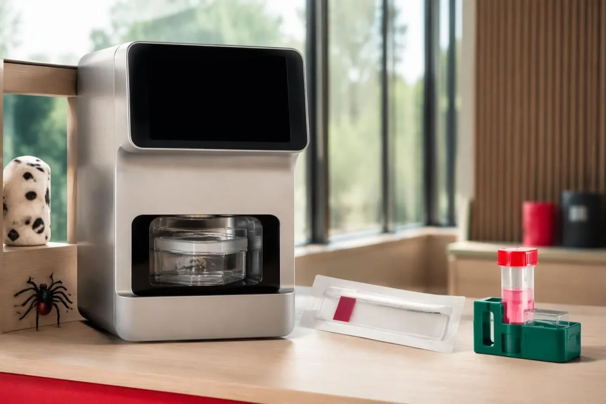
Unlike serology, PCR looks for the organism's genetic material rather than the host's immune response. Its usefulness depends on 3 practical variables: the specimen collected, illness severity and timing, and whether antibiotics have already reduced detectable organisms; there is no single sensitivity percentage applicable to every RMSF PCR order.
A clinician may arrange PCR or immunohistochemical testing of a rash skin biopsy through an appropriate laboratory, because affected tissue can provide evidence unavailable in circulating blood. Even when tissue testing is considered, collecting a specimen before the first doxycycline dose is desirable only if it does not delay urgent treatment.
Ask whether a panel actually includes R. rickettsii and whether it reports species-specific results or a broader rickettsial group. The PCR timing explanation for whooping cough demonstrates why molecular tests have specimen and treatment windows, but its respiratory sampling rules do not apply to RMSF.
Pse testet për RMSF dhe ehrlichiozën sillen ndryshe?
RMSF and ehrlichiosis require different diagnostic reasoning even when the same tick-exposure evaluation includes both. Whole-blood PCR is generally more useful early in ehrlichiosis because Ehrlichia organisms infect white cells, whereas R. rickettsii primarily affects vessel-lining cells; 1 negative tick-borne panel is not a universal exclusion test.

Fever, headache, muscle aches, and abnormal liver enzymes can occur in more than 1 tick-borne illness. Exposure location, platelet count, white-cell count, rash, and illness timing guide test selection; clinicians should not assume that an order called a tick panel covers every relevant organism or uses the best specimen for each.
I/E/Të/Të ehrlichiosis PCR and titer guide explains why acute PCR and later antibodies may disagree in that illness. For a hypothetical day-4 presentation, an Ehrlichia PCR positive and RMSF IgG negative need interpretation as separate findings—not evidence that all other tick-transmitted illnesses were conclusively excluded.
Kantesti është një platformë e interpretimit të analizave të gjakut AI that explains submitted laboratory reports, not a substitute for organism-specific diagnostic assessment. If a report contains 2 negative results from different methods, we emphasize the distinction between the method's target and the clinician's question rather than treating the word negative as a complete answer.
Cilat rezultate rutinë të analizave të gjakut mund të mbështesin dyshimin për RMSF?
Low platelets, mildly elevated AST or ALT, and low sodium can support suspicion of RMSF, but none confirms the diagnosis and early values may be normal. A platelet count of 110 × 10⁹/L and sodium of 130 mmol/L are contextual clues—not organism-specific thresholds or prerequisites for starting treatment.
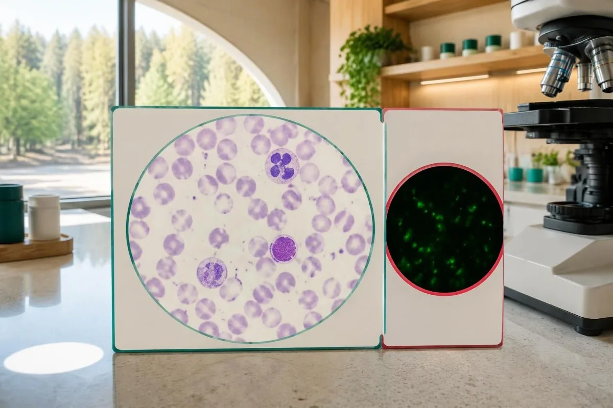
Typical adult platelet reference intervals are approximately 150–450 × 10⁹/L, and serum sodium is commonly 135–145 mmol/L, although local ranges vary. The reason clinicians consider falling platelets together with fever and low sodium is that the combination can suggest systemic illness; it does not distinguish RMSF from several infectious or noninfectious causes.
A hypothetical febrile traveler with platelets 95 × 10⁹/L, AST 82 U/L, and recent outdoor exposure deserves clinical assessment even if white cells are normal. Our krahasimi WBC dhe CRP explains why normal inflammatory markers cannot independently clear a person with concerning symptoms.
Unexpectedly low platelets may also reflect sample-related clumping, which requires laboratory verification rather than automatic attribution to RMSF; 38 x10^9/L explains that artifact. If systemic illness is accompanied by abnormal clotting or organ dysfunction, the udhëzuesi ynë për testet e koagulimit helps explain additional tests, but a repeat specimen must not postpone urgent care.
Pse duhet të fillojë trajtimi për RMSF të dyshuar përpara se të dalin rezultatet?
Suspected RMSF requires prompt empiric doxycycline, because early tests can be negative and delayed treatment increases the risk of severe illness and death. Clinicians should not wait for IgG seroconversion, a PCR report, or a convalescent sample 2–10 weeks later before treating a compatible acute presentation.

Treatment timing is a safety issue, not simply a preference for faster symptom relief. Kirkland et al. reported higher mortality with treatment initiated after the first 5 days of illness in their retrospective study; its observational design limits precise causal estimates, but the finding supports the urgency emphasized in CDC guidance (Kirkland et al., 1995).
CDC recommends doxycycline 100 mg every 12 hours for adults and children weighing at least 45 kg; children under 45 kg receive 2.2 mg/kg every 12 hours, up to 100 mg per dose. These are clinician-prescribed regimens, not instructions for self-treatment; oral versus intravenous therapy depends on severity and the ability to retain medication.
Treatment generally lasts at least 5–7 days and continues until fever has resolved for at least 3 days with clinical improvement; complicated cases may need longer care. Pregnancy or suspected doxycycline allergy requires urgent individualized advice rather than automatic delay, and urgent infection assessment illustrates the same need to coordinate testing and treatment, although Listeria requires different antibiotics.
Si duhen interpretuar rezultatet e zakonshme të testeve për RMSF?
RMSF test results must be interpreted using symptoms, illness dates, specimen types, and changes between samples. An early negative result may be uninformative, a single positive may reflect old exposure, and a fourfold paired IgG rise can support recent spotted fever group rickettsial infection without proving the species.

Example 1 is hypothetical: a person develops fever and severe headache after outdoor exposure, tests IgG negative on day 3, and begins clinician-directed doxycycline that day. An acute titer below 1:64 followed by a convalescent titer of 1:256 can provide supportive laboratory evidence, but the laboratory must verify the change against its starting dilution and reporting limits.
Example 2 is also hypothetical: an otherwise well person has IgG 1:256 on 2 specimens obtained several weeks apart, without a compatible intervening illness. Stable positivity does not prove new RMSF, and escalating antibiotics to make the titer disappear is not justified; Kantesti explains this as an interpretation limitation, not a declaration that every stable titer is harmless.
Example 3 involves fever, IgG 1:128, and no properly timed acute comparator, leaving recent versus previous exposure uncertain. The next step depends on clinical assessment and possible additional testing; antitrupat ndaj Babesia-s kundrejt infeksionit demonstrates a related distinction, but babesiosis needs its own organism-detection approach and is not treated with doxycycline alone.
Kur duhet që simptomat të kenë përparësi ndaj një rezultati negativ për RMSF?
A negative RMSF result should not override a concerning acute illness with fever, severe headache, gastrointestinal symptoms, or rash after plausible tick exposure. Symptoms typically begin 3–12 days after an infected tick bite, and neither an unremembered bite nor the absence of an early rash reliably excludes RMSF.

The classic rash often develops 2–4 days after fever begins, so waiting for wrist, ankle, palm, or sole spots can delay care. RMSF can occur without a recognized rash, and its name is geographically misleading: clinicians assess exposure beyond the Rocky Mountain region, including travel and locally circulating ticks.
Confusion, breathing difficulty, fainting, rapidly worsening illness, or a spreading purple or pinpoint rash warrant emergency assessment—not a plan to repeat antibodies in 2 weeks. A fever and severe headache after possible tick exposure merit prompt clinical contact even if an online portal marks the first RMSF test negative.
After a tick bite without symptoms, routine antibody screening and preventive antibiotics are not recommended specifically to prevent RMSF; CDC guidance advises symptom monitoring for approximately 14 days. Tick exposure can also lead to conditions with entirely different testing logic, such as alergji ndaj alfa-gal pas pickimit nga rriqrat, which involves allergy assessment rather than RMSF IgG interpretation.
A duhen ritestuar antitrupat ndaj RMSF pas trajtimit?
Repeat RMSF antibodies are useful for diagnostic confirmation, not as a routine test of cure. A convalescent sample collected 2–10 weeks after the acute specimen can document an antibody rise, but persistent IgG after recovery does not by itself mean treatment failed or that more antibiotics are needed.

Fever often improves within 24–48 hours after early appropriate doxycycline treatment, although severe illness may respond more slowly. Failure to improve or clinical deterioration requires reassessment for complications, an alternative diagnosis, inadequate medication absorption, or another illness; it should not be managed by waiting for a titer to fall.
Early antibiotic treatment can sometimes delay or diminish antibody responses in rickettsial illness, so the absence of a fourfold rise is not an absolute retrospective exclusion in every treated case. Do not stop effective treatment to improve test yield; the clinician should interpret the 2 specimens alongside treatment dates, sampling quality, and the original clinical presentation.
Kantesti AI can help organize submitted results for trend review, but a trend graph cannot establish that RMSF is cured or still active. Our udhëzuesi i afateve për analizat e gjakut explains why illness and medication dates matter; persistent symptoms after treatment need clinical reassessment, because lasting complications can occur without evidence of chronic ongoing RMSF infection.
Çfarë duhet të kontrolloni përpara se t’i kërkoni AI-së të shpjegojë një raport për RMSF?
A useful RMSF report explanation requires the assay method, exact titer, collection date, and symptom-onset date, not just the words positive or negative. Before seeking an AI explanation, identify whether 2 reports represent comparable acute and convalescent specimens; urgent symptoms still require clinical care before any upload or interpretation.

Record at least 5 details: symptom onset, sample collection, IgG versus IgM, IFA titer versus screening index, and the first antibiotic dose. Add recent travel and exposure information for the clinician; a numerical index of 2.1 from one assay cannot be mathematically converted into an IFA titer such as 1:128.
Kantesti është një Mjet për analizën e testeve të gjakut me AI that helps explain submitted laboratory information without replacing clinical diagnosis. Our Udhëzues teknologjik i AI-së describes the interpretation approach; for suspected RMSF, 1 negative early result must never become an AI-generated reassurance that postpones medical assessment.
An explanation should separate 3 categories: what the laboratory measured, what that finding can support, and what remains uncertain. Our kontrollet tona të burimeve të raporteve mjekësore describe that safety boundary; useful questions include whether paired IgG is planned, whether PCR covered the suspected species, and whether treatment has already started.
Publikimet kërkimore dhe kufizimet e provave diagnostikuese
RMSF diagnostic guidance and AI engineering publications answer different questions and should not be treated as interchangeable evidence. The 2016 CDC practical guide supports paired serology and immediate treatment when clinically indicated; 2 Figshare records listed below provide publication context, not proof that an AI system can diagnose or exclude RMSF.
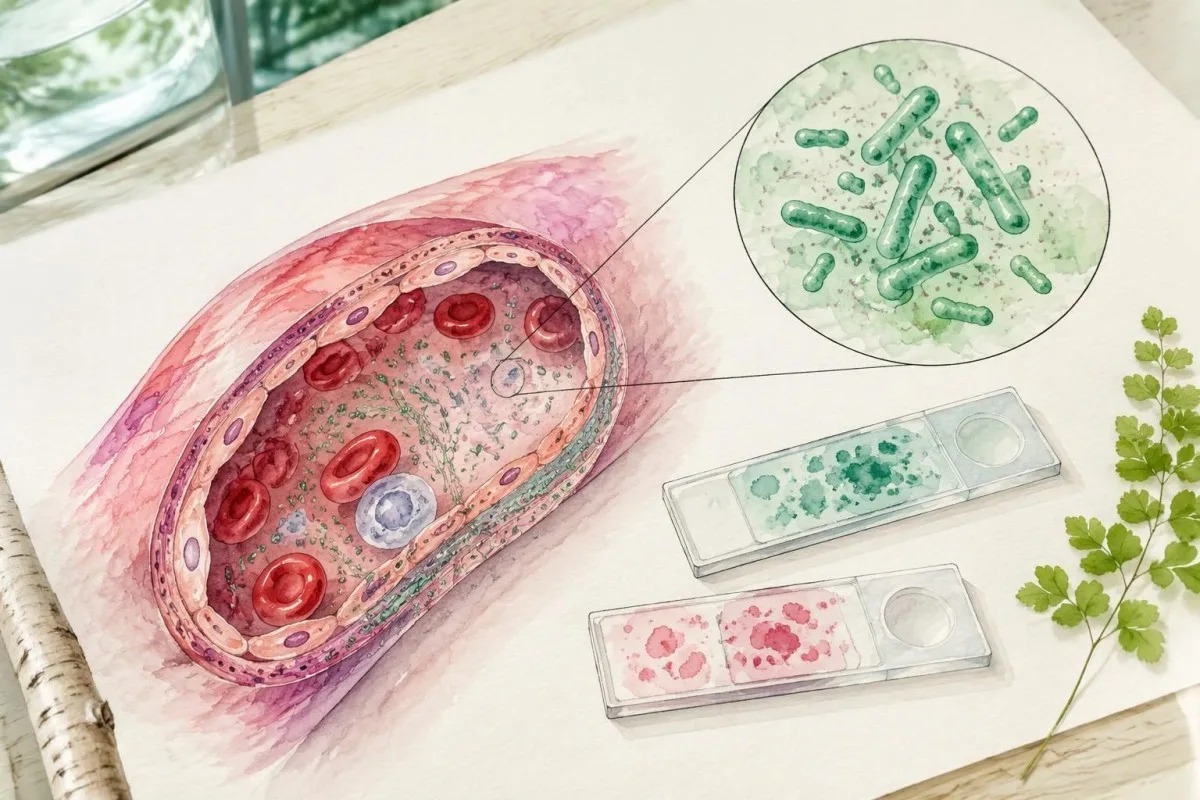
The 1995 therapeutic-delay study and the 2016 CDC recommendations directly address RMSF care, whereas a laboratory's assay validation addresses the particular test it performs. These are 3 different evidence levels—clinical outcomes, diagnostic guidance, and analytical performance—and none makes a first-week negative antibody result a reliable reason to delay treatment.
Kantesti's 2 requested Figshare publication records concern women's health and AI-assisted hantavirus triage, respectively; neither should be cited as an RMSF diagnostic-validation study. A DOI identifies a publication record rather than guaranteeing peer review, clinical accuracy, or applicability to another disease; our standardet e përmbajtjes mjekësore explain the need to distinguish directly relevant sources from contextual material.
For this topic, the practical evidence boundary is straightforward: a 4-fold paired IgG rise supports recent spotted fever group infection, while immediate care depends on clinical suspicion rather than a future titer. Our kornizë validimi klinik provides broader methodology context, but suspected RMSF still requires a clinician who can examine, prescribe, and arrange appropriate confirmatory testing.
Pyetjet e bëra shpesh
A mund të dalë negativ herët testi për ethet me njolla të Maleve Shkëmbore?
Po, testi i antitrupave kundër etheve me njolla të Malit Shkëmbor shpesh del negativ gjatë 7 ditëve të para të sëmundjes, sepse antitrupat e dallueshëm ende nuk janë zhvilluar. Prandaj, një rezultat negativ i hershëm i IgG IFA nuk e përjashton RMSF. Klinikët duhet të fillojnë menjëherë doksiciklinën kur sëmundja dhe historia e ekspozimit sugjerojnë RMSF, në vend që të presin përsëritjen e testimit. Një mostër e mëvonshme gjatë konvaleshencës mund të ndihmojë në përcaktimin e diagnozës.
A do të thotë një rezultat pozitiv i IgG për RMSF se kam një infeksion aktiv?
Një rezultat pozitiv për IgG ndaj RMSF-së nuk provon në mënyrë të pavarur një infeksion aktiv, edhe nëse titri është 1:256. Antitrupat mund të qëndrojnë për muaj ose vite dhe mund të reagojnë me rikecitë të tjera të grupit të etheve me njolla. Simptomat e fundit, ekspozimi dhe një rritje katërfish midis mostrave të çiftuara të krahasueshme e bëjnë më të mundshëm një infeksion të kohëve të fundit. Një rezultat i izoluar pozitiv te dikush pa simptoma të përputhshme nuk justifikon vetvetiu përdorimin e antibiotikëve.
Kur duhet të merret mostra e dytë për antitrupat ndaj RMSF-së?
Udhëzimet diagnostikuese të CDC-së për RMSF përshkruajnë mostra akute dhe konvaleshente të IgG-së, të marra me 2–10 javë diferencë. Udhëzuesi praktik i CDC-së i vitit 2016 përshkruan një mostër konvaleshente që, idealisht, merret 2–4 javë pas mostrës akute. Mjeku përgjegjës dhe laboratori duhet të zgjedhin intervalin e përshtatshëm dhe të përdorin analiza të krahasueshme. Trajtimi nuk duhet të presë marrjen e mostrës së dytë.
Çfarë është një rritje katërfish e titrit të antitrupave kundër etheve me njolla të Maleve Shkëmbore (RMSF)?
Një rritje katërfish do të thotë se titri i mëvonshëm i IgG-së është katër herë më i lartë se titri i mëparshëm, si p.sh. rritja nga 1:64 në 1:256. Në një seri të zakonshme hollimesh dyfish, kjo përfaqëson dy hapa hollimi. Në prani të një sëmundjeje klinikisht të pajtueshme, ky ndryshim mbështet një infeksion të kohëve të fundit nga rikeciet e grupit të etheve me njolla. Serologjia rutinë mund të mos jetë ende në gjendje të identifikojë në mënyrë specifike Rickettsia rickettsii ose të përcaktojë rëndësinë klinike të sëmundjes.
A e përjashton sëmundjen një PCR negative për ethet me njolla të Malit Shkëmbor (RMSF)?
Një PCR negative për RMSF në gjakun e plotë nuk e përjashton sëmundjen, sepse Rickettsia rickettsii prek kryesisht qelizat që veshin enët e gjakut dhe jo leukocitet qarkulluese. Një mostër e marrë në ditën e 3-të të sëmundjes mund të përmbajë sasi të pamjaftueshme të ADN-së bakteriale për t’u zbuluar, dhe antibiotikët mund ta ulin më tej rendimentin e testit. Testimi i specializuar i indit të prekur të lëkurës mund të ofrojë prova shtesë kur është e përshtatshme. Mjekët nuk duhet ta vonojnë trajtimin ndërsa marrin ose presin rezultatet e PCR-së.
A duhet t’u jepet fëmijëve doksiciklinë përpara se të dalin rezultatet për RMSF?
Children of all ages should receive clinician-prescribed doxycycline promptly when RMSF is suspected, without waiting for laboratory confirmation. CDC guidance recommends 2.2 mg/kg every 12 hours for children under 45 kg, with a maximum of 100 mg per dose. Short courses used for rickettsial disease have not been shown to cause the permanent tooth staining associated with older tetracycline concerns. The child's clinician determines route, duration, and whether hospital care is needed.
Pse IgG-ja ime për ethet me njolla të Maleve Shkëmbore është ende pozitive pas trajtimit?
IgG-ja ndaj etheve të njollosura të Maleve Shkëmbore (RMSF) mund të mbetet pozitive për muaj dhe ndonjëherë deri në 4 vjet pas sëmundjes, ndaj pozitiviteti i vazhdueshëm nuk tregon vetvetiu dështim të trajtimit. Testimi i antitrupave nuk është një test rutinë për të konfirmuar shërimin. Antibiotikët shtesë nuk duhet të përshkruhen vetëm për ta bërë titrin negativ. Simptomat e vazhdueshme ose që përkeqësohen kërkojnë rivlerësim klinik për komplikime ose një diagnozë tjetër, në vend që të interpretohen vetëm nga niveli i antitrupave.
Merrni shpjegimin e rezultateve të analizave të gjakut
Ngarkoni raportin tuaj laboratorik dhe AI e Kantesti shpjegon çdo biomarker me gjuhë të thjeshtë brenda pak sekondash. Përdoret nga më shumë se 2 milionë njerëz në mbarë botën.
Referencat
- Klein, T., Mitchell, S., & Weber, H. (2026). Udhëzuesi për shëndetin e grave: ovulacioni, menopauza dhe simptomat hormonale. Kërkim mjekësor me Kantesti AI. DOI
- Klein, T., Mitchell, S., & Weber, H. (2026). Mbështetje shumëgjuhëshe e asistuar nga IA-ja për vendimmarrjen klinike në triazhimin e hershëm të hantavirusit: projektimi, vërtetimi inxhinierik dhe zbatimi në botën reale në 50,000 raporte të interpretuara të analizave të gjakut. Kërkim mjekësor me Kantesti AI. DOI
- Biggs HM et al. (2016). Diagnosis and Management of Tickborne Rickettsial Diseases: Rocky Mountain Spotted Fever and Other Spotted Fever Group Rickettsioses, Ehrlichioses, and Anaplasmosis — United States. Rekomandimet dhe raportet e MMWR. DOI
- Kirkland KB et al. (1995). Therapeutic delay and mortality in cases of Rocky Mountain spotted fever. Sëmundjet Infektive Klinike.
- Qendrat për Kontrollin dhe Parandalimin e Sëmundjeve (n.d.). Clinical and Laboratory Diagnosis for Rocky Mountain Spotted Fever. Udhëzime klinike të CDC-së.
- 2M+Analizat e kryera
- 127+Vendet
- 100+Gjuhët
Rreth ekipit mjekësor


Sinjalet e Besimit E-E-A-T
Përvoja
Rishikim klinik i udhëhequr nga mjeku i flukseve të punës për interpretimin e analizave.
Ekspertizë
Fokus i mjekësisë laboratorike mbi mënyrën se si sillen biomarkerët në kontekstin klinik.
Autoritariteti
Rishikuar nga ana mjekësore nga Dr. Thomas Klein, me kontribute nga Dr. Sarah Mitchell dhe Prof. Dr. Hans Weber.
Besueshmëria
Interpretim i bazuar në prova, me rrugë të qarta për ndjekje për të reduktuar alarmin.





Mohim përgjegjësie mjekësore
Ky artikull është vetëm për qëllime edukative dhe nuk përbën këshilla mjekësore. Gjithmonë konsultohuni me një ofrues të kualifikuar të kujdesit shëndetësor për vendimet e diagnozës dhe trajtimit.
Botuar · Kontaktoni ekipin redaktues
Botuar nga Kantesti Ltd, e regjistruar në Angli dhe Uells, nr. i kompanisë. 17090423, Londër, Mbretëria e Bashkuar.
