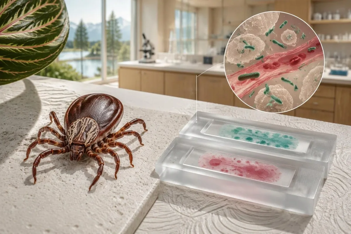
تاقیکردنەوەی تاوی خالخالدارەکانی چیای ڕۆکی لە هەفتەی یەکەمدا دەتوانێت نەرێنی بێت، چونکە هێشتا دژەتەنەکان دروست نەبوون. تیتری تاکی IgGی ئەرێنی ناتوانێت بوونی تووشبوونی حاد بسەلمێنێت؛ نموونە هاوتاکان کە زیادبوونێکی چوار هێندەیی پیشان دەدەن، پشتگیری تووشبوونی تازە دەکەن. پزیشکان کاتێک گومان لە RMSF دەکرێت، دەبێت دەستبەجێ چارەسەر دەست پێ بکەن—چاوەڕێی پشتڕاستکردنەوەی تاقیگەیی نەکەن.

خاڵە سەرەکییەکان
- ئەنجامی نەرێنیی هەڵەی سەرەتایی: تاقیکردنەوەکانی دژەتەنی RMSF زۆرجار لە ماوەی 7 ڕۆژی یەکەمی نەخۆشیدا نەرێنین، کە بڕیارەکانی چارەسەر لەو کاتەدا زۆرترین پەلەیان هەیە.
- شێوازی پەسەندکراوی تاقیکردنەوەی دژەتەنی: تاقیکردنەوەی دژەتەنی IgG بە شێوازی فڵۆرەسێنسی ناڕاستەوخۆ، یان IFA، تیتری دژەتەنەکان بەرامبەر ئەنتیجەنەکانی ڕیکێتسیای گروپی تاوی خالخالدار بەراورد دەکات.
- نموونە هاوتاکان: لە سەرەتادا نموونەیەکی حاد کۆ بکەرەوە و دواتر نموونەیەکی قۆناغی چاکبوونەوە کۆ بکەرەوە؛ ڕێنمایی CDC ماوەیەکی 2–10 هەفتەیی لەنێوان نموونەکاندا دیاری دەکات.
- زیادبوونێکی چوار هێندەیی: گۆڕانێک لە 1:64 بۆ 1:256 زیادبوونێکی چوار هێندەییە و لە نەخۆشییەکی گونجاودا پشتگیری تووشبوونی تازە بە ڕیکێتسیای گروپی تاوی خالخالدار دەکات.
- IgGی تاکی ئەرێنی: دژەجەستەکان دەتوانن بۆ ساڵانێک بمێننەوە؛ تەنانەت تایتەرێکی 1:256یش ناتوانێت بە تەنها دیاری بکات کە کەی تووشبوون ڕوویداوە یان جۆری ریکتسیایاکە بناسێنێت.
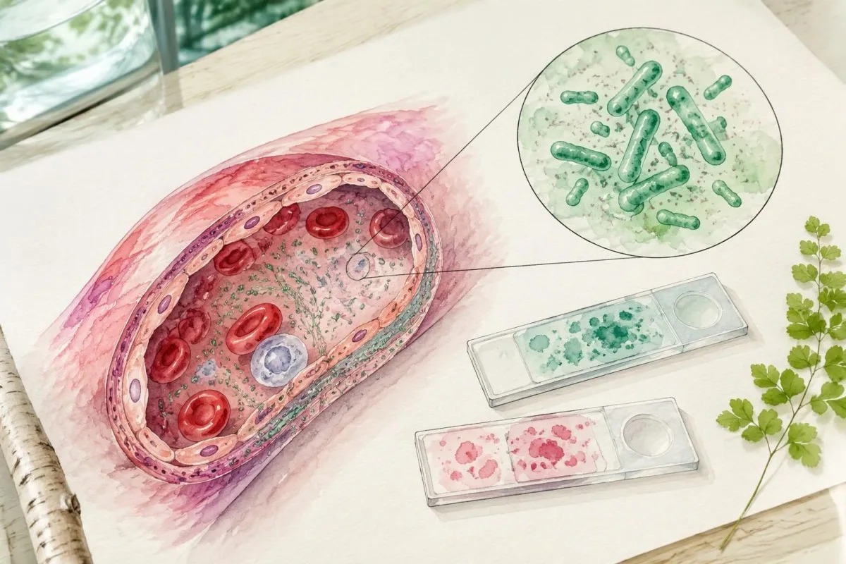
- PCRی نەرێنی: PCRی خوێنی تەواو دەتوانێت RMSF لەدەست بدات، چونکە Rickettsia rickettsii بە سەرەکی کاریگەری لەسەر خانە ئەندۆتیلییەکانی ڕەگە خوێنییەکان دادەنێت، نەک لەسەر خانە سپییە سوڕاوەکانی خوێن.
- کاتی چارەسەر: دۆکسایسایکلین چارەسەری هێڵی یەکەمە بۆ RMSFی گومانلێکراو لە گەورەساڵان و منداڵانی هەموو تەمەنەکان؛ پزیشکان نابێت چاوەڕێی ئەنجامی تاقیکردنەوەکان بکەن.
- ڕێژەی باوی گەورەساڵان: ڕێنماییەکانی CDC دۆکسایسایکلینی 100 mg هەر 12 کاتژمێر جارێک پێشنیار دەکەن، بە گشتی بۆ لانیکەم 5–7 ڕۆژ و تاوەکو تاو لانیکەم بۆ 3 ڕۆژ کۆتایی هاتبێت و باشبوونی کلینیکی هەبێت.
کام تاقیکردنەوەی تاوی خالخالدارەکانی چیای ڕۆکی سەرەتا ئەنجام دەدرێت؟
شێوازی باوی تاقیکردنەوەی دژەجەستە بۆ دەستنیشانکردنی RMSF تاقیکردنەوەی جوتکراوی IgG بە ڕێگەی تاقیکردنەوەی دژەجەستەی فلۆرەسەنتی ناڕاستەوخۆیە، کە بە شێوەیەکی باو پێی دەوترێت IFA. بەڵام پزیشک دەتوانێت پێش هاتنی ئەنجامی یەکەم دۆکسایسایکلین دەست پێ بکات، چونکە نموونەی دژەجەستەی کۆکراوە لە ڕۆژانی 1–7 زۆرجار ناتوانێت نەخۆشییەکە پشتڕاست بکاتەوە یان ڕەتی بکاتەوە.

RMSF دروست دەکرێت بەهۆی Rickettsia rickettsii, ، باکتریایەک کە بە کەنەوە دەگوازرێتەوە و دەتوانێت بە خێرایی زیان بە خوێنبەرە بچووکەکان بگەیەنێت. ڕێنمایی کرداری CDC دوو ئەرک لە یەک جیا دەکاتەوە: چارەسەرکردنی نەخۆشییەکی ئەگەری مەترسیدار لە ئێستادا و بەدەستهێنانی بەڵگەی تاقیگەیی بۆ پشتڕاستکردنەوە لە دواتردا؛ تێکەڵکردنی ئەم دوو ئەرکە دواخستنی خۆپارێزنەکراو دروست دەکات (Biggs et al., 2016).
ئەو داواکارییەی بە پەنێڵی RMSF ناودەبرێت، لەوانەیە IgG یان IgM یان هەردووکیان لەخۆ بگرێت، و لەوانەیە بەدوای ئەنجامی پشکنینەوە تایتەری IFA بێت. بپرسە کام شێواز هەر ئەنجامێکی بەرهەم هێناوە، چونکە 1 ئەنجامی پشکنینی پۆزەتیڤ و 1 تایتەری چەندایەتی هاوتا نین؛ ڕێنمایی ئێمە بۆ ئەنجامە ئەرێنییەکانی دژە جەستە ئەو جیاوازییە ڕوون دەکات.
Kantestî yek e Analyzerê testa xwînê ya AI بۆ ڕوونکردنەوەی ڕاپۆرتەکانی تاقیگەیە، نەک بۆ ڕەتکردنەوەی RMSF بە یەک ئەنجامی نەرێنی. ئێمە پێشینەی کۆمپانیا ئەو ڕێکخراوە دەناسێنێت کە لەپشت ئەو خزمەتگوزارییەوەیە؛ پشکنین، هەڵسەنگاندنی تووشبوون، داواکردنی تاقیکردنەوە و بڕیاردان لەبارەی ئەنتیبیۆتیک، هەموویان لە ئەستۆی پزیشکی چارەسەرکەرن.
بۆچی تاقیکردنەوەی دژەتەنی RMSF لە سەرەتادا نەرێنی دەردەکەوێت؟
Yek تاقیکردنەوەی هەڵە نەرێنیی RMSF زۆرجار ڕەنگدانەوەی ماوەی پەنجەرەی دژەجەستەیە: وەڵامی بەرگریی جەستە هێشتا بڕێکی پێویست لە دژەجەستەی دەتوانرێت بپێورێت بەرهەم نەهێناوە. IFAی IgG زۆرجار لە یەکەم 7 ڕۆژی نیشانەکاندا نەرێنییە، بۆیە ڕاپۆرتێکی نەرێنیی زوو، کاتێک نیشانە کلینیکییەکان گومانی RMSF دروست دەکەن، پاساو بۆ وەستاندنی چارەسەر نییە.

کاتی تاقیکردنەوە دەست پێ دەکات لە دەستپێکی نیشانەکان, ، نەک ئەو ڕۆژەی تاقیگە نموونەکە وەردەگرێت. بۆ نموونە، کەسێکی گریمانەیی کە لە ڕۆژی 3ی نەخۆشییەکەدا تاقی دەکرێتەوە، لەوانەیە بەڕاستی RMSFی هەبێت سەرەڕای IgGی نەرێنی؛ هەمان ئەنجامی نەرێنی چەند هەفتە دواتر مانایەکی جیاوازی دەبێت، بە تایبەتی ئەگەر تاقیکردنەوەی جوتکراو تەواو کرابێت.
بەرهەمهێنانی دژەجەستە لە هەموو کەسێکدا لەگەڵ ڕۆژێکی دیاریکراوی ساڵنامەدا هاوکات نییە. ڕێنمایی CDC بۆ دەستنیشانکردنی کلینیکی و تاقیگەیی ڕوون دەکاتەوە کە سیرۆلۆژیی هەفتەی یەکەم زۆرجار نەرێنییە؛ کارایی بەرزی تاقیگە لەسەر نموونە جێگیرەکان کە دژەجەستەیان پۆزەتیڤە، مانای ئەوە نییە کە هەمان هەستیاری لە ڕۆژانی 1–7یشدا هەیە.
دووبارەکردنەوەی تاقیکردنەوەی IgG تەنها 24–48 کاتژمێر دوای ئەنجامێکی نەرێنیی زوو، لەوانەیە سوودی کەمی هەبێت، چونکە ئەو ماوەیە زۆرجار زۆر کورتە بۆ پیشاندانی گۆڕانی چاوەڕوانکراوی دژەجەستە. ئەو Monospot testing window illustrates a similar timing principle, although its assay, illness, and recommended follow-up schedule differ from RMSF.
مانای ئەرێنیبوونی IgGی RMSF بەڕاستی چی لەخۆ دەگرێت؟
RMSF IgG positive meaning includes previous exposure, recent infection, or antibodies reacting with another spotted fever group rickettsia. A single positive titer—even 1:256 or higher—cannot independently establish acute RMSF, because antibody persistence and cross-reactivity prevent the result from acting as a reliable date stamp.
Antibodies against R. rickettsii can remain elevated for months and sometimes up to 4 years after illness, according to CDC diagnostic guidance. Persistent positivity does not mean bacteria remain active, and the height of a single titer does not reliably distinguish a new infection from an older immune response.
Background antibodies also affect interpretation: CDC notes that 10% or more of healthy people in some areas may have elevated titers from past exposure to spotted fever group rickettsiae. That figure is geographically dependent, not a universal prevalence estimate; testing someone without compatible symptoms increases the chance of finding a clinically misleading positive.
Consider a hypothetical asymptomatic adult with IgG 1:128 on an incidental panel and no recent febrile illness. That 1 result is not a reason by itself to prescribe doxycycline; antibody persistence after exposure is another example of why serology must be matched to the organism, symptoms, and sampling dates.
ئایا واکنشپێکەوەیی یان IgM دەتوانێت ئەنجامێکی گومڕاکەر بدات؟
وەڵامدانەوەی هاوبەش can produce positive RMSF serology after exposure to another spotted fever group rickettsia, and تاقیکردنەوەی سەرەتایی Lyme ـی نادرست-نەرێنی (false-negative) ڕوودەدات چونکە is less specific than IgG for diagnosing recent illness. A positive IgM result on day 5 is therefore not a dependable shortcut around an early negative IgG result.
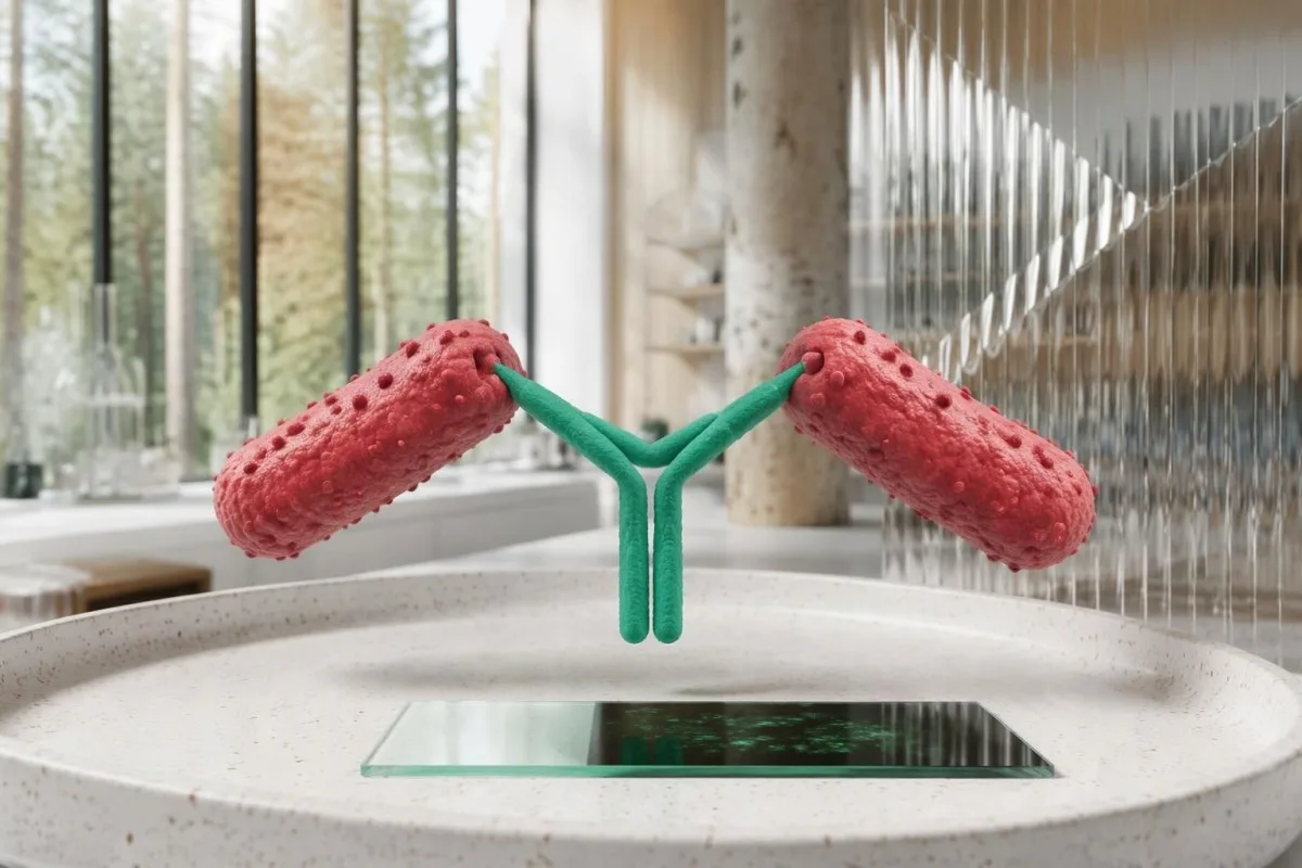
R. rickettsii, R. parkeri, and related organisms share antigenic features that routine commercial serology often cannot distinguish. A 4-fold IgG rise can establish recent spotted fever group rickettsial infection in a compatible illness, but identifying R. rickettsii specifically may require species-specific molecular testing or specialized reference-laboratory investigation.
IgM may appear without convincing evidence of recent rickettsial illness and should not be used as a stand-alone diagnostic marker. The CDC practical guide favors paired IgG IFA for confirmation rather than interpreting 1 isolated IgM positive as proof of acute RMSF (Biggs et al., 2016).
Not all antibody families behave alike: Q fever antibody patterns use phase-specific interpretation that should not be imported into RMSF. For a report showing IgM positive and IgG negative on day 4, ask about clinical suspicion, assay method, and the planned convalescent specimen rather than assuming the sequence proves infection.
نموونە هاوتاکان و زیادبوونێکی چوار هێندەیی چۆن پشتگیری تشخیص دەکەن؟
Paired IgG IFA samples support diagnosis by showing whether antibody titers rise substantially between acute illness and recovery. A rise from 1:64 to 1:256 is fourfold; with compatible symptoms, it supports recent spotted fever group rickettsial infection rather than merely documenting an antibody that was already present.
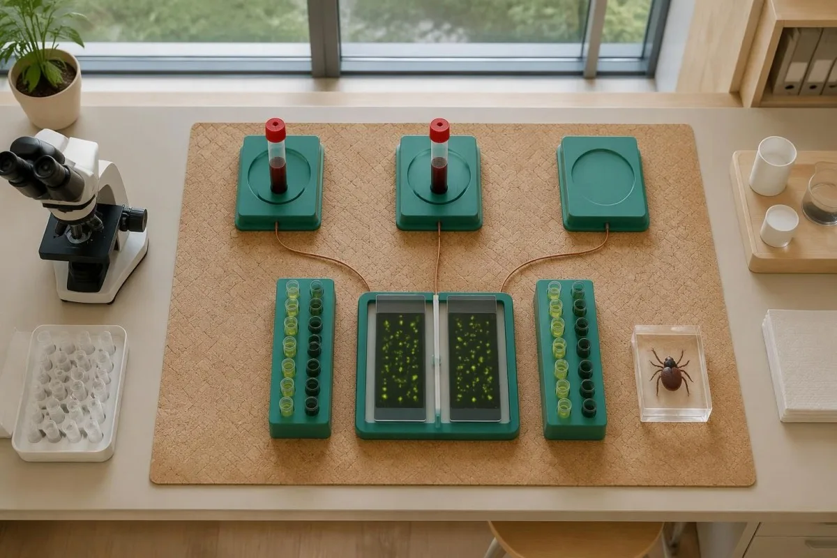
IFA titers describe the highest dilution at which antibody remains detectable; 1:256 means detection after greater dilution than 1:64. In a common 2-fold dilution series, 1:64 to 1:128 is one step, while 1:64 to 1:256 is two steps—the fourfold change used to reduce ambiguity from small fluctuations.
CDC RMSF guidance describes paired specimens collected 2–10 weeks apart, while the 2016 practical guide emphasizes an acute specimen and a convalescent specimen ideally 2–4 weeks later. These recommendations are not competing treatment deadlines; the clinician and laboratory should agree on follow-up timing that can capture the antibody response.
Use the same laboratory and assay when possible, and retain the original reports rather than comparing 2 abbreviated portal flags. Our explanations of ئەنجامی چەندایەتی بەرامبەر چۆنایەتی û گۆڕانکاری لە نێوان سەردانەکانی تاقیگەدا help distinguish a meaningful paired titer rise from unrelated reporting differences.
| ئاست | مەودا | مانای چییە |
|---|---|---|
| Early negative | Below laboratory cutoff on days 1–7 | Does not exclude RMSF; antibodies may not yet be detectable. |
| Single positive | Example: 1:128 on one specimen | Shows detectable antibody, not the date or species of infection. |
| Twofold rise | Example: 1:64 → 1:128 | Does not meet the standard fourfold-rise criterion. |
| زیادبوونی چوار ئەوەندە | نموونە: 1:64 → 1:256 | Supports recent spotted fever group infection with compatible illness; does not measure severity. |
ئایا PCR دەتوانێت RMSF پێش دەرکەوتنی دژەتەنەکان بدۆزێتەوە؟
PCR can detect rickettsial DNA before antibodies develop, but a negative whole-blood PCR does not exclude RMSF. R. rickettsii mainly targets vascular endothelial cells, so an illness-day-3 blood specimen may contain too little bacterial DNA for detection even when the disease is clinically significant.
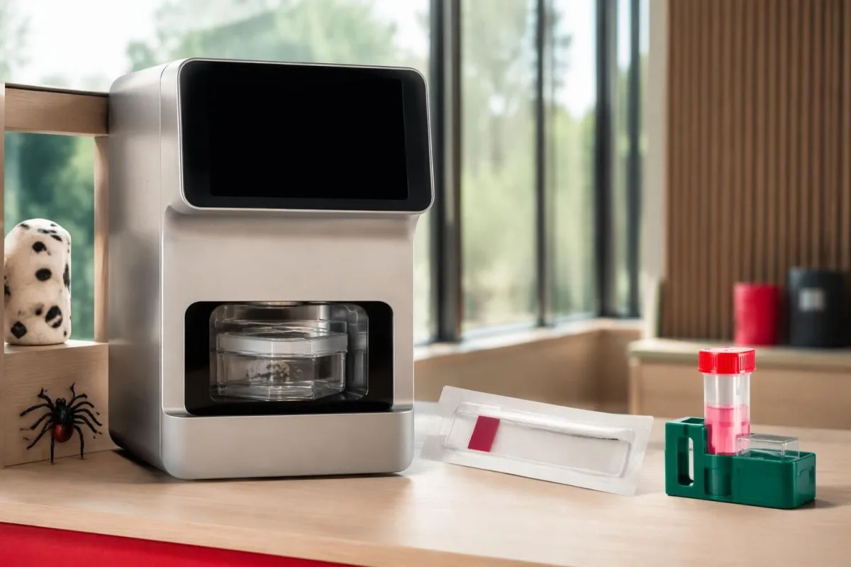
Unlike serology, PCR looks for the organism's genetic material rather than the host's immune response. Its usefulness depends on 3 practical variables: the specimen collected, illness severity and timing, and whether antibiotics have already reduced detectable organisms; there is no single sensitivity percentage applicable to every RMSF PCR order.
A clinician may arrange PCR or immunohistochemical testing of a rash skin biopsy through an appropriate laboratory, because affected tissue can provide evidence unavailable in circulating blood. Even when tissue testing is considered, collecting a specimen before the first doxycycline dose is desirable only if it does not delay urgent treatment.
Ask whether a panel actually includes R. rickettsii and whether it reports species-specific results or a broader rickettsial group. The PCR timing explanation for whooping cough demonstrates why molecular tests have specimen and treatment windows, but its respiratory sampling rules do not apply to RMSF.
بۆچی تاقیکردنەوەکانی RMSF و ئەهرلیخیۆز بە شێوەی جیاواز کار دەکەن؟
RMSF and ehrlichiosis require different diagnostic reasoning even when the same tick-exposure evaluation includes both. Whole-blood PCR is generally more useful early in ehrlichiosis because Ehrlichia organisms infect white cells, whereas R. rickettsii primarily affects vessel-lining cells; 1 negative tick-borne panel is not a universal exclusion test.

Fever, headache, muscle aches, and abnormal liver enzymes can occur in more than 1 tick-borne illness. Exposure location, platelet count, white-cell count, rash, and illness timing guide test selection; clinicians should not assume that an order called a tick panel covers every relevant organism or uses the best specimen for each.
Ew ehrlichiosis PCR and titer guide explains why acute PCR and later antibodies may disagree in that illness. For a hypothetical day-4 presentation, an Ehrlichia PCR positive and RMSF IgG negative need interpretation as separate findings—not evidence that all other tick-transmitted illnesses were conclusively excluded.
Kantestî yek e پلاتفۆرمی تێکست/وەشاندنی تاقیکردنی خوێنی AI that explains submitted laboratory reports, not a substitute for organism-specific diagnostic assessment. If a report contains 2 negative results from different methods, we emphasize the distinction between the method's target and the clinician's question rather than treating the word negative as a complete answer.
کام ئەنجامە ئاساییەکانی خوێن دەتوانن پشتگیری گومانی RMSF بکەن؟
Low platelets, mildly elevated AST or ALT, and low sodium can support suspicion of RMSF, but none confirms the diagnosis and early values may be normal. A platelet count of 110 × 10⁹/L and sodium of 130 mmol/L are contextual clues—not organism-specific thresholds or prerequisites for starting treatment.
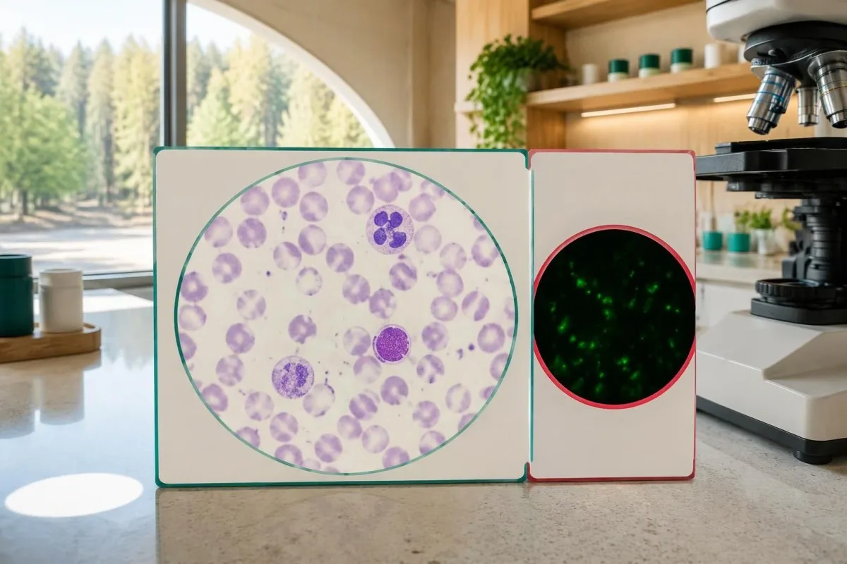
Typical adult platelet reference intervals are approximately 150–450 × 10⁹/L, and serum sodium is commonly 135–145 mmol/L, although local ranges vary. The reason clinicians consider falling platelets together with fever and low sodium is that the combination can suggest systemic illness; it does not distinguish RMSF from several infectious or noninfectious causes.
A hypothetical febrile traveler with platelets 95 × 10⁹/L, AST 82 U/L, and recent outdoor exposure deserves clinical assessment even if white cells are normal. Our بەراوردکردنی WBC و CRP explains why normal inflammatory markers cannot independently clear a person with concerning symptoms.
Unexpectedly low platelets may also reflect sample-related clumping, which requires laboratory verification rather than automatic attribution to RMSF; یەک دامەیەک کە هەر ماوەیەک جارێک دەبینم بریتییە لە explains that artifact. If systemic illness is accompanied by abnormal clotting or organ dysfunction, the ڕێنمای تاقیکردنەوەی کۆاگولیشن (coagulation testing)مان helps explain additional tests, but a repeat specimen must not postpone urgent care.
بۆچی دەبێت چارەسەری RMSFی گومانلێکراو پێش دەرکەوتنی ئەنجامەکان دەست پێ بکات؟
Suspected RMSF requires prompt empiric doxycycline, because early tests can be negative and delayed treatment increases the risk of severe illness and death. Clinicians should not wait for IgG seroconversion, a PCR report, or a convalescent sample 2–10 weeks later before treating a compatible acute presentation.

Treatment timing is a safety issue, not simply a preference for faster symptom relief. Kirkland et al. reported higher mortality with treatment initiated after the first 5 days of illness in their retrospective study; its observational design limits precise causal estimates, but the finding supports the urgency emphasized in CDC guidance (Kirkland et al., 1995).
CDC recommends doxycycline 100 mg every 12 hours for adults and children weighing at least 45 kg; children under 45 kg receive 2.2 mg/kg every 12 hours, up to 100 mg per dose. These are clinician-prescribed regimens, not instructions for self-treatment; oral versus intravenous therapy depends on severity and the ability to retain medication.
Treatment generally lasts at least 5–7 days and continues until fever has resolved for at least 3 days with clinical improvement; complicated cases may need longer care. Pregnancy or suspected doxycycline allergy requires urgent individualized advice rather than automatic delay, and urgent infection assessment illustrates the same need to coordinate testing and treatment, although Listeria requires different antibiotics.
ئەنجامە باوەکانی تاقیکردنەوەی RMSF چۆن دەبێت لێک بدرێنەوە؟
RMSF test results must be interpreted using symptoms, illness dates, specimen types, and changes between samples. An early negative result may be uninformative, a single positive may reflect old exposure, and a fourfold paired IgG rise can support recent spotted fever group rickettsial infection without proving the species.
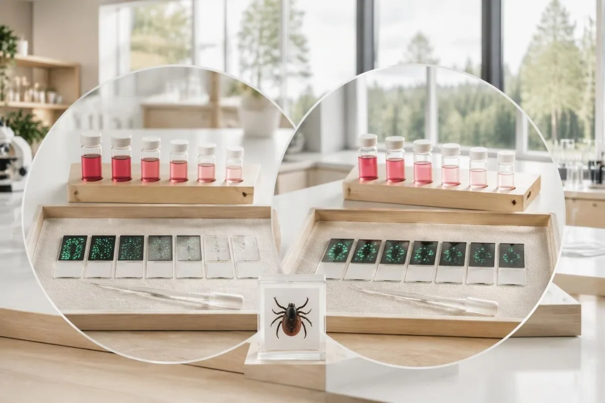
Example 1 is hypothetical: a person develops fever and severe headache after outdoor exposure, tests IgG negative on day 3, and begins clinician-directed doxycycline that day. An acute titer below 1:64 followed by a convalescent titer of 1:256 can provide supportive laboratory evidence, but the laboratory must verify the change against its starting dilution and reporting limits.
Example 2 is also hypothetical: an otherwise well person has IgG 1:256 on 2 specimens obtained several weeks apart, without a compatible intervening illness. Stable positivity does not prove new RMSF, and escalating antibiotics to make the titer disappear is not justified; Kantesti explains this as an interpretation limitation, not a declaration that every stable titer is harmless.
Example 3 involves fever, IgG 1:128, and no properly timed acute comparator, leaving recent versus previous exposure uncertain. The next step depends on clinical assessment and possible additional testing; دژەتەنییەکانی Babesia بەرامبەر تووشبوون demonstrates a related distinction, but babesiosis needs its own organism-detection approach and is not treated with doxycycline alone.
کەی دەبێت نیشانەکان لە ئەنجامی نەرێنی RMSF گرنگتر بن؟
A negative RMSF result should not override a concerning acute illness with fever, severe headache, gastrointestinal symptoms, or rash after plausible tick exposure. Symptoms typically begin 3–12 days after an infected tick bite, and neither an unremembered bite nor the absence of an early rash reliably excludes RMSF.
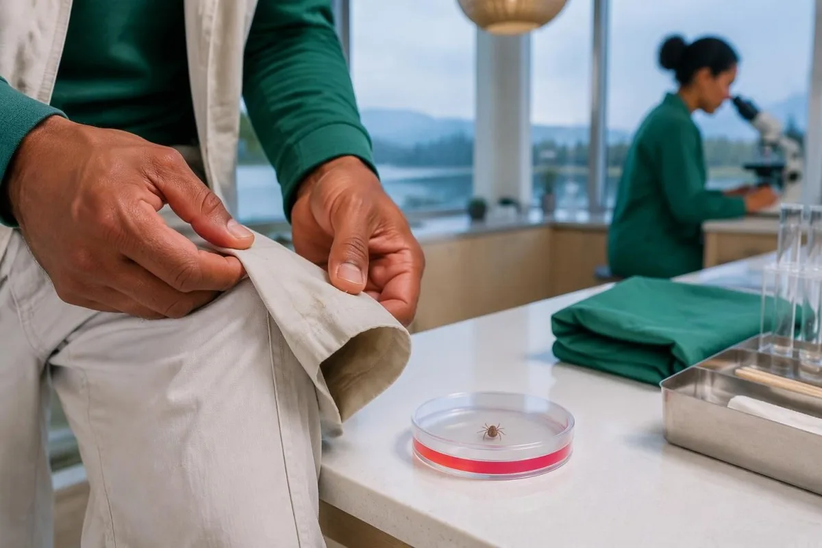
The classic rash often develops 2–4 days after fever begins, so waiting for wrist, ankle, palm, or sole spots can delay care. RMSF can occur without a recognized rash, and its name is geographically misleading: clinicians assess exposure beyond the Rocky Mountain region, including travel and locally circulating ticks.
Confusion, breathing difficulty, fainting, rapidly worsening illness, or a spreading purple or pinpoint rash warrant emergency assessment—not a plan to repeat antibodies in 2 weeks. A fever and severe headache after possible tick exposure merit prompt clinical contact even if an online portal marks the first RMSF test negative.
After a tick bite without symptoms, routine antibody screening and preventive antibiotics are not recommended specifically to prevent RMSF; CDC guidance advises symptom monitoring for approximately 14 days. Tick exposure can also lead to conditions with entirely different testing logic, such as هەستیاریی ئەلفا-گال دوای گازگرتنی کەنە دەکەن, which involves allergy assessment rather than RMSF IgG interpretation.
ئایا دەبێت دژەتەنەکانی RMSF دوای چارەسەر دووبارە تاقی بکرێنەوە؟
Repeat RMSF antibodies are useful for diagnostic confirmation, not as a routine test of cure. A convalescent sample collected 2–10 weeks after the acute specimen can document an antibody rise, but persistent IgG after recovery does not by itself mean treatment failed or that more antibiotics are needed.

Fever often improves within 24–48 hours after early appropriate doxycycline treatment, although severe illness may respond more slowly. Failure to improve or clinical deterioration requires reassessment for complications, an alternative diagnosis, inadequate medication absorption, or another illness; it should not be managed by waiting for a titer to fall.
Early antibiotic treatment can sometimes delay or diminish antibody responses in rickettsial illness, so the absence of a fourfold rise is not an absolute retrospective exclusion in every treated case. Do not stop effective treatment to improve test yield; the clinician should interpret the 2 specimens alongside treatment dates, sampling quality, and the original clinical presentation.
Kantesti AI can help organize submitted results for trend review, but a trend graph cannot establish that RMSF is cured or still active. Our ڕێنمای کاتەکانی تاقیکردنەوەی خوێن explains why illness and medication dates matter; persistent symptoms after treatment need clinical reassessment, because lasting complications can occur without evidence of chronic ongoing RMSF infection.
پێش داواکردن لە AI بۆ ڕوونکردنەوەی ڕاپۆرتی RMSF، چی دەبێت بپشکنیت؟
A useful RMSF report explanation requires the assay method, exact titer, collection date, and symptom-onset date, not just the words positive or negative. Before seeking an AI explanation, identify whether 2 reports represent comparable acute and convalescent specimens; urgent symptoms still require clinical care before any upload or interpretation.

Record at least 5 details: symptom onset, sample collection, IgG versus IgM, IFA titer versus screening index, and the first antibiotic dose. Add recent travel and exposure information for the clinician; a numerical index of 2.1 from one assay cannot be mathematically converted into an IFA titer such as 1:128.
Kantestî yek e ئامێری توێژینەوەی تاقیکردنەوەی خوێن بە پشتبەستن بە AI that helps explain submitted laboratory information without replacing clinical diagnosis. Our ڕێنمایی تەکنەلۆژیای AI describes the interpretation approach; for suspected RMSF, 1 negative early result must never become an AI-generated reassurance that postpones medical assessment.
An explanation should separate 3 categories: what the laboratory measured, what that finding can support, and what remains uncertain. Our پشکنینی سەرچاوەی ڕاپۆرتی پزیشکی describe that safety boundary; useful questions include whether paired IgG is planned, whether PCR covered the suspected species, and whether treatment has already started.
بڵاوکراوەکانی توێژینەوە و سنوورەکانی بەڵگەی تشخیصی
RMSF diagnostic guidance and AI engineering publications answer different questions and should not be treated as interchangeable evidence. The 2016 CDC practical guide supports paired serology and immediate treatment when clinically indicated; 2 Figshare records listed below provide publication context, not proof that an AI system can diagnose or exclude RMSF.
The 1995 therapeutic-delay study and the 2016 CDC recommendations directly address RMSF care, whereas a laboratory's assay validation addresses the particular test it performs. These are 3 different evidence levels—clinical outcomes, diagnostic guidance, and analytical performance—and none makes a first-week negative antibody result a reliable reason to delay treatment.
Kantesti's 2 requested Figshare publication records concern women's health and AI-assisted hantavirus triage, respectively; neither should be cited as an RMSF diagnostic-validation study. A DOI identifies a publication record rather than guaranteeing peer review, clinical accuracy, or applicability to another disease; our ستانداردەکانی ناوەڕۆکی پزیشکی explain the need to distinguish directly relevant sources from contextual material.
For this topic, the practical evidence boundary is straightforward: a 4-fold paired IgG rise supports recent spotted fever group infection, while immediate care depends on clinical suspicion rather than a future titer. Our چوارچێوەی ڕاستکردنەوەی کڵینیکی provides broader methodology context, but suspected RMSF still requires a clinician who can examine, prescribe, and arrange appropriate confirmatory testing.
پرسیاره باوهكان
ئایا تاقیکردنەوەی تاوی خاڵخاڵی چیای ڕۆکی دەتوانێت لە سەرەتادا نەرێنی بێت؟
0) بەڵێ، تاقیکردنەوەی ئەنتیبۆدی تایفی خاڵخاڵی چیای ڕۆکی ماونتەن زۆرجار لە ٧ ڕۆژی یەکەمی نەخۆشیدا نەرێنی دەبێت، چونکە هێشتا ئەنتیبۆدی دەتوانرێت پێشکەش نەبووە. بۆیە ئەنجامی نەرێنیی زووەختی IgG IFA، RMSF ڕەت ناکاتەوە. پزیشکان دەبێت بە خێرایی دۆکسایسایکلین دەست پێ بکەن، کاتێک نەخۆشی و مێژووی بەرکەوتن گومان بۆ RMSF دروست دەکەن، نەک چاوەڕێی تاقیکردنەوەی دووبارە بکەن. نموونەیەکی دواتری کاتی چاکبوونەوە دەتوانێت یارمەتی پشتڕاستکردنەوەی نەخۆشییەکە بدات.
ئایا IgGـی ئەرێنی RMSF واتە تووشبوونی چالاکم هەیە؟
ئەنجامی پۆزەتیڤی RMSF IgG بە تەنها سەلمێنەری تووشبوونی چالاک نییە، تەنانەت ئەگەر تیترەکە 1:256 بێت. دژەتەنەکان دەتوانن بۆ چەند مانگ یان ساڵێک بمێننەوە و لەگەڵ ڕیکێتسیای گرووپی تاوە خاڵدارەکانی دیکەدا وەڵام بدەنەوە. نیشانە تازەکان، بەرکەوتن و زیادبوونی چوار هێندە لەنێوان نموونە هاوتاکاندا، ئەگەری تووشبوونی تازە زیاتر دەکەن. ئەنجامی پۆزەتیڤی تاکە لە کەسێکدا کە نیشانەی گونجاوی نییە، بە تەنها هۆکارێک نییە بۆ بەکارهێنانی ئەنتیبیۆتیک.
نموونەی دووەمی ئەنتیبادیی RMSF کەی دەبێت وەربگیرێت؟
ڕێنماییەکانی CDC بۆ دەستنیشانکردنی RMSF، نموونەی IgGی حاد و دوای حاد باس دەکەن کە بە ماوەی 2–10 هەفتە لە یەکتر جیا دەکرێنەوە. ڕێنمایی کرداریی CDCی ساڵی 2016، نموونەیەکی دوای حاد باس دەکات کە باشترە 2–4 هەفتە دوای نموونەی حاد وەربگیرێت. پزیشکی چارەسەرکەر و تاقیگە دەبێت ماوەی گونجاو هەڵبژێرن و تاقیکردنەوەی هاوشێوە بەکاربهێنن. چارەسەر نابێت چاوەڕێی نموونەی دووەم بکات.
بەرزبوونەوەی چوار هێندەی تیتەری ئەنتیبادی RMSF چییە؟
0. بەرزبوونەوەی چوار هێندە واتە تایتەری دواتری IgG چوار هێندەی تایتەری پێشووترە، وەک زیادبوون لە 1:64 بۆ 1:256. لە زنجیرەیەکی باوی تەرقیقی دوو هێندەییدا، ئەمە نوێنەرایەتی دوو هەنگاوی تەرقیق دەکات. لەگەڵ نەخۆشییەکی گونجاودا، ئەو گۆڕانکارییە پشتگیری لە تووشبوونی تازە بە ڕیکێتسیای گروپی تاوی خاڵدار دەکات. تاقیکردنەوەی سیرۆلۆژیی ڕۆتین هێشتا ڕەنگە نەتوانێت Rickettsia rickettsii بە تایبەتی دیاری بکات یان توندیی نەخۆشییەکە بخاتە ڕوو.
ئایا ئەنجامی نەرێنی تاقیکردنەوەی PCR بۆ تبەی کێکەڵەکانی کۆتایی کێوەکان (RMSF)، نەخۆشییەکە ڕەت دەکاتەوە؟
نەتیجەی نەرێنی تاقیکردنەوەی PCRی خوێنی تەواو بۆ RMSF، نەخۆشییەکە ڕەت ناکاتەوە، چونکە Rickettsia rickettsii بە شێوەیەکی سەرەکی کاریگەری لەسەر خانەکانی پۆشەری ڕەگەکان دەکات، نەک خانە سپییەکانی خوێنی سووڕاو. نموونەی ڕۆژی سێیەمی نەخۆشی لەوانەیە DNAی بەکتریایی بە قەدەرێکی پێویست بۆ دۆزینەوە لەخۆ نەگرێت، و ئەنتیبیۆتیکەکان دەتوانن زیاتر دەرهێنانی تاقیکردنەوە کەم بکەنەوە. لە کاتی گونجاودا، تاقیکردنەوەی تایبەتی بافتی پێستی تووشبوو دەتوانێت بەڵگەی زیاتر دابین بکات. پزیشکان نابێت چارەسەر دوا بخەن لە کاتی وەرگرتن یان چاوەڕوانی ئەنجامەکانی PCR.
ئایا پێویستە پێش ئەوەی ئەنجامەکانی RMSF بگەڕێنەوە، دۆکسایسایکلین بە منداڵان بدرێت؟
Children of all ages should receive clinician-prescribed doxycycline promptly when RMSF is suspected, without waiting for laboratory confirmation. CDC guidance recommends 2.2 mg/kg every 12 hours for children under 45 kg, with a maximum of 100 mg per dose. Short courses used for rickettsial disease have not been shown to cause the permanent tooth staining associated with older tetracycline concerns. The child's clinician determines route, duration, and whether hospital care is needed.
بۆچی دوای چارەسەرکردن، تاقیکردنەوەی IgG بۆ RMSF ـم هێشتا ئەرێنییە؟
IgGی RMSF دەتوانێت بۆ چەند مانگێک و هەندێک جار تا ٤ ساڵ دوای نەخۆشی ئەرێنی بمێنێتەوە، بۆیە بەردەوامیی ئەرێنیبوون بە تەنیا نیشانەی شکستی چارەسەری نییە. تاقیکردنەوەی ئەنتیبۆدی تاقیکردنەوەیەکی ڕۆتینی بۆ دڵنیابوون لە چاکبوونەوە نییە. نابێت تەنها بۆ ئەوەی ئاستی ئەنتیبۆدی نەرێنی ببێت، ئەنتیبیۆتیکی زیاتر بنووسرێت. پشێوی بەردەوام یان خراپتربوون پێویستی بە هەڵسەنگاندنەوەی کلینیکی هەیە بۆ پێچیدەکان یان تشخیصێکی تر، نەک لێکدانەوە لەسەر بنەمای ژمارەی ئەنتیبۆدی بە تەنیا.
ئەنجامەکانی تاقیکردنەوەی خوێنت ڕوون بکەرەوە
ڕاپۆرتی تاقیگەکەت باربکە و AIی Kantesti لە چەند چرکەیەکدا هەر بایۆمارکەرێک بە زمانی سادە ڕوون دەکاتەوە. زیاتر لە 2 ملیۆن کەس لە سەرانسەری جیهان بەکاری دەهێنن.
Referans
- کلاین، تی.، میچڵ، ئێس.، و وێبەر، ھ. (٢٠٢٦). ڕێبەری تەندروستیی ژنان: هێلکەدان، پچڕانی مانگانە و نیشانە هۆرمۆنییەکان. توێژینەوەی پزیشکیی AIی Kantesti. DOI
- کلاین، تی.، میچڵ، ئێس.، و وێبەر، ھ. (٢٠٢٦). پشتگیریی بڕیاری کلینیکی بە یارمەتی AIی فرەزمانی بۆ پۆلێنکردنی سەرەتایی هانتاڤایرۆس: دیزاین، پشتڕاستکردنەوەی ئەندازیاری و جێبەجێکردنی لە ژیانی ڕاستەقینەدا لە 50,000 ڕاپۆرتی تاقیکردنەوەی خوێنی لێکدراوەوە. توێژینەوەی پزیشکیی AIی Kantesti. DOI
- Biggs HM et al. (2016). Diagnosis and Management of Tickborne Rickettsial Diseases: Rocky Mountain Spotted Fever and Other Spotted Fever Group Rickettsioses, Ehrlichioses, and Anaplasmosis — United States. ڕاسپاردە و ڕاپۆرتەکانی MMWR. DOI
- Kirkland KB et al. (1995). Therapeutic delay and mortality in cases of Rocky Mountain spotted fever. نەخۆشییە تووشبووە کلینیکییەکان.
- ناوەندەکانی کۆنترۆڵ و پێشگیری لە نەخۆشییەکان (بێ بەروار). Clinical and Laboratory Diagnosis for Rocky Mountain Spotted Fever. ڕێنمایی کلینیکی CDC.
- 2M+تاقیکردنەوە شیکراوەکان
- 127+Welat
- 100+Ziman
دەربارەی تیمی پزیشکی


سەنگەری باوەڕپێکردن E-E-A-T
Tecribe
ڕەوی پشکنینی کلینیکی لەلایەن پزیشکەوە بۆ ڕێکخستنی ڕووداوەکانی لێکدانەوەی ئازمایش.
Pisporî
گرێدانی لابراتۆرییەی پزیشکی بەوەی چۆن بایۆمارکەرەکان لە کۆنتێکستی کلینیکی دەگۆڕن.
Desthilatdarî
پێداچوونەوەی پزیشکی بۆ کراوە لەلایەن دکتۆر توماس کلاینەوە، بە بەشداریی دکتۆر سارا میچڵ و پرۆفیسۆر دکتۆر هانس ڤێبەر.
Bawerî
لێکدانەوە بە بنەمای دڵنیابوون (Evidence-based) بە ڕێڕەوی دوایینەوەی ڕوون بۆ کەمکردنەوەی هەست بە ترس/هەڵوەشاندن.




ڕاگەیاندنی بەرپرسیاریەتی پزیشکی
ئەم مادەیە تەنها بۆ. I think I must continue but user expects all items.
بڵاوکراوهتهوه · پەیوەندی بکە بە تیمی دەستەنووس
بڵاوکراوەتەوە لەلایەن Kantesti Ltd، تۆمارکراو لە ئینگلتەرا و وێڵز، ژمارەی کۆمپانیا. 17090423, ، لەندەن، شانشینی یەکگرتوو.
