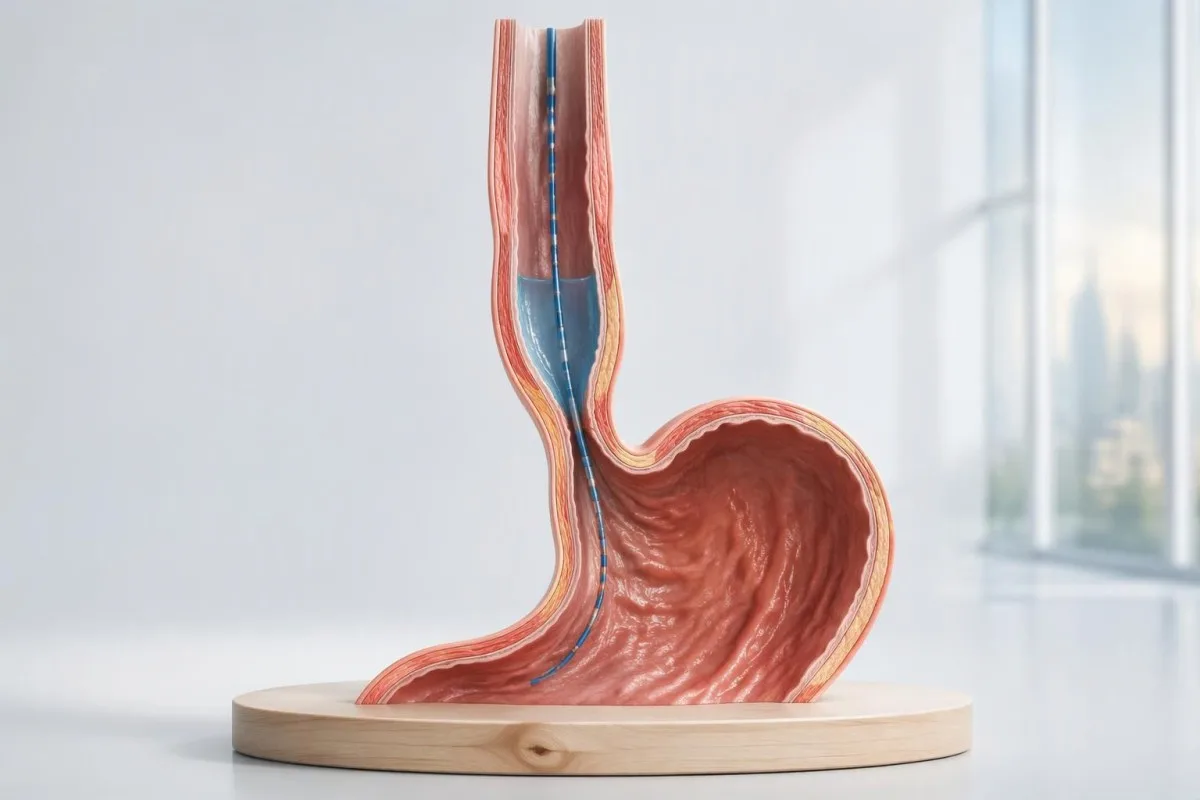
ค่าความดันสูงเพียงอย่างเดียวไม่เพียงพอที่จะวินิจฉัยว่าเป็นอะคาเลเซีย การวินิจฉัยขึ้นอยู่กับการคลายตัวของทางออกหลอดอาหาร การกลืนทำให้เกิดการบีบตัวที่ประสานกันหรือไม่ และมีภาวะอื่นที่อธิบายรูปแบบดังกล่าวได้หรือไม่.

ประเด็นสำคัญ
- การวินิจฉัยอะคาเลเซีย โดยทั่วไปต้องอาศัยการตรวจวัดความดันความละเอียดสูงที่แสดงการคลายตัวของทางออกที่บกพร่องและไม่มีการบีบตัวเป็นคลื่นตามปกติของหลอดอาหาร การส่องกล้องใช้ตรวจหาสาเหตุเชิงกล.
- ความดันการคลายตัวแบบบูรณาการ ใช้วัดการคลายตัวของทางออกในช่วงเวลา 4 วินาที แต่ค่าขีดจำกัดบนขึ้นอยู่กับอุปกรณ์และท่าทางของร่างกาย.
- อะคาเลเซียชนิดที่ I แสดงการบีบตัวเป็นคลื่นที่ล้มเหลว 100% โดยไม่มีแรงดันทั่วทั้งหลอดอาหารที่จำเป็นต่อการจัดเป็นชนิดที่ II.
- อะคาเลเซียชนิดที่ II แสดงการบีบตัวเป็นคลื่นที่ล้มเหลว 100% และแรงดันทั่วทั้งหลอดอาหารในการกลืนทดสอบอย่างน้อย 20% ครั้ง.
- อะคาเลเซียชนิดที่ III แสดงการหดตัวก่อนกำหนดในการกลืนอย่างน้อย 20% ครั้ง การคลายตัวของทางออกบกพร่อง และไม่มีการบีบตัวเป็นคลื่นตามปกติ.
- การหดตัวก่อนกำหนด โดยทั่วไปหมายถึงระยะเวลาแฝงส่วนปลายน้อยกว่า 4.5 วินาทีและมีแรงการหดตัวเพียงพอ สีที่ดูเหมือนแสดงการเคลื่อนไหวรวดเร็วเพียงอย่างเดียวไม่ได้ยืนยันว่ามีการเกร็ง.
- ผลตรวจที่อยู่ในเกณฑ์ก้ำกึ่ง อาจต้องตรวจภาพหลอดอาหารด้วยแบเรียมตามช่วงเวลาที่กำหนด หรือประเมินด้วย FLIP; แรงดันที่สูงขึ้นเพียงจุดเดียวไม่เพียงพอที่จะใช้เป็นเหตุผลในการรักษาที่ไม่อาจย้อนกลับได้.
- การหารือเรื่องการรักษา แตกต่างกันไปตามชนิดย่อย: ชนิดที่ III มักเหมาะกับการทำไมโอโทมีที่ยาวขึ้นและปรับให้เหมาะกับผู้ป่วย ส่วนชนิดที่ I และ II มีทางเลือกที่ได้ผลหลายวิธี.
- การตรวจเลือด สามารถใช้ประเมินภาวะขาดน้ำ ภาวะโลหิตจาง หรือภาวะแทรกซ้อนทางโภชนาการได้ แต่ไม่มีชุดตรวจทางห้องปฏิบัติการมาตรฐานที่ยืนยันหรือตัดภาวะอะคาเลเซียได้.
วินิจฉัยอะคาเลเซียด้วยการตรวจวัดความดันความละเอียดสูงได้อย่างไร?
การวินิจฉัยอะคาเลเซีย โดยทั่วไปวินิจฉัยจากการตรวจวัดการเคลื่อนไหวของหลอดอาหารด้วยความละเอียดสูง ซึ่งแสดงการคลายตัวของทางออกหลอดอาหารที่บกพร่องและไม่มีการบีบตัวเป็นคลื่นตามปกติ ชนิดที่ I มีการบีบตัวล้มเหลว ชนิดที่ II มีแรงดันทั่วทั้งหลอดอาหารเพิ่มขึ้นในการกลืนอย่างน้อย 20% ครั้ง และชนิดที่ III มีการหดตัวก่อนกำหนดในการกลืนอย่างน้อย 20% ครั้ง; การส่องกล้องช่วย排除การอุดกั้น.
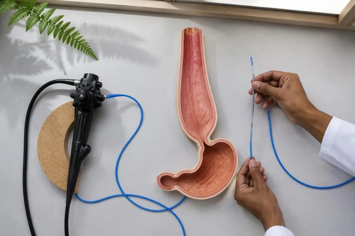
การตรวจวัดการเคลื่อนไหวของหลอดอาหารด้วยความละเอียดสูง หรือ HRM, วัดแรงดันแทนการถ่ายภาพการเคลื่อนที่ของอาหารโดยตรง คำถามหลัก 2 ข้อคือ รอยต่อหลอดอาหารกับกระเพาะอาหารเปิดได้อย่างเหมาะสมขณะกลืนหรือไม่ และตัวหลอดอาหารสร้างคลื่นที่เป็นระเบียบหรือไม่; แรงดันหูรูดขณะพักที่สูงเพียงอย่างเดียวไม่สามารถตอบคำถามทั้งสองข้อและไม่ยืนยันภาวะอะคาเลเซีย.
Chicago Classification ฉบับ 4.0 กำหนดรูปแบบจากการตรวจวัดการเคลื่อนไหวที่ใช้ระบุอะคาเลเซียทั้ง 3 ชนิดย่อย ขณะที่อาการและการตรวจอื่น ๆ ใช้กำหนดความหมายทางคลินิก (Yadlapati et al., 2021) รายงานที่ระบุเพียงว่ามีความผิดปกติของการเคลื่อนไหวให้ข้อมูลน้อยกว่ารายงานที่บันทึกแรงดันคลายตัวแบบบูรณาการ ผลการตรวจการบีบตัว ท่าทางขณะตรวจ และการจำแนกขั้นสุดท้าย.
คันเตสตีเป็น เครื่องวิเคราะห์ผลเลือด AI ซึ่งช่วยอธิบายผลกระทบทางห้องปฏิบัติการจากการรับประทานอาหารได้น้อย แต่ไม่สามารถวินิจฉัยอะคาเลเซียทั้ง 3 ชนิดย่อยจากผลตรวจเลือดได้ ข้อมูลเกี่ยวกับ องค์กรของเรา ควรอ่านแยกจากการประเมินโดยแพทย์เฉพาะทางที่จำเป็นต่อการแปลผลการตรวจวัดการเคลื่อนไหวและ排除โรคเชิงโครงสร้าง.
อาการเกี่ยวกับการกลืนแบบใดที่ควรได้รับการตรวจหาอะคาเลเซีย?
กลืนอาหารแข็งและของเหลวลำบาก, การสำรอกอาหารที่ค้างอยู่ และน้ำหนักลดโดยไม่ทราบสาเหตุควรนำไปสู่การประเมินภาวะอะคาเลเซีย อาการเพียงอย่างเดียวไม่สามารถระบุการวินิจฉัยหรือแยกอะคาเลเซียทั้ง 3 ชนิดย่อยได้ แม้ประวัติอาการจะชวนให้สงสัยก็ควรตรวจเพิ่มเติม เพราะภาวะอุดกั้น โรคที่เกี่ยวข้องกับกรดไหลย้อน และความผิดปกติของการกลืนชนิดอื่นอาจมีอาการซ้อนทับกัน.
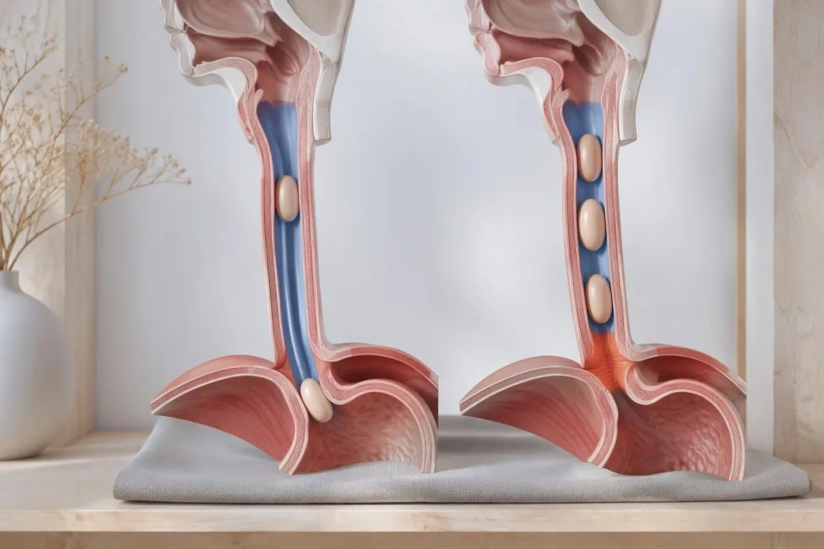
การสำรอกในภาวะอะคาเลเซียอาจมีอาหารที่รับประทานไปหลายชั่วโมงก่อนปะปนอยู่ ขณะที่กรดไหลย้อนมักทำให้มีของเหลวรสเปรี้ยวไหลย้อนขึ้นมา ความแตกต่างนี้ช่วยอธิบายว่าเหตุใดการรักษาเป็นเวลานานสำหรับ อาการคล้ายกรดไหลย้อน อาจไม่ช่วยแก้ปัญหาการกลืน อย่างไรก็ตาม ภาวะทั้ง 2 อย่างนี้อาจเกิดร่วมกันได้ และคำบรรยายอาการไม่สามารถแยกภาวะเหล่านี้ได้อย่างน่าเชื่อถือหากไม่มีการตรวจ.
การเริ่มกลืนลำบากบ่งชี้ตำแหน่งที่แตกต่างจากการที่อาหารติดหลังกลืนไปแล้วหลายวินาที อาการไอทันทีเมื่อจิบน้ำ เสียงขึ้นจมูก หรือกลืนลำบากมากขึ้นเมื่ออ่อนล้า อาจจำเป็นต้องประเมินช่องปากและคอหอยหรือระบบประสาท; ข้อจำกัดของการตรวจภาวะกล้ามเนื้ออ่อนแรงชนิดไมแอสทีเนีย มีความเกี่ยวข้องเมื่ออาการร่วมกับภาวะอ่อนแรง ไม่ใช่ชุดตรวจคัดกรองอะคาเลเซียตามปกติ.
An inability to swallow saliva or a suspected food impaction requires urgent assessment rather than waiting for elective manometry. Repeated nighttime regurgitation also raises concern for aspiration; for a hypothetical person losing 6 kg while meals become progressively slower, the weight trajectory and swallowing history both deserve attention even if routine laboratory results remain within range.
การตรวจวัดความดันในหลอดอาหารทำอย่างไร?
Esophageal manometry usually involves passing a thin pressure-sensing catheter through the nose into the stomach while the patient is awake. A standard Chicago Classification version 4.0 protocol commonly includes 10 supine and 5 upright water swallows, with additional maneuvers that test whether an apparent abnormality persists under different conditions.
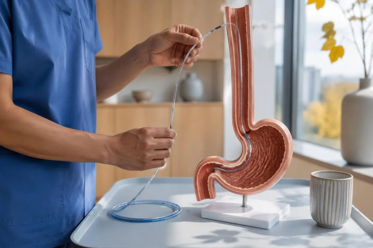
Preparation commonly includes at least 6 hours without food, but achalasia can leave material in the esophagus despite ordinary fasting. The testing center may therefore give a longer fasting or modified-diet plan; follow its instructions rather than assuming that an empty stomach guarantees an empty esophagus, especially if retained food was seen during endoscopy.
Medication review matters because opioids can produce impaired junction relaxation or spastic patterns that resemble type III achalasia. Provide the exact medicine, dose, and last-dose time; medication-related laboratory effects are a separate issue, but a single accurate medication list helps both investigations, and prescribed medicines should not be stopped without guidance.
Provocative testing may include 5 closely spaced 2 mL swallows or a rapid drink challenge of about 200 mL, if safe and appropriate. These maneuvers test different aspects of relaxation and contraction reserve; when standard swallows conflict with symptoms, their results can reveal a reproducible obstruction pattern or expose an artifact that disappears after changing position.
ผลการตรวจวัดความดันในหลอดอาหารใดสำคัญที่สุด?
สิ่งที่มีประโยชน์ที่สุด esophageal manometry results are median integrated relaxation pressure, the presence or absence of peristalsis, panesophageal pressurization, and distal latency. These measurements answer different questions: outlet opening, coordinated propulsion, pressure trapped within the esophagus, and contraction timing; none should be interpreted as an isolated severity score.
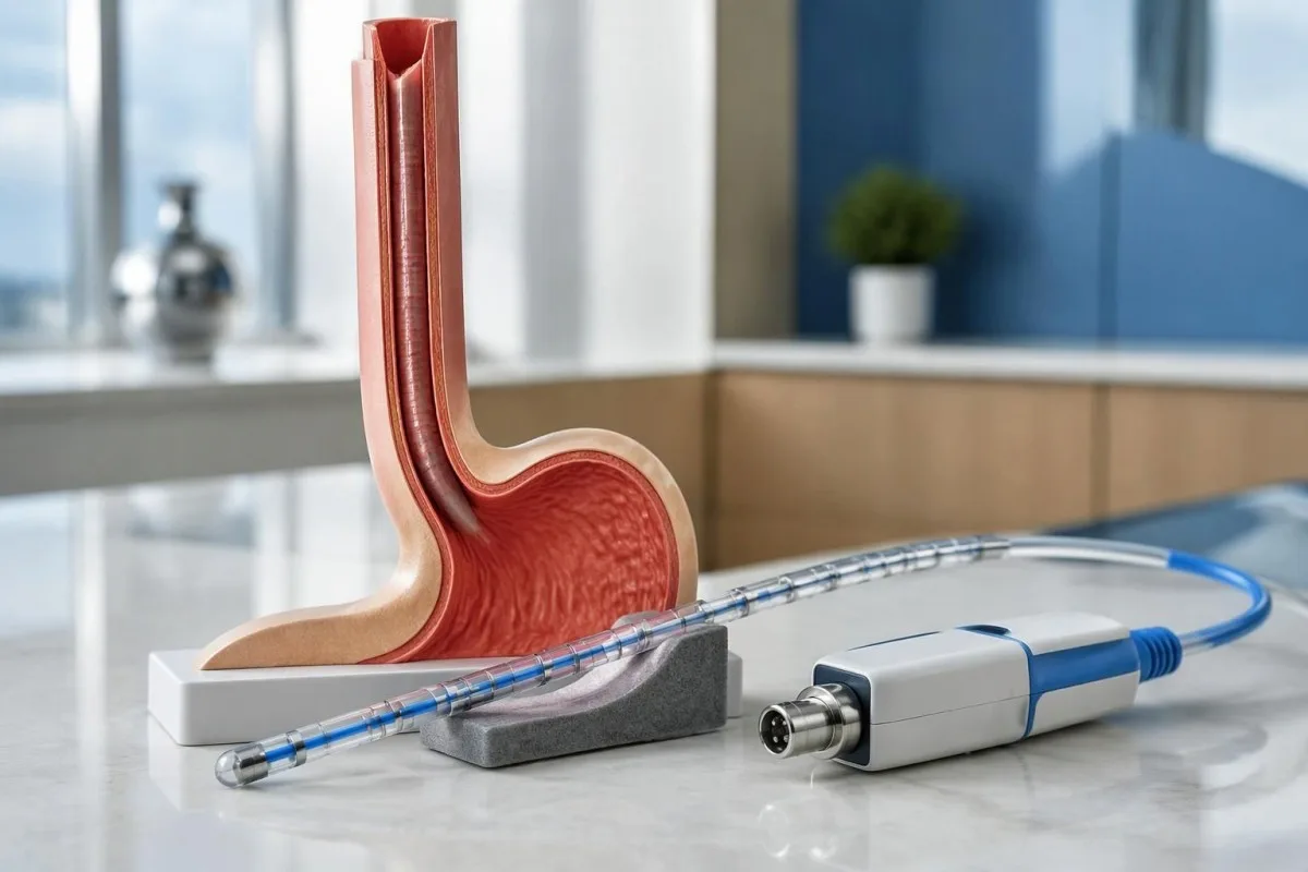
ความดันการคลายตัวแบบบูรณาการ, or IRP, summarizes junction pressure over a 4-second relaxation window within the post-swallow assessment period. It is not simply the lowest pressure reached, and its threshold changes with catheter system and posture; Chicago Classification version 4.0 gives different reference limits for commonly used systems rather than one universal 15 mmHg rule (Yadlapati et al., 2021).
Distal contractile integral, or DCI, combines contraction strength, duration, and length, using mmHg·s·cm. A failed swallow has DCI below 100 mmHg·s·cm; a sufficiently strong premature contraction generally requires DCI of at least 450 mmHg·s·cm, so a faint, early pressure blip should not automatically be classified as the spasm needed for type III.
Kantesti separates measurement units from clinical conclusions: a value in mmHg cannot be compared with a laboratory concentration in ng/mL. Our blood biomarker directory addresses a different measurement category; ask the manometry service which equipment-specific reference limit applies before treating a result such as IRP 17 mmHg as definitely abnormal.
| ระดับ | ช่วง | ความหมาย |
|---|---|---|
| Medtronic-system supine IRP | Upper reference limit approximately 15 mmHg | Chicago Classification version 4.0 reference; verify the laboratory's validated system and protocol. |
| Medtronic-system upright IRP | Upper reference limit approximately 12 mmHg | A lower positional limit applies; do not compare an upright result with a supine cutoff. |
| Laborie/Diversatek-system supine IRP | Upper reference limit approximately 22 mmHg | Different equipment can legitimately use a higher reference limit. |
| Laborie/Diversatek-system upright IRP | Upper reference limit approximately 15 mmHg | Interpret alongside peristalsis, symptoms, and supportive testing. |
| Premature contraction timing | Distal latency below 4.5 seconds | Supports a premature contraction when contraction strength is sufficient; does not independently diagnose achalasia. |
| Panesophageal pressurization | Uniform pressure contour at approximately 30 mmHg or above | Whole-esophagus pressurization in at least 20% of swallows helps define type II when the other achalasia criteria are met. |
อะคาเลเซียชนิดที่ I แสดงผลอย่างไรในการตรวจวัดความดัน?
อะคาเลเซียชนิดที่ I shows an abnormal median IRP and 100% failed peristalsis, without the panesophageal pressurization pattern required for type II. The esophageal body does not generate an effective propulsive wave; the absence of strong pressure does not mean that food passes normally or that the condition is mild.
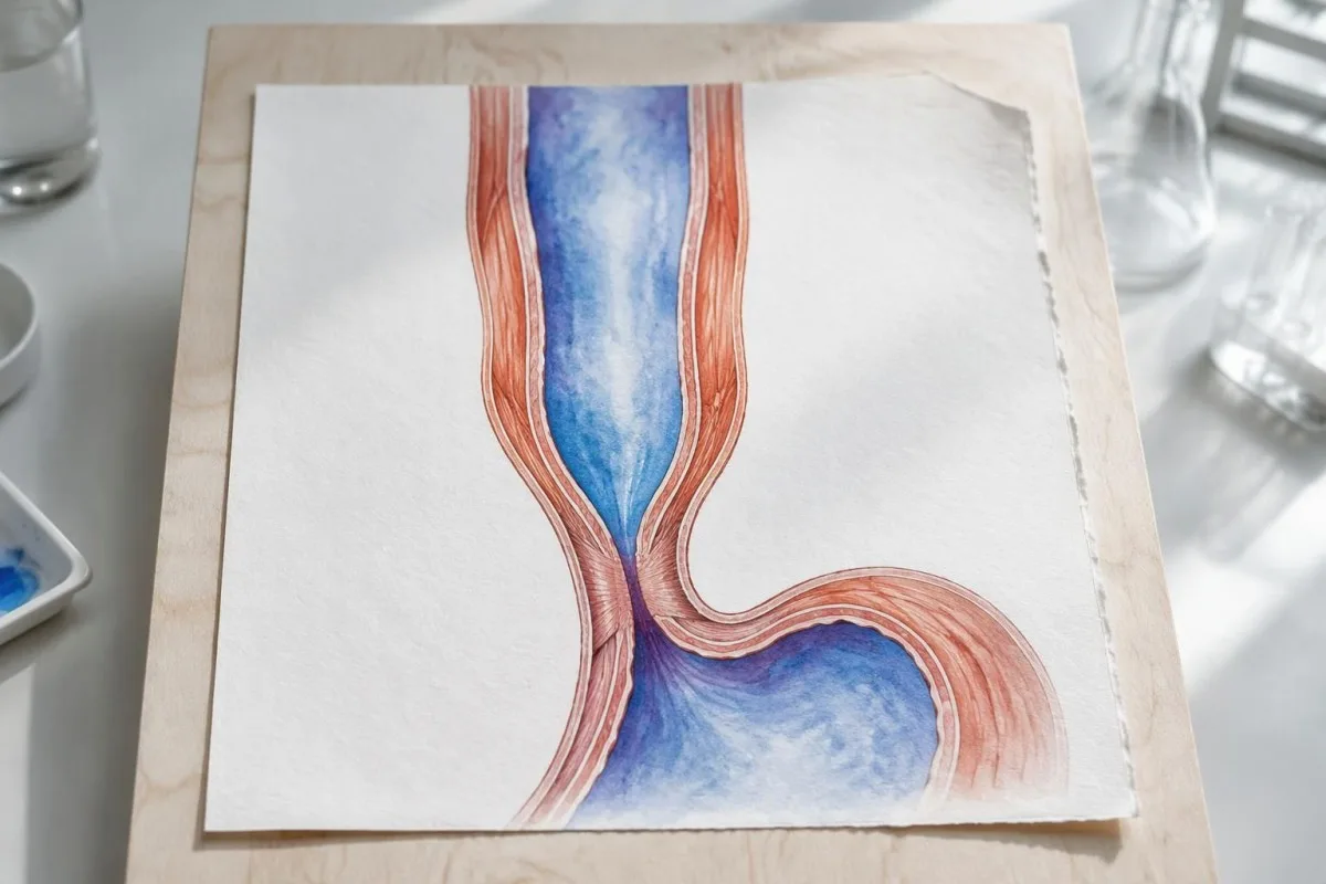
A flat-looking pressure plot can be misleading because failed contractions are not equivalent to successful relaxation. In a technically adequate study, 10 failed supine swallows combined with an elevated median IRP support type I, but the specialist still checks catheter placement, structural findings, and whether pressurization occurred frequently enough to meet type II criteria.
Esophageal dilation can reduce the ability to generate pressure, but type I is not a validated chronological stage that every person reaches after type II. The 3 subtypes describe current physiology rather than a guaranteed disease sequence; duration, esophageal shape, and previous treatment can affect the tracing without providing a reliable clock for when achalasia began.
Nutritional consequences depend on actual intake, not subtype alone. A hypothetical person with type I, declining weight, and ferritin 11 ng/mL needs attention to swallowing treatment and iron-deficiency symptoms; low iron stores do not prove achalasia, while a normal ferritin would not establish that nutrition or esophageal emptying is adequate.
อะไรทำให้อะคาเลเซียชนิดที่ II แตกต่างออกไป?
อะคาเลเซียชนิดที่ II combines an abnormal median IRP, 100% failed peristalsis, and panesophageal pressurization in at least 20% of swallows. Instead of a coordinated traveling contraction, pressure rises across much of the esophagus at once because swallowed material is compressed against an outlet that does not open adequately.
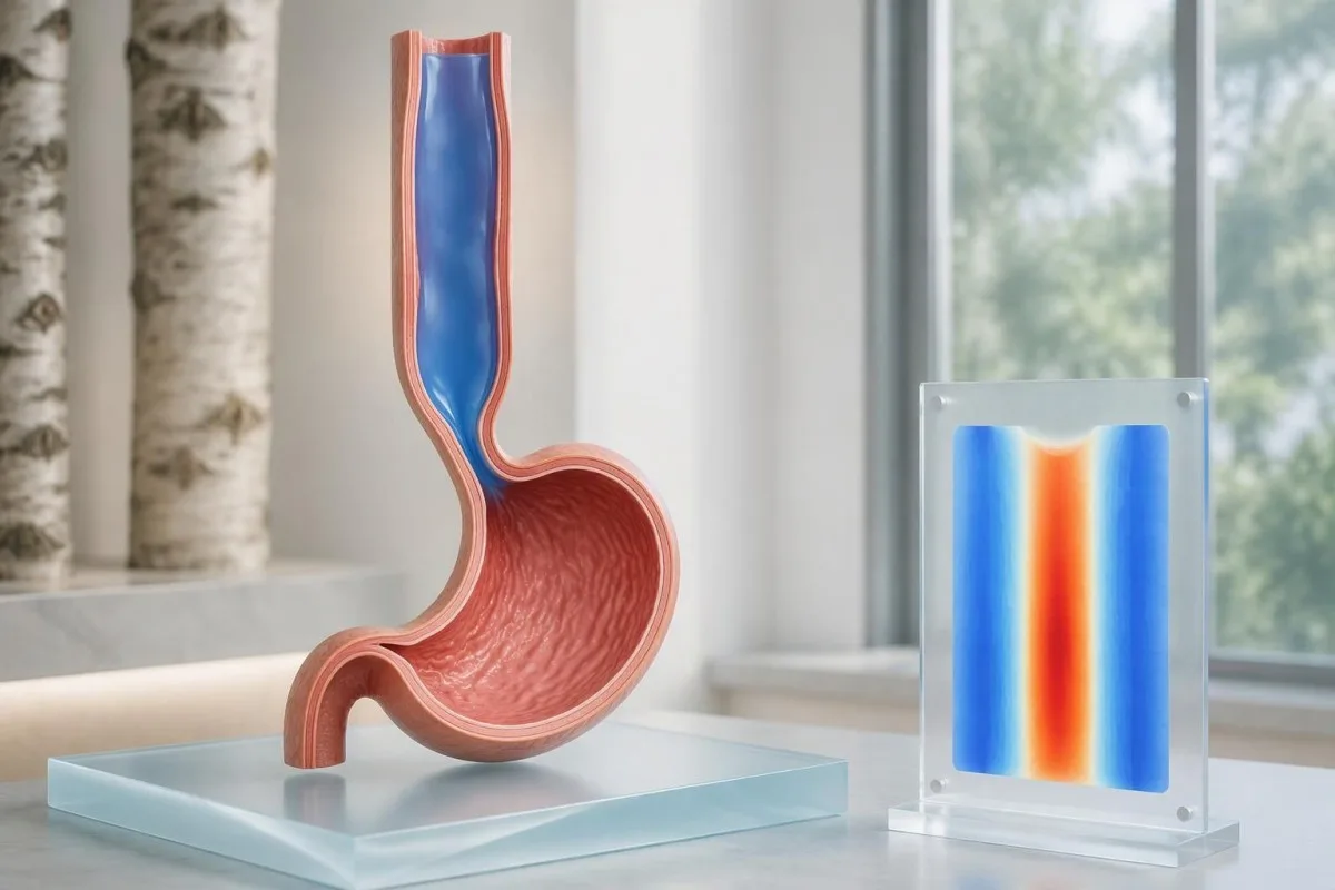
With 10 evaluable swallows, panesophageal pressurization in 2 swallows reaches the 20% threshold. That arithmetic is useful when reading a report, but it should not encourage self-classification from a screenshot: the interpreter must distinguish true pressure trapping from closely spaced swallows, movement artifacts, and pressure attributable to adjacent anatomy.
คำว่า panesophageal pressurization usually refers to a simultaneous pressure contour reaching approximately 30 mmHg across the esophageal body. A bright vertical band on a color map can represent this trapped pressure rather than a strong muscle wave; reading medical report explanations safely means checking the written interpretation rather than assuming warmer colors equal better propulsion.
Type II often has favorable outcomes after pneumatic dilation, Heller myotomy, or POEM, according to the ACG guideline (Vaezi et al., 2020). Nevertheless, the 20% diagnostic threshold is not a treatment-success percentage, and response still depends on anatomy, treatment technique, prior procedures, and follow-up; a subtype should inform discussion, not function as a personal guarantee.
การตรวจวัดความดันระบุอะคาเลเซียชนิดที่ III ได้อย่างไร?
อะคาเลเซียชนิดที่ III shows impaired junction relaxation, premature contractions in at least 20% of swallows, and no normal peristalsis. A premature contraction generally has distal latency below 4.5 seconds with adequate DCI; other swallows may fail, so type III does not require every swallow to produce a spasm.
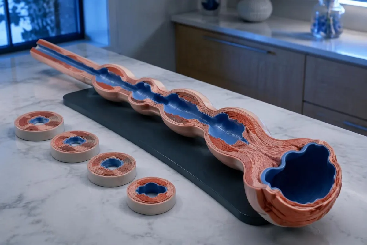
The absence of normal peristalsis separates conclusive type III achalasia from some other spastic patterns. If premature contractions coexist with otherwise preserved peristalsis and junction obstruction, the report may instead describe esophagogastric junction outflow obstruction with spastic features; the same 4.5-second timing cutoff does not erase that diagnostic distinction.
Chest pain can accompany spastic contractions, but it should not automatically be attributed to the esophagus. New severe chest pain, especially with breathlessness, sweating, or exertional onset, needs urgent assessment; chest-pain testing priorities differ from elective manometry, and no single esophageal measurement rules out a cardiac emergency.
Opioid exposure deserves particular scrutiny before an irreversible procedure for an apparent type III pattern. Chicago Classification version 4.0 recommends considering testing off opioids when feasible, with timing based on the medicine's half-life (Yadlapati et al., 2021); a fixed 24-hour pause is not appropriate for every drug, and any change requires the prescribing team's supervision.
รายงานผลการตรวจวัดความดันสมมติสามฉบับจะแตกต่างกันอย่างไร?
Hypothetical manometry examples show why an elevated IRP cannot determine the subtype by itself. Using a laboratory with a validated supine upper limit of 15 mmHg, the same median IRP of 24 mmHg could accompany type I, II, or III; the esophageal-body pattern supplies the distinguishing information.
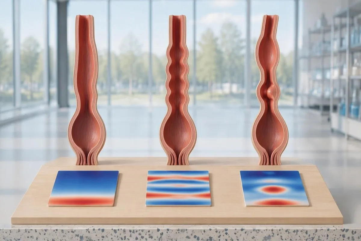
Example 1: a fictional report records median IRP 24 mmHg, DCI below 100 mmHg·s·cm in all 10 swallows, and no panesophageal pressurization. Assuming adequate technique, no structural explanation, and consistent symptoms, this is a type I pattern; the conclusion comes from failed propulsion plus impaired relaxation, not from the pressure value alone.
Example 2: another fictional report records IRP 24 mmHg, failed peristalsis in all 10 swallows, and panesophageal pressurization in 6 swallows. The pressurization frequency is 60%, exceeding the 20% threshold for type II; this does not mean the condition is three times more severe than a report with pressurization in 2 swallows.
Example 3: a fictional study records IRP 24 mmHg, 4 premature contractions with distal latency 3.8 seconds and DCI 900 mmHg·s·cm, and 6 failed swallows, with no normal peristalsis. This supports type III; Kantesti is an แพลตฟอร์มการแปลผลตรวจเลือดของ AI, และของเรา เวิร์กโฟลว์ตัวอย่างสำหรับการตีความ concern laboratory information rather than automated classification of these hypothetical motility tracings.
หาก IRP อยู่ในเกณฑ์ปกติ ก้ำกึ่ง หรือไม่สอดคล้องกัน จะเป็นอย่างไร?
A normal or borderline IRP does not always exclude achalasia, particularly when peristalsis is absent and symptoms strongly suggest impaired emptying. Conversely, elevated IRP with preserved peristalsis is not automatically achalasia; Chicago Classification version 4.0 requires careful separation of inconclusive achalasia, artifacts, and esophagogastric junction outflow obstruction.
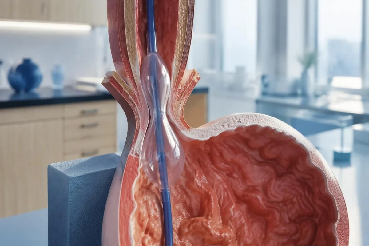
An elevated supine IRP that normalizes upright can reflect catheter contact or distortion around a hiatal hernia. This is one reason the standard protocol uses 2 positions: a reproducible obstruction pattern is more convincing than one abnormal posture-dependent number, although positional disagreement still needs specialist interpretation rather than an automatic normal label.
Esophagogastric junction outflow obstruction, or EGJOO, requires more than a high median IRP. Its manometric criteria include elevated IRP in both positions, preserved peristalsis, and elevated supine intrabolus pressure in at least 20% of swallows; clinically meaningful EGJOO also requires relevant symptoms and supportive evidence of obstruction on timed barium testing or FLIP.
Absent contractility with normal relaxation can occur in systemic sclerosis and is not the same as achalasia; scleroderma-related antibody clues cannot replace motility testing. Kantesti's ควรทำความเข้าใจอย่างรอบคอบเช่นกัน การแปลผลรายงานเลือดไม่ใช่การตรวจสอบความถูกต้องของคะแนนความเสี่ยงต่อการแพ้ยา และผลตรวจทางห้องปฏิบัติการที่เป็นบวกเพียง 1 รายการไม่อาจรับรองว่ามีคุณสมบัติเข้ารับการทดสอบกระตุ้น should not be extended to these 2 distinct pressure disorders, and an irreversible sphincter procedure should not follow from an unexplained borderline result alone.
เหตุใดจึงต้องส่องกล้องก่อนวินิจฉัยว่าเป็นอะคาเลเซีย?
Upper endoscopy helps exclude a mechanical obstruction or pseudoachalasia, including an obstructing growth at the esophagogastric junction. Manometry can demonstrate an achalasia-like pressure pattern without proving its cause; the ACG guideline recommends endoscopy in suspected achalasia because treatment decisions depend on distinguishing primary motility disease from structural disease (Vaezi et al., 2020).
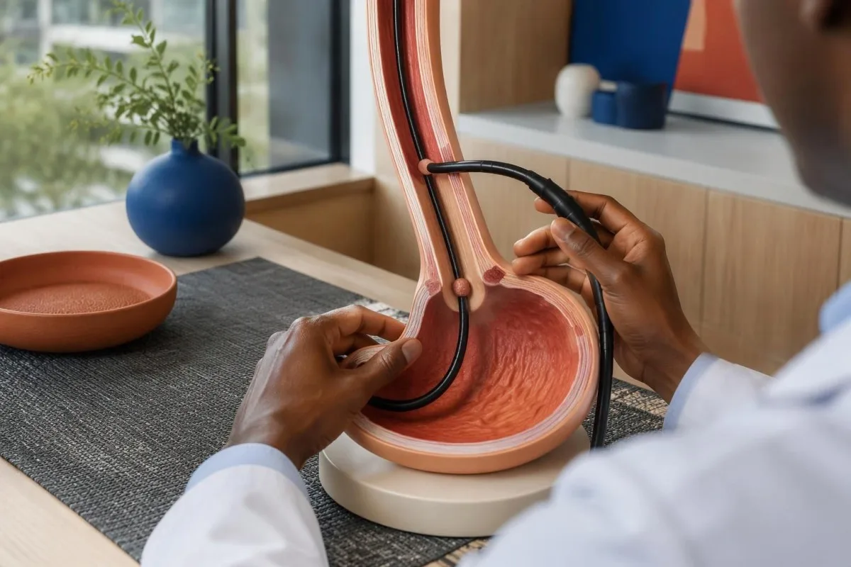
Retained saliva, retained food, and a tight-looking junction can support suspicion of achalasia, but none is diagnostic by itself. Early disease may have an almost normal endoscopic appearance; equally, resistance to passing the scope should not be dismissed as ordinary achalasia without considering whether a second test is needed to inspect the junction more closely.
Rapidly progressive symptoms, substantial short-term weight loss, older age at presentation, and unusual resistance at the junction increase concern for pseudoachalasia. There is no single age or kilogram cutoff that safely excludes it; a hypothetical 68-year-old with 3 months of progressive dysphagia and 9 kg weight loss warrants particular attention to the possibility of a structural cause.
A normal-looking mucosal surface does not always exclude a deeper or externally compressing growth, so CT or endoscopic ultrasound may be appropriate when suspicion persists. Normal CA 19-9 results cannot rule out this concern; ordering 1 tumor marker is not an alternative to imaging, endoscopy, or targeted tissue examination when clinically indicated.
การกลืนแบเรียมแบบจับเวลาและ FLIP ให้ข้อมูลเพิ่มเติมอย่างไร?
A timed barium esophagram measures esophageal emptying, while FLIP assesses how widely the esophagogastric junction opens under controlled distension. These tests provide information that pressure measurements alone cannot supply; a commonly used timed protocol obtains images at 1, 2, and 5 minutes after a standardized barium swallow.
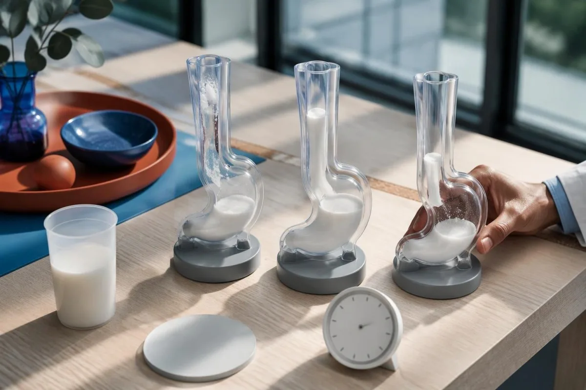
A standardized upright timed study often uses about 236 mL of barium; column heights above 5 cm at 1 minute or 2 cm at 5 minutes suggest impaired emptying in commonly used protocols. These are protocol-dependent abnormal-emptying thresholds, not stand-alone proof of achalasia, and the classic bird-beak appearance can be absent in early disease.
A 13 mm barium tablet can help reveal restricted passage when liquid contrast empties relatively well, provided the imaging team considers it appropriate. FLIP distensibility index expresses junction cross-sectional area divided by pressure in mm²/mmHg, but balloon volume, sedation, device protocol, and maximum opening diameter affect interpretation; treating every value below 2 as an interchangeable diagnosis would oversimplify the test.
An achalasia-like dilated esophagus does not establish that the disorder is idiopathic. In someone with relevant exposure history, confirmatory Chagas testing may be appropriate because chronic Trypanosoma cruzi infection can affect esophageal innervation; imaging and infection testing answer 2 different questions, namely how the esophagus empties and why its function may be impaired.
ประเภทของอะคาเลเซียจากการตรวจวัดความดันช่วยประกอบการรักษาอย่างไร?
Achalasia manometry types help clinicians choose how best to reduce resistance at the esophageal outlet. Pneumatic dilation, laparoscopic Heller myotomy, and POEM are established options for types I and II; type III often favors a longer, tailored myotomy that addresses the spastic segment as well as the junction.
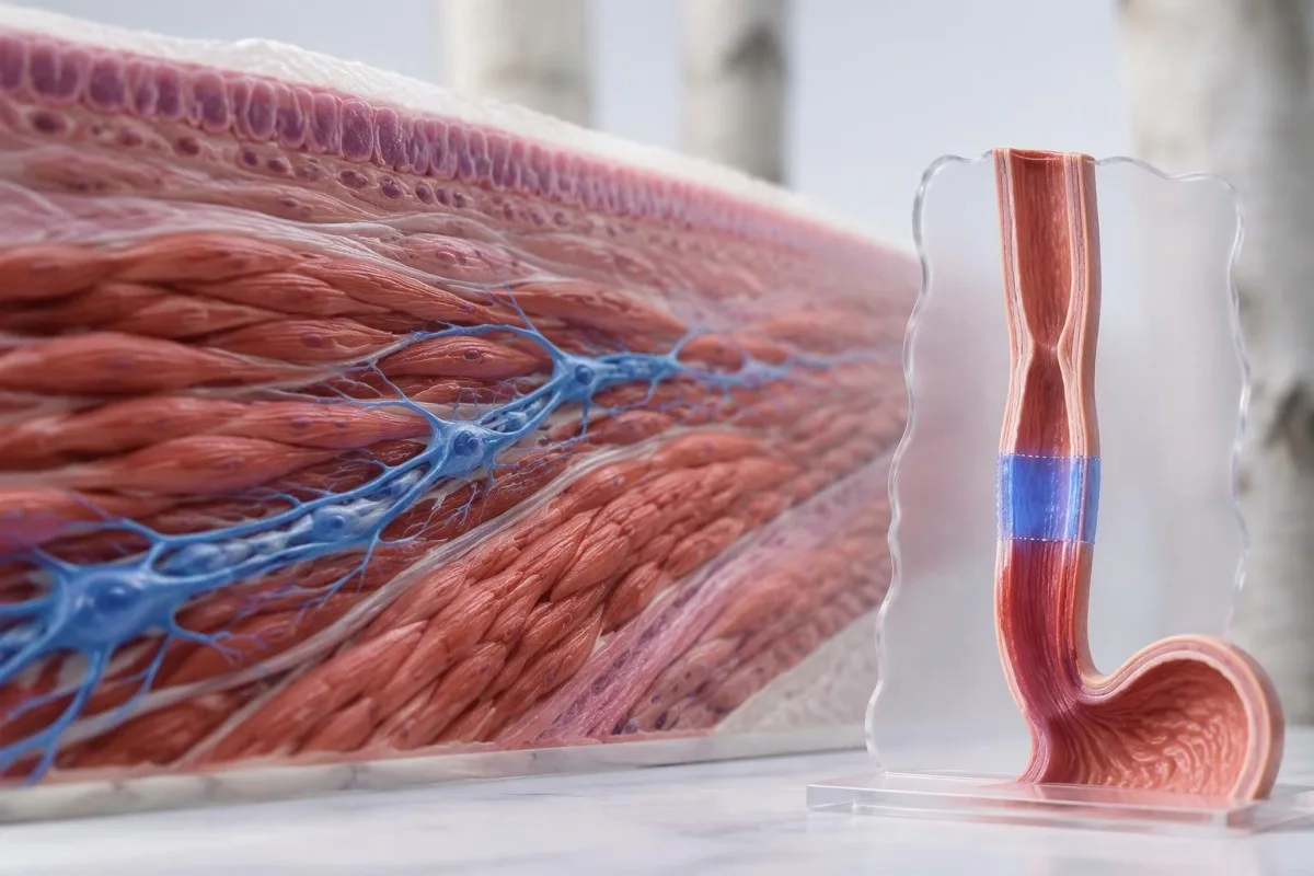
The ACG guideline considers pneumatic dilation, Heller myotomy, and POEM comparably effective options for appropriate patients with types I and II, while supporting tailored POEM or Heller myotomy over dilation for type III (Vaezi et al., 2020). The 3 procedures differ in anesthesia needs, recovery, retreatment possibilities, and reflux risk; local expertise and patient preference belong in the decision.
A randomized trial of 221 patients found clinical success at 2 years in 83.0% after POEM and 81.7% after Heller myotomy with Dor fundoplication (Werner et al., 2019). These overall results support both approaches but do not establish identical outcomes for every subtype; type III participants form a much smaller subgroup than the full trial population.
Reflux esophagitis at 2 years occurred in 44% after POEM versus 29% after Heller myotomy in that trial, illustrating a trade-off rather than a reason to reject POEM automatically. Acid-suppression treatment may subsequently affect gastrin blood results; meanwhile, pneumatic dilation carries a perforation risk of roughly 2% in experienced practice and requires an appropriate rescue pathway.
การติดตามผลและการตรวจเลือดหลังการรักษาอาจแสดงอะไรได้บ้าง?
Follow-up assesses swallowing, weight, esophageal emptying, and reflux, not just whether symptoms improve. Blood tests can identify nutritional or hydration complications but cannot confirm that the junction opens normally; the ACG guideline recommends timed barium esophagram as the first-line objective test when symptoms persist or recur after treatment (Vaezi et al., 2020).
การ Eckardt score adds dysphagia, regurgitation, chest pain, and weight loss, each scored from 0 to 3, for a total of 0–12. Trials often define success as a score of 3 or below, but symptom scores alone can miss retained material; reduced discomfort does not necessarily mean that esophageal clearance has normalized.
คันเตสตีเป็น แพลตฟอร์มการตีความไบโอมาร์กเกอร์ด้วย AI that can help explain a clinician-requested complete blood count, electrolytes, and iron studies alongside reduced intake. Ferritin below 15 ng/mL strongly supports depleted iron stores in adults without a confounding context; our การศึกษาธาตุเหล็กที่สมบูรณ์ explains why ferritin and transferrin saturation should be considered together rather than used to infer an achalasia subtype.
Albumin commonly has an adult reference interval around 35–50 g/L, but a normal value does not exclude inadequate intake and a low value is not specific for malnutrition. Our คู่มือการตีความโปรตีนในซีรั่ม explains those limitations; magnesium deserves separate attention when poor intake or long-term acid suppression is present, as discussed in สาเหตุของแมกนีเซียมต่ำ.
ควรถามอะไรต่อ และหลักฐานใดเกี่ยวข้อง?
Your next discussion should connect the manometry classification, structural assessment, and evidence of impaired emptying before selecting treatment. Ask 3 practical questions: which findings establish the subtype, whether obstruction or medication effects have been excluded, and how the proposed procedure's expected benefit compares with its reflux and retreatment risks.
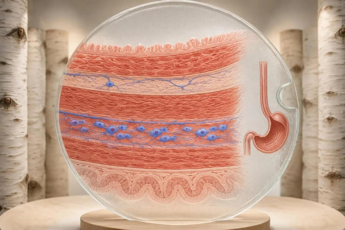
Request the full manometry report rather than only its conclusion, including the catheter system, body positions, median IRP, swallow counts, and any supportive tests. If you cannot swallow liquids or saliva, develop severe chest pain, or have respiratory difficulty, seek urgent care; waiting another 2 weeks for a routine report review is not an appropriate response to those warning signs.
The achalasia evidence cited here comes from Chicago Classification version 4.0, the ACG achalasia guideline, and the randomized POEM-versus-Heller trial. Kantesti's มาตรฐานเนื้อหาทางการแพทย์ distinguish evidence-based explanations from individualized diagnosis; the 2 Zenodo publications listed below concern coagulation and serum proteins, not studies validating manometry or achalasia treatment.
The serum-protein publication is relevant only to the laboratory-support discussion, while the coagulation publication provides separate background if a clinician requests clotting tests before a procedure. Neither DOI should be used to choose among types I, II, and III; when laboratory values change during recovery, blood-test timeline effects help explain why intake, illness, hydration, and medicines may alter results without indicating recurrent outlet obstruction.
คำถามที่พบบ่อย
วินิจฉัยภาวะอะคาเลเซียได้อย่างไร?
ภาวะอะคาเลเซียมักได้รับการวินิจฉัยด้วยการตรวจวัดความดันหลอดอาหารความละเอียดสูง ซึ่งแสดงให้เห็นการคลายตัวของรอยต่อหลอดอาหารและกระเพาะอาหารที่บกพร่องและไม่มีการบีบตัวเป็นคลื่นตามปกติ รูปแบบความดันจะแยกชนิดที่ I, II และ III การส่องกล้องทางเดินอาหารส่วนต้นช่วยคัดกรองการอุดกั้นเชิงโครงสร้าง ส่วนการกลืนแบเรียมแบบจับเวลาหรือ FLIP อาจช่วยให้ผลตรวจที่ไม่ชัดเจนกระจ่างขึ้น ไม่มีการตรวจเลือดเป็นกิจวัตรใดที่ยืนยันชนิดย่อยทั้ง 3 ชนิดได้.
การตรวจวัดความดันหลอดอาหารแสดงอะไรในภาวะอะคาเลเซีย?
Esophageal manometry shows how the junction relaxes after swallowing and whether the esophageal body produces coordinated contractions. Integrated relaxation pressure assesses outlet relaxation over a 4-second window, while contraction timing and pressure distribution help identify the subtype. Type II requires panesophageal pressurization in at least 20% of swallows, and type III requires premature contractions in at least 20%. Equipment-specific limits must be used because one universal IRP cutoff is not appropriate.
อะไรคือความแตกต่างระหว่างอะคาเลเซียชนิดที่ I, II และ III?
อะคาเลเซียชนิดที่ I มีการบีบตัวเป็นคลื่นที่ล้มเหลว โดยไม่มีแรงดันเพิ่มขึ้นทั่วหลอดอาหารซึ่งจำเป็นสำหรับชนิดที่ II ชนิดที่ II มีการบีบตัวเป็นคลื่นที่ล้มเหลวร่วมกับแรงดันเพิ่มขึ้นทั่วหลอดอาหารในการกลืนอย่างน้อย 20% ครั้ง ชนิดที่ III มีการหดตัวก่อนกำหนดในการกลืนอย่างน้อย 20% ครั้ง และไม่มีการบีบตัวเป็นคลื่นตามปกติ ร่วมกับการคลายตัวของรอยต่อที่บกพร่อง ลักษณะเหล่านี้เป็นรูปแบบทางสรีรวิทยา ไม่ใช่ระยะของการดำเนินโรคที่รับประกันว่าจะเกิดขึ้นตามลำดับ.
โรคอะคาเลเซียสามารถเกิดขึ้นได้โดยมีค่า IRP ปกติหรือไม่?
ภาวะอะคาเลเซียอาจยังเป็นข้อกังวลได้ในบางครั้ง แม้ค่า integrated relaxation pressure จะปกติหรืออยู่ในเกณฑ์ก้ำกึ่ง โดยเฉพาะเมื่อไม่มีการบีบตัวเป็นคลื่นและการระบายผ่านหลอดอาหารบกพร่อง ในสถานการณ์เช่นนี้ ผลการตรวจ manometry อาจสรุปไม่ได้ แทนที่จะถือว่าปกติอย่างชัดเจน การตรวจกลืนแบเรียมแบบจับเวลา ซึ่งโดยทั่วไปถ่ายภาพที่ 1, 2 และ 5 นาที หรือการตรวจ FLIP อาจให้หลักฐานสนับสนุน ผู้เชี่ยวชาญควรพิจารณาภาวะไม่มีการหดตัว ข้อจำกัดทางเทคนิค และการรักษาที่เคยได้รับด้วย.
หากเป็นโรคอะคาเลเซีย ผลการส่องกล้องอาจปกติได้หรือไม่?
การส่องกล้องอาจดูเกือบปกติในภาวะอะคาเลเซียระยะแรก เพราะตรวจดูโครงสร้างและเยื่อเมือก แทนที่จะวัดแรงดันขณะกลืนโดยตรง เศษอาหาร น้ำลาย หรือรอยต่อที่ตีบแน่นอาจทำให้สงสัยภาวะนี้ แต่ยังไม่สามารถยืนยันการวินิจฉัยได้ การตรวจวัดความดันหลอดอาหารช่วยระบุรูปแบบแรงดันที่เป็นลักษณะเฉพาะและแยกความแตกต่างของชนิดย่อยทั้ง 3 ชนิดได้ หากยังคงสงสัยภาวะอะคาเลเซียเทียม อาจจำเป็นต้องตรวจ CT หรืออัลตราซาวนด์ผ่านกล้อง แม้พื้นผิวเยื่อเมือกจะดูปกติก็ตาม.
อะคาเลเซียชนิดใดตอบสนองต่อการรักษาได้ดีที่สุด?
Type II achalasia generally has favorable responses to pneumatic dilation, Heller myotomy, and POEM, but subtype alone cannot predict an individual's result. Type III often benefits from a longer myotomy tailored to the spastic segment. A randomized trial reported 2-year success of 83.0% after POEM and 81.7% after Heller myotomy with fundoplication across its overall study population. Reflux, anatomy, prior treatment, and procedural expertise also influence the choice.
คุณจำเป็นต้องได้รับยาระงับประสาทสำหรับการตรวจวัดความดันในหลอดอาหารหรือไม่?
Standard esophageal manometry is usually performed awake, often with local nasal anesthetic rather than intravenous sedation. Patients complete a structured swallowing protocol, commonly 10 supine and 5 upright water swallows. Difficult catheter placement sometimes requires endoscopic assistance, but any sedation and recovery interval must be considered when interpreting the subsequent measurements. Follow the testing center's fasting instructions because achalasia can retain food despite an ordinary fast.
อ่านผลตรวจเลือดอย่างไร
อัปโหลดรายงานผลตรวจของคุณ แล้ว AI ของ Kantesti จะอธิบายตัวบ่งชี้ทางชีวภาพแต่ละรายการด้วยภาษาที่เข้าใจง่ายภายในไม่กี่วินาที มีผู้ใช้ทั่วโลกมากกว่า 2 ล้านคน.
เอกสารอ้างอิง
- ไคลน์, ที., มิตเชลล์, เอส., & เวเบอร์, เอช. (2026). ช่วงค่าปกติของ aPTT: คู่มือ D-Dimer และ Protein C เกี่ยวกับการแข็งตัวของเลือด. Kantesti AI การวิจัยทางการแพทย์. โดอิ
- ไคลน์, ที., มิตเชลล์, เอส., & เวเบอร์, เอช. (2026). คู่มือโปรตีนในซีรัม: การตรวจเลือดหาโกลบูลิน อัลบูมิน และอัตราส่วน A/G. Kantesti AI การวิจัยทางการแพทย์. โดอิ
- Yadlapati R et al. (2021). Esophageal motility disorders on high-resolution manometry: Chicago classification version 4.0. Neurogastroenterology & Motility. โดอิ
- Vaezi MF et al. (2020). ACG Clinical Guidelines: Diagnosis and Management of Achalasia. วารสารอเมริกันด้านโรคทางเดินอาหาร. โดอิ
- Werner YB et al. (2019). Endoscopic or Surgical Myotomy in Patients with Idiopathic Achalasia. The New England Journal of Medicine. โดอิ
- 2 ล้าน+การตรวจที่วิเคราะห์
- 127+ประเทศ
- 100+ภาษา
เกี่ยวกับทีมแพทย์


สัญญาณความน่าเชื่อถือ E-E-A-T
ประสบการณ์
การทบทวนเชิงคลินิกโดยแพทย์ที่นำโดยกระบวนการตีความผลตรวจในห้องแล็บ.
ความเชี่ยวชาญ
โฟกัสด้านเวชศาสตร์ห้องปฏิบัติการเกี่ยวกับพฤติกรรมของไบโอมาร์กเกอร์ในบริบททางคลินิก.
อำนาจ
ทบทวนทางการแพทย์โดย นพ. Thomas Klein พร้อมผลงานจาก นพ. Sarah Mitchell และศาสตราจารย์ ดร. Hans Weber.
ความน่าเชื่อถือ
การตีความที่อิงหลักฐาน พร้อมเส้นทางการติดตามที่ชัดเจนเพื่อลดความตื่นตระหนก.

ข้อจำกัดความรับผิดชอบทางการแพทย์
บทความนี้จัดทำเพื่อวัตถุประสงค์ด้านการศึกษาเท่านั้น และไม่ถือเป็นคำแนะนำทางการแพทย์ โปรดปรึกษาผู้ให้บริการด้านสุขภาพที่มีคุณสมบัติเหมาะสมเสมอสำหรับการตัดสินใจด้านการวินิจฉัยและการรักษา.
เผยแพร่แล้ว · ติดต่อทีมบรรณาธิการ
เผยแพร่โดย Kantesti Ltd จดทะเบียนในอังกฤษและเวลส์ เลขทะเบียนบริษัท. 17090423, ลอนดอน สหราชอาณาจักร.
