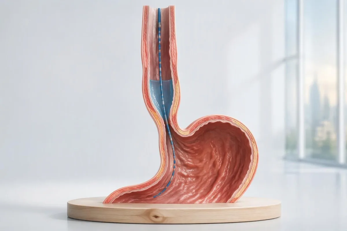
Kipimo cha shinikizo la juu pekee hakithibitishi akalasia. Utambuzi hutegemea jinsi njia ya kutoka kwenye umio inavyolegea, iwapo vitendo vya kumeza husababisha mikazo iliyoratibiwa, na iwapo hali nyingine inaeleza muundo huo.

Mambo muhimu ya kukumbuka
- Utambuzi wa akalasia kwa kawaida huhitaji manometria ya ubora wa juu inayoonyesha kulegea kwa njia ya kutoka kwenye umio kulikoharibika na kutokuwepo kwa peristalsisi ya kawaida ya umio; endoskopi hukagua uwezekano wa sababu ya kimitambo.
- Shinikizo la kulegea jumuishi hupima kulegea kwa njia ya kutoka kwenye umio katika kipindi cha sekunde 4, lakini kikomo chake cha juu hutegemea kifaa na mkao wa mwili.
- Akalasia ya aina ya I huonyesha 100% kutofanyika kwa peristalsisi bila shinikizo la juu katika umio mzima linalohitajika kwa aina ya II.
- Akalasia ya aina ya II huonyesha 100% kutofanyika kwa peristalsisi na shinikizo la juu katika umio mzima katika angalau 20% ya vitendo vya kumeza vya kipimo.
- Akalasiani ya Aina ya III huonyesha mikazo ya mapema katika angalau 20% ya vitendo vya kumeza, kulegea kwa tundu la kutoka kwa shida, na kutokuwepo kwa peristalsis ya kawaida.
- Mkazo wa mapema kwa kawaida humaanisha muda wa kuchelewa kwa sehemu ya mbali chini ya sekunde 4.5 pamoja na nguvu ya kutosha ya mkazo; rangi zinazoonekana kuwa za kasi pekee hazithibitishi mkazo wa ghafla.
- Matokeo yaliyo mpakani yanaweza kuhitaji esofagogramu ya bariamu ya muda au tathmini ya FLIP; shinikizo lililoinuka pekee halitoshi kuhalalisha matibabu yasiyoweza kutenduliwa.
- Majadiliano kuhusu matibabu hutofautiana kulingana na aina ndogo: aina ya III mara nyingi hupendelea myotomia ndefu iliyobinafsishwa, ilhali aina za I na II zina chaguo kadhaa zenye ufanisi.
- Vipimo vya damu inaweza kutathmini upungufu wa maji mwilini, anemia, au matatizo ya lishe, lakini hakuna jopo la kawaida la vipimo vya maabara linalothibitisha au kuondoa uwezekano wa akalasiani.
Akalasia hutambuliwaje kwa kutumia manometria ya ubora wa juu?
Utambuzi wa akalasia kwa kawaida hutegemea manometria ya azimio la juu inayoonyesha kulegea kwa shida kwa tundu la kutoka la umio na kutokuwepo kwa peristalsis ya kawaida. Aina ya I ina mikazo iliyoshindwa, aina ya II huongeza shinikizo katika umio mzima katika angalau 20% ya vitendo vya kumeza, na aina ya III huongeza mikazo ya mapema katika angalau 20%; endoskopi husaidia kuondoa uwezekano wa kizuizi.
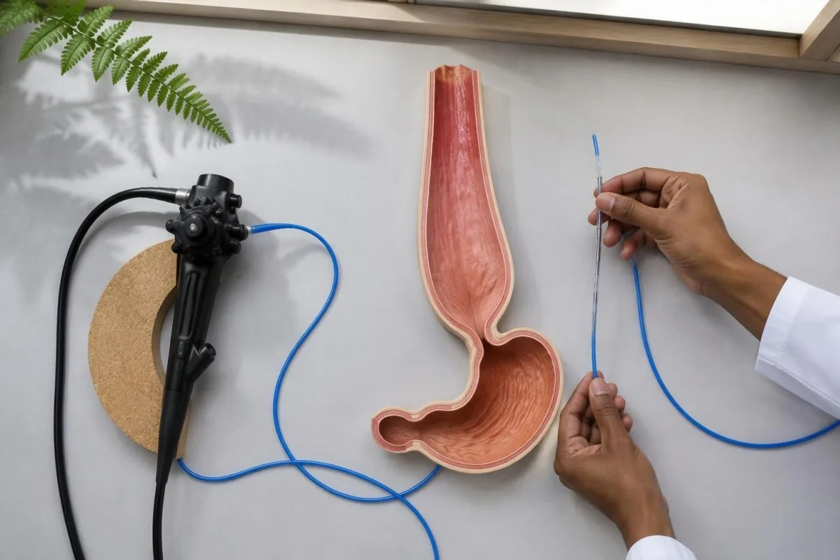
Manometria ya azimio la juu, au HRM, hupima shinikizo badala ya kurekodi moja kwa moja mwendo wa chakula. Maswali yake 2 ya msingi ni kama makutano ya umio na tumbo hufunguka ipasavyo wakati wa kumeza na kama mwili wa umio hutengeneza wimbi lililopangika; shinikizo kubwa la sphincter wakati wa kupumzika pekee halijibu swali lolote kati ya hayo na halithibitishi akalasiani.
Chicago Classification toleo la 4.0 hufafanua mifumo ya manometria inayotumiwa kutambua aina ndogo 3 za akalasiani, ilhali dalili na uchunguzi mwingine hubainisha maana yake ya kitabibu (Yadlapati et al., 2021). Ripoti inayosema tu ugonjwa wa motility haitoi taarifa nyingi kama ile inayoandika shinikizo jumuishi la kulegea, matokeo ya peristalsis, mkao wa uchunguzi, na uainishaji wa mwisho.
Kantesti ni Mchambuzi wa mtihani wa damu wa AI zinazoweza kusaidia kueleza matokeo ya maabara ya ulaji duni, lakini hakuna hata moja kati ya aina ndogo 3 za akalasiani inayoweza kutambuliwa kutokana na jopo la vipimo vya damu. Taarifa kuhusu shirika letu zinapaswa kusomwa kando na tathmini ya mtaalamu inayohitajika kutafsiri manometria na kuondoa uwezekano wa ugonjwa wa kimuundo.
Ni dalili zipi za kumeza zinazopaswa kupelekea kupimwa akalasia?
Ugumu wa kumeza vitu vigumu na vimiminika, kurudishwa kwa chakula kilichobaki, na kupungua uzito kusikoelezeka kunapaswa kuibua uchunguzi wa akalasiani. Dalili pekee haziwezi kubainisha utambuzi au kutofautisha aina zake ndogo 3; hata historia ya dalili inayoshawishi inahitaji vipimo kwa sababu kizuizi, ugonjwa unaohusiana na refluksi, na matatizo mengine ya kumeza yanaweza kufanana.
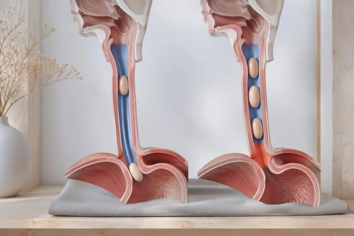
Kurudishwa kwa chakula katika akalasiani kunaweza kuwa na chakula kilicholiwa saa kadhaa mapema, ilhali refluksi ya asidi mara nyingi zaidi hurudisha kimiminika chachu juu. Tofauti hii husaidia kueleza kwa nini matibabu ya muda mrefu ya dalili zinazofanana na refluksi huenda yasisuluhishe matatizo ya kumeza; hata hivyo, hali hizi 2 zinaweza kutokea pamoja, na maelezo ya dalili hayawezi kuzitofautisha kwa uhakika bila uchunguzi.
Ugumu wa kuanzisha tendo la kumeza huashiria eneo tofauti na chakula kukwama sekunde kadhaa baada ya kumeza. Kukohoa mara moja unapokunywa funda, sauti ya puani, au uchovu wakati wa kumeza kunaweza kuhitaji tathmini ya orofaringe au ya mfumo wa neva; vikwazo vya vipimo vya myasthenia ni muhimu wakati udhaifu unaambatana na dalili, si kama jopo la kawaida la uchunguzi wa awali wa akalasiani.
An inability to swallow saliva or a suspected food impaction requires urgent assessment rather than waiting for elective manometry. Repeated nighttime regurgitation also raises concern for aspiration; for a hypothetical person losing 6 kg while meals become progressively slower, the weight trajectory and swallowing history both deserve attention even if routine laboratory results remain within range.
Nini hutokea wakati wa kipimo cha manometria ya umio?
Esophageal manometry usually involves passing a thin pressure-sensing catheter through the nose into the stomach while the patient is awake. A standard Chicago Classification version 4.0 protocol commonly includes 10 supine and 5 upright water swallows, with additional maneuvers that test whether an apparent abnormality persists under different conditions.
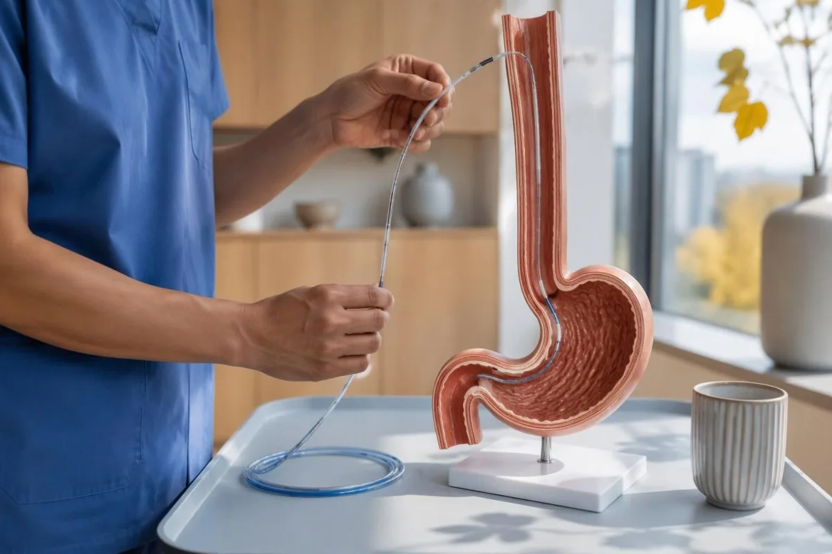
Preparation commonly includes at least 6 hours without food, but achalasia can leave material in the esophagus despite ordinary fasting. The testing center may therefore give a longer fasting or modified-diet plan; follow its instructions rather than assuming that an empty stomach guarantees an empty esophagus, especially if retained food was seen during endoscopy.
Medication review matters because opioids can produce impaired junction relaxation or spastic patterns that resemble type III achalasia. Provide the exact medicine, dose, and last-dose time; medication-related laboratory effects are a separate issue, but a single accurate medication list helps both investigations, and prescribed medicines should not be stopped without guidance.
Provocative testing may include 5 closely spaced 2 mL swallows or a rapid drink challenge of about 200 mL, if safe and appropriate. These maneuvers test different aspects of relaxation and contraction reserve; when standard swallows conflict with symptoms, their results can reveal a reproducible obstruction pattern or expose an artifact that disappears after changing position.
Ni matokeo gani ya manometria ya umio yaliyo muhimu zaidi?
Kiwango cha matumizi zaidi esophageal manometry results are median integrated relaxation pressure, the presence or absence of peristalsis, panesophageal pressurization, and distal latency. These measurements answer different questions: outlet opening, coordinated propulsion, pressure trapped within the esophagus, and contraction timing; none should be interpreted as an isolated severity score.
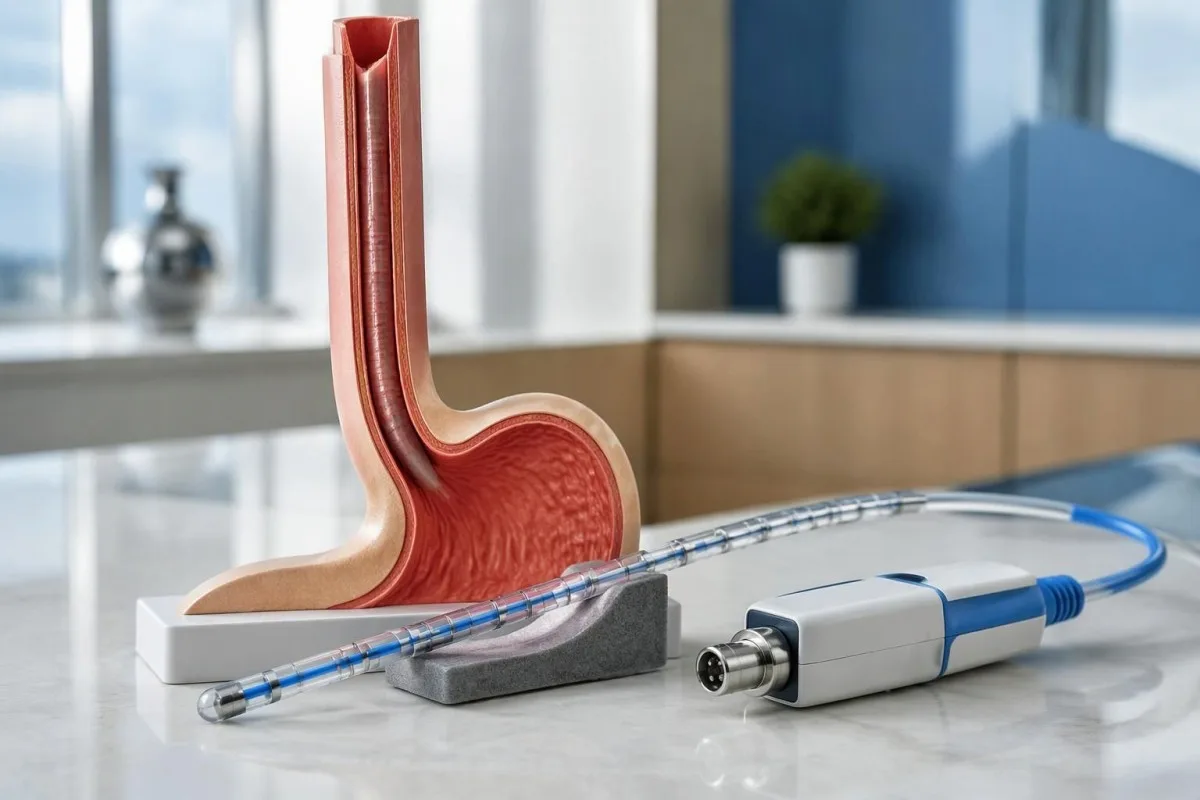
Shinikizo la kulegea jumuishi, or IRP, summarizes junction pressure over a 4-second relaxation window within the post-swallow assessment period. It is not simply the lowest pressure reached, and its threshold changes with catheter system and posture; Chicago Classification version 4.0 gives different reference limits for commonly used systems rather than one universal 15 mmHg rule (Yadlapati et al., 2021).
Distal contractile integral, or DCI, combines contraction strength, duration, and length, using mmHg·s·cm. A failed swallow has DCI below 100 mmHg·s·cm; a sufficiently strong premature contraction generally requires DCI of at least 450 mmHg·s·cm, so a faint, early pressure blip should not automatically be classified as the spasm needed for type III.
Kantesti separates measurement units from clinical conclusions: a value in mmHg cannot be compared with a laboratory concentration in ng/mL. Our blood biomarker directory addresses a different measurement category; ask the manometry service which equipment-specific reference limit applies before treating a result such as IRP 17 mmHg as definitely abnormal.
| Kiwango | Wigo | Maana yake |
|---|---|---|
| Medtronic-system supine IRP | Upper reference limit approximately 15 mmHg | Chicago Classification version 4.0 reference; verify the laboratory's validated system and protocol. |
| Medtronic-system upright IRP | Upper reference limit approximately 12 mmHg | A lower positional limit applies; do not compare an upright result with a supine cutoff. |
| Laborie/Diversatek-system supine IRP | Upper reference limit approximately 22 mmHg | Different equipment can legitimately use a higher reference limit. |
| Laborie/Diversatek-system upright IRP | Upper reference limit approximately 15 mmHg | Interpret alongside peristalsis, symptoms, and supportive testing. |
| Premature contraction timing | Distal latency below 4.5 seconds | Supports a premature contraction when contraction strength is sufficient; does not independently diagnose achalasia. |
| Panesophageal pressurization | Uniform pressure contour at approximately 30 mmHg or above | Whole-esophagus pressurization in at least 20% of swallows helps define type II when the other achalasia criteria are met. |
Akalasia ya aina ya I huonyesha nini kwenye manometria?
Akalasia ya aina ya I shows an abnormal median IRP and 100% failed peristalsis, without the panesophageal pressurization pattern required for type II. The esophageal body does not generate an effective propulsive wave; the absence of strong pressure does not mean that food passes normally or that the condition is mild.
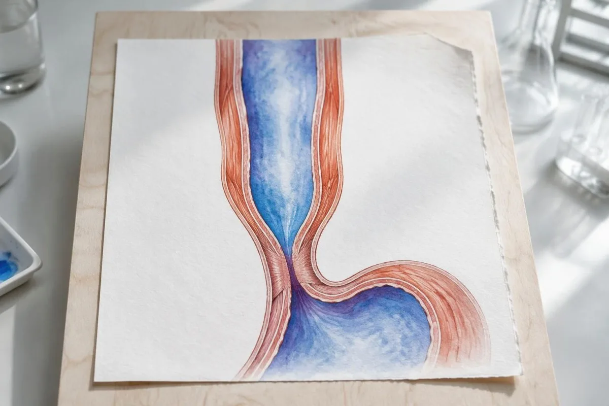
A flat-looking pressure plot can be misleading because failed contractions are not equivalent to successful relaxation. In a technically adequate study, 10 failed supine swallows combined with an elevated median IRP support type I, but the specialist still checks catheter placement, structural findings, and whether pressurization occurred frequently enough to meet type II criteria.
Esophageal dilation can reduce the ability to generate pressure, but type I is not a validated chronological stage that every person reaches after type II. The 3 subtypes describe current physiology rather than a guaranteed disease sequence; duration, esophageal shape, and previous treatment can affect the tracing without providing a reliable clock for when achalasia began.
Nutritional consequences depend on actual intake, not subtype alone. A hypothetical person with type I, declining weight, and ferritin 11 ng/mL needs attention to swallowing treatment and iron-deficiency symptoms; low iron stores do not prove achalasia, while a normal ferritin would not establish that nutrition or esophageal emptying is adequate.
Ni nini kinachotofautisha akalasia ya aina ya II?
Akalasia ya aina ya II combines an abnormal median IRP, 100% failed peristalsis, and panesophageal pressurization in at least 20% of swallows. Instead of a coordinated traveling contraction, pressure rises across much of the esophagus at once because swallowed material is compressed against an outlet that does not open adequately.
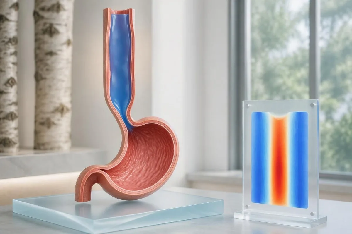
With 10 evaluable swallows, panesophageal pressurization in 2 swallows reaches the 20% threshold. That arithmetic is useful when reading a report, but it should not encourage self-classification from a screenshot: the interpreter must distinguish true pressure trapping from closely spaced swallows, movement artifacts, and pressure attributable to adjacent anatomy.
Neno panesophageal pressurization usually refers to a simultaneous pressure contour reaching approximately 30 mmHg across the esophageal body. A bright vertical band on a color map can represent this trapped pressure rather than a strong muscle wave; reading medical report explanations safely means checking the written interpretation rather than assuming warmer colors equal better propulsion.
Type II often has favorable outcomes after pneumatic dilation, Heller myotomy, or POEM, according to the ACG guideline (Vaezi et al., 2020). Nevertheless, the 20% diagnostic threshold is not a treatment-success percentage, and response still depends on anatomy, treatment technique, prior procedures, and follow-up; a subtype should inform discussion, not function as a personal guarantee.
Manometria hutambuaje akalasia ya aina ya III?
Akalasiani ya Aina ya III shows impaired junction relaxation, premature contractions in at least 20% of swallows, and no normal peristalsis. A premature contraction generally has distal latency below 4.5 seconds with adequate DCI; other swallows may fail, so type III does not require every swallow to produce a spasm.
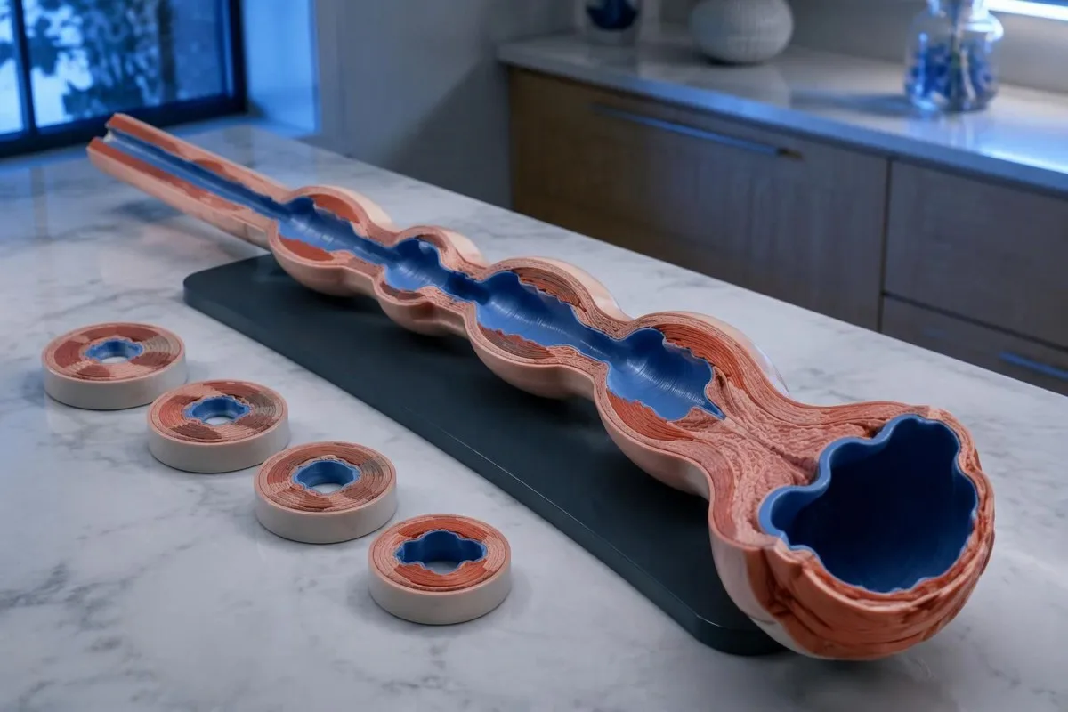
The absence of normal peristalsis separates conclusive type III achalasia from some other spastic patterns. If premature contractions coexist with otherwise preserved peristalsis and junction obstruction, the report may instead describe esophagogastric junction outflow obstruction with spastic features; the same 4.5-second timing cutoff does not erase that diagnostic distinction.
Chest pain can accompany spastic contractions, but it should not automatically be attributed to the esophagus. New severe chest pain, especially with breathlessness, sweating, or exertional onset, needs urgent assessment; chest-pain testing priorities differ from elective manometry, and no single esophageal measurement rules out a cardiac emergency.
Opioid exposure deserves particular scrutiny before an irreversible procedure for an apparent type III pattern. Chicago Classification version 4.0 recommends considering testing off opioids when feasible, with timing based on the medicine's half-life (Yadlapati et al., 2021); a fixed 24-hour pause is not appropriate for every drug, and any change requires the prescribing team's supervision.
Ripoti tatu za kubuni za manometria zingelinganishwaje?
Hypothetical manometry examples show why an elevated IRP cannot determine the subtype by itself. Using a laboratory with a validated supine upper limit of 15 mmHg, the same median IRP of 24 mmHg could accompany type I, II, or III; the esophageal-body pattern supplies the distinguishing information.
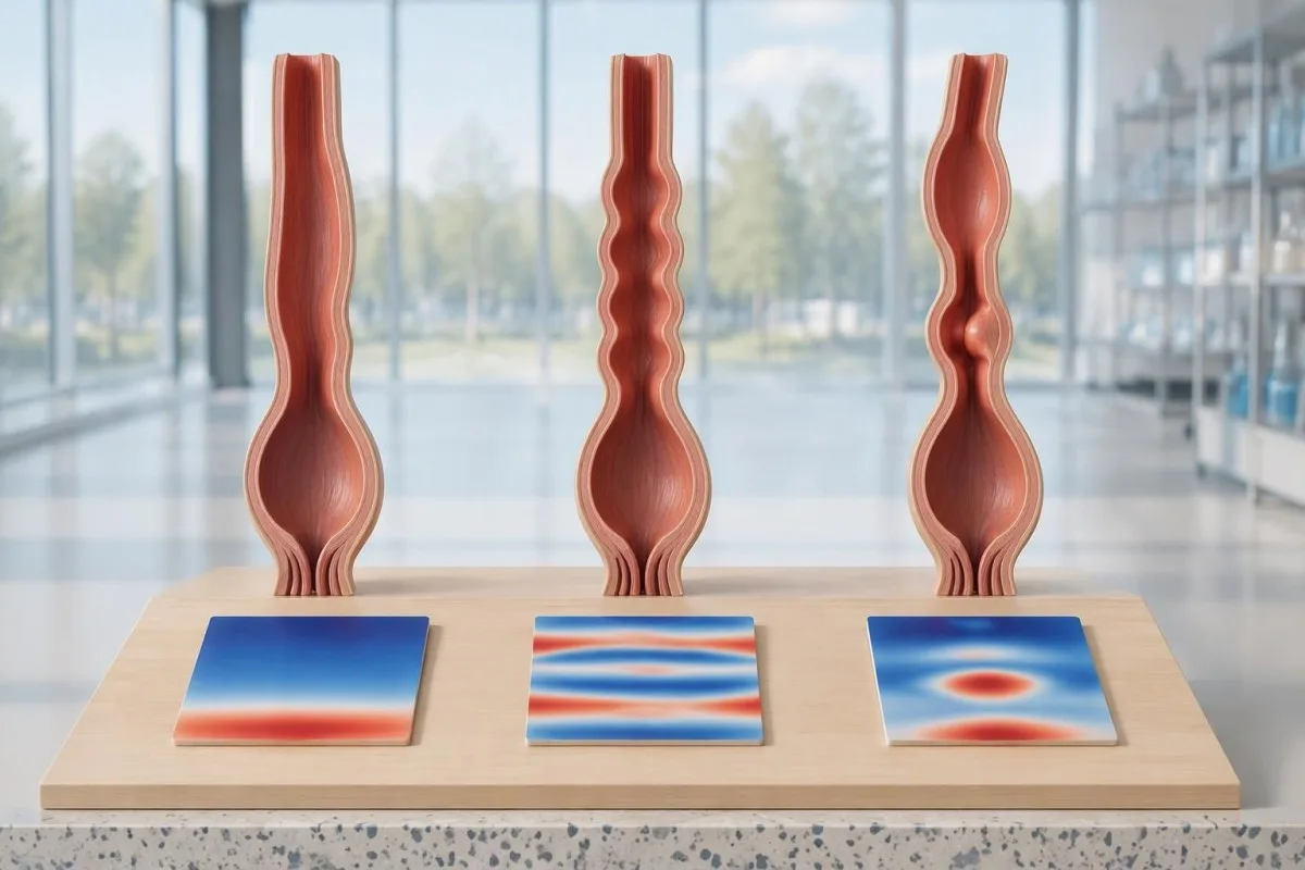
Example 1: a fictional report records median IRP 24 mmHg, DCI below 100 mmHg·s·cm in all 10 swallows, and no panesophageal pressurization. Assuming adequate technique, no structural explanation, and consistent symptoms, this is a type I pattern; the conclusion comes from failed propulsion plus impaired relaxation, not from the pressure value alone.
Example 2: another fictional report records IRP 24 mmHg, failed peristalsis in all 10 swallows, and panesophageal pressurization in 6 swallows. The pressurization frequency is 60%, exceeding the 20% threshold for type II; this does not mean the condition is three times more severe than a report with pressurization in 2 swallows.
Example 3: a fictional study records IRP 24 mmHg, 4 premature contractions with distal latency 3.8 seconds and DCI 900 mmHg·s·cm, and 6 failed swallows, with no normal peristalsis. This supports type III; Kantesti is an jukwaa la tafsiri ya vipimo vya damu la AI, na mifano ya mtiririko wa tafsiri (interpretation workflows) concern laboratory information rather than automated classification of these hypothetical motility tracings.
Vipi ikiwa IRP ni ya kawaida, iko mpakani, au hailingani?
A normal or borderline IRP does not always exclude achalasia, particularly when peristalsis is absent and symptoms strongly suggest impaired emptying. Conversely, elevated IRP with preserved peristalsis is not automatically achalasia; Chicago Classification version 4.0 requires careful separation of inconclusive achalasia, artifacts, and esophagogastric junction outflow obstruction.
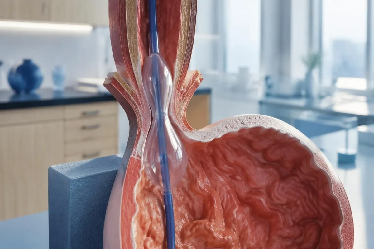
An elevated supine IRP that normalizes upright can reflect catheter contact or distortion around a hiatal hernia. This is one reason the standard protocol uses 2 positions: a reproducible obstruction pattern is more convincing than one abnormal posture-dependent number, although positional disagreement still needs specialist interpretation rather than an automatic normal label.
Esophagogastric junction outflow obstruction, or EGJOO, requires more than a high median IRP. Its manometric criteria include elevated IRP in both positions, preserved peristalsis, and elevated supine intrabolus pressure in at least 20% of swallows; clinically meaningful EGJOO also requires relevant symptoms and supportive evidence of obstruction on timed barium testing or FLIP.
Absent contractility with normal relaxation can occur in systemic sclerosis and is not the same as achalasia; scleroderma-related antibody clues cannot replace motility testing. Kantesti's inapaswa kueleweka kwa umakini huohuo: tafsiri ya ripoti ya damu si uthibitishaji wa alama ya hatari ya mzio, na hata matokeo 1 mazuri ya maabara hayawezi kuthibitisha kustahili kufanyiwa uchokozi. should not be extended to these 2 distinct pressure disorders, and an irreversible sphincter procedure should not follow from an unexplained borderline result alone.
Kwa nini endoskopi inahitajika kabla ya kuitambua kuwa akalasia?
Upper endoscopy helps exclude a mechanical obstruction or pseudoachalasia, including an obstructing growth at the esophagogastric junction. Manometry can demonstrate an achalasia-like pressure pattern without proving its cause; the ACG guideline recommends endoscopy in suspected achalasia because treatment decisions depend on distinguishing primary motility disease from structural disease (Vaezi et al., 2020).
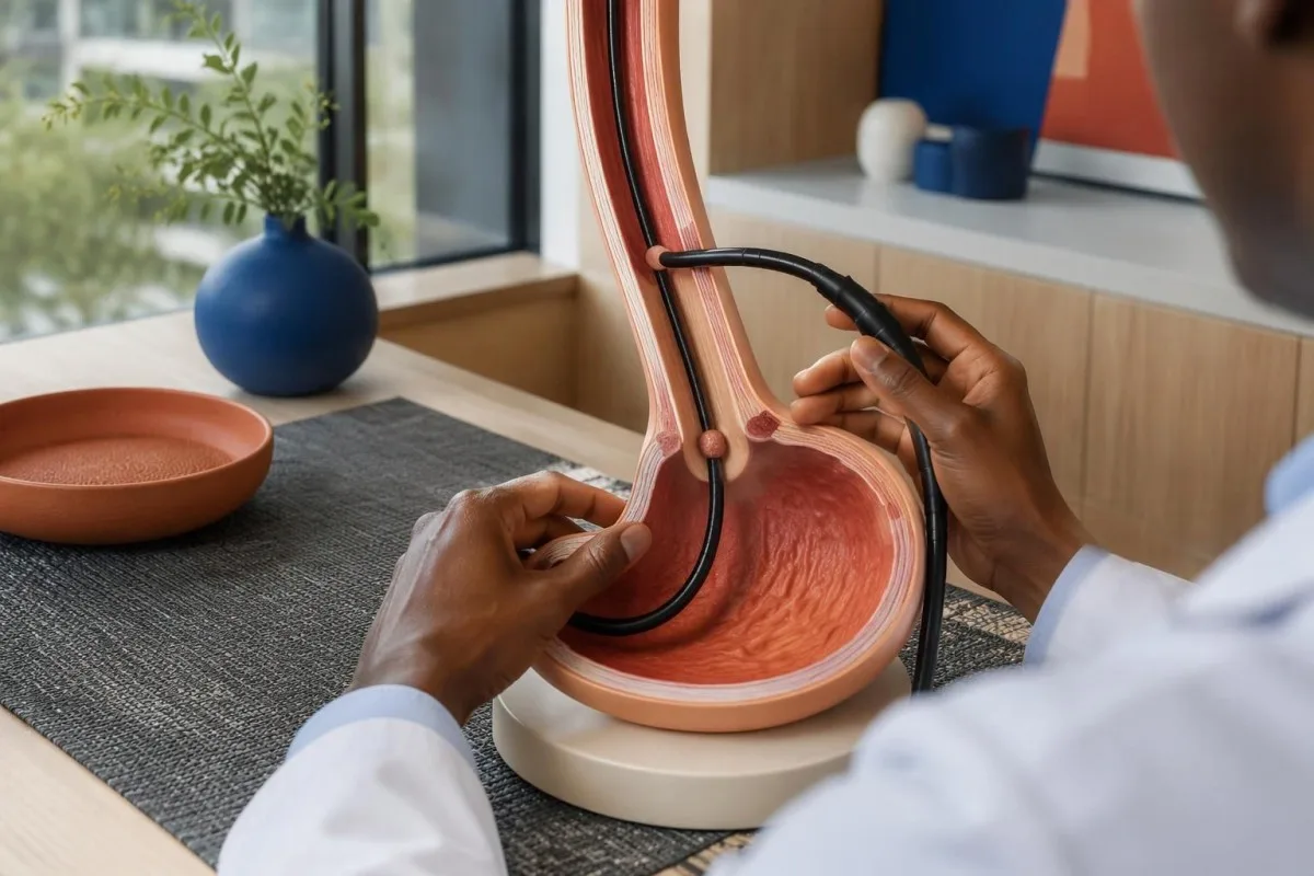
Retained saliva, retained food, and a tight-looking junction can support suspicion of achalasia, but none is diagnostic by itself. Early disease may have an almost normal endoscopic appearance; equally, resistance to passing the scope should not be dismissed as ordinary achalasia without considering whether a second test is needed to inspect the junction more closely.
Rapidly progressive symptoms, substantial short-term weight loss, older age at presentation, and unusual resistance at the junction increase concern for pseudoachalasia. There is no single age or kilogram cutoff that safely excludes it; a hypothetical 68-year-old with 3 months of progressive dysphagia and 9 kg weight loss warrants particular attention to the possibility of a structural cause.
A normal-looking mucosal surface does not always exclude a deeper or externally compressing growth, so CT or endoscopic ultrasound may be appropriate when suspicion persists. Normal CA 19-9 results cannot rule out this concern; ordering 1 tumor marker is not an alternative to imaging, endoscopy, or targeted tissue examination when clinically indicated.
Kumeza bariamu kwa muda maalumu na FLIP huongeza nini?
A timed barium esophagram measures esophageal emptying, while FLIP assesses how widely the esophagogastric junction opens under controlled distension. These tests provide information that pressure measurements alone cannot supply; a commonly used timed protocol obtains images at 1, 2, and 5 minutes after a standardized barium swallow.
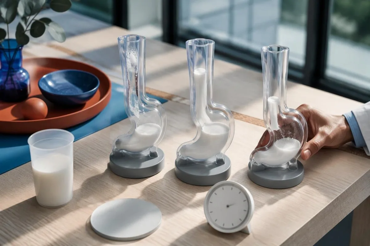
A standardized upright timed study often uses about 236 mL of barium; column heights above 5 cm at 1 minute or 2 cm at 5 minutes suggest impaired emptying in commonly used protocols. These are protocol-dependent abnormal-emptying thresholds, not stand-alone proof of achalasia, and the classic bird-beak appearance can be absent in early disease.
A 13 mm barium tablet can help reveal restricted passage when liquid contrast empties relatively well, provided the imaging team considers it appropriate. FLIP distensibility index expresses junction cross-sectional area divided by pressure in mm²/mmHg, but balloon volume, sedation, device protocol, and maximum opening diameter affect interpretation; treating every value below 2 as an interchangeable diagnosis would oversimplify the test.
An achalasia-like dilated esophagus does not establish that the disorder is idiopathic. In someone with relevant exposure history, confirmatory Chagas testing may be appropriate because chronic Trypanosoma cruzi infection can affect esophageal innervation; imaging and infection testing answer 2 different questions, namely how the esophagus empties and why its function may be impaired.
Aina za akalasia zinazoonekana kwenye manometria husaidiaje kuelekeza matibabu?
Achalasia manometry types help clinicians choose how best to reduce resistance at the esophageal outlet. Pneumatic dilation, laparoscopic Heller myotomy, and POEM are established options for types I and II; type III often favors a longer, tailored myotomy that addresses the spastic segment as well as the junction.
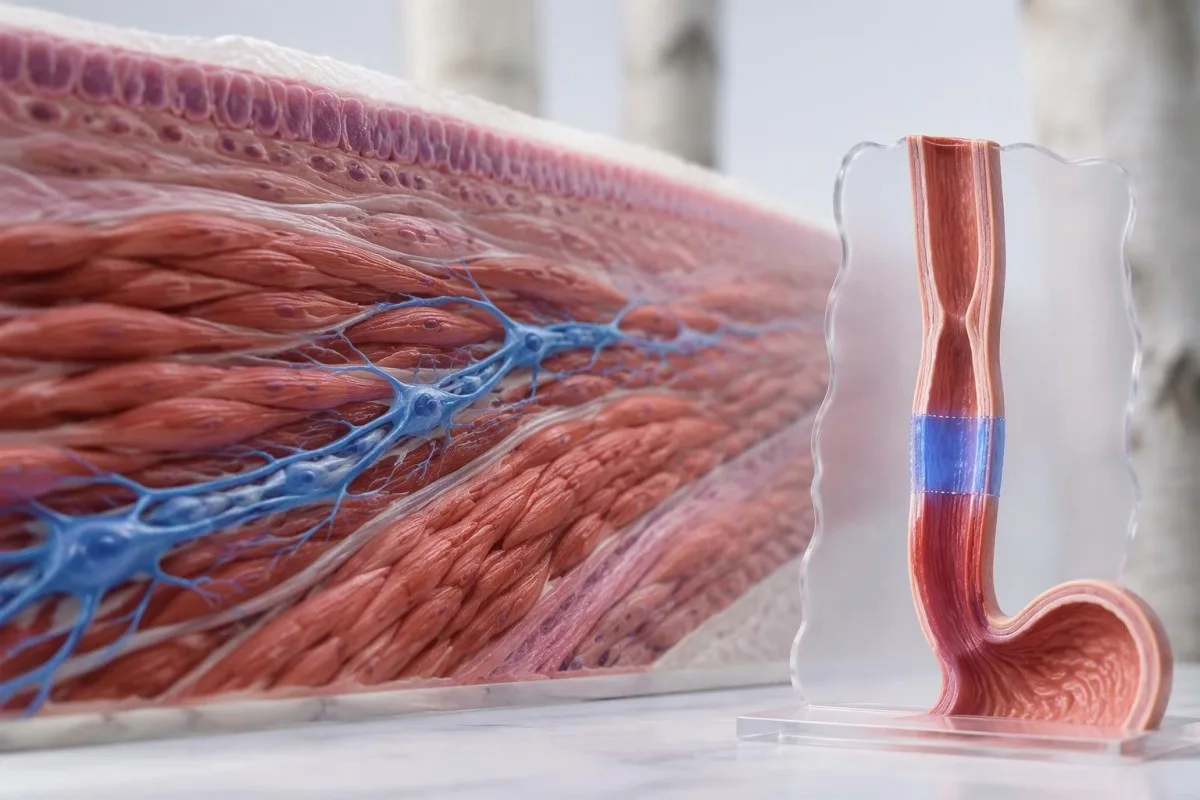
The ACG guideline considers pneumatic dilation, Heller myotomy, and POEM comparably effective options for appropriate patients with types I and II, while supporting tailored POEM or Heller myotomy over dilation for type III (Vaezi et al., 2020). The 3 procedures differ in anesthesia needs, recovery, retreatment possibilities, and reflux risk; local expertise and patient preference belong in the decision.
A randomized trial of 221 patients found clinical success at 2 years in 83.0% after POEM and 81.7% after Heller myotomy with Dor fundoplication (Werner et al., 2019). These overall results support both approaches but do not establish identical outcomes for every subtype; type III participants form a much smaller subgroup than the full trial population.
Reflux esophagitis at 2 years occurred in 44% after POEM versus 29% after Heller myotomy in that trial, illustrating a trade-off rather than a reason to reject POEM automatically. Acid-suppression treatment may subsequently affect gastrin blood results; meanwhile, pneumatic dilation carries a perforation risk of roughly 2% in experienced practice and requires an appropriate rescue pathway.
Ufuatiliaji na vipimo vya damu vinaweza kuonyesha nini baada ya matibabu?
Follow-up assesses swallowing, weight, esophageal emptying, and reflux, not just whether symptoms improve. Blood tests can identify nutritional or hydration complications but cannot confirm that the junction opens normally; the ACG guideline recommends timed barium esophagram as the first-line objective test when symptoms persist or recur after treatment (Vaezi et al., 2020).
The Eckardt score adds dysphagia, regurgitation, chest pain, and weight loss, each scored from 0 to 3, for a total of 0–12. Trials often define success as a score of 3 or below, but symptom scores alone can miss retained material; reduced discomfort does not necessarily mean that esophageal clearance has normalized.
Kantesti ni jukwaa la tafsiri ya viashiria vya AI that can help explain a clinician-requested complete blood count, electrolytes, and iron studies alongside reduced intake. Ferritin below 15 ng/mL strongly supports depleted iron stores in adults without a confounding context; our mwongozo wetu kamili wa tafiti za chuma explains why ferritin and transferrin saturation should be considered together rather than used to infer an achalasia subtype.
Albumin commonly has an adult reference interval around 35–50 g/L, but a normal value does not exclude inadequate intake and a low value is not specific for malnutrition. Our mwongozo wa tafsiri ya protini ya damu explains those limitations; magnesium deserves separate attention when poor intake or long-term acid suppression is present, as discussed in sababu za upungufu wa magnesium.
Unapaswa kuuliza nini baadaye, na ni ushahidi upi unaotumika?
Your next discussion should connect the manometry classification, structural assessment, and evidence of impaired emptying before selecting treatment. Ask 3 practical questions: which findings establish the subtype, whether obstruction or medication effects have been excluded, and how the proposed procedure's expected benefit compares with its reflux and retreatment risks.
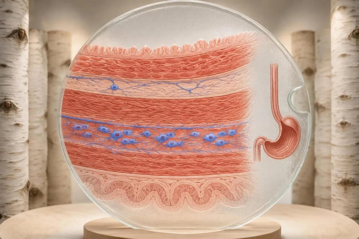
Request the full manometry report rather than only its conclusion, including the catheter system, body positions, median IRP, swallow counts, and any supportive tests. If you cannot swallow liquids or saliva, develop severe chest pain, or have respiratory difficulty, seek urgent care; waiting another 2 weeks for a routine report review is not an appropriate response to those warning signs.
The achalasia evidence cited here comes from Chicago Classification version 4.0, the ACG achalasia guideline, and the randomized POEM-versus-Heller trial. Kantesti's viwango vya maudhui ya matibabu distinguish evidence-based explanations from individualized diagnosis; the 2 Zenodo publications listed below concern coagulation and serum proteins, not studies validating manometry or achalasia treatment.
The serum-protein publication is relevant only to the laboratory-support discussion, while the coagulation publication provides separate background if a clinician requests clotting tests before a procedure. Neither DOI should be used to choose among types I, II, and III; when laboratory values change during recovery, blood-test timeline effects help explain why intake, illness, hydration, and medicines may alter results without indicating recurrent outlet obstruction.
Maswali ya mara kwa mara
Je, achalasia hutambuliwaje?
Achalasia kwa kawaida hutambuliwa kwa manometria ya umio yenye azimio la juu inayoonyesha kulegea kwa muunganiko wa umio na tumbo kulikoharibika na kutokuwepo kwa peristalsis ya kawaida. Kisha muundo wa shinikizo hutofautisha aina za I, II na III. Endoskopi ya njia ya juu ya chakula husaidia kuondoa uwezekano wa kuziba kwa kimuundo, huku kumeza bariamu kwa muda maalum au FLIP kukisaidia kufafanua matokeo yasiyo dhahiri. Hakuna kipimo cha kawaida cha damu kinachothibitisha mojawapo ya aina ndogo 3.
Manometria ya umio huonyesha nini katika achalasia?
Manometria ya umio huonyesha jinsi makutano yanavyolegea baada ya kumeza na ikiwa sehemu kuu ya umio huzalisha mikazo iliyoratibiwa. Shinikizo jumuishi la kulegea hupima kulegea kwa njia ya kutoka katika muda wa sekunde 4, huku muda wa mikazo na mgawanyo wa shinikizo vikisaidia kutambua aina ndogo. Aina ya II huhitaji shinikizo la juu katika umio mzima katika angalau 20% ya mienuko ya kumeza, na aina ya III huhitaji mikazo ya mapema katika angalau 20%. Viwango maalum vya kifaa husika lazima vitumike kwa sababu kigezo kimoja cha jumla cha IRP hakifai.
Kuna tofauti gani kati ya achalasia ya aina ya I, II, na III?
Achalasia ya aina ya I ina ukosefu wa peristalsis bila shinikizo la umio mzima linalohitajika kwa aina ya II. Aina ya II ina ukosefu wa peristalsis pamoja na shinikizo la umio mzima katika angalau 20% ya mamezeto. Aina ya III ina mikazo ya mapema katika angalau 20% ya mamezeto na haina peristalsis ya kawaida, pamoja na kulegea kwa muunganiko kuharibika. Hizi ni mifumo ya kifizikia badala ya hatua za uhakika za kuendelea kwa ugonjwa.
Je, achalasia inaweza kutokea huku IRP ikiwa ya kawaida?
0. Achalasia inaweza wakati mwingine kuendelea kuwa jambo la kutia wasiwasi licha ya shinikizo la kulegea lililounganishwa kuwa la kawaida au la mpakani, hasa peristalsis inapokosekana na utoaji wa yaliyomo unapokuwa na hitilafu. Katika hali hiyo, manometria inaweza kutotoa hitimisho la uhakika badala ya kuonyesha hali ya kawaida kwa uhakika. Uchunguzi wa bariamu wa muda maalum, ambao kwa kawaida hujumuisha picha za dakika 1, 2 na 5, au FLIP unaweza kutoa ushahidi wa kuunga mkono. Mtaalamu anapaswa pia kuzingatia kutokuwepo kwa mkazo wa misuli, vikwazo vya kiufundi na matibabu ya awali.
Je, endoskopi inaweza kuwa ya kawaida ikiwa una akalasia?
0 Endoskopi inaweza kuonekana karibu ya kawaida katika hatua za mwanzo za akalasia kwa sababu huchunguza muundo na utando wa mucosa badala ya kupima moja kwa moja shinikizo la kumeza. Chakula kilichobaki, mate au sehemu ya kuungana iliyobana vinaweza kuibua shaka, lakini havithibitishi utambuzi. Manometria hutambua mpangilio bainifu wa shinikizo na kutofautisha aina ndogo 3. Shaka inayoendelea ya pseudoakalasia inaweza kuhitaji CT au ultrasonografia ya endoskopi hata wakati uso wa mucosa unaonekana wa kawaida.
Ni aina gani ya akalasia inayojibu vyema zaidi matibabu?
Type II achalasia generally has favorable responses to pneumatic dilation, Heller myotomy, and POEM, but subtype alone cannot predict an individual's result. Type III often benefits from a longer myotomy tailored to the spastic segment. A randomized trial reported 2-year success of 83.0% after POEM and 81.7% after Heller myotomy with fundoplication across its overall study population. Reflux, anatomy, prior treatment, and procedural expertise also influence the choice.
Je, unahitaji kutulizwa kwa dawa ili kufanyiwa manometria ya umio?
Standard esophageal manometry is usually performed awake, often with local nasal anesthetic rather than intravenous sedation. Patients complete a structured swallowing protocol, commonly 10 supine and 5 upright water swallows. Difficult catheter placement sometimes requires endoscopic assistance, but any sedation and recovery interval must be considered when interpreting the subsequent measurements. Follow the testing center's fasting instructions because achalasia can retain food despite an ordinary fast.
Pata maelezo ya jinsi ya kusoma matokeo ya vipimo vya damu
Pakia ripoti yako ya maabara na AI ya Kantesti itaeleza kila kiashiria cha kibayolojia kwa lugha rahisi ndani ya sekunde chache. Inatumiwa na zaidi ya watu milioni 2 duniani kote.
Marejeleo
- Klein, T., Mitchell, S., & Weber, H. (2026). Kiwango cha Kawaida cha aPTT: Mwongozo wa D-Dimer na Protein C kuhusu Mgando wa Damu. Utafiti wa Kimatibabu wa AI ya Kantesti. DOI
- Klein, T., Mitchell, S., & Weber, H. (2026). Mwongozo wa Protini za Seramu: Globulini, Albumin na Kipimo cha Uwiano wa A/G. Utafiti wa Kimatibabu wa AI ya Kantesti. DOI
- Yadlapati R et al. (2021). Esophageal motility disorders on high-resolution manometry: Chicago classification version 4.0. Neurogastroenterology & Motility. DOI
- Vaezi MF et al. (2020). ACG Clinical Guidelines: Diagnosis and Management of Achalasia. Jarida la Marekani la Magonjwa ya Mfumo wa Mmeng'enyo wa Chakula. DOI
- Werner YB et al. (2019). Endoscopic or Surgical Myotomy in Patients with Idiopathic Achalasia. The New England Journal of Medicine. DOI
- 2M+Vipimo vilivyochanganuliwa
- 127+Nchi
- 100+Lugha
Kuhusu timu ya matibabu


E-E-A-T Trust Signals
Uzoefu
Mapitio ya kimatibabu inayoongozwa na daktari ya mifumo ya tafsiri ya maabara.
Utaalamu
Kuzingatia dawa za maabara kuhusu jinsi viashiria (biomarkers) vinavyobadilika katika muktadha wa kliniki.
Mamlaka
Imekaguliwa kimatibabu na Dkt. Thomas Klein, kwa michango kutoka kwa Dkt. Sarah Mitchell na Prof. Dkt. Hans Weber.
Uaminifu
Tafsiri inayotegemea ushahidi yenye njia zilizo wazi za ufuatiliaji ili kupunguza tahadhari za hofu.


Kanusho la kimatibabu
Makala haya ni kwa madhumuni ya elimu tu na si ushauri wa kimatibabu. Wasiliana na mtoa huduma aliyehitimu kila wakati kwa maamuzi ya utambuzi na matibabu.
Imechapishwa · Wasiliana na timu ya uhariri
Imechapishwa na Kantesti Ltd, imesajiliwa nchini Uingereza na Wales, nambari ya kampuni. 17090423, London, Uingereza.
