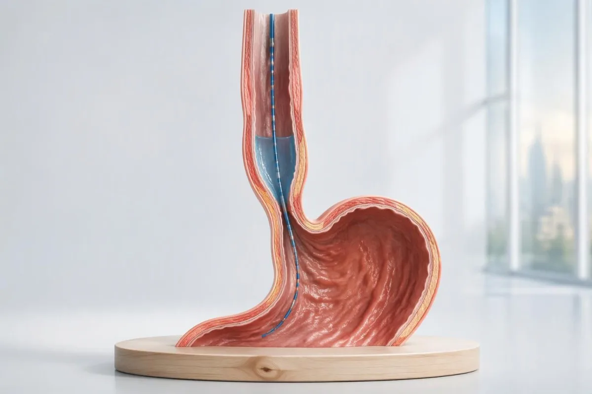
Viens pats augsta spiediena rādījums neapstiprina ahalāzijas diagnozi. Diagnoze ir atkarīga no tā, kā atslābst barības vada izeja, vai rīšanas laikā veidojas koordinētas kontrakcijas un vai konstatēto ainu izskaidro kāds cits stāvoklis.

Galvenie secinājumi
- Ahalāzijas diagnoze parasti tiek noteikta, ja augstas izšķirtspējas manometrija uzrāda traucētu izejas atslābšanu un normālas barības vada peristaltikas neesamību; endoskopijā pārbauda, vai nav mehāniska iemesla.
- Integrētā relaksācijas spiediena rādītājs mēra izejas atslābšanu 4 sekunžu intervālā, taču tā augšējā robežvērtība ir atkarīga no aprīkojuma un ķermeņa pozīcijas.
- I tipa ahalāzija uzrāda 100% neizdevušos peristaltiku bez panēzofageālās presurizācijas, kas nepieciešama II tipa noteikšanai.
- II tipa ahalāzija uzrāda 100% neizdevušos peristaltiku un panēzofageālu presurizāciju vismaz 20% testa rīšanas reižu.
- III tipa ahalāzija norāda uz priekšlaicīgām kontrakcijām vismaz 20% rīšanas reižu, traucētu sfinktera atslābšanu un normālas peristaltikas neesamību.
- Priekšlaicīga kontrakcija parasti nozīmē distālo latentumu, kas ir mazāks par 4,5 sekundēm, un pietiekamu kontrakcijas spēku; tikai ātri mainīgas krāsas neliecina par spazmu.
- Robežrezultātu gadījumā var būt nepieciešama laika kontrolēta barija ezofagogramma vai FLIP izmeklējums; atsevišķi konstatēts paaugstināts spiediens nav pietiekams pamatojums neatgriezeniskai ārstēšanai.
- Ārstēšanas apspriešana atšķiras atkarībā no apakštipa: III tipa gadījumā bieži dod priekšroku garākai, individuāli pielāgotai miotomijai, savukārt I un II tipa gadījumā ir vairākas efektīvas iespējas.
- Asins analīzes var palīdzēt novērtēt dehidratāciju, anēmiju vai uztura komplikācijas, taču neviena rutīnas laboratorisko analīžu kopa neapstiprina un neizslēdz ahalāziju.
Kā ar augstas izšķirtspējas manometriju diagnosticē ahalāziju?
Ahalāzijas diagnoze parasti balstās uz augstas izšķirtspējas manometrijas rezultātiem, kas liecina par traucētu barības vada izejas atslābšanu un normālas peristaltikas trūkumu. I tipam raksturīgas neveiksmīgas kontrakcijas, II tipam — spiediena palielināšanās visā barības vadā vismaz 20% rīšanas reižu, bet III tipam — priekšlaicīgas kontrakcijas vismaz 20% rīšanas reižu; endoskopija palīdz izslēgt nosprostojumu.
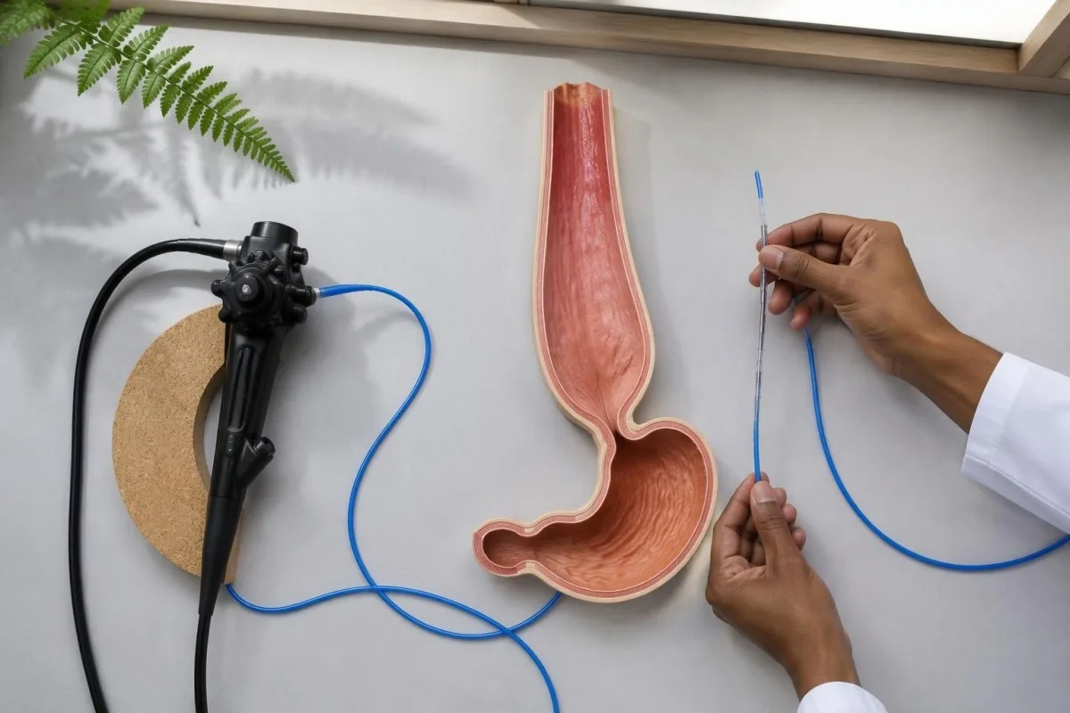
Augstas izšķirtspējas manometrija jeb HRM, mēra spiedienu, nevis tieši filmē pārtikas kustību. Tās 2 galvenie jautājumi ir, vai gastroezofageālais savienojums rīšanas laikā atveras atbilstoši un vai barības vada ķermenis rada koordinētu vilni; augsts sfinktera miera spiediens pats par sevi neatbild ne uz vienu no šiem jautājumiem un neapstiprina ahalāziju.
Čikāgas klasifikācijas 4.0. versija nosaka manometrisko raksturojumu, ko izmanto, lai identificētu 3 ahalāzijas apakštipus, savukārt to klīnisko nozīmi nosaka simptomi un citi izmeklējumi (Yadlapati et al., 2021). Slēdziens, kurā vienkārši minēti motilitātes traucējumi, ir mazāk informatīvs nekā tāds, kurā norādīts integrētās atslābšanas spiediens, peristaltikas atradne, izmeklēšanas pozīcija un galīgā klasifikācija.
Kantesti ir AI asins analīžu analizators kas var palīdzēt izskaidrot nepietiekamas uztura uzņemšanas radītās sekas laboratoriskajos rādītājos, taču nevienu no 3 ahalāzijas apakštipiem nevar diagnosticēt pēc asins analīzēm. Informācija par organizācijā jāskata atsevišķi no speciālista vērtējuma, kas nepieciešams manometrijas rezultātu interpretēšanai un strukturālas slimības izslēgšanai.
Kuriem rīšanas simptomiem vajadzētu būt par iemeslu ahalāzijas izmeklēšanai?
Rīšanas grūtības gan ar cietu, gan šķidru barību, atpakaļatraugas ar aizturētu barību un neizskaidrots svara zudums ir iemesls izvērtēt, vai nav ahalāzijas. Simptomi vien nevar noteikt diagnozi vai atšķirt tās 3 apakštipus; pat pārliecinoša anamnēze jāpapildina ar izmeklējumiem, jo nosprostojuma, ar refluksu saistītas slimības un citi rīšanas traucējumi var izpausties līdzīgi.
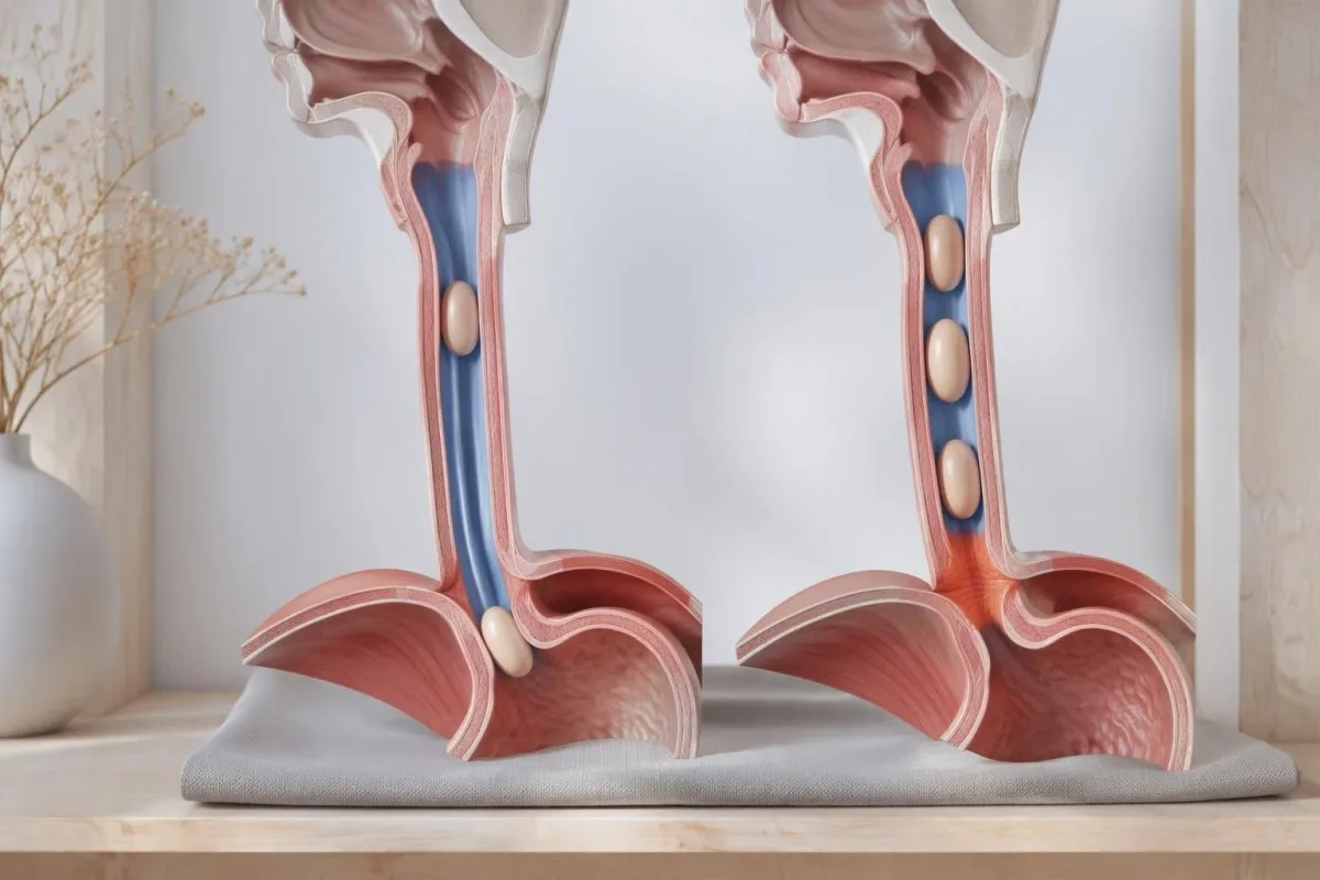
Ahalāzijas gadījumā atpakaļatraugās var būt barība, kas apēsta vairākas stundas iepriekš, savukārt skābes refluksa gadījumā biežāk augšup nonāk skābs šķidrums. Šī atšķirība palīdz izskaidrot, kāpēc ilgstoša ārstēšana refluksam līdzīgu simptomu gadījumā var nenovērst rīšanas grūtības; tomēr šie 2 procesi var pastāvēt vienlaikus, un simptomu apraksti bez izmeklēšanas neļauj tos droši atšķirt.
Grūtības sākt rīt liecina par citu lokalizāciju nekā sajūta, ka barība iestrēgst vairākas sekundes pēc norīšanas. Klepus tūlīt pēc malka, degunāla balss vai rīšanas nogurdināmība var būt iemesls izvērtēt mutes un rīkles rīšanas fāzi vai veikt neiroloģisku novērtējumu; miastēnijas testēšanas ierobežojumi ir būtiski, ja simptomus pavada vājums, bet nav paredzēti kā rutīnas ahalāzijas skrīninga analīžu kopa.
An inability to swallow saliva or a suspected food impaction requires urgent assessment rather than waiting for elective manometry. Repeated nighttime regurgitation also raises concern for aspiration; for a hypothetical person losing 6 kg while meals become progressively slower, the weight trajectory and swallowing history both deserve attention even if routine laboratory results remain within range.
Kā notiek barības vada manometrijas tests?
Esophageal manometry usually involves passing a thin pressure-sensing catheter through the nose into the stomach while the patient is awake. A standard Chicago Classification version 4.0 protocol commonly includes 10 supine and 5 upright water swallows, with additional maneuvers that test whether an apparent abnormality persists under different conditions.
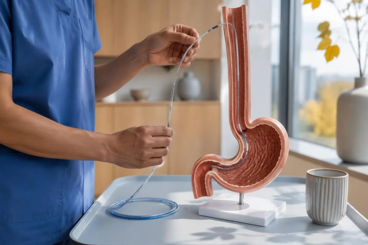
Preparation commonly includes at least 6 hours without food, but achalasia can leave material in the esophagus despite ordinary fasting. The testing center may therefore give a longer fasting or modified-diet plan; follow its instructions rather than assuming that an empty stomach guarantees an empty esophagus, especially if retained food was seen during endoscopy.
Medication review matters because opioids can produce impaired junction relaxation or spastic patterns that resemble type III achalasia. Provide the exact medicine, dose, and last-dose time; medication-related laboratory effects are a separate issue, but a single accurate medication list helps both investigations, and prescribed medicines should not be stopped without guidance.
Provocative testing may include 5 closely spaced 2 mL swallows or a rapid drink challenge of about 200 mL, if safe and appropriate. These maneuvers test different aspects of relaxation and contraction reserve; when standard swallows conflict with symptoms, their results can reveal a reproducible obstruction pattern or expose an artifact that disappears after changing position.
Kuri barības vada manometrijas rezultāti ir vissvarīgākie?
Visnoderīgākā esophageal manometry results are median integrated relaxation pressure, the presence or absence of peristalsis, panesophageal pressurization, and distal latency. These measurements answer different questions: outlet opening, coordinated propulsion, pressure trapped within the esophagus, and contraction timing; none should be interpreted as an isolated severity score.
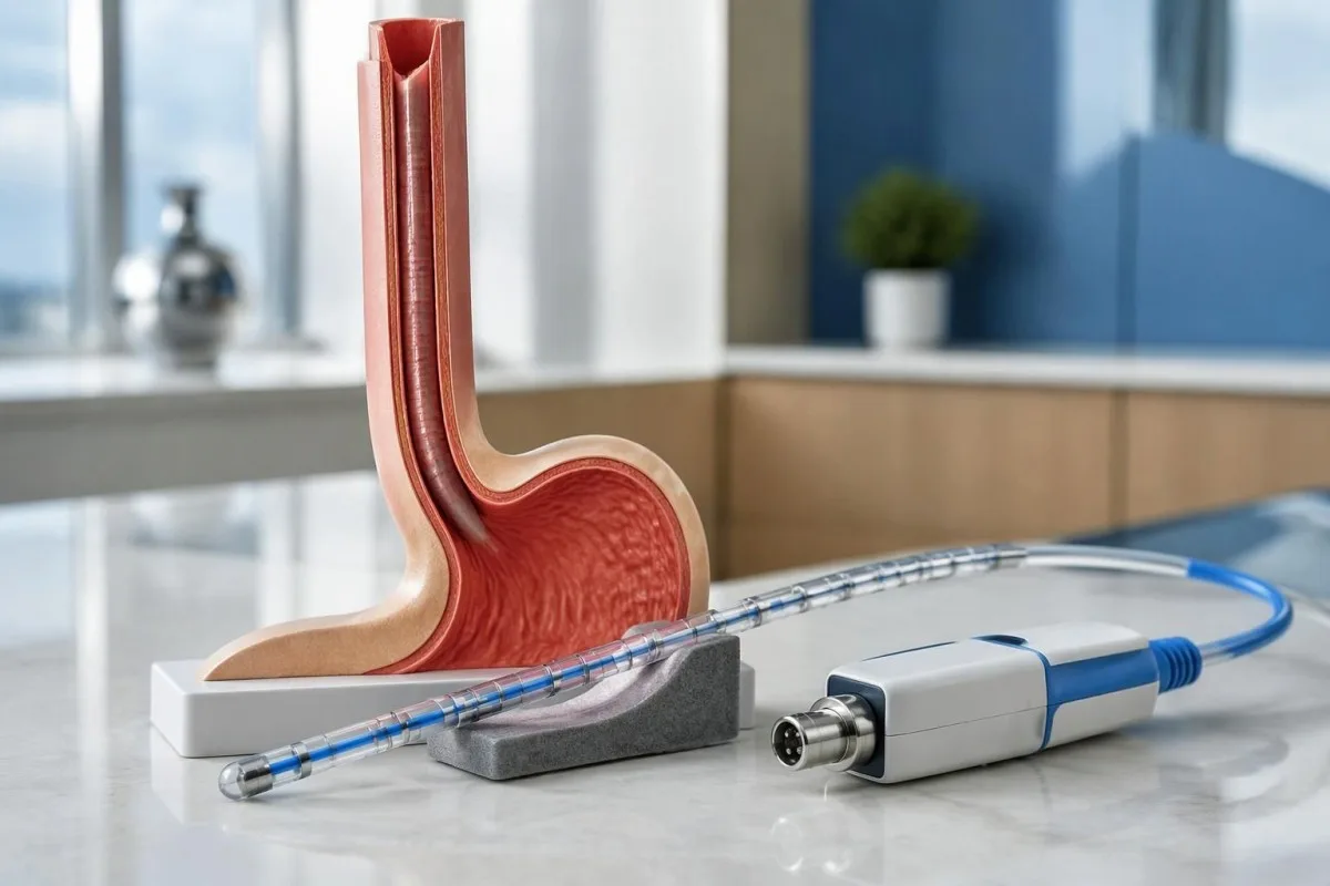
Integrētā relaksācijas spiediena rādītājs, or IRP, summarizes junction pressure over a 4-second relaxation window within the post-swallow assessment period. It is not simply the lowest pressure reached, and its threshold changes with catheter system and posture; Chicago Classification version 4.0 gives different reference limits for commonly used systems rather than one universal 15 mmHg rule (Yadlapati et al., 2021).
Distal contractile integral, or DCI, combines contraction strength, duration, and length, using mmHg·s·cm. A failed swallow has DCI below 100 mmHg·s·cm; a sufficiently strong premature contraction generally requires DCI of at least 450 mmHg·s·cm, so a faint, early pressure blip should not automatically be classified as the spasm needed for type III.
Kantesti separates measurement units from clinical conclusions: a value in mmHg cannot be compared with a laboratory concentration in ng/mL. Our blood biomarker directory addresses a different measurement category; ask the manometry service which equipment-specific reference limit applies before treating a result such as IRP 17 mmHg as definitely abnormal.
| Līmenis | Diapazons | Ko tas nozīmē |
|---|---|---|
| Medtronic-system supine IRP | Upper reference limit approximately 15 mmHg | Chicago Classification version 4.0 reference; verify the laboratory's validated system and protocol. |
| Medtronic-system upright IRP | Upper reference limit approximately 12 mmHg | A lower positional limit applies; do not compare an upright result with a supine cutoff. |
| Laborie/Diversatek-system supine IRP | Upper reference limit approximately 22 mmHg | Different equipment can legitimately use a higher reference limit. |
| Laborie/Diversatek-system upright IRP | Upper reference limit approximately 15 mmHg | Interpret alongside peristalsis, symptoms, and supportive testing. |
| Premature contraction timing | Distal latency below 4.5 seconds | Supports a premature contraction when contraction strength is sufficient; does not independently diagnose achalasia. |
| Panesophageal pressurization | Uniform pressure contour at approximately 30 mmHg or above | Whole-esophagus pressurization in at least 20% of swallows helps define type II when the other achalasia criteria are met. |
Kā manometrijā izpaužas I tipa ahalāzija?
I tipa ahalāzija shows an abnormal median IRP and 100% failed peristalsis, without the panesophageal pressurization pattern required for type II. The esophageal body does not generate an effective propulsive wave; the absence of strong pressure does not mean that food passes normally or that the condition is mild.
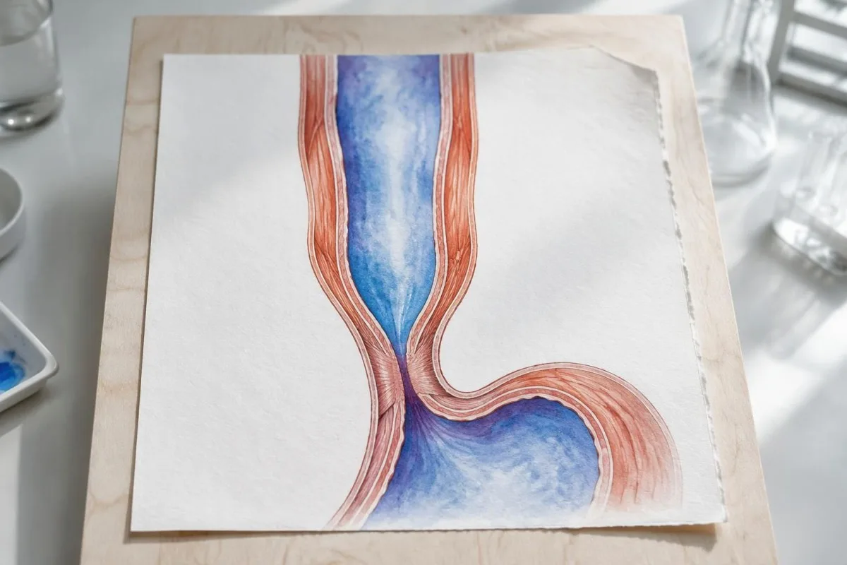
A flat-looking pressure plot can be misleading because failed contractions are not equivalent to successful relaxation. In a technically adequate study, 10 failed supine swallows combined with an elevated median IRP support type I, but the specialist still checks catheter placement, structural findings, and whether pressurization occurred frequently enough to meet type II criteria.
Esophageal dilation can reduce the ability to generate pressure, but type I is not a validated chronological stage that every person reaches after type II. The 3 subtypes describe current physiology rather than a guaranteed disease sequence; duration, esophageal shape, and previous treatment can affect the tracing without providing a reliable clock for when achalasia began.
Nutritional consequences depend on actual intake, not subtype alone. A hypothetical person with type I, declining weight, and ferritin 11 ng/mL needs attention to swallowing treatment and iron-deficiency symptoms; low iron stores do not prove achalasia, while a normal ferritin would not establish that nutrition or esophageal emptying is adequate.
Ar ko atšķiras II tipa ahalāzija?
II tipa ahalāzija combines an abnormal median IRP, 100% failed peristalsis, and panesophageal pressurization in at least 20% of swallows. Instead of a coordinated traveling contraction, pressure rises across much of the esophagus at once because swallowed material is compressed against an outlet that does not open adequately.
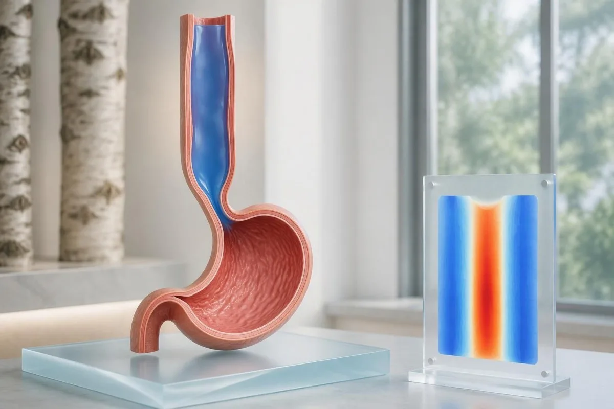
With 10 evaluable swallows, panesophageal pressurization in 2 swallows reaches the 20% threshold. That arithmetic is useful when reading a report, but it should not encourage self-classification from a screenshot: the interpreter must distinguish true pressure trapping from closely spaced swallows, movement artifacts, and pressure attributable to adjacent anatomy.
Termins panesophageal pressurization usually refers to a simultaneous pressure contour reaching approximately 30 mmHg across the esophageal body. A bright vertical band on a color map can represent this trapped pressure rather than a strong muscle wave; reading medical report explanations safely means checking the written interpretation rather than assuming warmer colors equal better propulsion.
Type II often has favorable outcomes after pneumatic dilation, Heller myotomy, or POEM, according to the ACG guideline (Vaezi et al., 2020). Nevertheless, the 20% diagnostic threshold is not a treatment-success percentage, and response still depends on anatomy, treatment technique, prior procedures, and follow-up; a subtype should inform discussion, not function as a personal guarantee.
Kā manometrijā nosaka III tipa ahalāziju?
III tipa ahalāzija shows impaired junction relaxation, premature contractions in at least 20% of swallows, and no normal peristalsis. A premature contraction generally has distal latency below 4.5 seconds with adequate DCI; other swallows may fail, so type III does not require every swallow to produce a spasm.
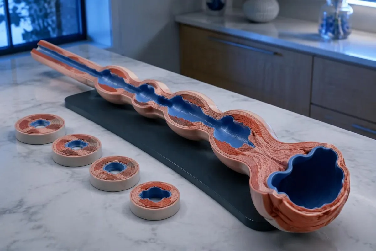
The absence of normal peristalsis separates conclusive type III achalasia from some other spastic patterns. If premature contractions coexist with otherwise preserved peristalsis and junction obstruction, the report may instead describe esophagogastric junction outflow obstruction with spastic features; the same 4.5-second timing cutoff does not erase that diagnostic distinction.
Chest pain can accompany spastic contractions, but it should not automatically be attributed to the esophagus. New severe chest pain, especially with breathlessness, sweating, or exertional onset, needs urgent assessment; chest-pain testing priorities differ from elective manometry, and no single esophageal measurement rules out a cardiac emergency.
Opioid exposure deserves particular scrutiny before an irreversible procedure for an apparent type III pattern. Chicago Classification version 4.0 recommends considering testing off opioids when feasible, with timing based on the medicine's half-life (Yadlapati et al., 2021); a fixed 24-hour pause is not appropriate for every drug, and any change requires the prescribing team's supervision.
Kā salīdzinātos trīs hipotētiski manometrijas izmeklējumu apraksti?
Hypothetical manometry examples show why an elevated IRP cannot determine the subtype by itself. Using a laboratory with a validated supine upper limit of 15 mmHg, the same median IRP of 24 mmHg could accompany type I, II, or III; the esophageal-body pattern supplies the distinguishing information.
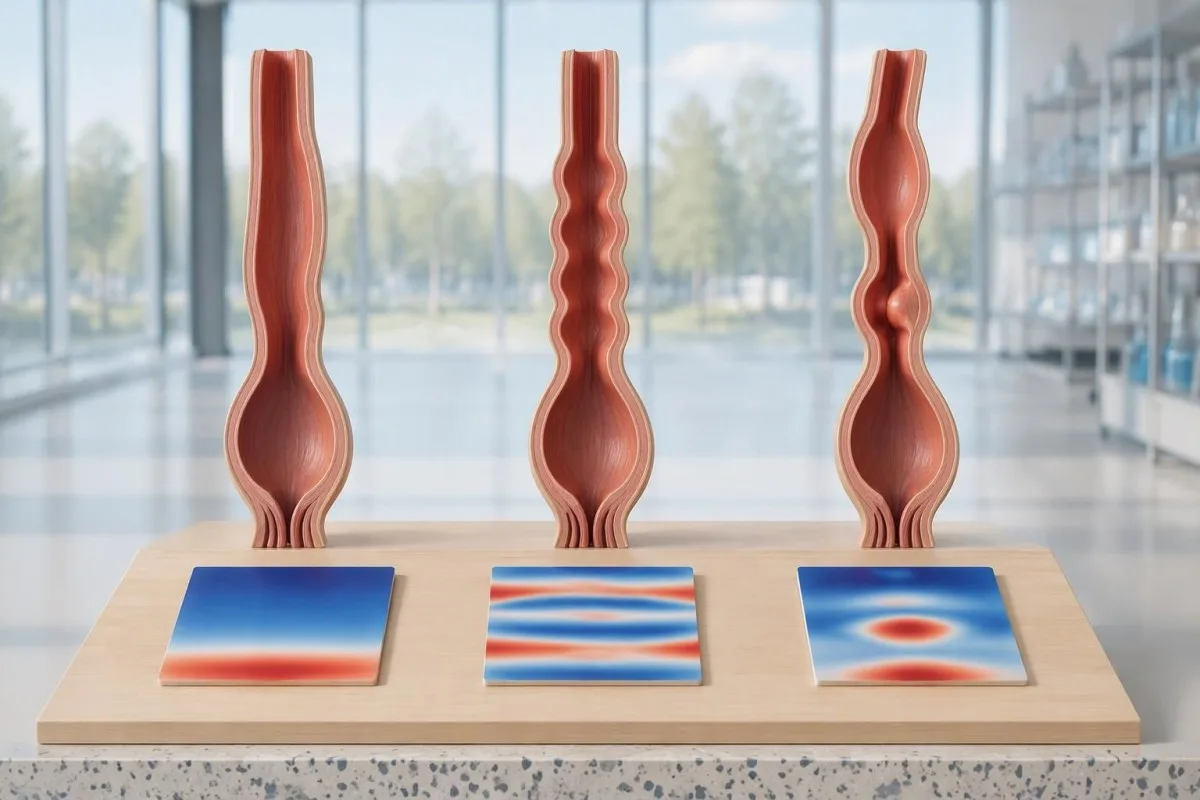
Example 1: a fictional report records median IRP 24 mmHg, DCI below 100 mmHg·s·cm in all 10 swallows, and no panesophageal pressurization. Assuming adequate technique, no structural explanation, and consistent symptoms, this is a type I pattern; the conclusion comes from failed propulsion plus impaired relaxation, not from the pressure value alone.
Example 2: another fictional report records IRP 24 mmHg, failed peristalsis in all 10 swallows, and panesophageal pressurization in 6 swallows. The pressurization frequency is 60%, exceeding the 20% threshold for type II; this does not mean the condition is three times more severe than a report with pressurization in 2 swallows.
Example 3: a fictional study records IRP 24 mmHg, 4 premature contractions with distal latency 3.8 seconds and DCI 900 mmHg·s·cm, and 6 failed swallows, with no normal peristalsis. This supports type III; Kantesti is an AI asins analīžu rezultātu platforma, un mūsu piemēra interpretācijas darbplūsmām concern laboratory information rather than automated classification of these hypothetical motility tracings.
Ko darīt, ja IRP ir normāls, robežvērtības vai nekonsekvents?
A normal or borderline IRP does not always exclude achalasia, particularly when peristalsis is absent and symptoms strongly suggest impaired emptying. Conversely, elevated IRP with preserved peristalsis is not automatically achalasia; Chicago Classification version 4.0 requires careful separation of inconclusive achalasia, artifacts, and esophagogastric junction outflow obstruction.
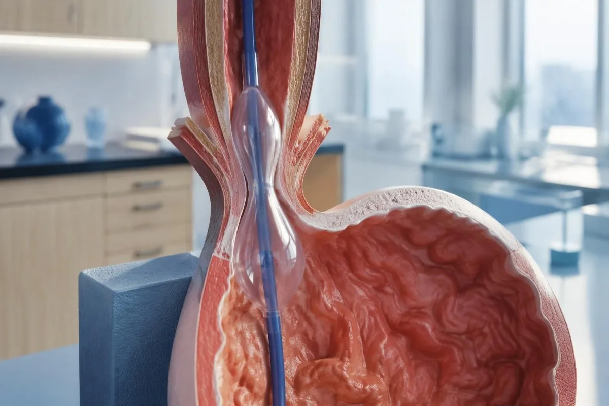
An elevated supine IRP that normalizes upright can reflect catheter contact or distortion around a hiatal hernia. This is one reason the standard protocol uses 2 positions: a reproducible obstruction pattern is more convincing than one abnormal posture-dependent number, although positional disagreement still needs specialist interpretation rather than an automatic normal label.
Esophagogastric junction outflow obstruction, or EGJOO, requires more than a high median IRP. Its manometric criteria include elevated IRP in both positions, preserved peristalsis, and elevated supine intrabolus pressure in at least 20% of swallows; clinically meaningful EGJOO also requires relevant symptoms and supportive evidence of obstruction on timed barium testing or FLIP.
Absent contractility with normal relaxation can occur in systemic sclerosis and is not the same as achalasia; scleroderma-related antibody clues cannot replace motility testing. Kantesti's klīniskās validācijas robežas should not be extended to these 2 distinct pressure disorders, and an irreversible sphincter procedure should not follow from an unexplained borderline result alone.
Kāpēc pirms ahalāzijas diagnosticēšanas nepieciešama endoskopija?
Upper endoscopy helps exclude a mechanical obstruction or pseudoachalasia, including an obstructing growth at the esophagogastric junction. Manometry can demonstrate an achalasia-like pressure pattern without proving its cause; the ACG guideline recommends endoscopy in suspected achalasia because treatment decisions depend on distinguishing primary motility disease from structural disease (Vaezi et al., 2020).
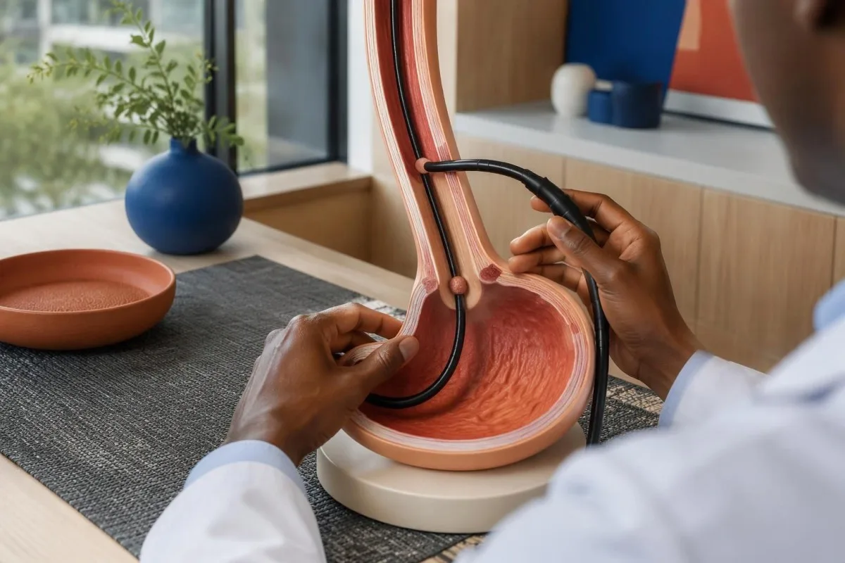
Retained saliva, retained food, and a tight-looking junction can support suspicion of achalasia, but none is diagnostic by itself. Early disease may have an almost normal endoscopic appearance; equally, resistance to passing the scope should not be dismissed as ordinary achalasia without considering whether a second test is needed to inspect the junction more closely.
Rapidly progressive symptoms, substantial short-term weight loss, older age at presentation, and unusual resistance at the junction increase concern for pseudoachalasia. There is no single age or kilogram cutoff that safely excludes it; a hypothetical 68-year-old with 3 months of progressive dysphagia and 9 kg weight loss warrants particular attention to the possibility of a structural cause.
A normal-looking mucosal surface does not always exclude a deeper or externally compressing growth, so CT or endoscopic ultrasound may be appropriate when suspicion persists. Normal CA 19-9 results cannot rule out this concern; ordering 1 tumor marker is not an alternative to imaging, endoscopy, or targeted tissue examination when clinically indicated.
Ko papildina laika bārija rīšanas izmeklējums un FLIP?
A timed barium esophagram measures esophageal emptying, while FLIP assesses how widely the esophagogastric junction opens under controlled distension. These tests provide information that pressure measurements alone cannot supply; a commonly used timed protocol obtains images at 1, 2, and 5 minutes after a standardized barium swallow.
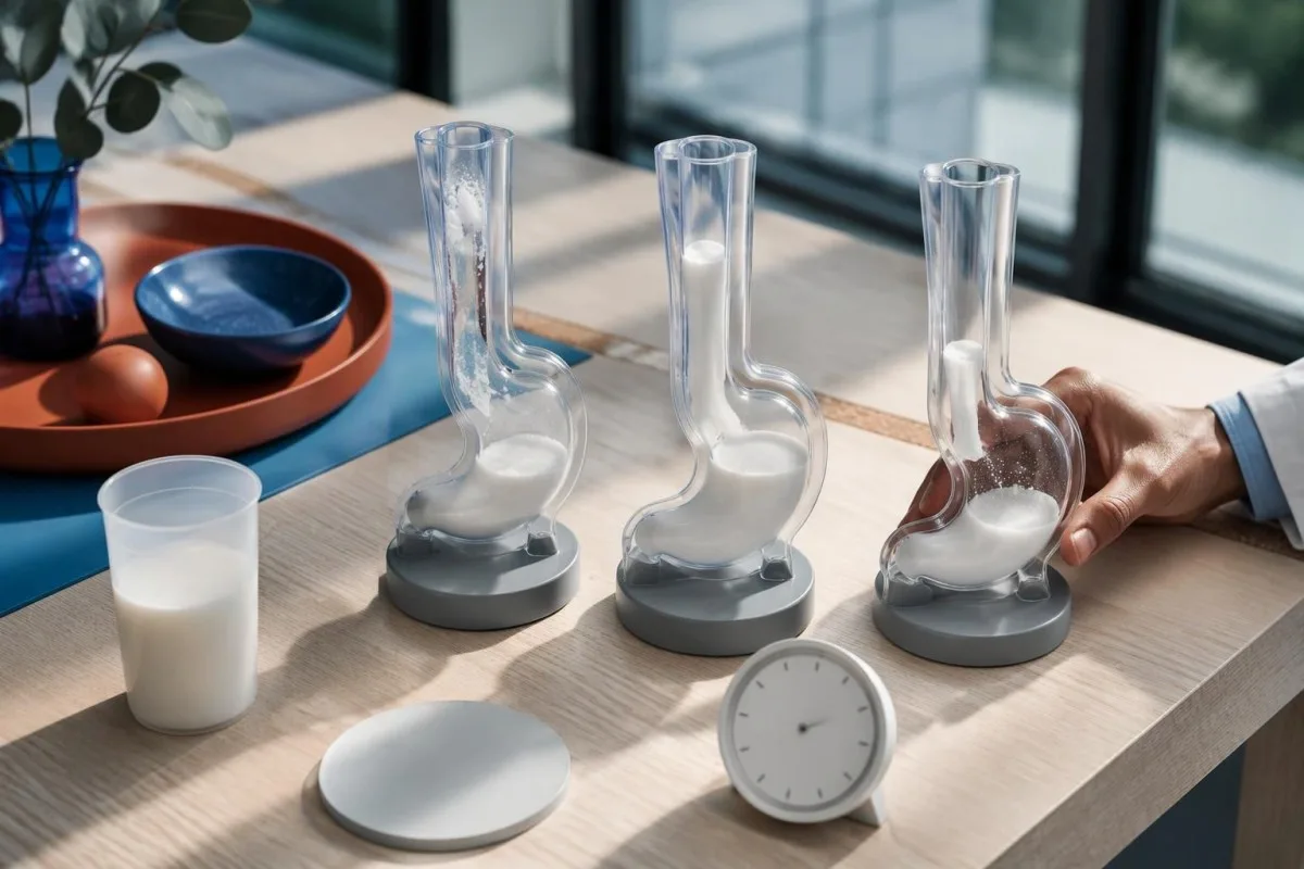
A standardized upright timed study often uses about 236 mL of barium; column heights above 5 cm at 1 minute or 2 cm at 5 minutes suggest impaired emptying in commonly used protocols. These are protocol-dependent abnormal-emptying thresholds, not stand-alone proof of achalasia, and the classic bird-beak appearance can be absent in early disease.
A 13 mm barium tablet can help reveal restricted passage when liquid contrast empties relatively well, provided the imaging team considers it appropriate. FLIP distensibility index expresses junction cross-sectional area divided by pressure in mm²/mmHg, but balloon volume, sedation, device protocol, and maximum opening diameter affect interpretation; treating every value below 2 as an interchangeable diagnosis would oversimplify the test.
An achalasia-like dilated esophagus does not establish that the disorder is idiopathic. In someone with relevant exposure history, confirmatory Chagas testing may be appropriate because chronic Trypanosoma cruzi infection can affect esophageal innervation; imaging and infection testing answer 2 different questions, namely how the esophagus empties and why its function may be impaired.
Kā ahalāzijas manometriskie tipi palīdz izvēlēties ārstēšanu?
Achalasia manometry types help clinicians choose how best to reduce resistance at the esophageal outlet. Pneumatic dilation, laparoscopic Heller myotomy, and POEM are established options for types I and II; type III often favors a longer, tailored myotomy that addresses the spastic segment as well as the junction.
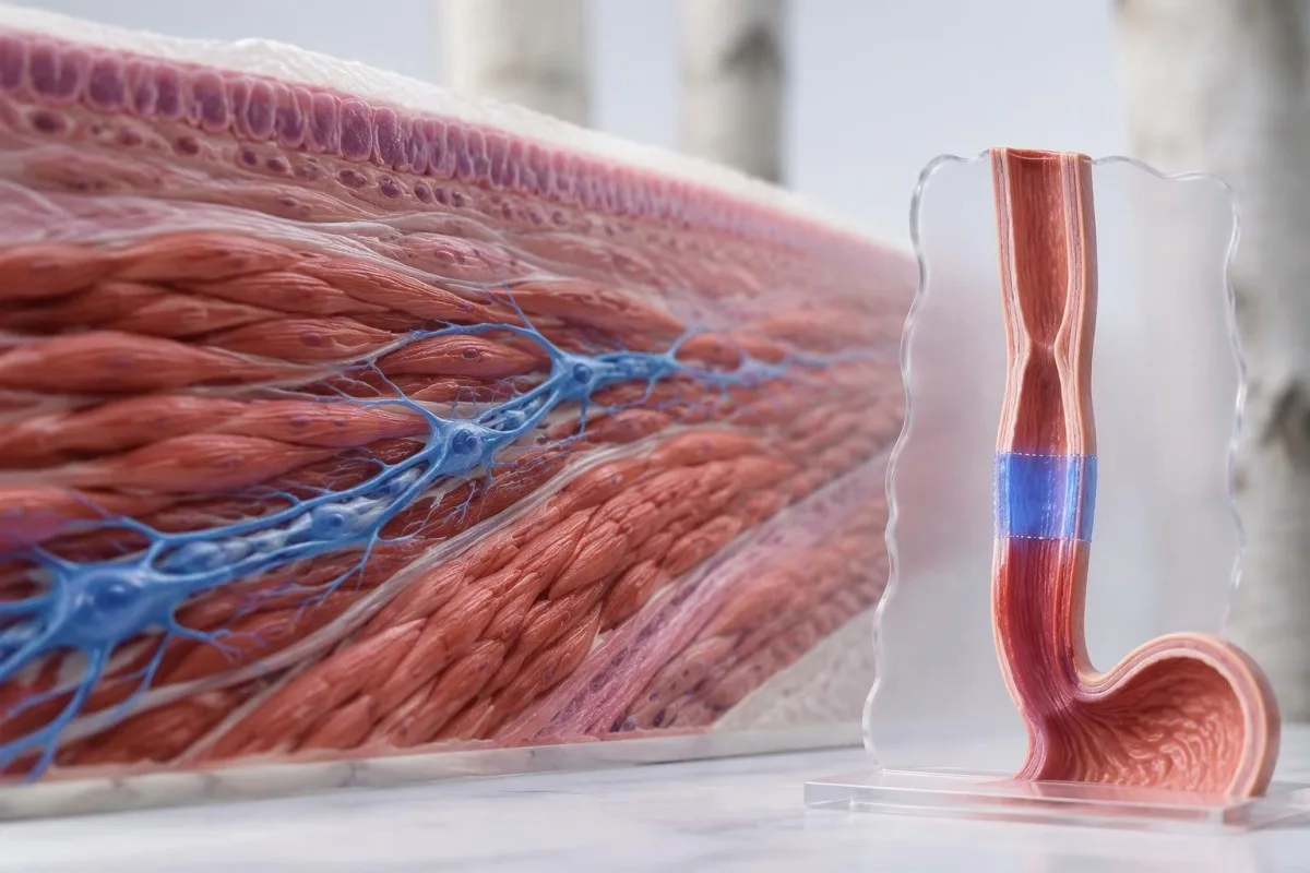
The ACG guideline considers pneumatic dilation, Heller myotomy, and POEM comparably effective options for appropriate patients with types I and II, while supporting tailored POEM or Heller myotomy over dilation for type III (Vaezi et al., 2020). The 3 procedures differ in anesthesia needs, recovery, retreatment possibilities, and reflux risk; local expertise and patient preference belong in the decision.
A randomized trial of 221 patients found clinical success at 2 years in 83.0% after POEM and 81.7% after Heller myotomy with Dor fundoplication (Werner et al., 2019). These overall results support both approaches but do not establish identical outcomes for every subtype; type III participants form a much smaller subgroup than the full trial population.
Reflux esophagitis at 2 years occurred in 44% after POEM versus 29% after Heller myotomy in that trial, illustrating a trade-off rather than a reason to reject POEM automatically. Acid-suppression treatment may subsequently affect gastrin blood results; meanwhile, pneumatic dilation carries a perforation risk of roughly 2% in experienced practice and requires an appropriate rescue pathway.
Ko pēc ārstēšanas var atklāt novērošana un asins analīzes?
Follow-up assesses swallowing, weight, esophageal emptying, and reflux, not just whether symptoms improve. Blood tests can identify nutritional or hydration complications but cannot confirm that the junction opens normally; the ACG guideline recommends timed barium esophagram as the first-line objective test when symptoms persist or recur after treatment (Vaezi et al., 2020).
The Eckardt score adds dysphagia, regurgitation, chest pain, and weight loss, each scored from 0 to 3, for a total of 0–12. Trials often define success as a score of 3 or below, but symptom scores alone can miss retained material; reduced discomfort does not necessarily mean that esophageal clearance has normalized.
Kantesti ir AI biomarķieru interpretācijas platforma that can help explain a clinician-requested complete blood count, electrolytes, and iron studies alongside reduced intake. Ferritin below 15 ng/mL strongly supports depleted iron stores in adults without a confounding context; our pilnais dzelzs izmeklējumu ceļvedis explains why ferritin and transferrin saturation should be considered together rather than used to infer an achalasia subtype.
Albumin commonly has an adult reference interval around 35–50 g/L, but a normal value does not exclude inadequate intake and a low value is not specific for malnutrition. Our seruma olbaltumvielu interpretācijas ceļvedis explains those limitations; magnesium deserves separate attention when poor intake or long-term acid suppression is present, as discussed in zema magnija līmeņa cēloņiem.
Ko jautāt tālāk un kuri pierādījumi ir attiecināmi?
Your next discussion should connect the manometry classification, structural assessment, and evidence of impaired emptying before selecting treatment. Ask 3 practical questions: which findings establish the subtype, whether obstruction or medication effects have been excluded, and how the proposed procedure's expected benefit compares with its reflux and retreatment risks.
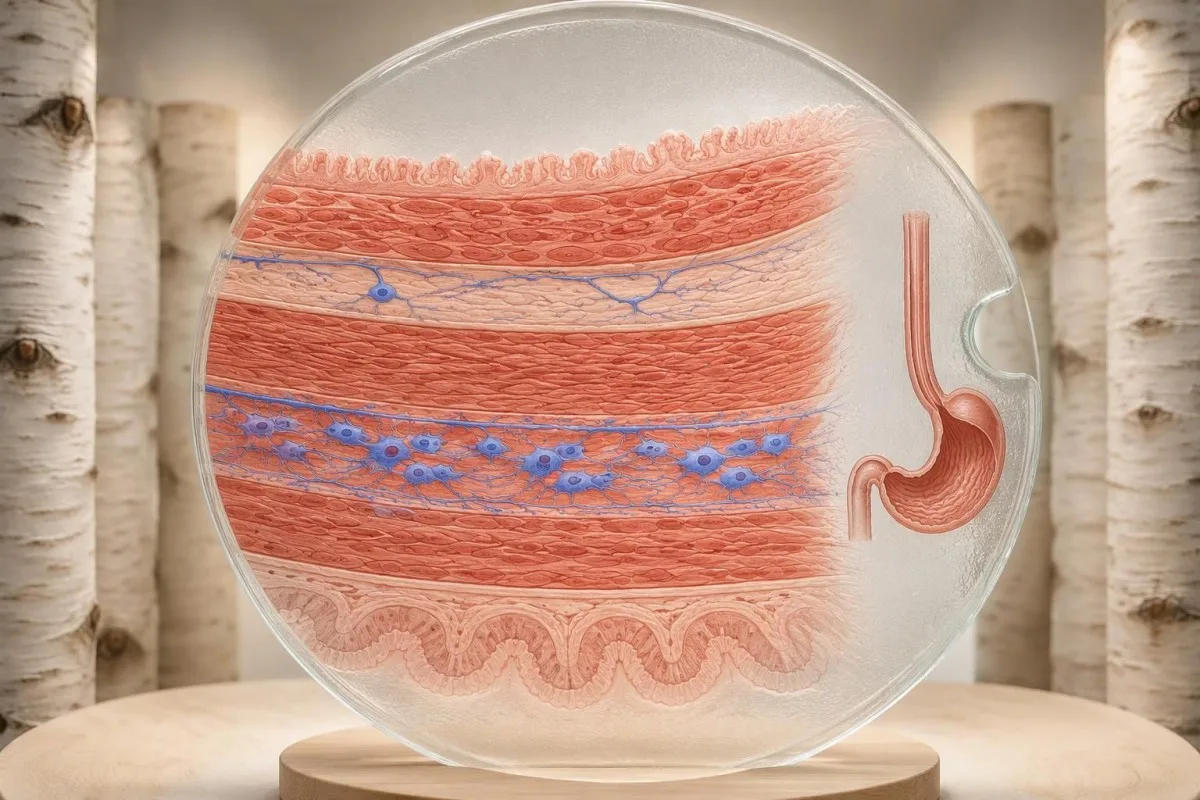
Request the full manometry report rather than only its conclusion, including the catheter system, body positions, median IRP, swallow counts, and any supportive tests. If you cannot swallow liquids or saliva, develop severe chest pain, or have respiratory difficulty, seek urgent care; waiting another 2 weeks for a routine report review is not an appropriate response to those warning signs.
The achalasia evidence cited here comes from Chicago Classification version 4.0, the ACG achalasia guideline, and the randomized POEM-versus-Heller trial. Kantesti's medicīniskā satura standarti distinguish evidence-based explanations from individualized diagnosis; the 2 Zenodo publications listed below concern coagulation and serum proteins, not studies validating manometry or achalasia treatment.
The serum-protein publication is relevant only to the laboratory-support discussion, while the coagulation publication provides separate background if a clinician requests clotting tests before a procedure. Neither DOI should be used to choose among types I, II, and III; when laboratory values change during recovery, blood-test timeline effects help explain why intake, illness, hydration, and medicines may alter results without indicating recurrent outlet obstruction.
Bieži uzdotie jautājumi
Kā diagnosticē ahalāziju?
Ahalāziju parasti diagnosticē ar augstas izšķirtspējas barības vada manometriju, kas uzrāda traucētu barības vada un kuņģa pārejas zonas relaksāciju un normālas peristaltikas neesamību. Pēc spiediena modeļa nosaka I, II un III tipu. Augšējā endoskopija palīdz izslēgt strukturālu nosprostojumu, savukārt laika intervālā veiktas bārija kontrastvielas norīšanas izmeklējums vai FLIP var palīdzēt precizēt neskaidrus atradumus. Neviena no 3 apakštipiem apstiprināšanai nav pieejama ikdienā veicama asins analīze.
Ko barības vada manometrija parāda ahalāzijas gadījumā?
Barības vada manometrija parāda, kā pēc norīšanas atslābst pārejas zona un vai barības vada ķermenī rodas koordinētas kontrakcijas. Integrētais relaksācijas spiediens novērtē izejas zonas atslābšanu 4 sekunžu intervālā, savukārt kontrakciju laiks un spiediena sadalījums palīdz noteikt apakštipu. II tipam nepieciešama pan-barības vada presurizācija vismaz 20% rīšanas reižu, bet III tipam — priekšlaicīgas kontrakcijas vismaz 20% rīšanas reižu. Jāizmanto konkrētajai iekārtai paredzētās robežvērtības, jo viena universāla IRP robežvērtība nav piemērota.
Kāda ir atšķirība starp I, II un III tipa ahalāziju?
I tipa ahalāzijai ir neefektīva peristaltika bez panesofageālās presurizācijas, kas nepieciešama II tipam. II tipam ir neefektīva peristaltika un panesofageāla presurizācija vismaz 20% rīšanas reižu. III tipam ir priekšlaicīgas kontrakcijas vismaz 20% rīšanas reižu un nav normālas peristaltikas, kā arī ir traucēta pārejas zonas atslābšana. Tie ir fizioloģiski raksti, nevis garantētas slimības progresēšanas stadijas.
Vai ahalāzija var rasties pie normāla IRP?
Ahalāzija dažkārt joprojām var radīt bažas, neraugoties uz normālu vai robežvērtībai tuvu integrēto relaksācijas spiedienu, īpaši tad, ja nav peristaltikas un ir traucēta iztukšošanās. Šādā situācijā manometrijas rezultāts var būt nepārliecinošs, nevis nepārprotami normāls. Laikā veiktā bārija izmeklējumā, kurā parasti tiek iegūti attēli pēc 1, 2 un 5 minūtēm, vai FLIP izmeklējumā var iegūt papildu apstiprinošus pierādījumus. Speciālistam jāapsver arī kontraktilitātes trūkums, tehniski ierobežojumi un iepriekš veikta ārstēšana.
Vai endoskopijas rezultāts var būt normāls, ja jums ir ahalāzija?
Endoskopijā agrīnas ahalāzijas gadījumā aina var būt gandrīz normāla, jo tā ļauj izmeklēt struktūru un gļotādu, nevis tieši noteikt rīšanas spiedienu. Aizdomas var radīt barības, siekalu vai ciešas pārejas zonas paliekas, taču tās neapstiprina diagnozi. Manometrijā tiek noteikts raksturīgais spiediena raksts un nošķirti 3 apakštipi. Ja joprojām pastāv aizdomas par pseidoahalāziju, var būt nepieciešama datortomogrāfija vai endoskopiskā ultrasonogrāfija, pat ja gļotādas virsma izskatās normāla.
Kurš ahalāzijas veids vislabāk reaģē uz ārstēšanu?
Type II achalasia generally has favorable responses to pneumatic dilation, Heller myotomy, and POEM, but subtype alone cannot predict an individual's result. Type III often benefits from a longer myotomy tailored to the spastic segment. A randomized trial reported 2-year success of 83.0% after POEM and 81.7% after Heller myotomy with fundoplication across its overall study population. Reflux, anatomy, prior treatment, and procedural expertise also influence the choice.
Vai barības vada manometrijai nepieciešama sedācija?
Standard esophageal manometry is usually performed awake, often with local nasal anesthetic rather than intravenous sedation. Patients complete a structured swallowing protocol, commonly 10 supine and 5 upright water swallows. Difficult catheter placement sometimes requires endoscopic assistance, but any sedation and recovery interval must be considered when interpreting the subsequent measurements. Follow the testing center's fasting instructions because achalasia can retain food despite an ordinary fast.
Uzziniet, kā lasīt asins analīžu rezultātus
Augšupielādējiet laboratorijas pārskatu, un Kantesti mākslīgais intelekts dažu sekunžu laikā vienkāršā valodā izskaidros katru biomarķieri. To izmanto vairāk nekā 2 miljoni cilvēku visā pasaulē.
Atsauces
- Klein, T., Mitchell, S., & Weber, H. (2026). aPTT normālais diapazons: D-dimēra un C proteīna rokasgrāmata par asins recēšanu. Kantesti mākslīgā intelekta medicīniskie pētījumi. DOI
- Klein, T., Mitchell, S., & Weber, H. (2026). Seruma proteīnu rokasgrāmata: globulīnu, albumīna un albumīna un globulīnu attiecības asins analīze. Kantesti mākslīgā intelekta medicīniskie pētījumi. DOI
- Yadlapati R et al. (2021). Esophageal motility disorders on high-resolution manometry: Chicago classification version 4.0. Neurogastroenterology & Motility. DOI
- Vaezi MF et al. (2020). ACG Clinical Guidelines: Diagnosis and Management of Achalasia. The American Journal of Gastroenterology. DOI
- Werner YB et al. (2019). Endoscopic or Surgical Myotomy in Patients with Idiopathic Achalasia. The New England Journal of Medicine. DOI
- 2M+Analizētie testi
- 127+Valstis
- 100+Valodas
Par medicīnas komandu


E-E-A-T uzticēšanās signāli
Pieredze
Ārstu vadīta klīniskā laboratorijas interpretācijas darbplūsmu pārskatīšana.
Ekspertīze
Laboratorijas medicīnas fokuss uz to, kā biomarķieri uzvedas klīniskā kontekstā.
Autoritāte
Medicīniski recenzējis Dr. Thomas Klein, piedaloties Dr. Sarah Mitchell un Prof. Dr. Hans Weber.
Uzticamība
Uz pierādījumiem balstīta interpretācija ar skaidriem turpmākās rīcības ceļiem, lai mazinātu trauksmi.

Medicīniskā atruna
Šis raksts ir paredzēts tikai izglītojošiem nolūkiem un nav medicīnisks padoms. Vienmēr konsultējieties ar kvalificētu veselības aprūpes speciālistu, lai pieņemtu lēmumus par diagnostiku un ārstēšanu.
Publicēts · Sazinieties ar redakcijas komandu
Publicējis Kantesti Ltd, reģistrēts Anglijā un Velsā, uzņēmuma reģistrācijas Nr. 17090423, Londona, Apvienotā Karaliste.
