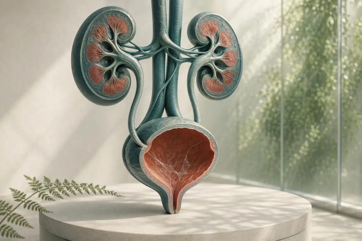
Śluz w badaniu osadu moczu zazwyczaj wynika z problemu ze zbiórką próbki, a nie jest diagnozą. Otaczające wyniki analizy moczu, objawy i jakość próbki decydują o potrzebie dalszej diagnostyki.


- Nitki śluzu same w sobie są zazwyczaj niegroźne, zwłaszcza w pojedynczej próbce pobranej metodą czystego chwytu, bez objawów ze strony układu moczowego.
- Próg ropomoczu przekraczający 5 białych krwinek na pole widzenia w powiększeniu, wspiera stan zapalny dróg moczowych, ale nie potwierdza bakteryjnej infekcji dróg moczowych.
- Wskazanie do posiewu to śluz plus pieczenie, nagląca potrzeba oddania moczu, gorączka, ból w boku, azotyny lub znacząca liczba białych krwinek – nie sam śluz.
- Komórki nabłonka płaskiego powyżej około 10 na pole widzenia w powiększeniu często wskazują na zanieczyszczenie wydzieliną narządów płciowych/skóry i obniżają wiarygodność wyniku badania moczu.
- Widoczna krew z bólem kolkowym boku wymaga pilnej oceny pod kątem kamienia, nawet jeśli zgłaszano również śluz.
- Ciąża zmienia próg dalszych działań, ponieważ bezobjawową bakteriurię zazwyczaj bada się za pomocą posiewu moczu, a nie samej mikroskopii.
- Mętny mocz może odzwierciedlać kryształy, zagęszczony mocz, wydzielinę pochwową, nasienie lub komórki; nie można nim zdiagnozować infekcji na podstawie wyglądu.
- Powtórz próbkę jest uzasadnione, gdy śluz jest jedynym nietypowym znaleziskiem, a pobranie nie było starannym pobraniem moczu ze środkowego strumienia.
Co zazwyczaj oznaczają nitki śluzu w analizie moczu
Włókna śluzu w moczu są zazwyczaj pasmami normalnej wydzieliny ochronnej lub zanieczyszczeniem narządów płciowych, a izolowany wynik rzadko sygnalizuje chorobę nerek. Wynik staje się znaczący, gdy pojawia się z objawami ze strony układu moczowego, białymi krwinkami, bakteriami, krwią lub białkiem.
Laboratoria identyfikują śluz jako cienkie, przezroczyste pasma widoczne pod mikroskopem; wiele raportów po prostu ocenia go jako rzadki, nieliczny, umiarkowany lub liczny, zamiast przypisywać zakres liczbowy. Mocz naturalnie zawiera niewielką ilość materiału bogatego w glikoproteiny z dróg moczowych, a wydzieliny z pochwy lub cewki moczowej mogą zwiększyć jego ilość podczas pobierania.
Sformułowanie raportu jest mniej alarmujące, niż brzmi. Stan na 26 sierpnia 2026 r., nie istnieje zweryfikowana liczba włókien śluzu, która diagnozowałaby ZUM, kamień, raka ani niewydolność nerek,; jest to obserwacja mikroskopowa kontekstowa. Aby uzyskać szerszą mapę innych elementów z raportu pasków testowych i mikroskopii, zobacz nasz kompletny przewodnik po analizie moczu.
Z mojego doświadczenia klinicznego wynika, że u osoby dorosłej bez objawów, u której śluz oznaczono jako “umiarkowany”, 0–2 białe krwinki na pole widzenia o dużej sile, azotyny ujemne i brak krwi, zazwyczaj najlepszym rozwiązaniem jest uspokojenie lub jedno prawidłowo pobrane powtórne badanie. Dr Thomas Klein często obserwuje niepotrzebny niepokój związany z antybiotykami, wynikający z błędnego utożsamiania znacznika laboratoryjnego z etykietą choroby.
Kantesti to platforma do interpretacji wyników badań krwi oparta na sztucznej inteligencji, która może umieścić markery krwi związane z nerkami, takie jak kreatynina i eGFR, obok wyników badania moczu – ale nie może zdiagnozować ZUM na podstawie samego śluzu. Wynik badania moczu pod mikroskopem wymaga historii klinicznej, która mu towarzyszyła.
Dlaczego laboratorium oznacza to jako problem
Systemy informatyczne laboratoriów oznaczają śluz jako problem, ponieważ jest widoczny, a nie dlatego, że laboratorium ustaliło niebezpieczny próg. Oznaczenie “problem” oznacza zatem “zaobserwowano”, a nie „nieprawidłowy marker choroby”, podobnie jak izolowany wynik śladowy, który może nie wymagać leczenia.
Skąd bierze się śluz w próbce moczu
Śluz w moczu może pochodzić z cewki moczowej, błony śluzowej pęcherza, szyjki macicy lub pochwy, nasienia lub zewnętrznej skóry narządów płciowych podczas pobierania. Jego źródło jest często domniemane na podstawie reszty obrazu mikroskopowego, a nie samych pasm.
Pęcherz i cewka moczowa mają ochronną warstwę powierzchniową zawierającą mucyny, w tym materiał związany z uroplakiną, który zmniejsza tarcie i przyleganie drobnoustrojów. Niewielkie ilości mogą się złuszczać do moczu po odwodnieniu, niedawnym aktywności seksualnej, łagodnym podrażnieniu lub po prostu dlatego, że skoncentrowana poranna próbka ułatwia zobaczenie pasm.
U osób miesiączkujących lub mających wydzielinę z pochwy, leukocyty i śluz mogą dostać się do kubeczka, nie pochodząc z pęcherza. Ponad 10 płaskich komórek nabłonkowych na pole widzenia o dużej sile zwiększa prawdopodobieństwo zanieczyszczenia, chociaż laboratoria stosują różne progi raportowania. Nasz przewodnik po komórkach nabłonkowych w moczu wyjaśnia, dlaczego zmienia to pewność wyniku.
Nasienie może również tworzyć włóknisty materiał lub zmętnienie przez kilka godzin po wytrysku. Nie czyni to moczu niebezpiecznym, ale może utrudniać mikroskopię; jeśli rozważa się posiew, zazwyczaj zalecam pobranie nowego materiału co najmniej 24 godziny później, jeśli to możliwe.
Dlaczego seks i anatomia wpływają na wynik badania
Pojemnik na mocz nie izoluje pęcherza od pobliskich tkanek. Dlatego “śluz obecny” jest częściej zgłaszany w próbkach z wydzielinami zewnętrznymi, podczas gdy próbka pobrana cewnikiem jest czasem używana, gdy lekarze potrzebują czystszego wyniku.
Kiedy nitki śluzu wskazują na infekcję dróg moczowych
Włókna śluzowe wspierają możliwość infekcji dróg moczowych (UTI) tylko wtedy, gdy towarzyszą im objawy i obiektywne cechy zapalne, w szczególności ropomocz, dodatni wynik testu na azotyny lub przekonujący wynik posiewu. Sam śluz nie jest testem na infekcję dróg moczowych (UTI).
Objawowa infekcja dolnych dróg moczowych (UTI) zazwyczaj powoduje pieczenie, parcie, częstomocz, dyskomfort nadłonowy, a czasem nowy zapach moczu. W prawidłowo pobranej próbce, ponad 5 białych krwinek na pole widzenia w dużym powiększeniu jest często nazywane ropomoczem; wspiera ono zapalenie dróg moczowych, ale może wystąpić również w przypadku kamieni, infekcji przenoszonych drogą płciową i zanieczyszczenia.
Azotyny są bardzo swoiste, gdy są dodatnie, ale nie wykrywają infekcji spowodowanych przez organizmy, które nie redukują azotanów, a ujemny wynik testu na azotyny nie wyklucza infekcji dróg moczowych (UTI). Esteraza leukocytarna wykrywa aktywność enzymu białych krwinek i może dawać fałszywie dodatnie wyniki, gdy wydzieliny z pochwy zanieczyszczają próbkę; nasza wyjaśnienie dotyczące wyników esterazy leukocytowej obejmuje te pułapki.
Wytyczne IDSA odradzają badanie przesiewowe lub leczenie bezobjawowego bakteriomoczu u zdrowych dorosłych kobiet niebędących w ciąży, ponieważ antybiotyki szkodzą bez korzyści (Nicolle et al., 2019). Ta zasada jest szczególnie istotna w odniesieniu do śluzu: brak pieczenia, brak gorączki i brak planowanego zabiegu urologicznego zazwyczaj oznacza brak antybiotyku tylko dlatego, że pole mikroskopowe wyglądało nieporządnie.
Kiedy posiew jest lepszym badaniem
Posiew moczu jest bardziej użyteczny niż ponowne badanie paskiem, gdy objawy utrzymują się, objawy nawracają w ciągu 4 tygodni, występuje ciąża, podejrzewa się odmiedniczkowe zapalenie nerek lub wcześniejsze antybiotyki mogły wpłynąć na wynik. Mieszany wzrost bakteryjny zazwyczaj wskazuje na zanieczyszczenie podczas pobierania próbki, a nie na pojedynczy patogen dróg moczowych; patrz interpretacja posiewu moczu.
Śluz, kryształy, kamienie i podrażnienia mechaniczne
Śluz z silnym falistym bólem w okolicy lędźwiowej lub krwią w moczu może wystąpić w przypadku kamienia moczowego, ale śluz ani nie potwierdza, ani nie wyklucza kamienia. Krew, kryształy, wzorzec bólu, obrazowanie i czynność nerek mają większe znaczenie.
Kamień może podrażniać lub blokować wyściółkę dróg moczowych, powodując powstawanie krwinek czerwonych, białych i dodatkowego śluzu. Klasycznym objawem jest nagły ból kolkowy promieniujący w kierunku pachwiny, często z nudnościami; widoczna czerwona lub herbaciana barwa moczu nasila potrzebę pilnego działania. Dowiedz się więcej o czerwonych flag związanych z krwią w moczu zamiast zakładać, że każda różowa próbka jest prostą infekcją.
Kryształy szczawianu wapnia mogą pojawiać się u zdrowych osób, szczególnie w zagęszczonym kwaśnym moczu, więc jeden rodzaj kryształów nie dowodzi aktywnego kamienia. U osoby z bólem, nawracającymi kamieniami lub uporczywą krwiomoczem, lekarze mogą zastosować ultrasonografię lub tomografię komputerową o niskiej dawce bez kontrastu, zamiast polegać wyłącznie na osadzie. Nasz zasób dotyczący kryształach szczawianu wapnia szczegółowo opisuje ograniczenia raportów dotyczących kryształów.
Ważnym wyjątkiem jest niedrożność z infekcją: gorączka 38,0°C lub wyższa, ból w okolicy lędźwiowej, wymioty i niemożność oddania moczu wymagają pilnej oceny. Problemem nie jest śluz; jest to zakażony zablokowany układ moczowy, który może szybko ulec pogorszeniu.
Podrażnienie bez kamienia
Niedawne użycie cewnika, zabiegi na pęcherzu, intensywne kolarstwo i radioterapia miednicy mogą podrażniać dolne drogi moczowe i zwiększać ilość śluzu lub białych krwinek. Lekarz powinien interpretować te wyniki w kontekście czasu, ponieważ próbka pobrana w ciągu 48 godzin od zabiegu nie jest równoważna z rutynowym badaniem przesiewowym.
Znaczenie mętnego moczu: co jego wygląd może, a czego nie może powiedzieć
Mętny mocz może być spowodowany przez zagęszczone sole, kryształy, śluz, komórki, wydzielinę z narządów płciowych lub bakterie, więc sam wygląd nie może zdiagnozować infekcji. Mocz, który jest mętny, ale bezbolesny i krótkotrwały, zazwyczaj nie jest pilny.
Kryształy fosforanów mogą powodować mętnienie alkalicznego moczu po jego ostygnięciu, podczas gdy kryształy moczanów mogą zmętnić kwaśny, skoncentrowany mocz. Próbka pozostawiona w temperaturze pokojowej przez ponad 2 godziny może stawać się coraz bardziej mętna z powodu degradacji komórek i namnażania się bakterii, dlatego ważne jest szybkie badanie lub przechowywanie w lodówce.
Mętność w połączeniu z dyzurią, naglącym parciem i ropomoczem wymaga badań; mętność po wysiłku fizycznym lub niedostatecznym nawodnieniu często ustępuje po zwykłym nawodnieniu. Staraj się o jasny żółty mocz, zamiast forsować nadmierne spożycie wody – bardzo przejrzysty mocz nie oznacza, że nerki “wypłukują” infekcję. Nasz szczegółowy przegląd przyczyn mętnnego moczu rozdziela wskazówki wizualne od wiarygodnych testów.
Kantesti jest Analizator do badań krwi AI przeznaczone do interpretacji markerów krwi w kontekście klinicznym; wysoki poziom kreatyniny lub obniżony eGFR w połączeniu z nieprawidłowościami moczu może uzasadniać konsultację lekarską, podczas gdy mętny mocz sam w sobie nie świadczy o upośledzonej filtracji nerkowej. Podstawowy panel metaboliczny może dostarczyć przydatnych informacji, gdy objawy wskazują na odwodnienie lub niedrożność.
Zapach nie jest hodowlą
Mocno pachnący mocz często odzwierciedla koncentrację, metabolity szparagów, witaminy z grupy B lub pojemnik, który stał zbyt długo. Nowy, nieprzyjemny zapach z towarzyszącą gorączką lub objawami ze strony układu moczowego jest powodem do konsultacji, ale zapach nie zastępuje mikroskopii i hodowli.
Wydzielina narządów płciowych i choroby przenoszone drogą płciową
Wydzielina z cewki moczowej lub pochwy może wyglądać jak śluz w moczu, a nowa wydzielina z pieczeniem podczas oddawania moczu wymaga badań w kierunku chorób przenoszonych drogą płciową, a także badania moczu. Rutynowa hodowla moczu może nie wykryć chlamydii i rzeżączki.
Chlamydia i rzeżączka mogą powodować dyzurię i jałową ropomoczu – obecność białych krwinek bez typowego wzrostu bakteryjnego – szczególnie po nowym kontakcie seksualnym. Testy amplifikacji kwasów nukleinowych, często wykorzystujące mocz z pierwszego strumienia zamiast ze środkowego, są odpowiednimi testami, ponieważ standardowa hodowla ukierunkowana jest na inne organizmy.
U osób z pochwą, bakteryjna waginoza, kandydoza i zapalenie szyjki macicy mogą dodawać wydzielinę do próbki ze środkowego strumienia; wymaz z pochwy lub badanie może być bardziej informacyjne niż powtarzanie mikroskopii moczu. U osób z penisem, widoczna wydzielina z cewki moczowej nie powinna być lekceważona jako “nici śluzowe”. Praktyczna różnica polega na tym, czy wydzielina jest zauważalna poza oddawaniem moczu, czy też nici są zgłaszane tylko przez laboratorium.
Wytyczne CDC dotyczące leczenia chorób przenoszonych drogą płciową z 2021 r. zalecają testowanie oparte na NAAT w odpowiednich miejscach anatomicznych, zgodnie z historią narażenia, a nie tylko na podstawie objawów (Workowski i wsp., 2021). W przypadku bólu w miednicy, bólu jąder, gorączki, ciąży lub podejrzenia napaści, należy zasięgnąć porady klinicznej tego samego dnia, zamiast leczyć się samemu pozostałościami antybiotyków.
Dlaczego antybiotyki mogą zaburzać obraz kliniczny
Przyjęcie nawet 1 lub 2 dawek antybiotyku przed hodowlą może zahamować normalny wzrost bakterii, podczas gdy objawy ze strony układu moczowego i białe krwinki utrzymują się. Należy dokładnie poinformować lekarza o przyjmowanym leku, dawce i czasie ostatniej dawki; ta informacja zmienia interpretację wyniku negatywnej hodowli.
Jak pobrać próbkę moczu, która odpowie na pytanie
Próbka ze środkowego strumienia pobrana po podmyciu lepiej redukuje śluz, komórki nabłonkowe i mieszany wzrost bakteryjny niż pobranie pierwszej lub ostatniej części strumienia moczu. Technika jest prosta, ale pierwsze 2 sekundy mają nieproporcjonalnie duże znaczenie.
Umyj ręce, rozchyl skórę narządów płciowych lub odciągnij napletek, jeśli jest to wygodne, oczyść zgodnie z instrukcją zestawu, zacznij oddawać mocz do toalety, a następnie zbierz środkową porcję, unikając kontaktu pojemnika ze skórą. Próbka o objętości 20–30 ml jest zazwyczaj wystarczająca; wypełnienie dużego pojemnika po brzeg nie poprawia wyniku testu.
Dostarcz pojemnik niezwłocznie, najlepiej w ciągu 1 godziny; jeśli opóźnienie jest nieuniknione, przechowuj w lodówce zgodnie z instrukcjami laboratorium i zwróć w ciągu 24 godzin. Chłodzenie spowalnia wzrost bakterii, ale nie przywraca próbki, która została już zanieczyszczona. Jest to szczególnie ważne, gdy zlecenie obejmuje hodowlę, a nie tylko badanie paskowe.
Kantesti jest Narzędzie do analizy wyników badań krwi oparte na AI stosowane w ponad 127+ krajach, a nasze procedury przeglądane przez klinicystów traktują jakość próbki jako część interpretacji, a nie jako dodatek. Ta sama zasada dotyczy każdej wartości laboratoryjnej: technicznie słaba próbka może dać przekonujący medycznie, ale mylący wynik. Zapoznaj się z naszą listą kontrolną dokładności laboratoryjnej z pytaniami, które należy zadać przed podjęciem działań na podstawie pojedynczego, nieprawidłowego wyniku.
Nie pobieraj próbek w tych sytuacjach, jeśli możesz poczekać
Unikaj rutynowych badań podczas obfitego krwawienia miesiączkowego, bezpośrednio po stosunku płciowym lub po zastosowaniu kremów dopochwowych, chyba że lekarz wyraźnie tego zażąda. Jeśli badanie nie może być odłożone, poinformuj laboratorium lub lekarza, ponieważ ten kontekst może wyjaśnić obecność śluzu, czerwonych krwinek lub komórek zewnętrznych.
Jak klinicyści interpretują śluz w kontekście całej analizy moczu
Najbardziej użytecznym wzorcem badania moczu jest połączenie objawów z liczbą białych krwinek, czerwonych krwinek, azotynu, esterazy leukocytów, białka, glukozy, ciężaru właściwego i komórek nabłonkowych. Śluz jest mniej istotną cechą pomocniczą w tym wzorcu.
Czysta próbka ze śluzem, 0–2 białych krwinek na pole widzenia z dużą mocą, negatywny azotyn i brak krwi zazwyczaj nie wymagają leczenia. Śluz z ponad 5 białymi krwinkami na pole widzenia z dużą mocą i dodatnia esteraza leukocytów zwiększa prawdopodobieństwo zapalenia; dodanie azotynu lub pojedynczej hodowli drobnoustroju zwiększa pewność, że za infekcję odpowiedzialne są bakterie.
Białko wymaga własnego podejścia. Śladowe ilości białka po gorączce, wysiłku fizycznym lub zagęszczonym moczu mogą być tymczasowe, ale utrzymujące się białko należy kwantyfikować za pomocą stosunku albuminy do kreatyniny, a nie przypisywać śluzowi. Nasz przewodnik do białka w moczu wyjaśnia, dlaczego białko na pasku testowym i ryzyko chorób nerek nie są wymienne.
Gęstość właściwa wynosząca 1,003 do 1,030 jest powszechne u dorosłych, chociaż zakresy referencyjne różnią się w zależności od laboratorium. Wysoki ciężar właściwy może zagęszczać śluz i tworzyć bardziej dramatycznie wyglądający osad, podczas gdy niski ciężar właściwy może liofilizować komórki i sprawić, że mikroskopia będzie zwodniczo łagodna; nasza przewodniku po ciężarze właściwym pokazuje, jak nawodnienie wpływa na interpretację.
Wartość negatywnych wyników
Negatywna krew, białko, azotyn i esteraza leukocytów znacząco obniżają poziom obaw u osoby bez objawów, nawet jeśli śluz jest zgłaszany jako umiarkowany. Żaden pojedynczy negatywny test nie jest doskonały, ale ta grupa jest bardziej uspokajająca niż niepokojąca ocena ilościowa śluzu.
Kiedy powtórzyć badanie, a kiedy zlecić posiew
Powtórzyć badanie moczu, gdy śluz jest izolowany, komórki nabłonka płaskiego sugerują zanieczyszczenie, lub pobranie było pośpieszne; zlecić hodowlę, gdy objawy są utrzymujące się, nawracające, ciężkie lub obarczone wysokim ryzykiem. Powtórne badanie nie jest “nicnierobieniem” – często jest to najbardziej diagnostyczny kolejny krok.
Dla dorosłej osoby niebędącej w ciąży, bez objawów i ze śluzem jako jedynym odchyleniem, powtórne badanie moczu metodą czystego uchwytu w ciągu 1–2 tygodni jest rozsądne, jeśli potrzebne jest uspokojenie; wielu klinicystów wcale by go nie powtarzało. Nie należy leczyć laboratoryjnego odchylenia antybiotykami w oczekiwaniu, chyba że lekarz zidentyfikuje wskazanie kliniczne.
Hodowla przed antybiotykami jest szczególnie przydatna w przypadku gorączki, bólu w okolicy lędźwiowej, ciąży, immunosupresji, przeszczepu nerki, stosowania cewnika moczowego, męskich objawów ze strony układu moczowego lub objawów, które powracają w ciągu 4 tygodni. Wytyczne Gupta et al. (2011) wspierają wykonanie hodowli w przypadku podejrzenia odmiedniczkowego zapalenia nerek oraz w sytuacjach, gdy bardziej prawdopodobna jest oporność lub alternatywna diagnoza.
Wyniki posiewów należy odczytywać z uwzględnieniem jakości pobrania materiału. Pojedynczy mikroorganizm przy 10^5 jednostkach tworzących kolonie na ml tradycyjnie potwierdzał obecność bakterii w moczu, ale u pacjentów objawowych mogą występować klinicznie istotne niższe liczby; “mieszana flora” zazwyczaj wymaga pobrania nowego próbki, a nie szerokiego spektrum antybiotyku. Porównaj przeznaczenie badania ogólnego moczu i posiewu przed zleceniem któregokolwiek z nich.
Ciąża wymaga innego progu
Ciąża jest wyjątkiem, ponieważ bezobjawowa bakteriuria może zwiększać ryzyko odmiedniczkowego zapalenia nerek i niekorzystnych wyników ciąży. W opiece prenatalnej powszechnie stosuje się badanie przesiewowe na początku ciąży; śluz w badaniu mikroskopowym nie może zastąpić tego posiewu.
Kiedy badania krwi dostarczają przydatnych informacji o nerkach i infekcji
Badania krwi są przydatne, gdy śluz w moczu występuje z gorączką, bólem w boku, nawracającymi infekcjami, obrzękami, zmniejszoną ilością wydalanego moczu lub uporczywym białkiem lub krwią. Kreatynina i eGFR oceniają filtrację, podczas gdy CBC i białko CRP mogą pomóc ocenić ogólnoustrojowy stan chorobowy.
eGFR poniżej 60 mL/min/1,73 m² przez co najmniej 3 miesiące spełnia jedno kryterium przewlekłej choroby nerek, ale pojedynczy niższy wynik podczas odwodnienia lub ostrej choroby nie wystarczy do postawienia tej diagnozy. Kreatynina jest zależna od masy mięśniowej, diety i niektórych leków, dlatego trendy i albuminuria są często bardziej informacyjne niż jedna liczba.
Podwyższony poziom białych krwinek lub CRP może wskazywać na proces zapalny, ale nie pozwala zidentyfikować dróg moczowych jako źródła. U osoby z gorączką i bólem w boku lekarze mogą sprawdzić kreatyninę przed wyborem obrazowania lub leków, zwłaszcza jeśli wymioty lub niedrożność mogą upośledzić funkcję nerek. Przegląd stadia przewlekłej choroby nerek dla kategorii eGFR i albuminy używanych przez lekarzy.
Kantesti jest usłudze interpretacji testów laboratoryjnych AI który uwypukla wzorce dla kreatyniny, eGFR, elektrolitów, CBC i markerów zapalnych, zamiast traktować pojedynczą notację śluzu jako diagnozę nerkową. Nasza metodologia podlega nadzorowi walidacji klinicznej, ale pilne objawy nadal wymagają bezpośredniej opieki medycznej, a nie interpretacji opartej na aplikacji.
Praktyczne rozróżnienie odwodnienia
Odwodnienie może zwiększyć ciężar właściwy moczu i tymczasowo zwiększyć poziom kreatyniny, szczególnie po biegunce, ekspozycji na ciepło lub intensywnym wysiłku fizycznym. Uporczywie zmniejszona produkcja moczu, zawroty głowy lub spadek eGFR po nawodnieniu wymagają konsultacji lekarskiej, a nie powtarzania testów domowych.
Pilne sygnały ostrzegawcze, które nie mogą czekać na powtórne badanie
Szukaj pilnej oceny tego samego dnia w przypadku śluzu w moczu z gorączką 38,0°C lub wyższą, bólem w boku, wymiotami, widoczną krwią, niemożnością oddania moczu, dezorientacją lub objawami ze strony układu moczowego związanymi z ciążą. Te kombinacje mają znaczenie, ponieważ mogą sygnalizować infekcję górnych dróg moczowych, niedrożność lub inny ostry stan.
Gorączka plus jednostronny ból pleców lub boku budzi większe obawy o odmiedniczkowe zapalenie nerek niż proste zapalenie pęcherza, szczególnie z dreszczami lub wymiotami. Opóźnione leczenie może prowadzić do odwodnienia, sepsy lub obciążenia nerek; prawidłowo wyglądająca próbka moczu w domu nie wyklucza bezpiecznie tego stanu.
Widoczna krew powinna zostać zbadana, nawet jeśli infekcja dróg moczowych wydaje się prawdopodobna, szczególnie po 35. roku życia, u palaczy lub jeśli krwawienie utrzymuje się po ustąpieniu objawów infekcji. Wytyczne AUA dotyczące mikrohematurii zalecają ocenę opartą na ryzyku po wykluczeniu łagodnych przyczyn (Barocas i in., 2020); uporczywe krwawienie nie jest wyjaśniane przez śluz.
Dzieci, osoby powyżej 65. roku życia, osoby z cukrzycą, osoby przyjmujące leki immunosupresyjne oraz osoby z pojedynczą nerką zasługują na niższy próg oceny. Prosta zasada w praktyce dr. Thomasa Kleina: jeśli objawy nasilają się w ciągu 6-12 godzin, nie czekaj na drugą filiżankę, aby uzyskać potwierdzenie.
Zadzwoń na pogotowie ratunkowe teraz w przypadku ciężkiej choroby
Zadzwoń na pogotowie ratunkowe w przypadku nowej dezorientacji, omdleń, silnego osłabienia, niebieskich lub szarych warg, ciężkiej duszności lub niemożności utrzymania płynów przy objawach ze strony układu moczowego. Są to ogólnoustrojowe oznaki niebezpieczeństwa, a nie rutynowe objawy UTI.
Śluz w moczu w ciąży, dzieciństwie i późniejszym życiu
Ciąża, dzieci i osoby starsze wymagają ostrożniejszej interpretacji, ponieważ zanieczyszczenie jest powszechne, ale konsekwencje pominiętej infekcji mogą być większe. Objawy i jakość posiewu pozostają cenniejsze niż stopień śluzu w każdej grupie wiekowej.
W ciąży częstomocz może być normalny, co sprawia, że objawy są mniej swoiste; gorączka, dyzuria lub ból pleców powinny skłaniać do pilnej oceny położniczej lub klinicznej. Posiew kontrolny jest zazwyczaj pobierany na początku opieki prenatalnej, a powtórny posiew może być zastosowany po leczeniu, w zależności od planu klinicysty.
U dzieci mocz pobrany do woreczka jest podatny na zanieczyszczenie i zazwyczaj nie powinien być stosowany samodzielnie do diagnozowania ZUM z posiewu. Cewnikowanie lub starannie pobrany czysty strumień moczu może być wymagany, gdy małe dziecko ma gorączkę bez wyraźnej przyczyny; śluz w próbce z woreczka jest szczególnie nieswoisty.
W późniejszym wieku bakteriuria i ropomocz stają się częstsze bez powodowania objawów. Nowe delirium powinno samo w sobie skłonić do szerokiej oceny medycznej pod kątem odwodnienia, leków, bólu, zaparć i źródeł infekcji, zamiast automatycznego leczenia ZUM; obowiązuje ta sama powściągliwość zalecana przez Nicolle i wsp. (2019).
Zmiany związane z miesiączką i hormonami
Krew miesiączkowa i śluz szyjkowy mogą wpływać na wyniki badania moczu przez kilka dni, a suchość pochwy po menopauzie może powodować miejscowe podrażnienie przypominające pieczenie dróg moczowych. Jeśli wyniki i objawy nie są zgodne, lekarz może ocenić przyczyny ginekologiczne zamiast przepisywać kolejne leczenie ZUM.
Typowe błędy po zauważeniu śluzu w wyniku badania laboratoryjnego
Nie należy przyjmować pozostałości antybiotyków, nie należy wewnętrznie się oczyszczać ani próbować “wypłukać” śluz przez ekstremalne spożycie wody po jednym nieprawidłowym wyniku badania moczu. Działania te mogą utrudnić wykonanie posiewu, zaburzyć normalną florę lub opóźnić właściwą diagnozę.
Antybiotyki przyjmowane bez posiewu mogą spowodować, że późniejsze badanie będzie fałszywie negatywne i mogą powodować biegunkę, wysypkę, objawy grzybicy lub oporność. Jeśli objawy są łagodne, ale utrzymują się, pobierz próbkę najpierw, gdy jest to praktyczne, a następnie postępuj zgodnie z planem leczenia lekarza opartym na całości obrazu klinicznego.
Produkty żurawinowe mogą nieznacznie zmniejszać ryzyko nawrotów niepowikłanych ZUM u niektórych osób, ale nie leczą gorączki, bólu w boku ani potwierdzonego zakażenia górnych dróg moczowych. Unikaj wysokich dawek witaminy C jako samoleczenia: może ona wpływać na niektóre reakcje pasków testowych i zwiększać obciążenie szczawianami u osób predysponowanych do kamieni szczawianowo-wapniowych.
Nie należy wielokrotnie sprawdzać muszli toaletowej w poszukiwaniu nitek. Włókna papieru toaletowego, pozostałości środków czyszczących, wydzieliny narządów płciowych i turbulencje wody mogą imitować śluz; odpowiednim miejscem do jego oceny jest próbka laboratoryjna pobrana do sterylnego pojemnika. W przypadku powiązanych zmian koloru, nasze przewodnik po kolorze moczu oferuje bardziej wiarygodny kontekst wizualny.
Lista leków ma znaczenie
Fenazopirydyna może zabarwić mocz na pomarańczowo i wpłynąć na interpretację wizualną, podczas gdy diuretyki mogą zagęszczać lub rozcieńczać mocz w zależności od pory podania. Na wizytę zabierz listę przepisanych leków, produktów dostępnych bez recepty i suplementów, podając dawki w mg.
Praktyczny plan dalszego postępowania w przypadku nici śluzu w moczu
Większość osób ze śluzem w moczu i bez objawów nie wymaga leczenia; staranne powtórne badanie moczu jest uzasadnione, jeśli próbka była wątpliwa. Objawy lub współistniejące nieprawidłowości decydują o tym, czy odpowiednie są posiew, badania na choroby przenoszone drogą płciową, obrazowanie czy badania krwi.
Krok 1: sprawdź pieczenie, parcie, gorączkę, ból w boku, widoczną krew, wydzielinę, ciążę, niedawne zabiegi na drogach moczowych i nowe narażenie seksualne. Krok 2: przeczytaj raport pod kątem białych krwinek, azotynów, esterazy leukocytów, czerwonych krwinek, białka, bakterii i komórek nabłonkowych – nie tylko śluzu.
Krok 3: jeśli czujesz się dobrze, a śluz jest izolowany, pobierz jedną próbkę środkowego strumienia czystego chwytu w ciągu 1-2 tygodni, tylko jeśli lekarz zaleci potwierdzenie. Krok 4: jeśli występują objawy, zapytaj, czy posiew moczu powinien być pobrany przed leczeniem i czy badania na choroby przenoszone drogą płciową są istotne; nawracające epizody wymagają bardziej przemyślanego przeglądu niż trzeci empiryczny antybiotyk.
Kantesti może pomóc w uporządkowaniu istotnych trendów wyników badań krwi i pytań na wizytę lekarską, podczas gdy nasze Rada doradcza ds. medycznych wspiera standardy bezpieczeństwa kierowane przez lekarza. Właściwym celem nie jest idealnie “czysty” raport mikroskopowy – ale wyjaśnienie, które pasuje do Twoich objawów, jakości próbki i czynników ryzyka.
Pytania do zabrania na wizytę
Zapytaj, czy w próbce obecne były komórki nabłonkowe, czy hodowla wykazała jeden organizm lub florę mieszaną, oraz czy krew lub białko utrzymywały się po ustąpieniu objawów. Te 3 pytania często dają bardziej użyteczne odpowiedzi niż pytanie o ilość widocznego śluzu.
Kontekst badawczy i ograniczenia raportowania śluzu
Ocena śluzu nie jest znormalizowana w różnych laboratoriach, dlatego klinicyści powinni unikać stosowania określeń “niewielki”, “umiarkowany” lub “duży” jako kategorii ciężkości choroby. Technika mikroskopowa, wiek próbki i oprogramowanie do raportowania mogą wpływać na sformułowania.
Niektóre laboratoria ręcznie analizują osad po odwirowaniu około 10–15 ml moczu, podczas gdy inne stosują zautomatyzowaną analizę cząstek stałych z ręczną weryfikacją wybranych flag. Ta zmienność oznacza, że wynik “umiarkowany śluz” z jednego laboratorium może nie zostać dokładnie powtórzony w innym laboratorium, nawet jeśli stan zdrowia pacjenta się nie zmienił.
Użytecznym podejściem laboratoryjnym jest zapytanie, czy wynik jest analitycznie prawdziwy, klinicznie znaczący i powtarzalny. Takie podejście jest znajome również z badań krwi: nasze przewodnik po biomarkerach krwi wyjaśnia, dlaczego zakresy referencyjne i warunki preanalityczne określają, czy oznaczony marker wymaga interwencji.
Kantesti AI wykorzystuje ustrukturyzowany kontekst laboratoryjny do wyjaśniania wzorców wyników w głównych panelach, ale mikroskopia moczu nadal wymaga osądu klinicznego specyficznego dla źródła. Dla czytelników zainteresowanych tym, jak oceniamy wydajność techniczną i granice kliniczne, nasze przewodnik technologii AI opisuje zabezpieczenia stojące za naszym podejściem interpretacyjnym.
Co powinien jasno wyjaśniać dobry raport
Przydatny raport zawiera identyfikację rodzaju próbki, daty pobrania, elementów mikroskopowych oraz wyników hodowli i antybiogramu, jeśli hodowla została wykonana. Jeśli raport zawiera tylko informację “obecne nitki śluzowe”, jest on niekompletny dla celów diagnostycznych, ale często jest całkowicie wystarczający w przypadku nieistotnego, mało prawdopodobnego znaleziska.
Często zadawane pytania
Czy niteczki śluzu w moczu są normalne?
Nitkowate śluzowatości w moczu są często prawidłowe lub wynikają z zanieczyszczenia wydzielinami narządów płciowych, szczególnie gdy osoba nie ma objawów ze strony układu moczowego. Izolowany wynik z obecnością 0–2 leukocytów na pole widzenia, ujemnym azotynem oraz brakiem krwi i białka zwykle wiąże się z niskim ryzykiem. Laboratoria nie stosują walidowanej liczby nitek śluzowych do diagnozowania ZUM ani chorób nerek. Powtórne pobranie próbki moczu ze środkowego strumienia jest uzasadnione, jeśli pierwotne pobranie było wykonane pośpiesznie lub było widocznie zanieczyszczone.
Czy śluz w moczu oznacza infekcję dróg moczowych?
Nici śluzowe w moczu same w sobie nie oznaczają infekcji dróg moczowych. Prawdopodobieństwo infekcji dróg moczowych wzrasta, gdy pieczeniu, parciu, częstomoczu, gorączce lub bólowi w nadłoniu towarzyszy obecność więcej niż 5 leukocytów na pole widzenia w wysokim powiększeniu, dodatni test na esterazę leukocytarną, azotyny lub dodatnia hodowla. Ujemny wynik testu na azotyny nie wyklucza całkowicie infekcji dróg moczowych, ponieważ nie wszystkie bakterie produkują azotyny. Hodowla jest zazwyczaj bardziej informatywna niż ocena ilości śluzu, gdy objawy utrzymują się lub nawracają.
Czy odwodnienie może powodować śluz w moczu?
Odwodnienie może ułatwić uwidocznienie śluzu, ponieważ zagęszczony mocz zawiera mniej wody wokół prawidłowych wydzielin i osadu. Podwyższone ciężar właściwy moczu, zazwyczaj w granicach 1,025–1,030, może towarzyszyć temu efektowi, chociaż sam w sobie nie dowodzi odwodnienia. Normalne nawodnienie i powtórzenie prawidłowo pobranej próbki może wyjaśnić wynik. Ekstremalne spożycie wody jest niepotrzebne i może stworzyć własne ryzyko związane z elektrolitami.
Co oznacza śluz i białe krwinki w moczu?
Śluz wraz z białymi krwinkami w moczu sugeruje zapalenie dróg moczowych lub narządów płciowych, ale nie dowodzi bakteryjnego zakażenia. Ponad 5 białych krwinek na pole widzenia w dużym powiększeniu może wystąpić w przypadku ZUM, kamicy, chorób przenoszonych drogą płciową, zanieczyszczenia pochwy lub niedawnego zabiegu na drogach moczowych. Objawy, azotyny, komórki nabłonkowe i wyniki posiewu decydują o dalszym postępowaniu. Gorączka 38,0°C lub wyższa z bólem w okolicy lędźwiowej wymaga pilnej oceny.
Czy upławy z pochwy mogą spowodować pojawienie się śluzu w badaniu moczu?
Wydzielina z pochwy może dostać się do próbki moczu ze środkowego strumienia i często powoduje pojawienie się śluzu, komórek nabłonka płaskiego, a czasem białych krwinek w obrazie mikroskopowym. Obecność ponad 10 komórek płaskich na pole widzenia pod dużym powiększeniem często zwiększa prawdopodobieństwo zanieczyszczenia, chociaż laboratoria różnią się w sposobie raportowania. Starannie pobrana próbka z czystego chwytu, pobrana poza okresem obfitego krwawienia menstruacyjnego, może zminimalizować ten problem. Jeśli występuje wydzielina, świąd, nieprzyjemny zapach lub ból miednicy, ocena pochwy lub wymaz z pochwy może być bardziej przydatny niż wielokrotne posiewy moczu.
Kiedy należy się martwić śluzowymi nitkami i krwią w moczu?
Nitki śluzu z widoczną krwią w moczu powinny być niezwłocznie ocenione, szczególnie gdy krew utrzymuje się, występuje ból w boku, skrzepy, gorączka lub trudności w oddawaniu moczu. Kamień, infekcja dróg moczowych, uraz, choroba nerek lub rzadziej guz dróg moczowych mogą powodować krew; śluz nie określa przyczyny. AUA zaleca ocenę opartą na ryzyku w przypadku utrzymującego się krwiomoczu mikroskopowego po wyeliminowaniu tymczasowych przyczyn. Należy rozważyć pilną ocenę w przypadku gorączki, wymiotów, silnego bólu lub zatrzymania moczu.
Uzyskaj analizę wyników badań krwi zasilaną przez AI już dziś
Dołącz do ponad 2 milionów użytkowników na całym świecie, którzy ufają Kantesti w zakresie natychmiastowej, dokładnej analizy badań laboratoryjnych. Prześlij swoje wyniki badań krwi i otrzymaj kompleksową interpretację biomarkerów 15,000+ w kilka sekund.
📚 Publikacje badawcze z odniesieniami
Klein, T., Mitchell, S., & Weber, H. (2026). Zakres normy aPTT: D-dimer, białko C – przewodnik po krzepnięciu krwi.Kantesti AI Medical Research.
Klein, T., Mitchell, S., & Weber, H. (2026). Przewodnik po białkach surowicy: badanie krwi globulin, albumin i stosunku A/G.Kantesti AI Medical Research.
📖 Zewnętrzne medyczne źródła odniesienia
⚕️ Zastrzeżenie medyczne
Ten artykuł ma wyłącznie charakter edukacyjny i nie stanowi porady medycznej. Zawsze konsultuj decyzje dotyczące diagnozy i leczenia z wykwalifikowanym pracownikiem ochrony zdrowia.
Sygnały zaufania E-E-A-T
Doświadczenie
Kliniczna weryfikacja procesów interpretacji przez lekarza.
Ekspertyza
Medycyna laboratoryjna skupiona na tym, jak zachowują się biomarkery w kontekście klinicznym.
Autorytatywność
Napisane przez dr. Thomasa Kleina, z recenzją dr Sarah Mitchell i prof. dr. Hansa Webera.
Solidność
Interpretacja oparta na dowodach, z jasnymi ścieżkami dalszego postępowania, aby ograniczyć alarm.