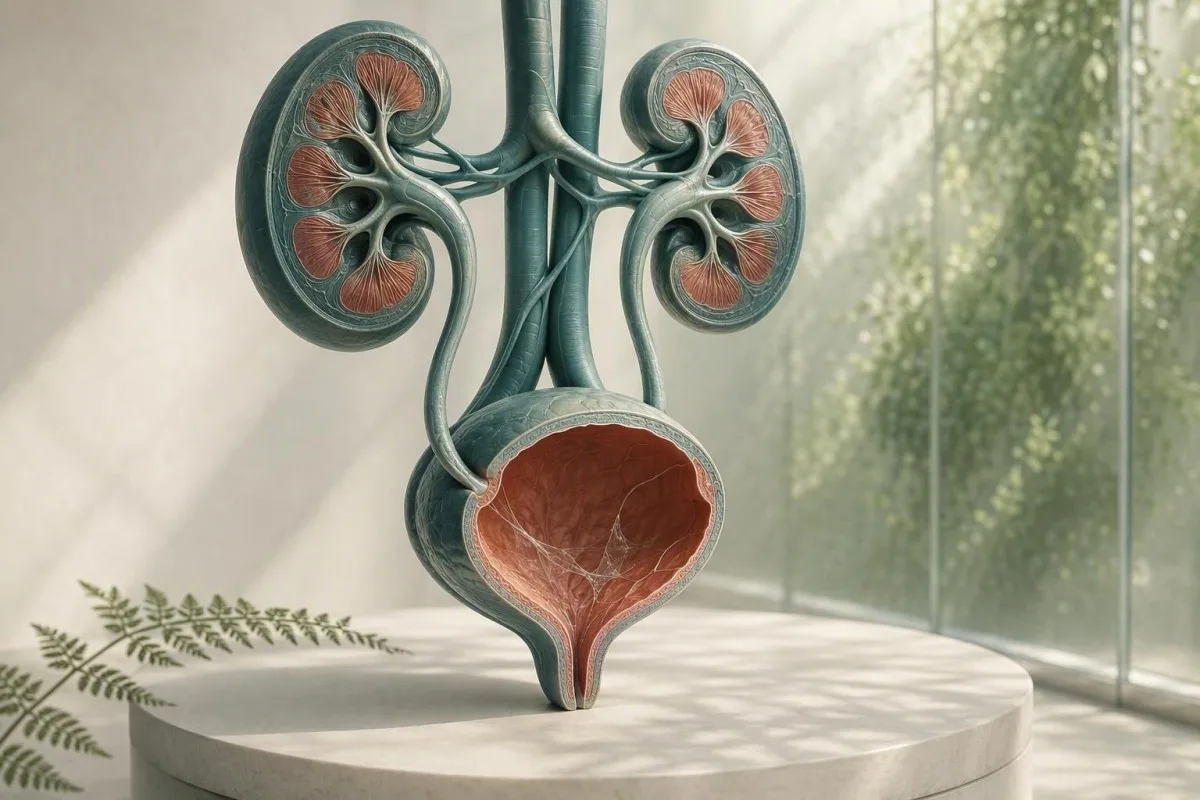
Mar as trice is e cùisean cruinneachaidh a th’ ann an caol-uisge air aithisg sgrùdaidh fual, chan e breithneachadh. Bidh na co-dhùnaidhean sgrùdaidh fual mun cuairt, comharran, agus càileachd na sampaill a’ dearbhadh a bheil feum air leantainn.


- Snàithleanan caol-uisge gu neo-eisimeileach mar as trice gun chron, gu sònraichte ann an aon sampaill glan-ghabhail gun chomharran urinary.
- Stairsneach pyuria de chòrr air 5 ceallan geal san raon cumhachdach àrd a’ toirt taic do sè slighe urinary ach chan eil e a’ dearbhadh UTI bacterial.
- brosnachadh cultair tha e na chaol-uisge a bharrachd air losgadh, èiginn, fiabhras, pian taobh, nitrites, no ceallan geal cudromach – chan e caol-uisge a-mhàin.
- Ceallan squamous os cionn timcheall air 10 gach raon cumhachdach àrd gu tric a’ comharrachadh truailleadh genital-skin agus a’ dèanamh toradh fual nas earbsaiche.
- Fuil fhaicsinneach le pian taobh colicky feumach air measadh sgiobalta airson clach, eadhon ged a tha caol-uisge air aithris cuideachd.
- Torrachas ag atharrachadh crìoch airson leanmhainn oir mar as trice bidh cruinneachaidhean bacteriach gun chomharran air an sgrìonadh le cultar fual, chan ann le microscopy a-mhàin.
- Fual sgòthach faodaidh e criostalan, fual dlùth, dìomhaireachd vaginal, seminal, no ceallan a nochdadh; chan urrainn dha galar a dhearbhadh le coltas.
- Sampall ath-aithris tha e ciallach nuair a tha mucus mar an aon rud neo-àbhaisteach agus cha robh cruinneachadh na chruinneachadh glan meadhan-sruth faiceallach.
Dè mar as trice a tha a’ ciallachadh snàithleanan caol-uisge air sgrùdadh fual
Mar as trice is e sreangan de secretion dìon àbhaisteach no truailleadh ginealach a th’ ann an snàithleanan mucus ann am fual, agus is ann ainneamh a bhios toradh iomallach a’ comharrachadh tinneas dubhaig. Tha an toradh a’ fàs ciallach nuair a nochdas e le comharran urinary, ceallan geala, bacteria, fuil, no pròtain.
Tha obair-lann a’ comharrachadh mucus mar shreangan tana, follaiseach a chithear fo microscopy; bidh mòran aithisgean ga mheas mar ainneamh, beag, meadhanach, no mòran an àite a bhith a’ toirt raon iomraidh àireamhach seachad. Tha beag-chuid de stuth làn glycoprotein bhon t-slighe urinary ann am fual gu nàdarra, agus faodaidh secretions vaginal no urethral barrachd a chuir ris rè cruinneachaidh.
Tha faclan na h-aithisg nas lugha de uamhasach na tha e coltach. Air 26 Lùnastal, 2026, chan eil cunntas snàithleach mucus air a dhearbhadh a dhearbhas UTI, clach, aillse, no fàilligeadh dubhaig; is e observation microscopic co-theacsa a th’ ann. Airson mapa nas fharsainge de na rudan eile air bileag dipstick agus microscope, faic ar stiùireadh iomlan air anailis urine.
Nam eòlas clionaigeach, is fheàrr le neach-aibidh gun chomharran le mucus air a chomharrachadh mar “meadhanach,” 0–2 ceallan geala gach achadh àrd-chumhachd, nitrite àicheil, agus gun fhuil, ath-dhearbhadh no aon shampall ath-chruinnichte a chaidh a chruinneachadh gu ceart. Tha Dr. Thomas Klein gu tric a’ faicinn dragh neo-riatanach antibiotics ag èirigh oir tha brataich obair-lann air a mhearachdachadh mar leubail galar.
is àrd-ùrlar mìneachadh deuchainn fala AI a dh’ fhaodas comharran fala co-cheangailte ri dubhaig, leithid creatinine agus eGFR, a chuir ri taobh toradh urinalysis—ach chan urrainn dha UTI a dhearbhadh bho mucus a-mhàin. Feumaidh toradh microscopy fual an sgeulachd clionaigeach a thàinig leis.
Carson a tha obair-lann ga bhratach
Bidh siostaman fiosrachaidh obair-lann a“ bratach mucus leis gu bheil e follaiseach, chan ann leis gu bheil an obair-lann air suidheachadh gearradh cunnartach a stèidheachadh. Mar sin tha brataich a” ciallachadh “air fhaicinn” an àite “comharra galar neo-àbhaisteach,” coltach ri toradh lorg eile a dh’ fhaodadh gun fheum air làimhseachadh.
Càit a’ tighinn caol-uisge ann an sampaill fual
Faodaidh mucus ann am fual tighinn bhon urethra, lìnigeadh an artaire, cervix no vagina, seminal, no an craiceann ginealach bhon taobh a-muigh rè cruinneachadh. Mar as trice bidh a thùs air a thuigsinn bho chòrr den phàtran meanbh-bheòthail seach bho na sreangan fhèin.
Tha sreath dìon air na h-artairean agus an urethra a tha a’ toirt a-steach mucins, a’ gabhail a-steach stuth co-cheangailte ri uroplakin, a lughdaicheas suathadh agus ceangal microbial. Faodaidh beag-chuid a bhith a’ crathadh a-steach do fual às deidh dìth uisge, gnìomhachd gnèitheasach o chionn ghoirid, irioslachd bheag, no dìreach leis gu bheil sampall cruinnichte sa mhadainn a’ dèanamh sreangan nas fhasa fhaicinn.
Dhaibhsan a tha a’ faighinn menstruation no a tha a’ faighinn dìomhaireachd vaginal, faodaidh leukocytes agus mucus a dhol a-steach don chupa gun a bhith a’ tighinn bhon artaire. Barrachd air 10 ceallan epithelial sgreamhach gach achadh àrd-chumhachd a’ dèanamh truailleadh nas dualtaiche, ged a bhios obair-lann a’ cleachdadh crìochan aithris eadar-dhealaichte. An stiùireadh againn gu ceallan epithelial ann am fual a’ mìneachadh carson a dh’ atharraicheas seo an earbsa ann an toradh.
Faodaidh seminal cuideachd stuth sreangach no neul a chruthachadh airson grunn uairean a thìde às deidh ejaculation. Chan eil sin a’ dèanamh am fual sàbhailte, ach faodaidh e microscopy a cheilt; ma tha cultar ga beachdachadh, mar as trice tha mi a’ moladh sampall ùr a chruinneachadh co-dhiù 24 uair às deidh sin nuair a ghabhas e dèanamh.
Carson a tha gnè agus anatamaidh a 'toirt buaidh air an aithisg
Chan eil cupa fual a “seApartachd na bladha bho nèapraigichean faisg air làimh. Is e sin as coireach gu bheil ”mùgan an làthair" air aithris nas trice ann an sampallan le rùintean a-muigh, fhad 'sa thathas a' cleachdadh sampall catheterichte uaireannan nuair a tha feum aig luchd-clionaigeach air freagairt nas glaine.
Cuin a tha snàithleanan caol-uisge a’ comharrachadh galar slighe urinary
Bidh snàithleanan mucus a 'toirt taic do UTI a dh'fhaodadh a bhith ann dìreach nuair a bhios iad a' dol còmhla ri comharraidhean agus fianais sèid, gu h-àraid pyuria, deimhinneachd nitrite, no cultar cunnartach. Chan eil mùgan leis fhèin na dheuchainn UTI.
Bidh UTI ìosal le comharraidhean gu tric ag adhbhrachadh losgadh, èiginn, tricead, mì-chofhurtachd suprapubic, agus uaireannan fàileadh ùr de dh'òl. Ann an sampall air a chruinneachadh gu ceart, barrachd air 5 ceallan fola geal gach raon àrd-chumhachd thèid gu tric a ghairm pyuria; tha e a 'toirt taic do sèid an t-siostam urinary ach faodaidh e tachairt cuideachd le clachan, galairean a tha air an sgaoileadh gu gnè, agus truailleadh.
Tha Nitrite glè shònraichte nuair a tha e dearbhach ach bidh e a 'call ghalaran air adhbhrachadh le buidhnean nach eil a' lughdachadh nitrate, agus chan eil toradh dearbhach nitrite a 'riaghladh a-mach UTI. Tha leukocyte esterase a 'lorg gnìomhachd enzyme nan ceallan geala agus faodaidh e a bhith dearbhach gu bràth nuair a bhios rùintean vaginal a' truailleadh an sampall; ar mìneachadh air toraidhean esterase leukocyte a’ còmhdach nan ribeachan sin.
Tha an stiùireadh IDSA a 'toirt comhairle an aghaidh sgrìonadh no làimhseachadh bacteriuria gun chomharraidhean ann an daoine inbheach neo-torrach fallain oir bidh antibiotics a' cur cron gun bhuannachd (Nicolle et al., 2019). Tha am prionnsapal sin gu sònraichte buntainneach ri mùgan: gun losgadh, gun fhiabhras, agus gun phòsadh urològach clàraichte mar as trice a 'ciallachadh gun antibiotic dìreach air sgàth gu robh raon meanbh-sgrùdaidh a' coimhead neo-chothromach.
Nuair a tha cultar na dheuchainn nas fheàrr
Tha cultar fual nas fheumail na ath-dheuchainn dipstick nuair a mhaireas comharraidhean, bidh comharraidhean a 'tighinn a-rithist taobh a-staigh 4 seachdainean, tha torrachas an làthair, tha pyelonephritis air a bhith ann, no is dòcha gu bheil antibiotics roimhe air an toradh atharrachadh. Tha fàs bacterial measgaichte mar as trice a 'nochdadh truailleadh cruinneachaidh seach aon pathogen urinary; faic mìneachadh air cultar na fual.
Caol-uisge, criostalan, clachan agus irioslachd meacanaigeach
Faodaidh mùgan le pian mòr-ghaoil-lìn no fuil ann an fual a bhith a 'tachairt le clach urinary, ach chan eil mùgan a' dearbhadh no a 'dùnadh a-mach clach. Tha fuil, criostalan, pàtran pian, ìomhaigh, agus gnìomh dubhaig nas cudromaiche.
Faodaidh clach a bhith a 'sgrìobadh no a' bacadh lìnigeadh urinary, a 'toirt a-mach ceallan dearga, ceallan geala, agus mùgan a bharrachd. Is e an comharra clasaigeach pian obann colicky a tha a 'sgaoileadh a dh'ionnsaigh an groin, gu tric le nausea; tha fuil no fual dath tì a' togail èiginn. Leugh tuilleadh mu dheidhinn fuil san fhualach comharran draghail seach a bhith a 'gabhail ris gu bheil a h-uile sampall pinc na galar sìmplidh.
Faodaidh criostalan calcium oxalate nochdadh ann an daoine fallain, gu h-àraid ann an fual searbh dlùth, mar sin chan eil aon sheòrsa criostail a 'dearbhadh clach gnìomhach. Ann an neach le pian, clachan a thig a-rithist, no haematuria a mhaireas, faodaidh luchd-clionaigeach ultrasound no CT neo-iomlaid ìosal-dose a chleachdadh an àite a bhith an urra ris an t-saidean a-mhàin. Ar goireas air criostalan calcium oxalate a' mìneachadh na crìochan a tha aig aithisgean criostail.
Is e eisgeachd chudromach bacadh le galar: fiabhras de 38.0 °C no nas àirde, pian taobh, cur a-mach, agus neo-chomas urination a thoirt seachad a 'gealltainn measadh èiginn. Chan e an dragh na mùgan; is e siostam urinary air a bhacadh air a ghalarachadh, a dh'fhaodas a dhol sìos gu luath.
Irritation às aonais clach
Faodaidh cleachdadh catheter o chionn ghoirid, modhan bladder, rothaireachd làidir, agus rèididheachd pelvic an t-slighe ìosal a bhrosnachadh agus àrdachadh ann am mùgan no ceallan geala. Bu chòir do neach-clionaigeach na toraidhean sin a mhìneachadh an aghaidh na h-ùine, oir chan eil sampall air a chruinneachadh taobh a-staigh 48 uair bho ionnstramaid co-ionann ri sampall sgrìonaidh cunbhalach.
Ciall fual neòlach: dè as urrainn agus nach urrainn nochdadh innse dhut
Faodaidh fual sgòthach a bhith air adhbhrachadh le salann dlùth, criostalan, mùgan, ceallan, rùintean ginealach, no bacteria, mar sin chan urrainn do choltas leis fhèin galar a dhearbhadh. Tha fual a tha sgòthach ach gun phian agus goirid mar as trice neo-èiginneach.
Faicheadan fosfair faodaidh iad uisge alcaileach a bhith a' coimhead neulach an dèidh dha fuarachadh, fhad 's a dh' fhaodas faicheadan searbhagach, dùmhail an uisge a dhèanamh neulach. Faodaidh sampall a dh' fhàgar aig teòthachd an t-seòmair airson còrr air 2 uair a thìde fàs nas neulaiche mar a bhios ceallan a' lobhadh agus bacteria a' tighinn am meud, is e sin as coireach gu bheil giollachd ùr no fuarachadh cudromach.
Bidh neulaiche còmhla ri dysuria, èiginn, agus pyuria a“ toirt air falbh deuchainn; bidh neulaiche às dèidh eacarsaich no droch ghabhail a-steach lionn gu tric a” leasachadh le uisgeachadh àbhaisteach. Amas air uisge a tha buidhe bàn an àite a bhith a' sparradh cus gabhail a-steach uisge - chan eil uisge glan gu tur a' ciallachadh gu bheil na dubhagan a' "sruthadh a-mach" galar. An lèirmheas mionaideach againn air adhbàr urrainne sgòthach a' sgaradh comharran lèirsinneach bho dheuchainnean earbsach.
Tha Kantesti na Anailisiche deuchainn fala AI air a dhealbhadh gus comharran fala a mhìneachadh ann an co-theacsa clionaigeach; faodaidh creatinine àrd no eGFR lùghdaichte còmhla ri neo-riaghailteachdan urinary a bhith a' fìreanachadh lèirmheas meidigeach, fhad 's nach eil uisge neulach leis fhèin a' stèidheachadh sìoltachain dubhaig lag. Faodaidh pannal meatabolach bunaiteach cur ri co-theacsa feumail nuair a tha comharraidhean a' moladh dìth uisge no bacadh.
Chan eil fàileadh na chultar
Bidh fàileadh làidir ann an uisge gu tric a' nochdadh dùmhlachd, metabol anns asparagus, vitamain B, no soitheach a dh' fhuirich ro fhada. Is e fàileadh ùr dona le fiabhras no comharraidhean urinary adhbhar airson measadh iarraidh, ach chan eil fàileadh na àite airson microscopy agus cultar.
Casg genital agus galairean a thèid a sgaoileadh gu gnèitheasach
Faodaidh dùnadh urethral no vaginal coimhead coltach ri mucus ann an uisge, agus feumaidh dùnadh ùr le losgadh urinary deuchainn slàinte gnè a bharrachd air measadh uisge. Faodaidh cultar urinary àbhaisteach a bhith ag ionndrainn chlamydia agus gonorrhoea.
Faodaidh Chlamydia agus gonorrhoea adhbhrachadh dysuria agus pyuria sterile - ceallan geala gun fhàs bacteria àbhaisteach - gu sònraichte às dèidh gnìomh gnè ùr. Tha deuchainn amplification searbhag nucleic, gu tric a’ cleachdadh uisge a’ chiad ghlacadh an àite sampall meadhan-shruth, na dheuchainn iomchaidh oir tha cultar àbhaisteach a’ targaid diofar aingheilean.
Ann an daoine le vagina, faodaidh vaginosis bacteria, candidiasis, agus sè meanbh-chraobhan cur ri dùnadh ann an cupa meadhan-shruth; faodaidh swab vaginal no sgrùdadh a bhith nas fhiosaiche na bhith ag ath-aithris microscopy urinary. Ann an daoine le peanas, cha bu chòir dùnadh urethral follaiseach a bhith air a leigeil seachad mar “snàithleanan mucus.” Is e an dearbh-aithne practaigeach dùnadh a chithear taobh a-muigh urination an aghaidh sreathan a dh’ innseas an obair-lann a-mhàin.
Tha stiùireadh làimhseachadh STI CDC 2021 a' moladh deuchainn stèidhichte air NAAT aig làraich anatomach iomchaidh a rèir eachdraidh nochdaidh, chan e comharraidhean a-mhàin (Workowski et al., 2021). Ma tha pian pelvic, pian testiculach, fiabhras, torrachas, no ionnsaigh a dh’ fhaodadh a bhith ann, iarr comhairle clionaigeach air an aon latha an àite fèin-làimhseachaidh le antibiotics air fhàgail.
Carson a dh’ fhaodas antibiotics an dealbh a dhèanamh neònaiche
Faodaidh eadhon 1 no 2 dòs antibiotic a ghabhail ron chultar fàs bacteria àbhaisteach a shlaodadh fhad ‘s a mhaireas comharraidhean urinary agus ceallan geala. Innis don neach-clionaigeach dìreach dè an druga, an dòs, agus an ùine mu dheireadh a chaidh a chleachdadh; tha am mion-fhiosrachadh sin ag atharrachadh mar a thèid cultar àicheil a mhìneachadh.
Mar a chruinnicheas tu sampaill fual a fhreagras air a’ cheist
Bidh sampall glan-catch meadhan-shruth a' lughdachadh mucus, ceallan còmhnard, agus fàs bacteria measgaichte nas fheàrr na bhith a' cruinneachadh a' chiad no a' phàirt mu dheireadh den t-sruth. Tha an dòigh sìmplidh, ach tha a’ chiad 2 diog a’ dèanamh eadar-dhealachadh neo-chuibheasach.
Nigh làmhan, sgar an craiceann gnèitheach no tarraing air ais an foreskin ma tha comhfhurtachd ann, glan a rèir stiùireadh a’ phacaid, tòisich a’ slugadh a-steach don taigh-beag, an uairsin cruinnich am meadhan-phàirt gun an cupa a’ suathadh craiceann. Mar as trice bidh sampall 20–30 mL gu leòr; chan eil lìonadh soitheach mòr gu mullach a’ leasachadh na deuchainn.
Lìbhrig an cupa sa bhad, gu h-àraid taobh a-staigh 1 uair; ma tha dàil riatanach, fuaraich e a rèir stiùireadh na h-obair-lann agus till e taobh a-staigh 24 uairean. Bidh fuarachadh a’ slaodadh fàs bacteria ach cha dèan e ath-nuadhachadh sampall a bha mu thràth air a thruailleadh. Tha seo gu sònraichte cudromach nuair a tha an iarrtas a’ toirt a-steach cultar an àite dipstick a-mhàin.
Tha Kantesti na Inneal mion-sgrùdaidh deuchainn fala le cumhachd AI air a chleachdadh thairis air 127+ dùthaich, agus bidh na pròiseasan againn a chaidh ath-sgrùdadh le clionaigean a’ làimhseachadh càileachd sampall mar phàirt den mhìneachadh seach mar notaichean cas. Tha an aon phrionnsapal a’ buntainn ri luach obair-lann sam bith: faodaidh sampall droch-theicneachach toradh a tha persuasive gu meidigeach ach a tha a’ clàradh a chruthachadh. Faic ar liosta-sgrùdaidh cruinneas obair-lann airson ceistean a chur mus gabh thu gnìomh air bratach iomallach.
Na cruinnich rè na suidheachaidhean seo ma tha thu a' feitheamh
Seachain deuchainnean àbhaisteach rè sruthadh menstrual trom, dìreach às dèidh gnè, no às dèidh crèamhan vaginal a chleachdadh mura h-iarr an neach-clionaigeach gu sònraichte e. Nuair nach urrainn deuchainn feitheamh, innis don obair-lann no don neach-clionaigeach, oir faodaidh an co-theacsa sin mìneachadh air mucus, ceallan dearga, no ceallan bhon taobh a-muigh.
Mar a leughas luchd-clionaigeach caol-uisge leis a’ chòrr den sgrùdadh fual
Tha an ìomhaigh urinalysis as fheumail a’ cothlamadh comharraidhean le ceallan geala, ceallan dearga, nitrite, leukocyte esterase, pròtain, glùcois, dùmhlachd sònraichte, agus ceallan epithelial. 'S e re a ghnìomhaireachd bheag taiceil anns an phàtran sin.
Chan fheum sampall glan le slàinteachas, 0–2 cealla geal gach raon àrd-mhasaidh, nitrit àicheil, agus gun fhuil leigheas mar as trice. Tha slàinteachas le còrr air 5 ceallan geal gach raon àrd-mhasaidh agus esterase leukocyte adhartach ag àrdachadh coltachd sèid; tha cur ris an nitrit no cultar aon-organismach a’ cur misneach ris gu bheil bacteria an urra.
Feumaidh pròtain an slighe fhèin. Faodaidh pròtain lorg às dèidh fiabhras, eacarsaich, no urine dlùth a bhith sealach, ach bu chòir pròtain leantainneach a thomhas le co-mheas albumin-gu-creatinine an àite a bhith air a chur ris an t-slàinteachas. An stiùireadh againn gu pròtain san fhuaim a’ mìneachadh carson nach eil pròtain dipstick agus cunnart dubhaig eadar-mhalairteach.
Bhiodh dùmhlachd shònraichte de 1.003 gu 1.030 cumanta ann an inbhich, ged a tha na h-eileamaidean iomraidh ag atharrachadh a rèir obairlann. Faodaidh dùmhlachd sònraichte àrd dùmhlachd a dhèanamh air slàinteachas agus sìoltachain nas dràmaiche a dhèanamh, fhad ‘s as urrainn do dhùmhlachd sònraichte ìosal ceallan a sgaradh agus microscopy a dhèanamh gu meallta sìmplidh; ar stiùireadh dùmhlachd shònraichte a’ sealltainn mar a tha uisgeachadh a’ toirt buaidh air mìneachadh.
Luach nan toraidhean àicheil
Tha fuil, pròtain, nitrit, agus esterase leukocyte àicheil a’ lughdachadh dragh gu ciallach ann an neach gun chomharraidhean, eadhon ged a tha slàinteachas air aithris mar mheadhanach. Chan eil deuchainn àicheil sam bith foirfe, ach tha a’ chruinneachadh seo nas misneachail na tha ìre slàinteachais a’ cur dragh.
Cuin a nì thu deuchainn a-rithist agus cuin a dh’ iarras tu cultar
Dèan ath-aithris air sampall urine nuair a tha slàinteachas air a sgaradh, tha ceallan còmhnard a’ moladh truailleadh, no chaidh cruinneachadh a luathachadh; iarr cultar nuair a tha comharraidhean leantainneach, ath-chuairteach, dona, no àrd-chunnart. Chan eil ath-aithris deuchainn na rud a tha “gun a bhith a” dèanamh dad ”- is e gu tric an ath cheum as motha a bheir breithneachadh.
Airson neach-inbheach neo-torrach gun chomharraidhean agus slàinteachas mar an aon bhratach, tha ath-aithris deuchainn urine glan-ghabhail taobh a-staigh 1–2 seachdainean reusanta ma tha feum air misneachd; cha dhèanadh mòran dhaoine clionaigeach ath-aithris idir. Na bi a’ làimhseachadh bratach obair-lann le antibiotics fhad ‘s a tha thu a’ feitheamh mura h-eil neach-òrdaidh a’ comharrachadh adhbhar clionaigeach.
Tha cultar mus tèid antibiotics a chleachdadh gu sònraichte feumail airson fiabhras, pian san t-sàil, torrachas, dìonachd lag, tar-chur dubhaig, cleachdadh cathetar urinary, comharraidhean urinary fireann, no comharraidhean a thilleas taobh a-staigh 4 seachdainean. Tha an stiùireadh le Gupta et al. (2011) a’ toirt taic do chultar ann am fìor pyelonephritis agus suidheachaidhean far a bheil strì no breithneachadh eile nas dualtaiche.
Feumaidh cunntasan cultair a bhith air an leughadh le càileachd cruinneachaidh. Organism singilte aig 10^5 aonadan a' cruthachadh coloin airson gach mL air taic a thoirt gu traidiseanta do bacteriuria ann an urine glan meadhan-sruth, ach faodaidh cunntasan nas ìsle a tha buntainneach gu clionaigeach a bhith aig euslaintich le comharraidhean; mar as trice bidh “flòra mheasgaichte” a’ brosnachadh sampall ùr seach antibiotic farsaing. Dèan coimeas eadar na h-adhbharan airson urinalysis agus cultar mus iarr thu gin dhiubh.
Tha torrachas ag iarraidh stairsneach eadar-dhealaichte
Tha torrachas na eisgeachd leis gum faod bacteriuria gun chomharraidhean an cunnart bho pyelonephritis agus droch thoraidhean torrachais a mheudachadh. Bidh cùram màthaireil gu tric a’ cleachdadh cultar sgrìonaidh tràth ann an torrachas; chan urrainn mucus air microscopy an cultar sin a chuir an àite.
Cuin a chuireas deuchainnean fala tuilleadh co-theacsa cudromach air dubhaig agus galar
Tha deuchainnean fala feumail nuair a nochdas mucus ann an urine le fiabhras, pian taobh, galar ath-chuairteach, sèid, lughdachadh toradh urine, no pròtain no fuil leantainneach. Bidh Creatinine agus eGFR a’ measadh sìoladh, fhad ‘s a dh’ fhaodadh CBC agus C-reactive protein cuideachadh le bhith a’ breithneachadh tinneas siostaim.
An eGFR nas ìsle 60 mL/min/1.73 m² airson co-dhiù 3 mìosan a’ coinneachadh ri aon chrìtar airson tinneas dubhaig cronach, ach chan eil aon toradh nas ìsle rè dìth uisge no tinneas geur gu leòr airson an dearbhadh sin a dhèanamh. Tha creatinine air a thoirt air buaidh le mòr-chuid de fhèith, daithead, agus cuid de chungaidhean, mar sin bidh gluasadan agus albumin urine gu tric nas fiosrachail na aon àireamh.
Faodaidh àrdachadh ann an cunntas ceallan fala geal no CRP taic a thoirt do phròiseas sèididh ach chan urrainn dha an t-slighe urinary a chomharrachadh mar an tùs. Ann an neach le fiabhras agus pian taobh, is dòcha gun dèan luchd-clionaigeach sgrùdadh air creatinine mus tagh iad ìomhaigh no cungaidhean, gu sònraichte ma dh’ fhaodadh vomiting no bacadh casg a chuir air gnìomh dubhaig. Ath-sgrùdadh ìrean galar dubhaig leantainneach airson na roinnean eGFR agus albumin a chleachdas luchd-clionaigeach.
Tha Kantesti na seirbheis eadar-mhìneachaidh deuchainn-lann AI a tha a’ soilleireachadh phàtranan thar creatinine, eGFR, electrolytes, CBC, agus comharran sèididh an àite a bhith a’ làimhseachadh aon nota mucus mar dhearbhadh dubhaig. Tha ar modh-obrach fo ùmhlachd stiùireadh dearbhaidh clionaigeach, ach feumaidh comharran èiginneach fhathast cùram meidigeach dìreach, chan e mìneachadh stèidhichte air aplacaid.
Eadar-dhealachadh practaigeach dìth uisge
Faodaidh dìth uisge àrdachadh a dhèanamh air dùmhlachd sònraichte urine agus àrdachadh sealach ann an creatinine, gu sònraichte às deidh a’ bhuinneach, nochdadh teas, no eacarsaich dian. Tha toradh leantainneach ìosal de dh’ fhual, ceann aotrom, no lùghdachadh eGFR às deidh ath-uisgeachadh a’ cur feum air ath-sgrùdadh clionaigeach an àite deuchainn dachaigh ath-aithris.
Comharran rabhaidh èiginn nach bu chòir feitheamh ri deuchainn a-rithist
Iarr measadh èiginneach air an aon latha airson mucus ann an urine le fiabhras de 38.0°C no nas àirde, pian taobh, vomiting, fuil a chithear, neo-chomas urination, troimh-chèile, no comharraidhean urinary co-cheangailte ri torrachas. Tha na cothlamadh sin cudromach leis gum faod iad a bhith a’ comharrachadh gabhaltachd àrd-shligheach, bacadh, no suidheachadh geur eile.
Tha fiabhras a bharrachd air pian aon-thaobhach air ais no taobh nas draghail airson pyelonephritis na cystitis sìmplidh, gu sònraichte le fuachd crathaidh no vomiting. Faodaidh dàil ann an làimhseachadh leantainn gu dìth uisge, sepsis, no cuideam dubhaig; chan eil sampall urine a tha a’ coimhead àbhaisteach aig an taigh a’ cur às dha gu sàbhailte.
Bu chòir fuil a chithear a mheasadh eadhon nuair a tha UTI a’ coimhead comasach, gu sònraichte às deidh aois 35, ann an smocadairean, no ma mhaireas bleeding nuair a bhios comharraidhean gabhaltachd a’ suidheachadh. Tha an stiùireadh microhaematuria AUA a’ moladh measadh stèidhichte air cunnart às deidh mìneachaidhean fàbharach a bhith air an dèiligeadh (Barocas et al., 2020); chan eil fuil leantainneach air a mhìneachadh air falbh le mucus.
Feumaidh clann, inbhich thairis air 65, daoine le tinneas an t-siùcair, daoine a tha a’ gabhail cungaidhean immunosuppressing, agus an fheadhainn le aon dubhaig stairsneach nas ìsle airson measadh. Tha riaghailt Dr. Thomas Klein ann an cleachdadh sìmplidh: ma tha comharraidhean a’ fàs thairis air 6–12 uairean, na bi a’ feitheamh ri dàrna cupa gus fois a thoirt seachad.
Cuir fòn gu seirbheisean èiginn a-nis airson droch thinneas
Cuir fòn gu seirbheisean èiginn airson troimh-chèile ùr, faochadh, laigse mòr, bilean gorm no liath, fìor ghann anail, no neo-chomas cumail ri lionn le comharraidhean urinary. Is e so na comharran cunnart siostaim, chan e comharraidhean UTI àbhaisteach.
Caol-uisge ann am fual rè torrachas, leanabas agus nas fhaide air adhart nam beatha
Feumaidh torrachas, clann, agus seann daoine mìneachadh nas faiceallach leis gu bheil truailleadh cumanta ach faodaidh builean gabhaltachd a chaillte a bhith nas motha. Tha comharran agus càileachd cultair fhathast nas luachmhoire na a’ chruinneachd bhleith anns a h-uile buidheann aoise.
Nuair a tha thu trom, faodaidh tricead urinary a bhith àbhaisteach, a nì na comharran nas lugha de shònraichte; bu chòir fiabhras, dysuria, no pian cùil a bhith a’ brosnachadh measadh clionaigeach no clionaigeach obsteetrach sgiobalta. Mar as trice bidh cultar sgrìonaidh air fhaighinn tràth ann an cùram ro-bhreith, agus faodar cultar ath-aithris a chleachdadh às dèidh làimhseachadh a rèir plana an dotair.
Ann an clann, tha fual a chaidh a chruinneachadh ann am baga buailteach do thruailleadh agus cha bu chòir a chleachdadh leis fhèin gus UTI a dhearbhadh le cultar. Faodar sampaill air a thoirt le catheter no air a thoirt gu faiceallach a bhith air a chruinneachadh nuair a tha fiabhras air leanabh òg às aonais stòr soilleir; tha cruinneachd ann an sampaill baga gu sònraichte neo-shònraichte.
Nas fhaide air adhart nam beatha, bidh bacteriuria agus pyuria a’ fàs nas cumanta gun a bhith a’ toirt comharraidhean. Bu chòir ùr-dòchas leis fhèin a bhith a’ brosnachadh measadh meidigeach farsaing airson dìth uisge, cungaidhean-leigheis, pian, giorrachadh, agus stòran gabhaltachd an àite làimhseachadh UTI fèin-ghluasadach; tha an aon chuingealachadh a chaidh a mholadh le Nicolle et al. (2019) a’ buntainn.
Caidheamh agus atharrachaidhean co-cheangailte ri hormonaichean
Faodaidh fuil caidheamh agus cruinneachd amhach atharrachadh air mion-sgrùdadh urinary airson grunn làithean, agus faodaidh tiormachd bhànachais às dèidh menopause adhbhrachadh irioslachaidh ionadail a tha coltach ri losgadh urinary. Mura h-eil na toraidhean agus na comharraidhean a’ maidseadh, faodaidh dotair adhbharan ginealach a mheasadh an àite a bhith ag òrdachadh làimhseachadh UTI ath-aithris.
Mearachdan cumanta an dèidh faicinn caol-uisge air aithisg obair-lann
Na tòisich antibiotics a tha air fhàgail, glanadh a-staigh, no feuchainn ri cruinneachd a “ghlanadh a-mach” le cus uisge a ghabhail às dèidh aon mhion-sgrùdadh urinary neo-àbhaisteach. Faodaidh na gnìomhan seo cultar a dhèanamh neo-shoilleir, cron a dhèanamh air an fhàs-bheairt àbhaisteach, no dàil a chuir air a’ mhì-thuigse cheart.
Faodaidh antibiotics a chaidh a ghabhail às aonais cultair deuchainn a dhèanamh nas fhaide air adhart gu ceàrr neo-dhligheach agus faodaidh e adhbhrachadh a’ bhuinneach, broth, comharraidhean dhruim, no strì. Ma tha na comharraidhean tlàth ach leantainneach, faigh an sampaill an toiseach nuair a ghabhas sin dèanamh, an uairsin lean plana làimhseachaidh an dotair stèidhichte air an dealbh iomlan.
Faodaidh toraidhean a’ chraobhach rùsgach gu modhail an cunnart bho UTI neo-fhillte ath-chuairteach a lughdachadh dha cuid de dhaoine, ach cha bhith iad a’ làimhseachadh fiabhras, pian taobh, no gabhaltachd air a dhearbhadh san àrd-shlighe. Seachain àrd-dòs de bhiotamain C mar fhèin-làimhseachadh: faodaidh e cuid de dh’ ath-bheachdan dipstick atharrachadh agus faodaidh e an cuideam oxalate a mheudachadh ann an daoine a tha buailteach do chlachan oxalate calcium.
Na dèan sgrùdadh a-rithist air bobhla an taigh-beag airson sreangan. Faodaidh freumhagan pàipear taigh-beag, fuigheall glanaidh, secreted ginealach, agus turbulence uisge a bhith coltach ri cruinneachd; is e sampaill obair-lann a chaidh a chruinneachadh ann an soitheach sterile an t-àite iomchaidh airson a mheasadh. Airson atharrachaidhean dath co-cheangailte, ar stiùireadh dath fual a’ tabhann co-theacsa lèirsinneach nas earbsaiche.
Tha an liosta de chungaidh-leigheis a’ cunntadh
Faodaidh phenazopyridine fual orains a thionndadh agus casg a chuir air mìneachadh lèirsinneach, fhad ‘s a dh’ fhaodas diuretics fual a dhèanamh nas dùmhail no nas tana a rèir ùine. Thoir liosta de chungaidh-leigheis òrdaichte, toraidhean thairis air a’ chunntair, agus stuthan cur-ris chun dreuchd, a ’toirt a-steach dòsan ann an mg.
Plana leantainn practaigeach airson snàithleanan caol-uisge ann am fual
Chan fheum a’ mhòr-chuid de dhaoine le sreangan cruinneachaidh san fhual agus gun chomharraidhean làimhseachadh sam bith; tha ath-mhion-sgrùdadh urinary faiceallach reusanta ma bha an sampaill teagmhach. Bidh comharraidhean no neo-riaghailteachdan companaidh a’ dearbhadh a bheil cultar, deuchainn STI, ìomhaighean, no deuchainnean fala iomchaidh.
Ceum 1: thoir sùil air losgadh, èiginn, fiabhras, pian taobh, fuil ri fhaicinn, dìomhaireachd, torrachas, modhan urinary o chionn ghoirid, agus nochd gnèitheasach ùr. Ceum 2: leugh an aithisg airson ceallan geala, nitrite, leukocyte esterase, ceallan dearga, pròtain, bacteria, agus ceallan squamous—chan ann dìreach cruinneachd.
Ceum 3: ma tha thu a’ faireachdainn gu math agus gu bheil cruinneachd air a sgaradh, cruinnich aon sampaill glan-catch meadhan-shrutha taobh a-staigh 1–2 seachdainean dìreach ma tha do dhotair a’ moladh dearbhadh. Ceum 4: ma tha comharraidhean an làthair, faighnich a bheil cultar fual air a chruinneachadh ro làimhseachadh agus a bheil deuchainn STI buntainneach; tha tachartasan ath-chuairteach airidh air ath-sgrùdadh nas miosa na treas antibiotic empiric.
Faodaidh Kantesti treallaich toraidhean fala buntainneach agus ceistean a chuir air dòigh airson dreuchd meidigeach, fhad ‘s a tha ar Bòrd Comhairleachaidh Meidigeach a“ toirt taic do phrionnsabalan sàbhailteachd fo stiùir an dotair. Chan e an ceann-uidhe ceart aithisg mhiocroscopaidh a tha gu tur ”glan”—is e mìneachadh a tha a’ freagairt air do chomharran, càileachd sampaill, agus factaran cunnairt.
Ceistean airson an toirt don dreuchd agad
Faighnich an robh ceallan squamous anns an sampaill, an robh aon bhuidheann no measgachadh de fhàs-bheairtean aig a’ chultar, agus an robh fuil no pròtain a’ leantainn às deidh na comharraidhean a bhith air an rèiteachadh. Bidh na 3 cheistean sin gu tric a’ toirt freagairtean nas feumail na bhith a’ faighneachd dè an ìre cruinneachaidh a chaidh fhaicinn.
Co-theacs rannsachaidh agus crìochan aithris caol-uisge
Chan eil sgèileadh nam biastagan air a stiadhú thar obair-lann, is e sin as coireach gum bu chòir do luchd-clionaigeach seachainn “beag,” “meadhanach,” no “mòran” a chleachdadh mar roinnean-seòrsa de dhroch thinneas. Faodaidh teicneòlas microscopaidh, aois sampaill, agus bathar-bog aithris uile atharrachadh a dhèanamh air na facail.
Bidh cuid de dh’obair-lannan a“ sgrùdadh nan cailcean gu làimhe an dèidh timcheall air 10–15 mL de dh’uisge a thionndadh, agus cuid eile a” cleachdadh anailis inneal air pàirtean le sgrùdadh làimhe air bratagan a chaidh a thaghadh. Tha an eadar-dhealachadh sin a’ ciallachadh gum faod toradh “biastagan meadhanach” bho aon obair-lann gun a bhith ag ath-riochdachadh gu dìreach aig obair-lann eile eadhon nuair a tha slàinte an duine gun atharrachadh.
Is e dòigh smaoineachaidh feumail obair-lann faighneachd a bheil toradh fìor ann an anailis, ciallach ann an clionaigeach, agus comasach air ath-riochdachadh. Tha an dòigh sin eòlach bho dheuchainn fala cuideachd: ar stiùireadh bith-chomharra fala mìneachadh carson a tha raointean iomraidh agus suidheachaidhean ro-anailis a’ dearbhadh a bheil comharra a chaidh a bhratadh airidh air gnìomh.
Bidh Kantesti AI a’ cleachdadh co-theacsa obair-lann structaraichte gus mìneachadh a thoirt air pàtrain toraidh thairis air pannalan mòra, ach feumaidh microscopaidh fual fhathast breithneachadh clionaigeach a tha sònraichte don stòr. Dha luchd-leughaidh aig a bheil ùidh ann mar a nì sinn measadh air coileanadh teicnigeach agus crìochan clionaigeach, tha ar stiùireadh teicneòlais AI a’ toirt cunntas air na dìonan a tha air cùl ar dòigh-obrach mìneachaidh.
Na bu chòir do dheagh aithisg a dhèanamh soilleir
Tha aithisg feumail ag aithneachadh an seòrsa sampaill, ceann-latha cruinneachaidh, eileamaidean microscopach, agus buidhnean cultair sam bith agus toraidhean fulangas nuair a thèid cultar a dhèanamh. Mura h-eil an aithisg ag ràdh ach “snàithleanan biastagan an làthair,” tha i neo-iomlan airson breithneachadh ach gu tric tha i gu tur freagarrach airson toraidhean neo-dhìreach cunnairt ìosal.
Ceistean Bitheanta
A bheil snàthadan mhucais san fhuaimneachadh àbhaisteach?
Tha snàithichean rùsg ann an fual gu tric àbhaisteach no air sgàth truailleadh bho dhìomhaireachd ginteil, gu sònraichte nuair nach eil comharraidhean sam bith aig neach. Is e suidheachadh singilte le 0–2 ceallan geal gach raon àrd-chumhachd, nitrite àicheil, agus gun fhuil no pròtain, mar as trice cunnart ìosal. Chan eil obair-lann a' cleachdadh àireamh snàithle rùsg air a dhearbhadh gus UTI no galair nan dubhagan a dhearbhadh. Tha e ciallach sampall ath-aithris de'n ionad-sruth glan a ghabhail ma bha a' chruinneachadh tùsail ro luath no air a thruailleadh gu follaiseach.
A bheil snàithleanan mòran anns an fhualas a' ciallachadh gu bheil UTI agam?
Chan fuaim a’ chroite ann an fual chan eil e a’ ciallachadh UTI leis fhèin. Tha UTI a’ fàs nas coltaiche nuair a tha losgadh, cabhag, tricse, fiabhras, no pian suprapubic a’ tachairt le còrr air 5 ceallan geal gach raon àrd-chumhachd, esterase leukocyte deimhinneach, nitrite, no cultar deimhinneach. Chan eil nitrite àicheil a’ dùnadh a-mach UTI gu tur leis nach eil a h-uile bacteria a’ dèanamh nitrite. Mar as trice tha cultar nas fhiosraiche na ìre mucous nuair a mhaireas comharraidhean no ma thig iad air ais.
An urrain dehydration adhbhar mucus san fhualas?
Faodaidh dìth uisge mucus a dhèanamh nas fhasa fhaicinn oir tha nas lugha de dh'uisge aig fual dùmhail timcheall air dìomhaireachdan agus cruinneachaidhean àbhaisteach. Faodaidh àrd dùmhlachd fual, mar as trice timcheall air 1.025–1.030, a bhith an cois a' bhuaidh seo, ged nach eil e a' dearbhadh dìth uisge leis fhèin. Faodaidh ath-uisgeachadh mar as àbhaist agus ath-aithris air sampall a chaidh a chruinneachadh gu ceart an toradh a shoilleireachadh. Chan eil feum air cus uisge òl agus faodaidh e a chunnartan electrolyte fhèin a chruthachadh.
Dè tha ciallachadh mucus agus ceallan fala geal ann an urine?
Tha casan is na ceallan fola geala anns an fhualas a' nochdadh sè anns an t-slighe-uisge no an t-slighe-ghinealach, ach cha dhearbh iad galar bacterial. Faodaidh barrachd air 5 ceallan fola geala gach achadh àrd-chumhachd tachairt le UTI, clachan, galairean a thèid a sgaoileadh gu gnèitheasach, truailleadh bhànagach, no ionnsramaid urological o chionn ghoirid. Thèid ceumannan a-rithist a dhearbhadh le comharran, nitrite, ceallan epithelial, agus toraidhean cultair. Tha fiabhras de 38.0°C no nas àirde le pian taobh ag iarraidh measadh èiginneach.
An urrain bhànach an urrainn cnatan-mòr adhbhrachadh ann an deuchainn fual?
Faighidh an t-ur sgaraidh bhon fhèin a-steach do shamhla fual meadhan-shrutha agus bidh e gu tric ag adhbhrachadh gum bi slàbagan, ceallan epithelial sgiamach, agus uaireannan ceallan geala a’ nochdadh air micreascopaidh. Bidh barrachd air 10 ceallan sgiamach gach raon àrd-chumhachd gu tric a’ dèanamh truailleadh nas dualtaiche, ged a dh’atharraicheas obair-lann san aithris aca. Faodaidh sampla faiceallach de chobhair ghlan air a chruinneachadh taobh a-muigh sruth trom mìosach an duilgheadas seo a lughdachadh. Ma tha sgaraidhean, tachas, fàileadh, no pian anns a’ chliabh an làthair, dh’fhaodadh measadh no swab bhon fhèin a bhith nas feumail na cultaran fual ath-aithris.
Cuin bu chòir dhomh a bhith draghail mu snàithleanan mucus agus fuil anns an fhual?
Bu chòir snàithleanan dìsne leis an fhuil fhaicsinneach ann an fual a mheasadh gu sgiobalta, gu sònraichte nuair a tha an fhuil a’ leantainn, tha pian taobh, cnapan-fala, fiabhras, no trioblaid a’ dol seachad air fual. Faodaidh clach, UTI, leòn, galar dubhaig, no nas teirce fàs anns an t-slighe urinary adhbhrachadh fuil; chan eil dìsne a’ comharrachadh dè an adhbhar a tha an làthair. Tha an AUA a’ moladh measadh stèidhichte air cunnart airson haematuria meanbh-bheag leantainneach às dèidh do adhbharan sealach a bhith air an dèiligeadh. Tha measadh èiginn iomchaidh airson fiabhras, cuir a-mach, pian mòr, no gleidheadh urinary.
Faigh Mion-sgrùdadh Deuchainn Fala le Cumhachd AI an-diugh
Thig còmhla ri còrr is 2 mhillean neach air feadh an t-saoghail a tha a’ earbsa Kantesti airson mion-sgrùdadh sa bhad, ceart air deuchainnean obair-lann. Luchdaich suas na toraidhean deuchainn fala agad agus faigh mìneachadh coileanta air biomarcair 15,000+ ann an diogan.
📚 Foillseachaidhean Rannsachaidh le Iomraidhean
Klein, T., Mitchell, S., & Weber, H. (2026). Raon Àbhaisteach aPTT: D-Dimer, Pròtain C Stiùireadh air Clotadh Fuil. Rannsachadh Leigheis AI Kantesti.
Klein, T., Mitchell, S., & Weber, H. (2026). Stiùireadh Pròtainean Serum: Deuchainn Fuil Globulins, Albumin & Co-mheas A/G. Rannsachadh Leigheis AI Kantesti.
📖 Iomraidhean Meidigeach Taobh a-muigh
⚕️ Àicheadh Meidigeach
Tha an artaigil seo dìreach airson adhbharan foghlaim agus chan eil e a’ dèanamh comhairle mheidigeach. Cuir fios an-còmhnaidh gu solaraiche cùram slàinte teisteanasach airson co-dhùnaidhean breithneachaidh is leigheis.
Comharran earbsa E-E-A-T
Eòlas
Lèirmheas clionaigeach air a stiùireadh le lighiche air sruthan-obrach mìneachaidh obair-lann.
Eòlas
Fòcas air leigheas obair-lann air mar a bhios bith-chomharraidhean (biomarkers) a’ giùlan ann an co-theacsa clionaigeach.
Ùghdarrasachd
Air a sgrìobhadh le Dr. Thomas Klein le ath-sgrùdadh le Dr. Sarah Mitchell agus Prof. Dr. Hans Weber.
Earbsachd
Mìneachadh stèidhichte air fianais le slighean leanmhainn soilleir gus dragh a lughdachadh.