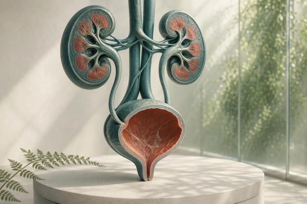
El moco en un informe de microscopía de orina suele ser un problema de recolección, no un diagnóstico. Los hallazgos circundantes del análisis de orina, los síntomas y la calidad de la muestra determinan si necesita seguimiento.


- Hebras de moco por sí solas son comúnmente inofensivas, especialmente en una sola muestra de chorro limpio sin síntomas urinarios.
- Umbral de piuria de más de 5 células blancas por campo de gran aumento apoya la inflamación del tracto urinario pero no prueba una ITU bacteriana.
- Desencadenante de cultivo es moco más ardor, urgencia, fiebre, dolor en el flanco, nitritos o células blancas significativas, no solo moco.
- Células escamosas por encima de aproximadamente 10 por campo de gran aumento a menudo indican contaminación genital-cutánea y hacen que un resultado de orina sea menos confiable.
- Sangre visible con dolor de costado cólico necesita una evaluación rápida para un cálculo, incluso si también se informa moco.
- Embarazo cambia el umbral de seguimiento porque la bacteriuria asintomática generalmente se examina con urocultivo, no solo con microscopía.
- Orina turbia puede reflejar cristales, orina concentrada, secreción vaginal, semen o células; no puede diagnosticar una infección por su apariencia.
- Repetir muestra es sensato cuando el moco es el único hallazgo inusual y la recolección no fue una técnica de chorro medio limpia y cuidadosa.
Lo que las hebras de moco en el análisis de orina suelen significar
Los hilos de moco en la orina suelen ser hebras de secreción protectora normal o contaminación genital, y un resultado aislado rara vez indica enfermedad renal. El hallazgo cobra significado cuando aparece junto con síntomas urinarios, leucocitos, bacterias, sangre o proteínas.
Los laboratorios identifican el moco como hebras translúcidas y finas vistas al microscopio; muchos informes simplemente lo clasifican como raras, pocas, moderadas o muchas en lugar de asignar un rango de referencia numérico. La orina contiene naturalmente una pequeña cantidad de material rico en glicoproteínas del tracto urinario, y las secreciones vaginales o uretrales pueden añadir más durante la recolección.
La redacción del informe es menos alarmante de lo que suena. A partir del 26 de agosto de 2026, no existe un recuento validado de hilos de moco que diagnostique una ITU, cálculo, cáncer o insuficiencia renal; es una observación microscópica contextual. Para un mapa más amplio de los otros elementos en un informe de tira reactiva y microscopía, consulte nuestra guía completa de análisis de orina.
En mi experiencia clínica, un adulto sin síntomas con moco marcado como “moderado”, 0-2 leucocitos por campo de gran aumento, nitrito negativo y sin sangre, generalmente se beneficia de tranquilización o de una muestra repetida recogida correctamente. El Dr. Thomas Klein a menudo observa una ansiedad innecesaria por antibióticos debido a que una señal de laboratorio se confunde con una etiqueta de enfermedad.
Kantesti es una plataforma de interpretació n de análisis de sangre con IA que puede colocar marcadores sanguíneos relacionados con los riñones, como la creatinina y el eGFR, junto con un hallazgo de urianálisis, pero no puede diagnosticar una ITU solo por el moco. Un resultado de microscopía de orina necesita la historia clínica que lo acompaña.
Por qué un laboratorio lo señala
Los sistemas de información de laboratorio señalan el moco porque está visualmente presente, no porque el laboratorio haya establecido un punto de corte peligroso. Por lo tanto, una señal significa “observado” en lugar de “marcador de enfermedad anormal”, muy parecido a como un hallazgo de traza aislado puede no requerir tratamiento.
De dónde proviene el moco en una muestra de orina
El moco en la orina puede provenir de la uretra, el revestimiento de la vejiga, el cuello uterino o la vagina, el semen o la piel genital externa durante la recolección. Su origen a menudo se infiere del resto del patrón microscópico en lugar de las hebras en sí.
La vejiga y la uretra tienen una capa superficial protectora que contiene mucinas, incluido material asociado a la uroplakina, que reduce la fricción y la adhesión microbiana. Pequeñas cantidades pueden desprenderse en la orina después de la deshidratación, actividad sexual reciente, irritación leve o simplemente porque un espécimen concentrado de la primera mañana facilita la visualización de las hebras.
En personas que menstruan o tienen secreción vaginal, leucocitos y moco pueden entrar en la copa sin provenir de la vejiga. Más de 10 células epiteliales escamosas por campo de gran aumento hace que la contaminación sea más probable, aunque los laboratorios utilizan diferentes umbrales de informe. Nuestra guía sobre células epiteliales en orina explica por qué esto cambia la confianza en un resultado.
El semen también puede crear material filamentoso o turbidez durante varias horas después de la eyaculación. Eso no hace que la orina sea insegura, pero puede dificultar la microscopía; si se considera el cultivo, generalmente aconsejo recolectar una nueva muestra al menos 24 horas después, si es factible.
Por qué el sexo y la anatomía afectan el informe
Una copa de orina no aísla la vejiga del tejido cercano. Es por eso que el “moco presente” se informa con mayor frecuencia en muestras con secreciones externas, mientras que a veces se utiliza un espécimen cateterizado cuando los médicos necesitan una respuesta más limpia.
Cuándo las hebras de moco apuntan hacia una infección del tracto urinario
Los hilos de moco apoyan una posible ITU solo cuando acompañan síntomas y hallazgos inflamatorios objetivos, particularmente piuria, positividad para nitritos o un cultivo convincente. El moco por sí solo no es una prueba de ITU.
Una ITU sintomática del tracto inferior comúnmente causa ardor, urgencia, frecuencia, molestia suprapúbica y, a veces, un nuevo olor en la orina. En una muestra recolectada adecuadamente, más de 5 glóbulos blancos por campo de alta potencia a menudo se denomina piuria; apoya la inflamación del tracto urinario, pero también puede ocurrir con cálculos, infecciones de transmisión sexual y contaminación.
El nitrito es muy específico cuando es positivo, pero no detecta infecciones causadas por organismos que no reducen el nitrato, y un resultado negativo de nitrito no descarta una ITU. La esterasa leucocitaria detecta la actividad enzimática de los glóbulos blancos y puede ser falsamente positiva cuando las secreciones vaginales contaminan el espécimen; nuestra explicación de resultados de esterasa leucocitaria cubre esas trampas.
La guía de la IDSA desaconseja la detección o el tratamiento de bacteriuria asintomática en adultos sanos no embarazadas porque los antibióticos causan daño sin beneficio (Nicolle et al., 2019). Ese principio es especialmente relevante para el moco: sin ardor, sin fiebre y sin un procedimiento urológico planificado, generalmente no hay antibiótico simplemente porque un campo de microscopía se vea desordenado.
Cuándo el cultivo es la mejor prueba
El cultivo de orina es más útil que la tira reactiva repetida cuando los síntomas persisten, los síntomas recurren dentro de las 4 semanas, hay embarazo, se sospecha pielonefritis o los antibióticos previos pueden haber alterado el resultado. El crecimiento bacteriano mixto comúnmente indica contaminación en la recolección en lugar de un patógeno urinario único; consulte interpretación del urocultivo.
Moco, cristales, cálculos e irritación mecánica
El moco con dolor lumbar severo en forma de oleada o sangre en la orina puede ocurrir con un cálculo urinario, pero el moco ni confirma ni excluye un cálculo. La sangre, los cristales, el patrón del dolor, las imágenes y la función renal son más importantes.
Un cálculo puede raspar u obstruir el revestimiento urinario, produciendo glóbulos rojos, glóbulos blancos y moco adicional. El síntoma clásico es un dolor cólico abrupto que irradia hacia la ingle, a menudo con náuseas; la orina roja o de color té visible aumenta la urgencia. Lea más sobre señales de alarma de sangre en la orina en lugar de asumir que cada muestra rosada es una infección simple.
Los cristales de oxalato de calcio pueden aparecer en personas sanas, particularmente en orina ácida concentrada, por lo que un tipo de cristal no demuestra un cálculo activo. En una persona con dolor, cálculos recurrentes o hematuria persistente, los médicos pueden usar ecografía o tomografía computarizada de baja dosis sin contraste en lugar de depender solo del sedimento. Nuestro recurso sobre cristales de oxalato de calcio detalla los límites de los informes de cristales.
Una excepción importante es la obstrucción con infección: fiebre de 38.0 °C o superior, dolor lumbar, vómitos e incapacidad para orinar justifican una evaluación de emergencia. La preocupación no es el moco; es un sistema urinario bloqueado infectado, que puede deteriorarse rápidamente.
Irritación sin un cálculo
El uso reciente de catéter, los procedimientos vesicales, el ciclismo vigoroso y la radiación pélvica pueden irritar el tracto inferior y aumentar el moco o los glóbulos blancos. Un médico debe interpretar estos resultados en relación con el momento, porque una muestra recolectada dentro de las 48 horas posteriores a la instrumentación no es equivalente a un espécimen de detección de rutina.
Significado de la orina turbia: lo que la apariencia puede y no puede decirle
La orina turbia puede ser causada por sales concentradas, cristales, moco, células, secreción genital o bacterias, por lo que la apariencia sola no puede diagnosticar una infección. La orina turbia pero indolora y de corta duración suele ser de baja urgencia.
Los cristales de fosfato pueden hacer que la orina alcalina se vea turbia después de enfriarse, mientras que los cristales de urato pueden enturbiar la orina ácida concentrada. Una muestra dejada a temperatura ambiente durante más de 2 horas puede volverse cada vez más turbia a medida que las células se degradan y las bacterias se multiplican, razón por la cual el procesamiento fresco o la refrigeración son importantes.
La turbidez junto con disuria, urgencia y piuria merece ser analizada; la turbidez después del ejercicio o una ingesta inadecuada de líquidos a menudo mejora con la hidratación ordinaria. Busque una orina de color amarillo pálido en lugar de forzar una ingesta excesiva de agua: la orina muy clara no significa que los riñones estén “eliminando” la infección. Nuestra revisión detallada de causas de orina turbia separa las pistas visuales de las pruebas fiables.
Kantesti es un Analizador de sangre con inteligencia artificial diseñado para interpretar marcadores sanguíneos en contexto clínico; una creatinina alta o una eGFR reducida junto con anomalías urinarias pueden justificar una revisión médica, mientras que la orina turbia por sí sola no establece una filtración renal deficiente. Un panel metabólico básico puede añadir contexto útil cuando los síntomas sugieren deshidratación u obstrucción.
El olor no es un cultivo
La orina con olor fuerte a menudo refleja la concentración, metabolitos del espárrago, vitaminas B o un recipiente que estuvo demasiado tiempo. Un olor fétido nuevo con fiebre o síntomas urinarios es motivo para buscar evaluación, pero el olor no sustituye a la microscopía y el cultivo.
Flujo genital e infecciones de transmisión sexual
Las secreciones uretrales o vaginales pueden parecer mucosidad en la orina, y una secreción nueva con ardor urinario requiere pruebas de salud sexual, así como una evaluación de orina. El cultivo de orina de rutina puede no detectar clamidia y gonorrea.
La clamidia y la gonorrea pueden causar disuria y piuria estéril (glóbulos blancos sin crecimiento bacteriano habitual), especialmente después de una nueva exposición sexual. Las pruebas de amplificación de ácidos nucleicos, que a menudo utilizan orina de la primera micción en lugar de una muestra de chorro medio, son la prueba adecuada porque el cultivo estándar se dirige a diferentes organismos.
En personas con vagina, la vaginosis bacteriana, la candidiasis y la inflamación cervical pueden añadir secreción a una taza de chorro medio; un hisopado vaginal o un examen pueden ser más informativos que repetir la microscopía de orina. En personas con pene, la secreción uretral visible no debe descartarse como “hilos de mucosidad”. La distinción práctica es la secreción notada fuera de la micción frente a hebras reportadas solo por el laboratorio.
La guía de tratamiento de ITS de los CDC de 2021 recomienda pruebas basadas en NAAT en los sitios anatómicos relevantes según el historial de exposición, no solo los síntomas (Workowski et al., 2021). Si hay dolor pélvico, dolor testicular, fiebre, embarazo o posible agresión, busque asesoramiento clínico el mismo día en lugar de autotratarse con antibióticos sobrantes.
Por qué los antibióticos pueden confundir el cuadro
Tomar incluso 1 o 2 dosis de antibióticos antes de un cultivo puede suprimir el crecimiento bacteriano habitual mientras persisten los síntomas urinarios y los glóbulos blancos. Informe al médico exactamente qué fármaco, dosis y hora de la última dosis se usaron; ese detalle cambia la forma en que se interpreta un cultivo negativo.
Cómo recolectar una muestra de orina que responda a la pregunta
Una muestra de chorro medio de captura limpia reduce la mucosidad, las células escamosas y el crecimiento bacteriano mixto mejor que recolectar la primera o la última parte del flujo. La técnica es simple, pero los primeros 2 segundos marcan una diferencia desproporcionada.
Lávese las manos, separe la piel genital o retraiga el prepucio si se siente cómodo, limpie según las instrucciones del kit, comience a orinar en el inodoro, luego recolecte la porción media sin que la taza toque la piel. Una muestra de 20-30 mL suele ser suficiente; llenar un recipiente grande hasta el borde no mejora la prueba.
Entregue la taza con prontitud, idealmente dentro de 1 hora; si el retraso es inevitable, refrigérela según las instrucciones del laboratorio y devuélvala dentro de las 24 horas. La refrigeración ralentiza el crecimiento bacteriano, pero no restaura una muestra que ya estaba contaminada. Esto es particularmente importante cuando la solicitud incluye cultivo en lugar de solo tira reactiva.
Kantesti es un Herramienta de análisis de pruebas de sangre con IA utilizado en [127+] países, y nuestros flujos de trabajo revisados por médicos tratan la calidad de la muestra como parte de la interpretación en lugar de una nota al pie de página. El mismo principio se aplica a cualquier valor de laboratorio: una muestra técnicamente deficiente puede generar un resultado médicamente persuasivo pero engañoso. Consulte nuestro lista de verificación de precisión de laboratorio para preguntas que hacer antes de actuar sobre una señal aislada.
No recolecte durante estas situaciones si puede esperar
Evite las pruebas de rutina durante el flujo menstrual abundante, inmediatamente después de las relaciones sexuales o después de usar cremas vaginales, a menos que el médico lo solicite específicamente. Cuando las pruebas no pueden esperar, informe al laboratorio o al médico, ya que ese contexto puede explicar la mucosidad, los glóbulos rojos o las células externas.
Cómo los médicos interpretan el moco junto con el resto del análisis de orina
El patrón de análisis de orina más útil combina síntomas con glóbulos blancos, glóbulos rojos, nitrito, esterasa leucocitaria, proteína, glucosa, gravedad específica y células epiteliales. El moco es una característica secundaria menor en ese patrón.
Una muestra limpia con moco, 0–2 leucocitos por campo de gran aumento, nitrito negativo y sin sangre generalmente no necesita tratamiento. El moco con más de 5 leucocitos por campo de gran aumento y esterasa leucocitaria positiva aumenta la probabilidad de inflamación; la adición de nitrito o un cultivo de organismo único aumenta la confianza de que las bacterias son las responsables.
La proteína necesita su propia vía. Trazas de proteína después de fiebre, ejercicio o orina concentrada pueden ser temporales, pero la proteína persistente debe cuantificarse con una relación albúmina/creatinina en lugar de atribuirse al moco. Nuestra guía para proteína en orina explica por qué la proteína en tira reactiva y el riesgo renal no son intercambiables.
Una gravedad específica de 1,003 a 1,030 es común en adultos, aunque los intervalos de referencia varían según el laboratorio. Una gravedad específica alta puede concentrar el moco y crear un sedimento de apariencia más dramática, mientras que una gravedad específica baja puede lisis las células y hacer que la microscopia sea engañosamente insípida; nuestra guía de densidad específica muestra cómo la hidratación afecta la interpretación.
El valor de los hallazgos negativos
La sangre, proteína, nitrito y esterasa leucocitaria negativos reducen significativamente la preocupación en una persona sin síntomas, incluso si el moco se informa como moderado. Ninguna prueba negativa es perfecta, pero este grupo es más tranquilizador que una calificación de moco es preocupante.
Cuándo repetir la prueba y cuándo solicitar un cultivo
Repita una muestra de orina cuando el moco esté aislado, las células escamosas sugieran contaminación o la recolección se hiciera apresuradamente; solicite cultivo cuando los síntomas sean persistentes, recurrentes, graves o de alto riesgo. Una prueba repetida no es “no hacer nada”, a menudo es el siguiente paso más diagnóstico.
Para un adulto no embarazada sin síntomas y con moco como única señal de alerta, un análisis de orina repetido de chorro medio en 1–2 semanas es razonable si se necesita tranquilidad; muchos médicos no lo repetirían en absoluto. No trate una señal de laboratorio con antibióticos mientras espera, a menos que un prescriptor identifique una indicación clínica.
El cultivo antes de los antibióticos es especialmente útil para fiebre, dolor en el flanco, embarazo, inmunosupresión, trasplante de riñón, uso de catéter urinario, síntomas urinarios en hombres o síntomas que regresan dentro de las 4 semanas. La guía de Gupta et al. (2011) apoya el cultivo en casos de sospecha de pielonefritis y situaciones donde la resistencia o un diagnóstico alternativo es más probable.
Los recuentos de cultivo deben leerse con la calidad de la recolección. Un solo organismo en 10^5 unidades formadoras de colonias por mililitro tradicionalmente ha apoyado la bacteriuria en orina limpia de chorro medio, pero los pacientes sintomáticos pueden tener recuentos más bajos clínicamente relevantes; la “flora mixta” generalmente requiere una nueva muestra en lugar de un antibiótico de amplio espectro. Compare los propósitos de análisis de orina y cultivo antes de solicitar cualquiera de ellos.
El embarazo requiere un umbral diferente
El embarazo es una excepción porque la bacteriuria asintomática puede aumentar el riesgo de pielonefritis y resultados adversos del embarazo. La atención prenatal utiliza comúnmente un cultivo de detección temprano en el embarazo; la mucosidad en la microscopía no puede reemplazar ese cultivo.
Cuándo los análisis de sangre agregan contexto útil sobre riñones e infección
Los análisis de sangre son útiles cuando la mucosidad en la orina se presenta con fiebre, dolor en el flanco, infecciones recurrentes, hinchazón, disminución de la producción de orina o proteína o sangre persistentes. La creatinina y la eGFR evalúan la filtración, mientras que un hemograma completo (CBC) y la proteína C reactiva (CRP) pueden ayudar a juzgar la enfermedad sistémica.
Un eGFR por debajo de 60 mL/min/1.73 m² durante al menos 3 meses cumple un criterio para enfermedad renal crónica, pero un único resultado bajo durante la deshidratación o enfermedad aguda no es suficiente para hacer ese diagnóstico. La creatinina se ve influenciada por la masa muscular, la dieta y algunos medicamentos, por lo que las tendencias y la albúmina en orina a menudo son más informativas que un solo número.
Un recuento elevado de glóbulos blancos o CRP puede respaldar un proceso inflamatorio pero no puede identificar el tracto urinario como la fuente. En una persona febril con dolor en el flanco, los médicos pueden verificar la creatinina antes de elegir imágenes o medicamentos, especialmente si el vómito u obstrucción podrían afectar la función renal. Revisar de los estadios de la enfermedad renal crónica para las categorías de eGFR y albúmina que utilizan los médicos.
Kantesti es un servicio de interpretación de pruebas de laboratorio de IA que destaca patrones en la creatinina, eGFR, electrolitos, hemograma completo (CBC) y marcadores inflamatorios en lugar de tratar una sola anotación de mucosidad como un diagnóstico renal. Nuestra metodología está sujeta a supervisión de validación clínica, pero los síntomas urgentes aún requieren atención médica directa, no interpretación basada en aplicaciones.
Una distinción práctica de deshidratación
La deshidratación puede elevar la gravedad específica de la orina y aumentar temporalmente la creatinina, particularmente después de diarrea, exposición al calor o ejercicio intenso. La producción de orina baja persistente, el mareo o una disminución de la eGFR después de la rehidratación justifican la revisión médica en lugar de pruebas caseras repetidas.
Señales de advertencia urgentes que no deben esperar una repetición de la prueba
Busque evaluación urgente el mismo día para mucosidad en la orina con fiebre de 38.0 °C o más, dolor en el flanco, vómitos, sangre visible, incapacidad para orinar, confusión o síntomas urinarios relacionados con el embarazo. Estas combinaciones son importantes porque pueden indicar infección del tracto superior, obstrucción u otra afección aguda.
La fiebre más dolor unilateral en la espalda o el flanco es más preocupante para la pielonefritis que la cistitis simple, particularmente con escalofríos o vómitos. El tratamiento tardío puede provocar deshidratación, sepsis o estrés renal; una muestra de orina de aspecto normal en casa no la excluye de manera segura.
La sangre visible debe evaluarse incluso cuando una ITU parece plausible, especialmente después de los 35 años, en fumadores, o si el sangrado continúa una vez que los síntomas de infección se resuelven. La guía de microhematuria de la AUA recomienda una evaluación basada en el riesgo después de abordar las explicaciones benignas (Barocas et al., 2020); la sangre persistente no se explica por la mucosidad.
Los niños, los adultos mayores de 65 años, las personas con diabetes, las personas que toman medicamentos inmunosupresores y aquellos con un riñón solitario merecen un umbral más bajo para la evaluación. La regla práctica del Dr. Thomas Klein es simple: si los síntomas empeoran en 6 a 12 horas, no espere una segunda muestra para brindar tranquilidad.
Llame a los servicios de emergencia ahora en caso de enfermedad grave
Llame a los servicios de emergencia en caso de confusión nueva, desmayo, debilidad severa, labios azules o grises, dificultad para respirar severa o incapacidad para retener líquidos con síntomas urinarios. Estos son signos de peligro sistémico, no síntomas de ITU de rutina.
Moco en la orina durante el embarazo, la infancia y la vida posterior
El embarazo, los niños y los adultos mayores necesitan una interpretación más cuidadosa porque la contaminación es común, pero las consecuencias de una infección no detectada pueden ser mayores. Los síntomas y la calidad del cultivo siguen siendo más valiosos que el grado de moco en todos los grupos de edad.
Durante el embarazo, la frecuencia urinaria puede ser normal, lo que hace que los síntomas sean menos específicos; la fiebre, la disuria o el dolor de espalda deben ser evaluados por un obstetra o médico. El cultivo de cribado se obtiene típicamente al principio de la atención prenatal y se puede utilizar un cultivo repetido después del tratamiento, dependiendo del plan del médico.
En niños, la orina recogida con bolsa es propensa a la contaminación y generalmente no debe usarse sola para diagnosticar una ITU con cultivo. Puede ser necesaria una muestra cateterizada o obtenida cuidadosamente de chorro medio cuando un niño pequeño tiene fiebre sin una fuente clara; el moco en una muestra de bolsa es particularmente inespecífico.
En la vida posterior, la bacteriuria y la piuria se vuelven más comunes sin causar síntomas. El nuevo delirio por sí solo debe desencadenar una evaluación médica amplia para deshidratación, medicamentos, dolor, estreñimiento y fuentes de infección, en lugar de un tratamiento automático para la ITU; se aplica la misma moderación recomendada por Nicolle et al. (2019).
Menstruación y cambios relacionados con las hormonas
La sangre menstrual y el moco cervical pueden alterar el análisis de orina durante varios días, y la sequedad vaginal posmenopáusica puede causar irritación local que se asemeja a ardor urinario. Si los resultados y los síntomas no concuerdan, un médico puede evaluar causas genitales en lugar de prescribir un tratamiento repetido para la ITU.
Errores comunes después de ver moco en un informe de laboratorio
No comience con antibióticos sobrantes, no se limpie internamente ni intente “eliminar” el moco con una ingesta extrema de agua después de un análisis de orina de aspecto anormal. Estas acciones pueden oscurecer un cultivo, alterar la flora normal o retrasar el diagnóstico correcto.
Los antibióticos tomados sin un cultivo pueden hacer que una prueba posterior sea falsamente negativa y pueden causar diarrea, erupción cutánea, síntomas de levadura o resistencia. Si los síntomas son leves pero persistentes, obtenga la muestra primero siempre que sea posible, luego siga el plan de tratamiento de un médico basado en el panorama general.
Los productos de arándanos rojos pueden reducir modestamente el riesgo de ITU no complicada recurrente en algunas personas, pero no tratan la fiebre, el dolor en el flanco o una infección confirmada del tracto superior. Evite la vitamina C en dosis altas como autotratamiento: puede alterar algunas reacciones de las tiras reactivas y aumentar la carga de oxalato en personas propensas a cálculos de oxalato de calcio.
No inspeccione repetidamente el inodoro en busca de filamentos. Las fibras de papel higiénico, los residuos de limpieza, las secreciones genitales y la turbulencia del agua pueden imitar el moco; una muestra de laboratorio recogida en un recipiente estéril es el lugar apropiado para evaluarlo. Para cambios de color relacionados, nuestra guía de color de la orina ofrece un contexto visual más fiable.
La lista de medicamentos es importante
La fenazopiridina puede volver la orina naranja e interferir con la interpretación visual, mientras que los diuréticos pueden concentrar o diluir la orina según el momento. Lleve una lista de medicamentos recetados, productos de venta libre y suplementos a la cita, incluidas las dosis en mg.
Un plan de seguimiento práctico para hebras de moco en la orina
La mayoría de las personas con hilos de moco en la orina y sin síntomas no necesitan tratamiento; un análisis de orina repetido cuidadoso es razonable si la muestra fue cuestionable. Los síntomas o las anomalías asociadas determinan si son apropiados el cultivo, las pruebas de ITS, las pruebas de imagen o los análisis de sangre.
Paso 1: verificar la presencia de ardor, urgencia, fiebre, dolor en el flanco, sangre visible, secreción, embarazo, procedimientos urinarios recientes y exposición sexual reciente. Paso 2: leer el informe para detectar glóbulos blancos, nitrito, esterasa leucocitaria, glóbulos rojos, proteínas, bacterias y células escamosas, no solo moco.
Paso 3: si se siente bien y el moco es aislado, recolecte una muestra de chorro medio limpio y capturado dentro de 1 a 2 semanas solo si su médico recomienda la confirmación. Paso 4: si hay síntomas, pregunte si se debe recolectar un cultivo de orina antes del tratamiento y si las pruebas de ITS son relevantes; los episodios recurrentes merecen una revisión más deliberada que un tercer antibiótico empírico.
Kantesti puede organizar tendencias y preguntas relevantes de resultados de sangre para una cita médica, mientras que nuestro Consejo Asesor Médico apoya los estándares de seguridad dirigidos por el médico. El resultado correcto no es un informe de microscopía perfectamente “limpio”, sino una explicación que se ajuste a sus síntomas, la calidad de la muestra y los factores de riesgo.
Preguntas para llevar a su cita
Pregunte si la muestra tenía células escamosas, si el cultivo creció un organismo o flora mixta, y si la sangre o la proteína persistieron después de que los síntomas se resolvieron. Estas 3 preguntas a menudo producen respuestas más útiles que preguntar cuánto moco se vio.
Contexto de investigación y los límites de la notificación de moco
La graduación del moco no está estandarizada en los laboratorios, por lo que los médicos deben evitar usar “pocos”, “moderados” o “muchos” como categorías de gravedad de la enfermedad. La técnica de microscopía, la antigüedad de la muestra y el software de informes pueden cambiar la redacción.
Algunos laboratorios inspeccionan manualmente el sedimento después de centrifugar aproximadamente 10-15 mL de orina, mientras que otros utilizan análisis automatizado de partículas con revisión manual de indicadores seleccionados. Esa variación significa que un resultado de “moco moderado” de un laboratorio puede no ser exactamente reproducible en otro laboratorio, incluso cuando la salud de la persona no ha cambiado.
Una mentalidad de laboratorio útil es preguntarse si un resultado es analíticamente real, clínicamente significativo y reproducible. Ese enfoque es familiar también en las pruebas de sangre: nuestros guía de biomarcadores en sangre explican por qué los intervalos de referencia y las condiciones preanalíticas determinan si un marcador señalado merece una acción.
Kantesti AI utiliza el contexto estructurado del laboratorio para explicar los patrones de resultados en los paneles principales, pero la microscopía de orina aún requiere juicio clínico específico de la fuente. Para los lectores interesados en cómo evaluamos el rendimiento técnico y los límites clínicos, nuestros Guía de tecnología de IA describen las salvaguardias detrás de nuestro enfoque interpretativo.
Lo que un buen informe debería dejar claro
Un informe útil identifica el tipo de espécimen, la fecha de recolección, los elementos microscópicos y cualquier organismo de cultivo y resultados de susceptibilidad cuando se realiza el cultivo. Si el informe solo dice “hilos de moco presentes”, está incompleto para el diagnóstico, pero a menudo es totalmente adecuado para un hallazgo incidental de bajo riesgo.
Preguntas frecuentes
¿Son normales los hilos de moco en la orina?
Los hilos de moco en la orina a menudo son normales o se deben a la contaminación por secreciones genitales, particularmente cuando una persona no tiene síntomas urinarios. Un hallazgo aislado con 0-2 leucocitos por campo de alta potencia, nitrito negativo y sin sangre ni proteínas suele ser de bajo riesgo. Los laboratorios no utilizan un número validado de hilos de moco para diagnosticar ITU o enfermedad renal. Una muestra repetida de chorro medio con recolección limpia es razonable si la recolección original fue apresurada o visiblemente contaminada.
¿Significan los hilos de moco en la orina que tengo una ITU?
Los hilos de moco en la orina no significan por sí solos una infección del tracto urinario (ITU). Una ITU se vuelve más probable cuando el ardor, la urgencia, la frecuencia, la fiebre o el dolor suprapúbico ocurren junto con más de 5 glóbulos blancos por campo de alta potencia, esterasa leucocitaria positiva, nitrito o un cultivo positivo. Un nitrito negativo no descarta completamente la ITU porque no todas las bacterias producen nitrito. El cultivo suele ser más informativo que la gradación de moco cuando los síntomas persisten o recurren.
¿Puede la deshidratación causar mucosidad en la orina?
La deshidratación puede hacer que el moco sea más fácil de ver porque la orina concentrada tiene menos agua alrededor de las secreciones normales y los sedimentos. Una gravedad específica de orina alta, comúnmente hacia 1.025–1.030, puede acompañar este efecto, aunque no prueba deshidratación por sí sola. La rehidratación normal y la repetición de una muestra recolectada adecuadamente pueden aclarar el hallazgo. El consumo extremo de agua es innecesario y puede crear sus propios riesgos electrolíticos.
¿Qué significa tener mucosidad y glóbulos blancos en la orina?
El moco junto con glóbulos blancos en la orina sugiere inflamación del tracto urinario o genital, pero no prueba una infección bacteriana. Más de 5 glóbulos blancos por campo de gran aumento pueden ocurrir con IVU, cálculos, infecciones de transmisión sexual, contaminación vaginal o instrumentación urinaria reciente. Los síntomas, el nitrito, las células epiteliales y los resultados del cultivo determinan el siguiente paso. La fiebre de 38.0 °C o superior con dolor en el flanco requiere una evaluación urgente.
¿Puede la secreción vaginal causar mucosidad en un análisis de orina?
La secreción vaginal puede entrar en una muestra de orina de chorro medio y comúnmente causa que aparezcan moco, células epiteliales escamosas y, a veces, glóbulos blancos en la microscopia. Más de 10 células escamosas por campo de gran aumento a menudo hace que la contaminación sea más probable, aunque los laboratorios varían en sus informes. Una muestra cuidadosa de chorro medio recogida fuera del flujo menstrual abundante puede reducir este problema. Si hay secreción, picazón, olor o dolor pélvico, una evaluación vaginal o un cultivo vaginal pueden ser más útiles que los cultivos de orina repetidos.
¿Cuándo debo preocuparme por hilos de moco y sangre en la orina?
Los hilos de moco con sangre visible en la orina deben evaluarse de inmediato, especialmente cuando la sangre persiste, hay dolor en el flanco, coágulos, fiebre o dificultad para orinar. Una piedra, una infección del tracto urinario, una lesión, una enfermedad renal o, con menos frecuencia, un crecimiento en el tracto urinario pueden causar sangre; el moco no identifica la causa presente. La AUA recomienda una evaluación basada en el riesgo para la hematuria microscópica persistente después de abordar las causas temporales. La evaluación de emergencia es apropiada para fiebre, vómitos, dolor intenso o retención urinaria.
Obtén hoy un análisis de sangre con IA
Únete a más de 2 millones de usuarios en todo el mundo que confían en Kantesti para el análisis instantáneo y preciso de pruebas de laboratorio. Sube tus resultados de análisis de sangre y recibe una interpretación completa de los biomarcadores de 15,000+ en segundos.
📚 Publicaciones de investigación citadas
Klein, T., Mitchell, S., & Weber, H. (2026). Rango normal de aPTT: dímero D, proteína C Guía de coagulación sanguínea. Investigación médica con IA de Kantesti.
Klein, T., Mitchell, S., & Weber, H. (2026). Guía de proteínas séricas: análisis de sangre de globulinas, albúmina y relación A/G. Investigación médica con IA de Kantesti.
📖 Referencias médicas externas
⚕️ Descargo de responsabilidad médica
Este artículo es solo con fines educativos y no constituye asesoramiento médico. Consulta siempre a un profesional sanitario cualificado para decisiones de diagnóstico y tratamiento.
Señales de confianza E-E-A-T
Experiencia
Revisión clínica dirigida por un médico de los flujos de interpretación de análisis.
Pericia
Enfoque en medicina de laboratorio sobre cómo se comportan los biomarcadores en el contexto clínico.
Autoridad
Escrito por el Dr. Thomas Klein, con revisión de la Dra. Sarah Mitchell y el Prof. Dr. Hans Weber.
Integridad
Interpretación basada en la evidencia con vías de seguimiento claras para reducir la alarma.