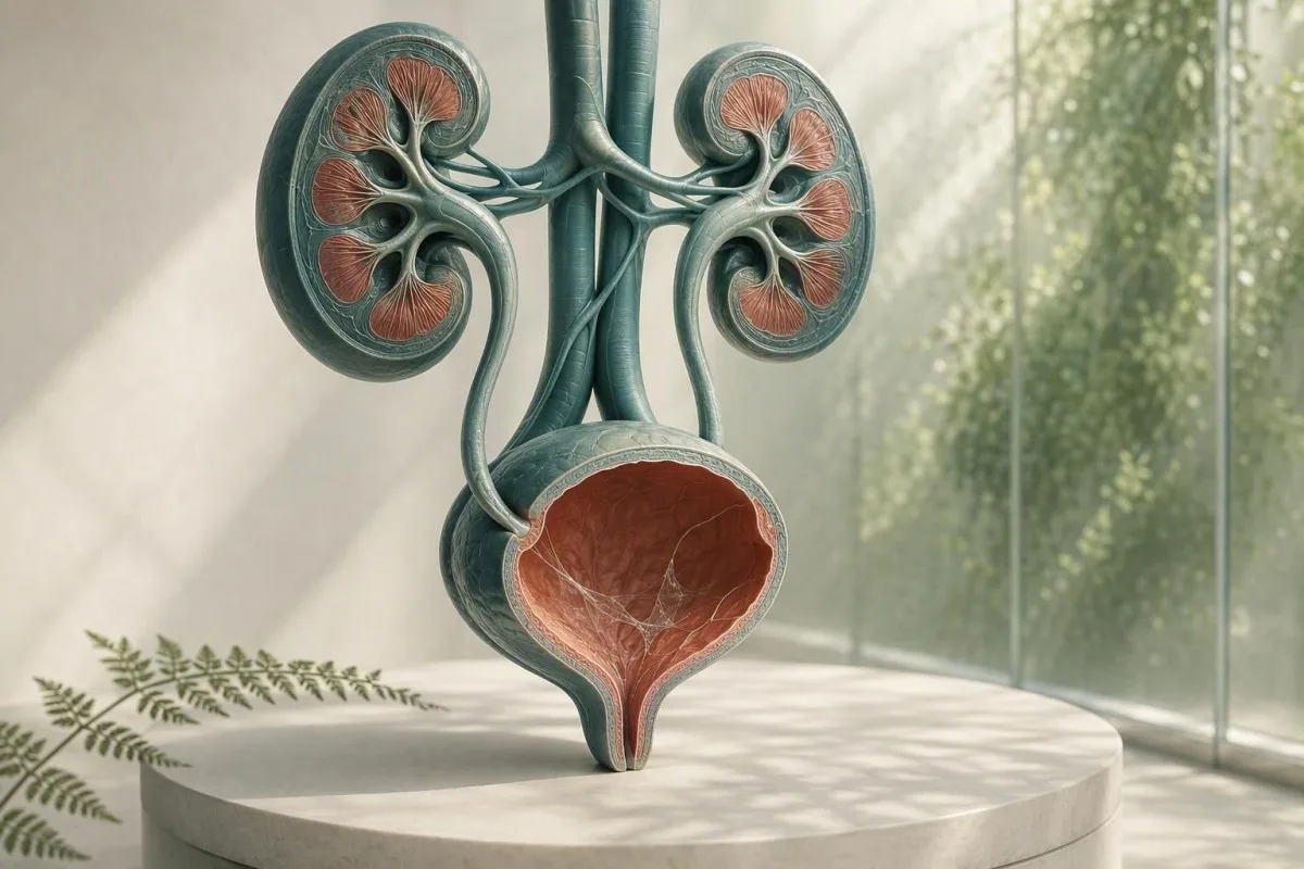
Sluz u mikroskopskom nalazu urina obično je problem pri uzimanju uzorka, a ne dijagnoza. Nalazi urina, simptomi i kvaliteta uzorka utvrđuju treba li daljnju obradu.


- Nitice sluzi same po sebi su često bezopasne, osobito u jednom uzorku čistog hvata bez simptoma mokraćnog sustava.
- Prag za piuriju veći od 5 bijelih krvnih stanica po vidnom polju jake rezolucije podupire upalu mokraćnog sustava, ali ne dokazuje bakterijsku UTI.
- Okidač za kulturu je sluz uz peckanje, žurbu, groznicu, bol u boku, nitrite ili značajan broj bijelih krvnih stanica—ne sama sluz.
- Pločaste stanice iznad otprilike 10 po vidnom polju jake rezolucije često ukazuju na kontaminaciju genitalne kože i čine rezultat urina manje pouzdanim.
- Vidljiva krv s kolikoidnim bolom u boku zahtijeva hitnu procjenu na prisustvo kamenca, čak i ako se prijavi i sluz.
- Trudnoća mijenja prag za praćenje jer se asimptomatska bakteriurija obično skrininguje urinskom kulturom, a ne samo mikroskopijom.
- Zamućen urin može odražavati kristale, koncentrisani urin, vaginalni iscjedak, sjeme ili ćelije; izgledom ne može dijagnostikovati infekciju.
- Ponoviti uzorak je razuman kada je sluz jedini neuobičajeni nalaz, a sakupljanje nije bilo pažljivo srednjim mlazom.
Što nitice sluzi u analizi urina obično znače
Niti sluzi u urinu su obično pramenovi normalnog zaštitnog sekreta ili genitalne kontaminacije, a izolovani nalaz rijetko ukazuje na bolest bubrega. Nalaz postaje značajan kada se pojavi sa urinarnim simptomima, bijelim ćelijama, bakterijama, krvlju ili proteinima.
Laboratorije identifikuju sluz kao tanke, prozirne pramenove viđene pod mikroskopom; mnogi izvještaji je jednostavno ocjenjuju kao rijetko, malo, umjereno ili mnogo, umjesto da dodjeljuju numerički referentni raspon. Urin prirodno sadrži malu količinu materijala bogatog glikoproteinima iz urinarnog trakta, a vaginalni ili uretralni sekreti mogu dodati više tokom sakupljanja.
Formulacija izvještaja je manje alarmantna nego što zvuči. Od 26. avgusta 2026., ne postoji validiran broj niti sluzi koji dijagnostikuje UTI, kamenac, rak ili otkazivanje bubrega; to je kontekstualno mikroskopsko zapažanje. Za širu mapu ostalih stavki u izvještaju o test trakama i mikroskopiji, pogledajte naš kompletan vodič za analizu urina.
Po mom kliničkom iskustvu, odrasloj osobi bez simptoma sa sluzi označenom kao “umjereno”, 0-2 bijele krvne ćelije po vidnom polju visoke rezolucije, negativan nitrit i bez krvi, obično je najvažnije umirenje ili jedno pravilno sakupljeno ponovljeno uzorak. Dr. Thomas Klein često viđa nepotrebnu anksioznost zbog antibiotika koja nastaje jer se laboratorijska oznaka pogrešno smatra etiketom bolesti.
Kantesti je AI platforma za tumačenje krvne slike koja može staviti bubrežne markere, kao što su kreatinin i eGFR, pored nalaza analize urina—ali ne može dijagnostikovati UTI samo na osnovu sluzi. Mikroskopski nalaz urina zahtijeva kliničku priču koja je došla uz njega.
Zašto laboratorija označava
Laboratorijski informacioni sistemi označavaju sluz jer je vizuelno prisutna, a ne zato što je laboratorija uspostavila opasan prag. Oznaka stoga znači “uočeno” umjesto “abnormalni marker bolesti”, slično kao što izolovani trag nalaza može zahtijevati bez liječenja.
Odakle potječe sluz u uzorku urina
Sluz u urinu može potjecati iz uretre, sluznice mokraćne bešike, grlića materice ili vagine, sjemena ili vanjske genitalne kože tokom sakupljanja. Njegov izvor se često pretpostavlja na osnovu ostatka mikroskopskog obrasca, a ne na osnovu samih pramenova.
Mokraćna bešika i uretra imaju zaštitni površinski sloj koji sadrži mucine, uključujući materijal povezan s uroplakinom, koji smanjuje trenje i vezivanje mikroba. Male količine mogu dospjeti u urin nakon dehidracije, nedavnog seksualnog odnosa, manje iritacije, ili jednostavno zato što koncentrovani prvi jutarnji uzorak lakše prikazuje pramenove.
Kod osoba koje menstruiraju ili imaju vaginalni iscjedak, leukociti i sluz mogu ući u posudu bez dolaska iz mokraćne bešike. Više od 10 skvamoznih epitelnih ćelija po vidnom polju visoke rezolucije čini kontaminaciju vjerovatnijom, iako laboratorije koriste različite pragove izvještavanja. Naš vodič za epitelne ćelije u urinu objašnjava zašto ovo mijenja povjerenje u rezultat.
Sjeme može također stvoriti nitasti materijal ili zamućenost nekoliko sati nakon ejakulacije. To ne čini urin nesigurnim, ali može zamagliti mikroskopiju; ako se razmatra kultura, obično savjetujem prikupljanje novog uzorka najmanje 24 sata kasnije kada je to moguće.
Zašto spol i anatomija utječu na izvještaj
Šalica za urin ne izolira mjehur od obližnjeg tkiva. Zbog toga se “prisutna sluz” češće izvještava u uzorcima s vanjskim izlučevinama, dok se kateterizirani uzorak ponekad koristi kada kliničari trebaju čistiji odgovor.
Kada nitice sluzi ukazuju na infekciju mokraćnog sustava
Niti sluzi podržavaju moguću UTI samo kada prate simptome i objektivne upalne nalaze, posebno piuriju, pozitivnost nitrita ili uvjerljivu kulturu. Sama sluz nije test za UTI.
Simptomatska infekcija donjeg urinarnog trakta obično uzrokuje peckanje, hitnost, učestalost, suprapubičnu nelagodu i ponekad novi miris urina. U pravilno prikupljenom uzorku, više od 5 bijelih krvnih stanica po vidnom polju visoke snage često se naziva piurija; podržava upalu mokraćnog trakta, ali se može pojaviti i kod kamenaca, spolno prenosivih infekcija i kontaminacije.
Nitrit je visoko specifičan kada je pozitivan, ali propušta infekcije uzrokovane organizmima koji ne reduciraju nitrat, a negativan rezultat nitrita ne isključuje UTI. Leukocitna esteraza otkriva aktivnost enzima bijelih stanica i može biti lažno pozitivna kada vaginalni sekret kontaminira uzorak; naše objašnjenje o rezultate leukocitne esteraze pokriva te zamke.
Smjernice IDSA savjetuju protiv probira ili liječenja asimptomatske bakteriurije kod zdravih, ne-trudnih odraslih osoba jer antibiotici donose štetu bez koristi (Nicolle et al., 2019). Taj princip je posebno relevantan za sluz: bez peckanja, bez vrućice i bez planiranog urološkog zahvata obično znači bez antibiotika samo zato što je mikroskopsko polje izgledalo neuredno.
Kada je kultura bolji test
Kultura urina je korisnija od ponovljenog urinskog testa kada simptomi potraju, simptomi se ponove unutar 4 tjedna, prisutna je trudnoća, sumnja se na pijelonefritis, ili su prethodni antibiotici možda promijenili rezultat. Mješoviti bakterijski rast često ukazuje na kontaminaciju prikupljanja, a ne na pojedinačni urinarni patogen; pogledajte tumačenje urinske kulture.
Sluz, kristali, kamenci i mehanička iritacija
Sluz sa jakim valovitim bolovima u boku ili krvlju u urinu može se pojaviti kod urinarnog kamena, ali sluz niti potvrđuje niti isključuje kamen. Krv, kristali, obrazac boli, snimanje i funkcija bubrega su važniji.
Kamenac može ostružiti ili začepiti urinarni epitel, proizvodeći crvene stanice, bijele stanice i dodatnu sluz. Klasični simptom je iznenadna kolikativna bol koja se širi prema preponama, često sa mučninom; vidljivo crveni ili čajne boje urin povećava hitnost. Pročitajte više o crvenih zastavica za krv u urinu umjesto pretpostavke da je svaki ružičasti uzorak jednostavna infekcija.
Kristali kalcijevog oksalata mogu se pojaviti kod zdravih ljudi, posebno u koncentriranom kiselom urinu, tako da jedna vrsta kristala ne dokazuje aktivni kamenac. Kod osobe s bolovima, ponavljajućim kamencima ili upornom hematurijom, kliničari mogu koristiti ultrazvuk ili CT s niskom dozom bez kontrasta umjesto da se oslanjaju samo na talog. Naš resurs o kristale kalcij-oksalata detaljno opisuje ograničenja izvještaja o kristalima.
Važan izuzetak je opstrukcija s infekcijom: vrućica od 38,0°C ili viša, bol u boku, povraćanje i nemogućnost mokrenja zahtijevaju hitnu procjenu. Briga nije sluz; to je zaražen blokiran urinarni sustav, koji se može brzo pogoršati.
Iritacija bez kamenca
Nedavna upotreba katetera, postupci na mjehuru, snažno bicikliranje i zračenje zdjelice mogu iritirati donji trakt i povećati sluz ili bijele stanice. Kliničar bi trebao tumačiti ove rezultate u odnosu na vrijeme, jer uzorak prikupljen unutar 48 sati od instrumentacije nije ekvivalentan rutinskom probirnom uzorku.
Značenje mutnog urina: što izgled može, a što ne može reći
Zamagljen urin mogu uzrokovati koncentrirane soli, kristali, sluz, stanice, genitalni iscjedak ili bakterije, tako da sam izgled ne može dijagnosticirati infekciju. Urin koji je zamućen, ali bezbolan i kratkotrajan, obično nije hitan.
Kristali fosfata mogu učiniti alkalni urin zamućenim nakon što se ohladi, dok kristali urata mogu zamutiti kiseli koncentrirani urin. Uzorak ostavljen na sobnoj temperaturi duže od 2 sata može postati sve zamućeniji kako se stanice degradiraju i bakterije razmnožavaju, zbog čega je obrada svježeg uzorka ili hlađenje važno.
Zamućenost praćena dizurijom, hitnošću i piurijom zahtijeva testiranje; zamućenost nakon vježbanja ili nedovoljnog unosa tekućine često se poboljšava običnom hidratacijom. Ciljajte na urin koji je blijedožut, a ne forsirajte prekomjeran unos vode – vrlo bistar urin ne znači da bubrezi “ispiru” infekciju. Naš detaljni pregled uzroci mutne mokraće odvaja vizualne tragove od pouzdanih testova.
Kantesti je AI analizator krvi dizajniran za tumačenje krvnih markera u kliničkom kontekstu; visoki kreatinin ili smanjeni eGFR uz urinarne abnormalnosti mogu opravdati medicinski pregled, dok sama zamućenost urina ne utvrđuje oštećenu bubrežnu filtraciju. Osnovna metabolička panelna pretraga može pružiti koristan kontekst kada simptomi ukazuju na dehidraciju ili opstrukciju.
Miris nije kultura
Jaki miris urina često odražava koncentraciju, metabolite šparoga, B vitamine ili posudu koja je stajala predugo. Novi neugodan miris s povišenom temperaturom ili urinarnim simptomima razlog je za traženje procjene, ali miris nije zamjena za mikroskopiju i kulturu.
Genitalni iscjedak i spolno prenosive bolesti
Izlučevine iz uretre ili vagine mogu izgledati kao sluz u urinu, a nove izlučevine s peckanjem pri mokrenju zahtijevaju testiranje spolnog zdravlja, kao i analizu urina. Rutinska kultura urina može promašiti klamidiju i gonoreju.
Klamidija i gonoreja mogu uzrokovati dizuriju i sterilnu piuriju – bijele krvne stanice bez uobičajenog bakterijskog rasta – posebno nakon novog spolnog izlaganja. Testiranje amplifikacije nukleinske kiseline, često korištenjem urina prvog mlaza umjesto uzorka srednjeg mlaza, je odgovarajući test jer standardna kultura cilja na različite organizme.
Kod osoba s vaginom, bakterijska vaginoza, kandidijaza i cervikalna upala mogu dodati izlučevine u posudu za srednji mlaz; vaginalni bris ili pregled mogu biti informativniji od ponovljene mikroskopije urina. Kod osoba s penisom, vidljive izlučevine iz uretre ne treba odbaciti kao “niti sluzi”. Praktična razlika je izlučevina primijećena izvan mokrenja u odnosu na niti prijavljene samo od strane laboratorija.
Smjernica CDC-a o liječenju spolno prenosivih bolesti iz 2021. preporučuje testiranje temeljeno na NAAT-u na relevantnim anatomskim mjestima prema povijesti izlaganja, a ne samo na simptomima (Workowski et al., 2021.). Ako su uključeni bolovi u zdjelici, bolovi u testisima, vrućica, trudnoća ili moguća agresija, potražite savjet liječnika istog dana umjesto samoliječenja zaostalim antibioticima.
Zašto antibiotici mogu zbuniti sliku
Uzimanje čak i 1 ili 2 doze antibiotika prije kulture može suzbiti uobičajeni bakterijski rast, dok urinarni simptomi i bijele krvne stanice perzistiraju. Recite liječniku točno koji lijek, dozu i vrijeme posljednje doze ste koristili; taj detalj mijenja način na koji se tumači negativna kultura.
Kako prikupiti uzorak urina koji daje odgovor
Uzorak srednjeg mlaza iz čistog hvata bolje smanjuje sluz, skvamozne stanice i mješoviti bakterijski rast nego skupljanje prvog ili zadnjeg dijela mlaza. Tehnika je jednostavna, ali prvih 2 sekunde čine nesrazmjernu razliku.
Operite ruke, razdvojite genitalnu kožu ili povucite kožicu ako vam je ugodno, očistite prema uputama kompleta, počnite mokriti u toalet, a zatim sakupite srednji dio bez da posuda dodiruje kožu. Uzorak od 20–30 mL je obično dovoljan; punjenje velike posude do vrha ne poboljšava test.
Dostavite posudu promptno, idealno unutar 1 sata; ako je odgoda neizbježna, ohladite je prema uputama laboratorija i vratite je u roku od 24 sata. Hlađenje usporava rast bakterija, ali ne obnavlja uzorak koji je već bio kontaminiran. Ovo je posebno važno kada zahtjev uključuje kulturu umjesto samo kemijske analize urina.
Kantesti je Alat za analizu krvnih nalaza uz pomoć AI-a koristi se u cijelom svijetu, a naši radni postupci koje pregledaju liječnici tretiraju kvalitetu uzorka kao dio interpretacije, a ne kao fusnotu. Isto načelo vrijedi za bilo koju laboratorijsku vrijednost: tehnički loš uzorak može stvoriti medicinski uvjerljiv, ali pogrešan rezultat. Pogledajte naš popis za provjeru točnosti laboratorija za pitanja koja treba postaviti prije poduzimanja radnji na temelju izoliranog upozorenja.
Nemojte skupljati u tim situacijama ako možete pričekati
Izbjegavajte rutinsko testiranje tijekom obilnog menstrualnog krvarenja, neposredno nakon spolnog odnosa ili nakon upotrebe vaginalnih krema, osim ako liječnik to izričito ne zatraži. Kada testiranje ne može čekati, obavijestite laboratorij ili liječnika, jer taj kontekst može objasniti sluz, crvena krvna zrnca ili vanjske stanice.
Kako kliničari tumače sluz u kontekstu analize urina
Najkorisniji obrazac urinoanalize kombinuje simptome sa bijelim krvnim zrncima, crvenim krvnim zrncima, nitritima, leukocit esterazom, proteinima, glukozom, specifičnom težinom i epitelijalnim ćelijama. Sluz je manji podržavajući element u tom obrascu.
Čist uzorak sa sluzi, 0–2 bijela krvna zrnca po vidnom polju pod velikim povećanjem, negativni nitriti i bez krvi obično ne zahtijevaju liječenje. Sluz sa više od 5 bijelih krvnih zrnaca po vidnom polju pod velikim povećanjem i pozitivna leukocit esteraza povećava vjerovatnoću upale; dodavanje nitrita ili kulture sa jednim organizmom povećava sigurnost da su bakterije odgovorne.
Proteini zahtijevaju sopstveni pristup. Trag proteina nakon groznice, vježbanja ili koncentrisanog urina može biti privremen, ali uporni proteini trebaju biti kvantifikovani omjerom albumin-kreatinin umjesto da se pripisuju sluzi. Naš vodič za protein u urinu objašnjava zašto dipstick proteini i bubrežni rizik nisu zamjenjivi.
Specifična težina od 1.003 do 1.030 je uobičajeno kod odraslih, iako se referentni intervali razlikuju u zavisnosti od laboratorije. Visoka specifična težina može koncentrisati sluz i stvoriti dramatičniji sediment, dok niska specifična težina može lizirati ćelije i učiniti mikroskopiju varljivo blagom; naš vodiču za specifičnu težinu pokazuje kako hidratacija utječe na interpretaciju.
Vrijednost negativnih nalaza
Negativna krv, proteini, nitriti i leukocit esteraza značajno smanjuju zabrinutost kod osobe bez simptoma, čak i ako je sluz prijavljena kao umjerena. Nijedan pojedinačni negativni test nije savršen, ali ovaj skup je umirujućiji nego što je ocjena sluzi zabrinjavajuća.
Kada ponoviti pretragu, a kada zatražiti kulturu
Ponoviti uzorak urina kada je sluz izolirana, skvamozne ćelije ukazuju na kontaminaciju, ili je uzorkovanje bilo žurno; zatražiti kulturu kada su simptomi uporni, rekurentni, teški ili visokog rizika. Ponovljeni test nije “nerad” – često je najdijagnostičkiji sljedeći korak.
Za trudnicu bez simptoma i sa sluzi kao jedinim pokazateljem, ponovljena urinoanaliza čistim hvatanjem unutar 1–2 sedmice je razumna ako je potrebno umirenje; mnogi kliničari je uopšte ne bi ponovili. Ne liječiti laboratorijski pokazatelj antibioticima dok se čeka, osim ako ljekar ne utvrdi kliničku indikaciju.
Kultura prije antibiotika je posebno korisna kod groznice, bolova u slabinama, trudnoće, imunosupresije, transplantacije bubrega, upotrebe mokraćnog katetera, muških urinarnih simptoma ili simptoma koji se vraćaju unutar 4 sedmice. Smjernice Gupta et al. (2011) podržavaju kulturu kod sumnje na pijelonefritis i situacija gdje je otpornost ili alternativna dijagnoza vjerovatnija.
Kultura mora biti očitana u odnosu na kvalitet prikupljanja. Jedan organizam na 10^5 jedinica koje formiraju kolonije po mL tradicionalno je podržavao bakteriuriju u čistom uzorku urina srednjeg mlaza, ali simptomatski pacijenti mogu imati klinički relevantne niže brojeve; “mješovita flora” obično nalaže novi uzorak umjesto širokog spektra antibiotika. Uporedite svrhe analizu urina i kulturu prije nego što zatražite bilo šta od navedenog.
Trudnoća zahtijeva drugačiji prag
Trudnoća je izuzetak jer asimptomatska bakteriurija može povećati rizik od pijelonefritisa i neželjenih trudnoćnih ishoda. Antenatalna njega obično koristi skrining kulturu rano u trudnoći; sluz pod mikroskopom ne može zamijeniti tu kulturu.
Kada krvne pretrage daju koristan kontekst za bubrege i infekcije
Krvni testovi su korisni kada se sluz u urinu pojavi sa groznicom, bolom u boku, ponovljenim infekcijama, oticanjem, smanjenim izlučivanjem urina, ili upornim proteinima ili krvlju. Kreatinin i eGFR procjenjuju filtraciju, dok CBC i C-reaktivni protein mogu pomoći u procjeni sistemske bolesti.
eGFR ispod 60 mL/min/1.73 m² tokom najmanje 3 mjeseca ispunjava jedan kriterij za hroničnu bubrežnu bolest, ali jedan niži rezultat tokom dehidracije ili akutne bolesti nije dovoljan za postavljanje te dijagnoze. Kreatinin je pod utjecajem mišićne mase, prehrane i nekih lijekova, tako da su trendovi i bubrežni albumin često informativniji od jednog broja.
Povišen broj bijelih krvnih zrnaca ili CRP može podržati upalni proces, ali ne može identificirati urinarni trakt kao izvor. Kod osobe sa groznicom i bolom u boku, kliničari mogu provjeriti kreatinin prije odabira snimanja ili lijekova, posebno ako povraćanje ili opstrukcija mogu narušiti bubrežnu funkciju. Pregledajte stadije hronične bubrežne bolesti za kategorije eGFR i albumina koje kliničari koriste.
Kantesti je usluzi za tumačenje laboratorijskih testova AI koji naglašava obrasce kreatinina, eGFR, elektrolita, CBC i inflamatornih markera, umjesto da se jedna notacija sluzi tretira kao bubrežna dijagnoza. Naša metodologija podliježe nadzoru kliničke validacije, ali hitni simptomi i dalje zahtijevaju direktnu medicinsku njegu, a ne interpretaciju putem aplikacije.
Praktično razlikovanje dehidracije
Dehidracija može povisiti specifičnu težinu urina i privremeno povećati kreatinin, posebno nakon proljeva, izlaganja vrućini ili intenzivnog vježbanja. Uporno nisko izlučivanje urina, vrtoglavica, ili pad eGFR-a nakon rehidracije zahtijeva pregled kliničara umjesto ponovljenog kućnog testiranja.
Hitni znakovi upozorenja koji ne smiju čekati ponovnu pretragu
Potražite hitnu procjenu istog dana za sluz u urinu sa temperaturom od 38.0°C ili višom, bolom u boku, povraćanjem, vidljivom krvlju, nemogućnošću mokrenja, konfuzijom ili simptomima urinarnog trakta povezanima s trudnoćom. Ove kombinacije su važne jer mogu signalizirati infekciju gornjeg dijela urinarnog trakta, opstrukciju ili drugo akutno stanje.
Groznica praćena bolom u jednom dijelu leđa ili boka je zabrinjavajuća za pijelonefritis nego za jednostavnu cistitis, posebno uz drhtavicu ili povraćanje. Odloženo liječenje može dovesti do dehidracije, sepse ili bubrežnog stresa; normalno izgledajući uzorak urina kod kuće ga ne isključuje sigurno.
Vidljivu krv treba procijeniti čak i kada je UTImoguća, posebno nakon 35. godine života, kod pušača, ili ako krvarenje nastavi nakon smirivanja simptoma infekcije. Smjernica AUA za mikrohematuriju preporučuje procjenu zasnovanu na riziku nakon što se riješe benigni razlozi (Barocas et al., 2020); uporna krv se ne objašnjava sluzi.
Djeca, odrasli stariji od 65 godina, osobe sa dijabetesom, osobe koje uzimaju imunosupresivne lijekove i oni sa jednim bubregom zaslužuju niži prag za procjenu. Pravilo dr. Thomasa Kleina u praksi je jednostavno: ako se simptomi pogoršavaju tokom 6-12 sati, ne čekajte drugu čašu da biste pružili uvjerenje.
Pozovite hitnu pomoć odmah zbog teške bolesti
Pozovite hitnu pomoć zbog nove konfuzije, nesvjestice, jake slabosti, plavih ili sivih usana, teške kratkoće daha, ili nemogućnosti zadržavanja tečnosti uz urinarne simptome. Ovo su sistemski znaci opasnosti, a ne rutinski simptomi UTI.
Sluz u urinu tijekom trudnoće, djetinjstva i kasnije u životu
Trudnoća, djeca i starije osobe zahtijevaju pažljiviju interpretaciju jer je kontaminacija česta, ali posljedice propuštene infekcije mogu biti veće. Simptomi i kvalitet kulture ostaju vrjedniji od stepena sluzi u svakoj starosnoj grupi.
Tokom trudnoće, učestalo mokrenje može biti normalno, što čini simptome manje specifičnim; vrućica, dizurija ili bol u leđima trebaju potaknuti hitnu akušersku ili kliničku procjenu. Probirna kultura se obično uzima rano tokom prenatalne njege, a ponovljena kultura se može koristiti nakon liječenja ovisno o planu kliničara.
Kod djece, urin prikupljen vrećicom je sklon kontaminaciji i obično se ne bi trebao koristiti samostalno za dijagnosticiranje UTI kulturom. Kateterizirani ili pažljivo uzeti uzorak iz čistog hvata može biti potreban kada mlado dijete ima vrućicu bez jasnog izvora; sluz u uzorku iz vrećice je posebno nespecifična.
U kasnijem životnom dobu, bakteriurija i pijurija postaju češći bez izazivanja simptoma. Novi deliro sam po sebi trebao bi pokrenuti široku medicinsku procjenu dehidracije, lijekova, bola, zatvora i izvora infekcije, a ne automatsko liječenje UTI; ista uzdržanost koju preporučuju Nicolle et al. (2019) primjenjuje se.
Menstruacija i promjene povezane s hormonima
Menstrualna krv i cervikalna sluz mogu promijeniti analizu urina na nekoliko dana, a postmenopauzalna suhoća vagine može uzrokovati lokalnu iritaciju koja podsjeća na peckanje pri mokrenju. Ako se rezultati i simptomi ne poklapaju, kliničar može procijeniti genitalne uzroke umjesto da propisuje ponovljeno liječenje UTI.
Uobičajene pogreške nakon viđenja sluzi u laboratorijskom nalazu
Nemojte započinjati preostale antibiotike, ne čistiti iznutra niti pokušavati “isprati” sluz ekstremnim unosom vode nakon jedne abnormalne analize urina. Ove radnje mogu zamagliti kulturu, narušiti normalnu floru ili odgoditi ispravnu dijagnozu.
Antibiotici uzeti bez kulture mogu učiniti naknadni test lažno negativnim i mogu uzrokovati proljev, osip, simptome gljivica ili otpornost. Ako su simptomi blagi, ali uporni, uzmite uzorak prvi kada je to praktično, a zatim slijedite plan liječenja kliničara zasnovan na cjelokupnoj slici.
Proizvodi od brusnice mogu skromno smanjiti rizik od rekurentnih nekomplikovanih UTI kod nekih ljudi, ali ne liječe vrućicu, bol u boku ili potvrđenu infekciju gornjeg urinarnog trakta. Izbjegavajte vitamin C u visokim dozama kao samoliječenje: može promijeniti neke reakcije test traka i može povećati opterećenje oksalatom kod osoba sklonih kamencima kalcijum oksalata.
Nemojte više puta pregledavati WC školjku na niti. Vlakna toaletnog papira, ostaci sredstava za čišćenje, genitalni sekreti i turbulencija vode mogu imitirati sluz; laboratorijski uzorak prikupljen u sterilnoj posudi je odgovarajuće mjesto za procjenu. Za povezane promjene boje, naša vodič za boju urina nudi pouzdaniji vizuelni kontekst.
Lista lijekova je bitna
Fenazopiridin može učiniti urin narandžastim i ometati vizuelnu interpretaciju, dok diuretici mogu koncentrirati ili razrijediti urin ovisno o vremenu. Ponesite sa sobom listu lijekova na recept, proizvoda bez recepta i suplemenata, uključujući doze u mg.
Praktični plan daljnje obrade za nitice sluzi u urinu
Većina ljudi sa nitima sluzi u urinu i bez simptoma ne treba liječenje; pažljiva ponovljena analiza urina je razumna ako je uzorak bio upitan. Simptomi ili prateće abnormalnosti utvrđuju da li su kultura, testiranje na spolno prenosive infekcije, snimanje ili krvni testovi prikladni.
Korak 1: provjerite peckanje, hitnost, vrućicu, bol u boku, vidljivu krv, iscjedak, trudnoću, nedavne urinarne procedure i novo seksualno izlaganje. Korak 2: pročitajte izvještaj za bijele ćelije, nitrite, leukocit esterazu, crvene ćelije, proteine, bakterije i skvamozne ćelije—ne samo sluz.
Korak 3: ako se osjećate dobro i sluz je izolirana, sakupite jedan uzorak iz srednjeg mlaza čistog hvata u roku od 1-2 sedmice samo ako vaš kliničar preporuči potvrdu. Korak 4: ako su simptomi prisutni, pitajte da li treba prikupiti kulturu urina prije liječenja i da li je testiranje na SPI relevantno; rekurentne epizode zaslužuju pažljiviji pregled nego treći empirijski antibiotik.
Kantesti može organizirati relevantne trendove krvnih nalaza i pitanja za medicinski pregled, dok naš Medicinski savjetodavni odbor podržava standarde sigurnosti vođene kliničarom. Pravi krajnji cilj nije savršeno “čist” mikroskopski izvještaj—to je objašnjenje koje odgovara vašim simptomima, kvalitetu uzorka i faktorima rizika.
Pitanja koja treba ponijeti na vaš pregled
Pitajte da li je u uzorku bilo pločastih ćelija, da li je kultura pokazala jedan organizam ili mješovitu floru, te da li su krv ili protein perzistirali nakon što su simptomi prestali. Ova 3 pitanja često daju korisnije odgovore nego pitati koliko je sluzi viđeno.
Kontekst istraživanja i ograničenja izvješćivanja o sluzi
Ocjenjivanje sluzi nije standardizirano među laboratorijama, zbog čega bi kliničari trebali izbjegavati korištenje “malo”, “umjereno” ili “mnogo” kao kategorije težine bolesti. Tehnika mikroskopije, starost uzorka i softver za izvještavanje mogu promijeniti formulaciju.
Neke laboratorije ručno pregledavaju talog nakon centrifugiranja otprilike 10–15 mL urina, dok druge koriste automatizovanu analizu čestica sa ručnim pregledom odabranih zastavica. Ta razlika znači da se rezultat “umjerena sluz” iz jedne laboratorije možda neće tačno ponoviti u drugoj laboratoriji čak i kada je zdravlje osobe nepromijenjeno.
Koristan laboratorijski pristup je pitati da li je rezultat analitički stvaran, klinički značajan i ponovljiv. Taj pristup je poznat i iz testiranja krvi: naša vodič za krvne biomarkere objašnjava zašto referentni intervali i preanalitički uslovi određuju da li označeni marker zaslužuje akciju.
Kantesti AI koristi strukturirani laboratorijski kontekst za objašnjenje obrazaca rezultata preko glavnih panela, ali mikroskopija urina i dalje zahtijeva kliničku procjenu specifičnu za izvor. Za čitatelje zainteresirane za to kako procjenjujemo tehničke performanse i kliničke granice, naša Vodič kroz AI tehnologiju opisuje zaštitne mjere iza našeg interpretativnog pristupa.
Šta dobar izvještaj treba jasno pokazati
Koristan izvještaj identifikuje vrstu uzorka, datum prikupljanja, mikroskopske elemente i rezultate kulture organizama i osjetljivosti kada se radi kultura. Ako izvještaj samo kaže “prisutne sluzne niti”, on je nepotpun za dijagnozu, ali često potpuno adekvatan za slučajni nalaz niskog rizika.
Često postavljana pitanja
Jesu li sluzavi konci u urinu normalni?
Sluzava vlakna u urinu su često normalna ili posljedica kontaminacije genitalnim sekretima, posebno kada osoba nema urinarnih simptoma. Izolovan nalaz sa 0-2 bijele krvne ćelije po vidnom polju visoke rezolucije, negativan nitrit, te bez krvi ili proteina, obično je niskog rizika. Laboratorije ne koriste validiran broj sluzavih vlakana za dijagnosticiranje urinarne infekcije (UTI) ili bolesti bubrega. Ponovljeni uzorak srednjeg mlaza prikupljen čistom tehnikom je razuman ako je originalno prikupljanje bilo na brzinu ili vidljivo kontaminirano.
Da li sluzavi konci u urinu znače da imam UTI?
Sluz u urinu sam po sebi ne znači infekciju urinarnog trakta (UTI). UTI postaje vjerovatniji kada se javi peckanje, nagon, učestalost mokrenja, groznica ili suprapubični bol uz prisustvo više od 5 bijelih krvnih zrnaca po vidnom polju, pozitivna leukocitna esteraza, nitrit ili pozitivna kultura. Negativan nitrit ne isključuje u potpunosti UTI jer svi bakterije ne proizvode nitrite. Kultura je obično informativnija od ocjene sluzi kada simptomi traju ili se ponavljaju.
Može li dehidracija uzrokovati sluz u mokraći?
Dehidracija može učiniti sluz lakšom za uočavanje jer koncentrirani urin ima manje vode oko normalnih sekreta i sedimenta. Visoka specifična težina urina, obično prema 1,025–1,030, može pratiti ovaj efekat, iako sam po sebi ne dokazuje dehidraciju. Normalna rehidratacija i ponavljanje pravilno prikupljenog uzorka mogu pojasniti nalaz. Ekstremni unos vode nije neophodan i može stvoriti vlastite rizike od elektrolita.
Šta znači sluz i bijela krvna zrnca u urinu?
Sluz plus bijelih krvnih zrnaca u urinu ukazuje na upalu mokraćnog ili polnog trakta, ali ne dokazuje bakterijsku infekciju. Više od 5 bijelih zrnaca po vidnom polju visoke rezolucije može se javiti kod infekcije mokraćnog trakta (UTI), kamenaca, polno prenosivih infekcija, vaginalne kontaminacije ili nedavne instrumentalizacije urinarnog trakta. Simptomi, nitriti, epitelne ćelije i rezultati kulture određuju sljedeći korak. Temperatura od 38.0°C ili viša uz bol u boku zahtijeva hitnu procjenu.
Može li vaginalni iscjedak uzrokovati sluz u urinu za vrijeme testiranja?
Vaginalni iscjedak može ući u uzorak urina prikupljen srednjim mlazom i često uzrokuje pojavu sluzi, skvamoznih epitelnih stanica, a ponekad i bijelih krvnih stanica na mikroskopiji. Više od 10 skvamoznih stanica po vidnom polju visoke rezolucije često ukazuje na veću mogućnost kontaminacije, iako se laboratoriji razlikuju u izvještavanju. Pažljivo prikupljen uzorak čistog ulova izvan perioda obilnog menstrualnog krvarenja može smanjiti ovaj problem. Ako su prisutni iscjedak, svrab, neugodan miris ili bol u zdjelici, vaginalni pregled ili bris može biti korisniji od ponovljenih urinokultura.
Kada treba da brinem o nitima sluzi i krvi u mokraći?
Mukozne niti sa vidljivom krvlju u urinu treba odmah procijeniti, posebno kada krv perzistira, postoji bol u boku, krvni ugrušci, groznica ili poteškoće s mokrenjem. Kamenac, infekcija mokraćnih puteva, povreda, bolest bubrega ili, rjeđe, rast u mokraćnim putevima, mogu uzrokovati krv; sluz ne utvrđuje koji je uzrok prisutan. AUA preporučuje procjenu zasnovanu na riziku za perzistentnu mikroskopsku hematuriju nakon što se otklone privremeni uzroci. Hitna procjena je prikladna za groznicu, povraćanje, jake bolove ili retenciju urina.
Nabavite analizu krvne slike uz AI već danas
Pridružite se više od 2 miliona korisnika širom svijeta koji vjeruju Kantesti-u za trenutnu i tačnu analizu laboratorijskih testova. Otpremite svoje rezultate krvne slike i dobijte sveobuhvatno tumačenje 15,000+ biomarkera za nekoliko sekundi.
📚 Referisane naučne publikacije
Klein, T., Mitchell, S., & Weber, H. (2026). Normalni raspon aPTT-a: D-dimer, protein C, vodič za zgrušavanje krvi. Kantesti AI Medical Research.
Klein, T., Mitchell, S., & Weber, H. (2026). Vodič za serumske proteine: Test krvi za globuline, albumine i odnos A/G. Kantesti AI Medical Research.
📖 Eksterne medicinske reference
⚕️ Medicinska izjava o odricanju odgovornosti
Ovaj članak je samo u edukativne svrhe i ne predstavlja medicinski savjet. Za odluke o dijagnozi i liječenju uvijek se posavjetujte s kvalifikovanim zdravstvenim radnikom.
E-E-A-T signal(i) povjerenja
Iskustvo
Klinička revizija radnih tokova tumačenja laboratorijskih nalaza koju vodi ljekar.
Stručnost
Fokus laboratorijske medicine na to kako se biomarkeri ponašaju u kliničkom kontekstu.
Autoritativnost
Napisao dr. Thomas Klein, uz recenziju dr. Sarah Mitchell i prof. dr. Hans Weber.
Pouzdanost
Tumačenje zasnovano na dokazima, s jasnim sljedećim koracima kako bi se smanjila uzbuna.