Pain can be real and clinically significant even when CRP and ESR are normal. The most useful tests depend on whether the pattern is joint, muscle, bone, nerve, abdominal, or whole-body pain.



- CRP and ESR are screening clues, not pain detectors; normal values do not rule out fibromyalgia, nerve compression, osteoarthritis, or early inflammatory arthritis.
- Creatine kinase (CK) above 1,000 IU/L with dark urine or weakness needs same-day assessment for significant muscle injury.
- Ferritin below 15 ng/mL strongly supports depleted iron stores, although fatigue and diffuse aching can occur before anaemia develops.
- Vitamin D below 12 ng/mL (30 nmol/L) indicates deficiency and can contribute to bone pain and proximal muscle weakness.
- TSH above 10 mIU/L with low free T4 supports overt hypothyroidism, a reversible cause of cramps, stiffness, and neuropathic symptoms.
- Calcium above 12 mg/dL (3.0 mmol/L) or below 7.5 mg/dL (1.9 mmol/L) can cause clinically important neuromuscular symptoms and needs urgent review.
- A normal ANA or rheumatoid factor does not exclude every autoimmune disease; the history, examination, urine findings, and pattern of joints matter.
- Chest pain, a hot swollen joint, new weakness, fever, black stools, or loss of bladder control should never wait for routine outpatient blood work.
What a blood test for unexplained pain can actually answer
A blood test for unexplained pain can identify systemic clues—such as anaemia, thyroid dysfunction, muscle breakdown, mineral imbalance, infection, or immune activity—but it cannot locate every source of pain. In my experience, the best first panel is driven by the pain pattern and examination, not by ordering every available antibody. Kantesti AI is an AI blood test analyzer that places common pain-related results into their wider laboratory context rather than treating a single flag as a diagnosis.
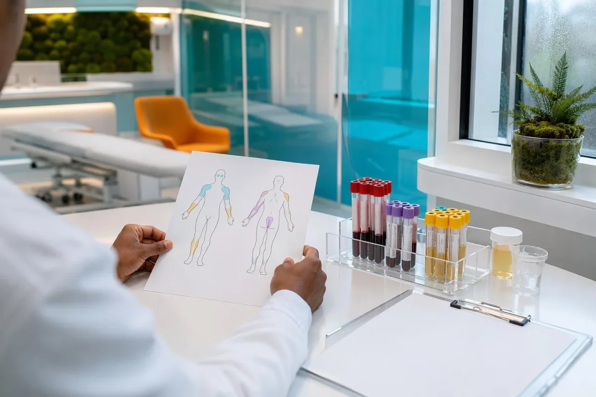
Pain is a signal from tissue, nerves, joints, bone, or internal organs; blood testing mainly detects conditions that affect the whole body. A complete blood count, renal and liver profile, calcium, glucose, thyroid-stimulating hormone, CRP or ESR, and urine testing often give more value than a broad autoimmune panel on day one. A normal panel is useful too: it shifts attention toward mechanical, neurological, sleep-related, or centrally amplified pain.
I reviewed a 41-year-old teacher with six months of shoulder, hip, and thigh aching whose first CRP was 2 mg/L. Her real clue was a TSH of 18.6 mIU/L and low free T4, not “hidden inflammation.” Conversely, a 52-year-old runner with an AST of 89 IU/L after a hilly race had CK 1,420 IU/L and normal bilirubin—an exercise-related muscle pattern rather than primary liver disease.
The practical question for a clinician is not “Which blood test proves my pain?” but “What dangerous, reversible, or treatable explanation would this result make more likely?” Our biomarker reference guide explains why the laboratory’s own range, units, timing, medicines, and prior values all change that answer.
Pain location is the first laboratory filter
Diffuse aching with fatigue points toward CBC, ferritin, thyroid, calcium, vitamin D, kidney function, glucose, and medication review. A single red, swollen joint calls for joint aspiration more urgently than an expanded blood panel; severe abdominal pain may need imaging even when liver enzymes and lipase are normal. This distinction prevents the common mistake of mistaking normal blood work for proof that nothing is wrong.
Match initial tests to the pattern of pain
The distribution and timing of pain determine which unexplained body pain blood tests have a reasonable chance of helping. Symmetric small-joint stiffness lasting more than 30 to 60 minutes after waking raises a different set of questions than burning feet, nighttime leg cramps, or deep bone pain.
For widespread pain with poor sleep and non-restorative fatigue, I usually begin with CBC, ferritin, TSH, calcium, renal profile, liver enzymes, glucose or HbA1c, and CRP or ESR when the history warrants it. Haemoglobin below 12.0 g/dL in most non-pregnant adult women or 13.0 g/dL in most adult men meets the WHO definition of anaemia, but iron depletion can precede that threshold. The CBC and differential guide helps readers see why MCV and RDW matter alongside haemoglobin.
Muscle pain plus objective weakness—difficulty rising from a chair, lifting a kettle, or climbing stairs—makes CK, potassium, phosphate, calcium, TSH, AST, ALT, and urine dipstick more relevant. CK reference limits vary considerably by sex, ancestry, training status, and laboratory; a value of 350 IU/L after unfamiliar exercise is not equivalent to 3,500 IU/L with weakness. This is one reason I ask patients to avoid unusually hard training for 48 to 72 hours before a non-urgent repeat.
Burning, pins-and-needles, or electric pain points more toward glucose or HbA1c, vitamin B12 with methylmalonic acid when appropriate, TSH, renal function, and selected infection or autoimmune testing based on risk. A fasting glucose of 126 mg/dL (7.0 mmol/L) or more on two separate tests meets diabetes criteria, while an HbA1c of 6.5% or more can do the same when assay conditions are valid. Nerve pain with normal blood results may still require neurological examination, nerve conduction studies, or spine imaging.
Write down the timing before your appointment
Morning stiffness, pain after exertion, pain that wakes you from sleep, and pain relieved by movement are not interchangeable clues. A symptom diary covering 14 days, including new medicines, supplements, fever, rashes, bowel changes, and activity, can be more diagnostic than adding ten low-probability assays. This is a small effort that often changes the quality of the consultation.
Inflammation tests for pain: what CRP and ESR can miss
CRP and ESR are non-specific markers of systemic inflammatory activity, and neither test can diagnose the cause of pain on its own. A CRP below 5 mg/L and an ESR within the laboratory range make major systemic inflammation less likely, but they do not exclude localized infection, early autoimmune disease, osteoarthritis, endometriosis, fibromyalgia, or nerve-root pain.
CRP usually rises within 6 to 8 hours of a substantial inflammatory stimulus and falls relatively quickly when that stimulus settles. ESR changes more slowly because it is influenced by fibrinogen, red-cell shape, age, pregnancy, anaemia, and immunoglobulin levels. An ESR of 45 mm/hour with haemoglobin 9.8 g/dL may partly reflect anaemia, whereas a rising CRP from 8 to 62 mg/L over 48 hours deserves more immediate clinical attention.
A discordant pattern can be informative. High ESR with normal CRP occurs in some people with anaemia, kidney disease, pregnancy, or increased immunoglobulins; high CRP with normal ESR can appear early in bacterial illness or after tissue injury. The pattern is explored in our guide to high ESR with normal CRP, but neither result should be used to self-treat with steroids or antibiotics.
The 2021 NICE guideline on chronic pain advises assessment for an underlying condition while also recognizing that persistent pain can continue without a clear structural or inflammatory marker (NICE, 2021). That distinction matters clinically: “normal inflammation tests” are not a verdict on the legitimacy or intensity of a patient’s symptoms. For persistent stiffness with rashes, mouth ulcers, kidney findings, or cold-induced colour changes, targeted ANA and complement testing may be more appropriate than simply repeating CRP.
When autoimmune tests help—and when they create noise
Autoimmune blood tests are most useful when pain comes with objective swelling, prolonged morning stiffness, rashes, mouth ulcers, Raynaud-type colour changes, eye inflammation, unexplained low blood counts, or abnormal urine. Ordering ANA, rheumatoid factor, anti-CCP, ANCA, and multiple rare antibodies for non-specific aching alone produces false positives that can cause months of anxiety.
Rheumatoid factor is not a rheumatoid arthritis test in isolation. It can be positive in older age, chronic infections, other autoimmune conditions, and a small proportion of healthy people; anti-CCP is more specific but still requires compatible joint symptoms. The 2021 American College of Rheumatology guideline emphasizes early clinical assessment and treatment for established rheumatoid arthritis, not diagnosis from a laboratory number alone (Fraenkel et al., 2021).
ANA is particularly prone to misinterpretation. Low-titre ANA results can occur in healthy adults, after viral illnesses, and with some medicines; the staining pattern and titre only become meaningful alongside a specific symptom cluster. I have seen patients referred in alarm over an ANA of 1:80 when their examination, urine protein, complements, and repeat history offered no evidence of connective-tissue disease.
A normal rheumatoid factor does not rule out inflammatory arthritis. Seronegative disease exists, and a clinician may use joint ultrasound, repeat examination, or imaging when swollen joints persist for more than 6 weeks. Readers with ongoing hand or foot joint symptoms can review seronegative rheumatoid arthritis before assuming a negative result closes the case.
Why broad panels often mislead
Every added test carries a chance of an incidental abnormality. If ten low-prevalence antibody tests each have 95% specificity, at least one false-positive result becomes surprisingly likely in an otherwise low-risk person. A focused question produces a focused panel—and a cleaner route to a real answer.
Muscle pain tests: CK, electrolytes, kidney function and urine
CK, potassium, phosphate, calcium, creatinine, AST, and urine testing are the core tests when muscle pain accompanies weakness, severe cramps, dark urine, or recent exertion. CK above 5 times the laboratory upper limit, particularly with reduced urine output or brown urine, should be assessed urgently because muscle breakdown can stress the kidneys.
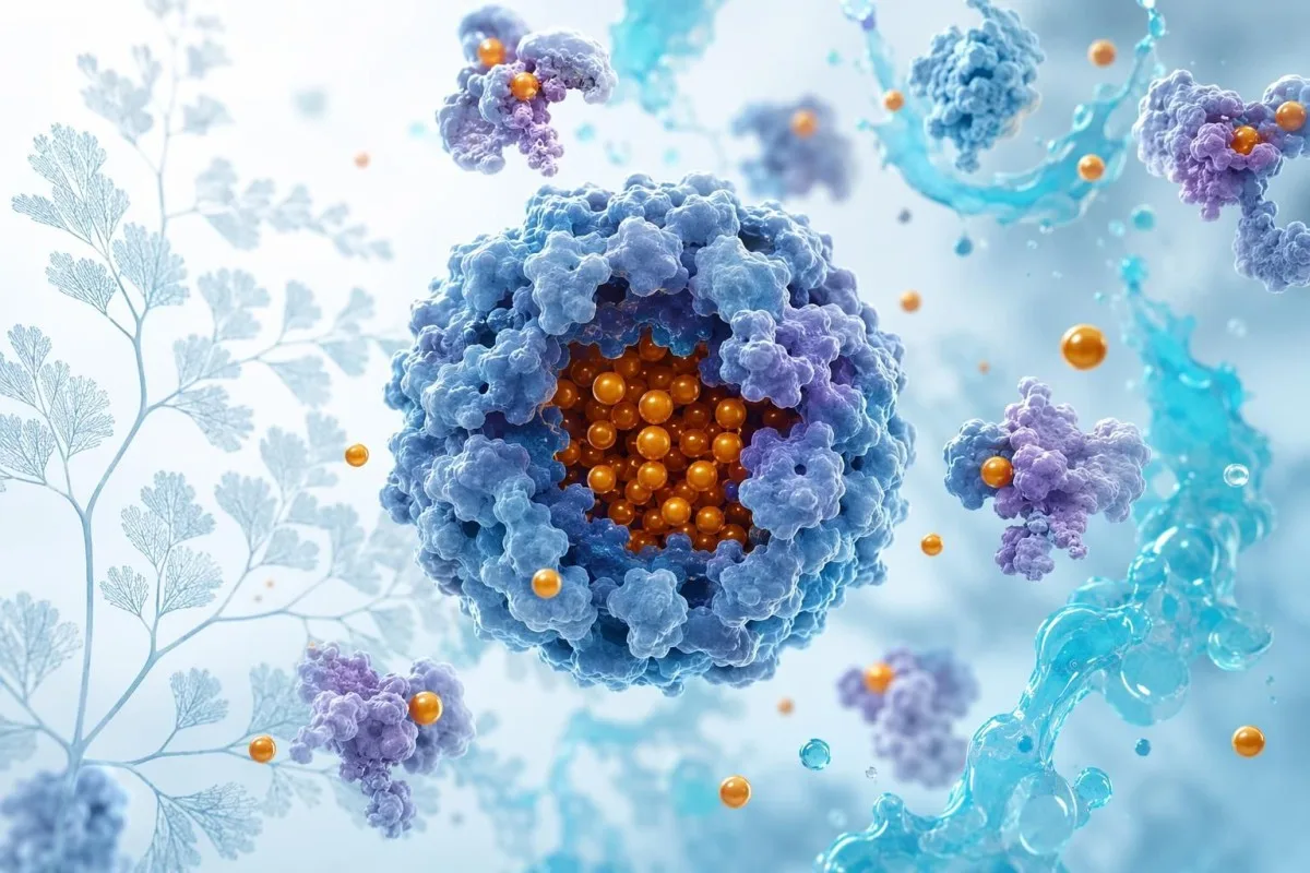
CK is an enzyme concentrated in skeletal muscle, so it rises after strenuous exercise, seizures, trauma, intramuscular injections, hypothyroidism, some medicines, and inflammatory muscle disorders. In a resting adult, many laboratories use an upper limit around 200 IU/L, but a result above 1,000 IU/L warrants active clinical context rather than casual dismissal. CK can peak 24 to 72 hours after exercise, which explains why the first sample may underestimate the later result.
Potassium below 3.0 mmol/L or above 6.0 mmol/L can cause weakness, cramps, or dangerous cardiac rhythm effects; severe symptoms require urgent care, not a home supplement experiment. Phosphate below 1.0 mg/dL (0.32 mmol/L) can impair muscle function, particularly with malnutrition, alcohol excess, uncontrolled diabetes, or refeeding. The practical interpretation of these results is covered in our muscle weakness lab guide.
Urine dipstick matters because myoglobin from substantial muscle injury can produce a positive “blood” reaction with few or no red cells on microscopy. Creatinine may remain normal early, especially in muscular people, so repeat renal testing and hydration advice must be individualized. New muscle pain after a medicine change should always prompt a list review, including statins, antipsychotics, antivirals, colchicine, and supplements.
Nutrient and bone clues that ordinary inflammation tests miss
Iron deficiency, vitamin D deficiency, vitamin B12 deficiency, magnesium loss, and phosphate disorders can contribute to fatigue, cramps, weakness, bone discomfort, or neuropathic pain without raising CRP. These tests are best targeted to diet, menstrual history, digestive symptoms, medication use, kidney function, and the character of pain.

A ferritin below 15 ng/mL usually indicates absent or near-absent iron stores in otherwise well adults, while inflammation can falsely elevate ferritin and obscure deficiency. Transferrin saturation below 20% supports restricted iron availability, especially when ferritin sits in the ambiguous 30 to 100 ng/mL range. Our detailed guide to ferritin levels in women explains why heavy periods are only one possible cause.
A 25-hydroxyvitamin D concentration below 12 ng/mL (30 nmol/L) is widely accepted as deficiency associated with impaired mineralization; 12 to 20 ng/mL is often considered inadequate, though national cutoffs differ. Vitamin D-related symptoms tend to be diffuse bone tenderness or proximal muscle weakness rather than sharp focal pain. Checking calcium, phosphate, alkaline phosphatase, creatinine, and parathyroid hormone can reveal whether the issue is simple deficiency or altered mineral metabolism.
Vitamin B12 below approximately 200 pg/mL (148 pmol/L) suggests deficiency, but borderline values between 200 and 300 pg/mL may need methylmalonic acid or homocysteine in patients with numbness or gait change. Serum magnesium is an imperfect proxy for total-body stores, yet a level below 1.46 mg/dL (0.60 mmol/L) is clinically meaningful. Kantesti is an AI-powered blood test analysis tool that can compare ferritin, MCV, B12, renal function, and CRP together when a single nutrient result looks misleading.
Thyroid and glucose results that can masquerade as pain disorders
Overt hypothyroidism and diabetes can cause muscle aching, stiffness, cramps, and nerve pain, while routine inflammation tests may remain normal. A TSH above 10 mIU/L with low free T4 is a much stronger clue to overt hypothyroidism than a mildly borderline TSH alone.
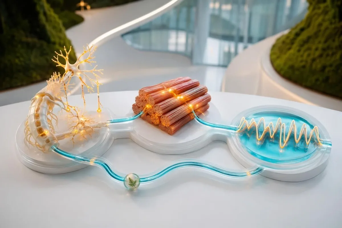
Hypothyroidism can raise CK, worsen carpal tunnel symptoms, slow reflexes, and produce proximal muscle discomfort. TSH values are measured in mIU/L, but their interpretation depends on free T4, pregnancy status, pituitary history, and medicines such as amiodarone or lithium. A mildly high TSH of 5.5 mIU/L does not explain every ache, whereas TSH 22 mIU/L with low free T4 and elevated CK deserves prompt treatment planning.
Diabetes-related neuropathy often begins as burning, tingling, numbness, or sensitivity in both feet, not as a raised CRP. HbA1c of 6.5% (48 mmol/mol) or greater supports diabetes when confirmed appropriately; prediabetes spans 5.7% to 6.4% in many guidelines. Diabetes can coexist with B12 deficiency in people taking metformin, which is why symptoms and medication duration matter. See our overview of thyroid test abbreviations for the common components of a thyroid panel.
Cortisol, sex hormones, and DHEA are frequently marketed as explanations for non-specific pain, but random testing has limited value without a focused clinical suspicion. In my 15 years of clinical practice, I have found that sleep loss, untreated sleep apnoea, depression, and medication effects are more common contributors than an occult “adrenal” disorder. Test only what would change a next clinical decision.
Kidney, liver and calcium patterns that change the pain work-up
Kidney impairment, liver disease, and calcium disorders can cause itching, cramps, weakness, bone pain, or abdominal discomfort and are often first detected on a metabolic panel. An eGFR below 60 mL/min/1.73 m² for at least 3 months meets a chronic kidney disease criterion, although one isolated low value needs context and repeat testing.
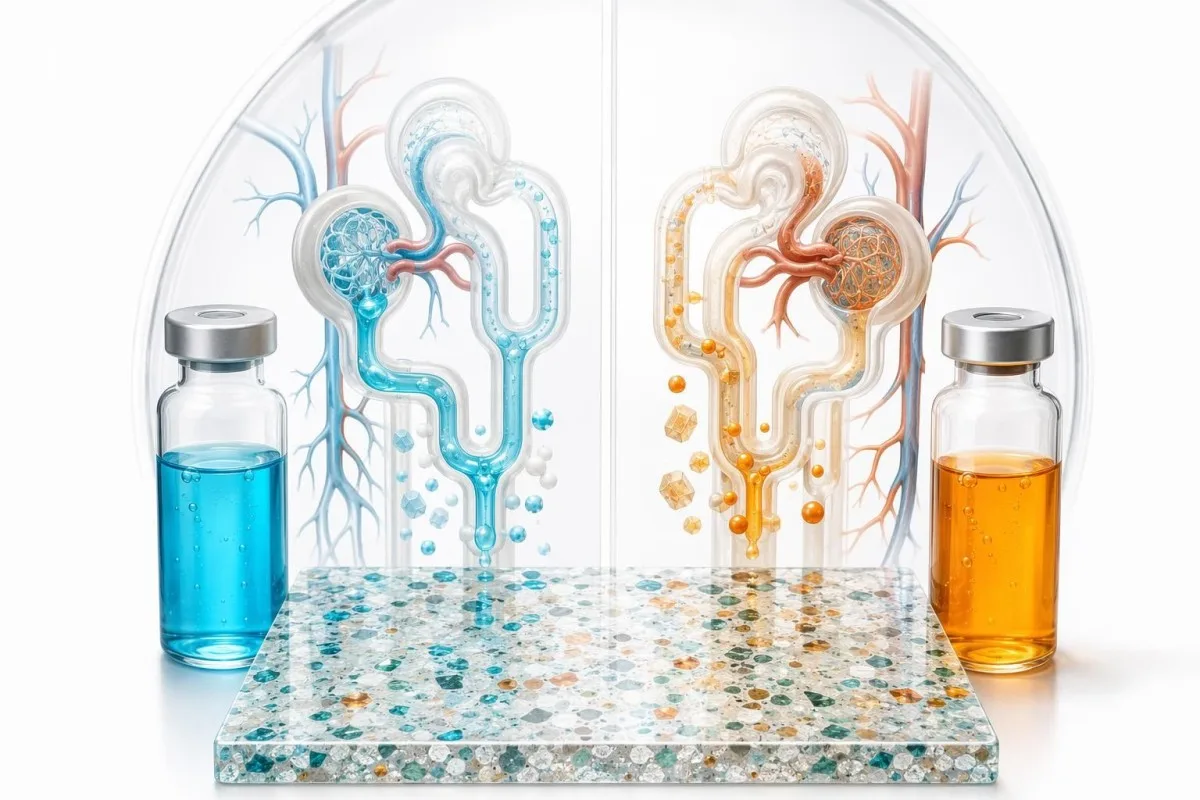
Kidney disease can alter vitamin D activation, phosphate handling, acid-base balance, and medicine clearance. Calcium should be interpreted with albumin or as ionized calcium when protein levels are unusual; a total calcium of 10.7 mg/dL may be less alarming with high albumin, whereas symptoms with calcium above 12 mg/dL require timely assessment. The basic metabolic panel guide explains the practical relationships among creatinine, bicarbonate, sodium, potassium, and calcium.
Liver enzymes are not pain markers, but their pattern can distinguish muscle release from a hepatobiliary problem. AST can rise with exercise and muscle injury; ALT is more liver-weighted, while alkaline phosphatase and GGT together suggest a cholestatic pattern when elevated. Right upper abdominal pain with jaundice, fever, pale stool, or dark urine needs urgent clinical review regardless of the exact enzyme level.
Low albumin can contribute to swelling and may indicate kidney protein loss, liver dysfunction, poor intake, or systemic illness. A urine albumin-creatinine ratio of 30 mg/g or more is abnormal and can identify kidney injury even when eGFR looks preserved. Tests should trigger a search for the cause, not just a dietary response or a repeat draw.
Joint pain: uric acid, infection clues and the test blood cannot replace
For a suddenly hot, red, swollen joint, aspiration of joint fluid is more decisive than uric acid, CRP, or rheumatoid factor. Serum urate can be normal during an acute gout flare, while a high urate level alone does not establish that gout is causing today’s pain.
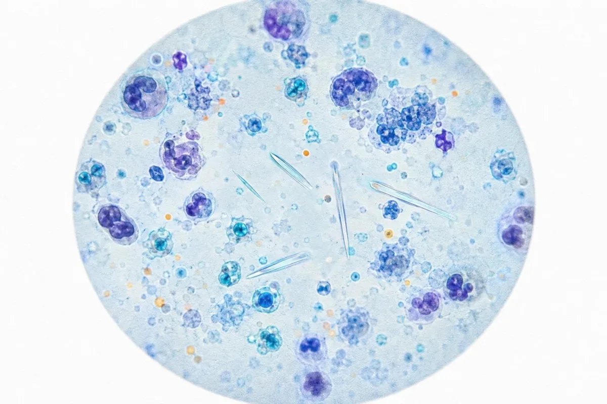
Urate above 6.8 mg/dL (404 µmol/L) exceeds its approximate solubility threshold and increases the probability of monosodium urate crystal deposition, yet many people with that level never develop gout. During a flare, the value may transiently fall as urate shifts into tissue. The most reliable confirmation remains identification of crystals in joint fluid when the diagnosis is uncertain.
Septic arthritis is a clinical emergency because joint damage can progress rapidly. Fever may be absent in older adults, people using immune-suppressing medicine, and those with diabetes; a normal peripheral white count does not rule it out. New inability to bear weight, a markedly swollen joint, fever, or recent joint procedure should prompt same-day emergency assessment rather than waiting for outpatient blood tests.
Rheumatoid arthritis more often causes persistent symmetrical small-joint swelling, whereas gout typically has abrupt attacks, commonly affecting a foot or ankle. These are tendencies, not rules. If urate is elevated outside an attack, our urate testing guide explains why kidney function, flare frequency, stones, and treatment history guide the next step.
Pain conditions blood work cannot diagnose
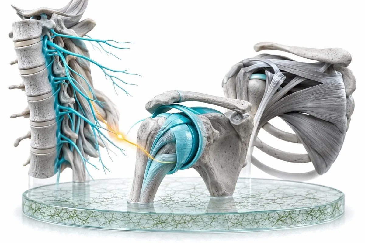
Fibromyalgia, osteoarthritis, migraine, many back-pain syndromes, tendinopathy, nerve entrapment, and most myofascial pain cannot be diagnosed from blood work. Normal blood tests should narrow the differential and reduce concern for some systemic illnesses; they should never be used to tell a patient that pain is imagined.
Fibromyalgia is a clinical diagnosis characterized by widespread pain with symptoms such as unrefreshing sleep, fatigue, cognitive difficulty, and sensory sensitivity. There is no confirmatory CRP, ANA, vitamin level, or proprietary biomarker. The EULAR recommendations describe education, graded physical activity, and individualized symptom care as central treatments, while cautioning against unnecessary testing once major mimics have been considered (Häuser et al., 2017).
Osteoarthritis is diagnosed from the story, examination, and sometimes radiography—not an “arthritis blood test.” Osteoarthritic pain commonly worsens with loading and improves with rest, while inflammatory arthritis often brings longer morning stiffness and visible swelling. But mixed disease is possible: I have cared for people with both hand osteoarthritis and rheumatoid arthritis, so new swelling changes the threshold for reassessment.
Persistent pain also changes the nervous system’s threat processing, particularly after poor sleep, trauma, infection, or prolonged stress. That mechanism is biological, not a character flaw. A thoughtful burnout and symptom lab guide can help distinguish sensible screening from indiscriminate repeat testing.
Why repeating normal panels can be harmful
Repeating the same normal tests every few weeks rarely creates clarity unless symptoms have changed or a result was near a decision threshold. It increases the chance of incidental abnormalities and may delay rehabilitation, pain education, sleep treatment, physiotherapy, or a neurological assessment. Reassessment should follow a new clue, not frustration alone.
Escalation red flags: when pain needs urgent assessment
Pain needs urgent or emergency assessment when it is paired with chest pressure, breathing difficulty, fainting, new neurological weakness, loss of bladder or bowel control, a hot swollen joint, high fever, black stools, or rapidly worsening symptoms. Blood tests may be part of emergency care, but they must not delay an ECG, imaging, examination, cultures, or specialist assessment.
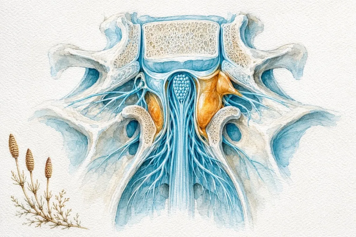
New severe back pain with saddle numbness, urinary retention, faecal incontinence, or progressive leg weakness may indicate cauda equina compression and requires emergency assessment. ESR and CRP can support investigation for spinal infection or cancer, but normal values do not make these neurological red flags safe to ignore. This is one of the clearest examples of why symptoms outrank a reassuring laboratory screen.
Headache with jaw pain, scalp tenderness, or visual disturbance in a person over 50 raises concern for giant cell arteritis. ESR can exceed 50 mm/hour and CRP may rise, but treatment decisions are clinical because visual loss can occur early. Review the symptom-and-lab combination in our article on ESR and back pain red flags if pain is severe or progressive.
Dark urine with severe muscle pain, persistent vomiting with abdominal pain, or fever with a painful swollen joint all warrant same-day advice. A D-dimer is not a general pain test: it is only useful when a clinician has already judged that a clot is plausible. The more targeted the question, the safer the result.
How exercise, illness and supplements distort pain-related results
Hard exercise, dehydration, a recent viral illness, alcohol, biotin, and supplements can change pain-related blood results enough to imitate disease or hide it. A repeat sample is often sensible when the result conflicts with the clinical picture, provided no urgent symptoms are present.
Running a marathon or starting intense resistance training can transiently raise CK, AST, creatinine, white cells, and CRP. A sample drawn 24 hours after a race may look startling without signifying organ disease; symptoms, hydration, urine findings, and the size of rise determine whether to repeat or escalate. Our article on exercise-related lab changes gives a practical recovery-time framework.
Biotin doses of 5 to 10 mg per day, common in hair and nail products, can interfere with some immunoassays and falsely alter thyroid, troponin, and hormone results. Patients should tell the laboratory and prescriber about every supplement, including creatine, high-dose vitamin D, magnesium laxatives, herbal products, and pre-workout mixtures. Do not stop prescribed medicine simply to “clean up” a panel without professional advice.
Kantesti AI compares a result with prior values, collection context, and related markers, which can help identify a biologically plausible shift versus a number that merits confirmation. A meaningful change is not always a value outside range: a creatinine rise from 0.7 to 1.2 mg/dL after dehydration may matter in context. Our lab-change comparison guide explains why laboratories use expected biological variation rather than eyeballing a graph.
How to prepare for a productive pain and blood-test review
The most useful appointment brings a pain timeline, full medication and supplement list, family history, previous results, and a clear description of what function has changed. A clinician can interpret a ferritin of 24 ng/mL or CRP of 9 mg/L far more accurately when they know whether the person has heavy periods, recent infection, endurance training, weight loss, fever, or new bowel symptoms.
Record onset date, sites of pain, morning stiffness duration, swelling, rashes, numbness, fever, weight change, sleep, and triggers such as exercise or meals. Bring exact product names and doses: “a magnesium supplement” is less useful than magnesium citrate 300 mg nightly, and “a vitamin” misses the crucial biotin dose. A pattern over 4 to 6 weeks is often more revealing than one difficult day.
Kantesti AI is an AI biomarker interpretation platform designed to organize result patterns and longitudinal changes for discussion with a clinician; it does not replace a physical examination or urgent evaluation. A result outside range may be harmless variation, while a within-range value can be meaningful if it has changed sharply from a person’s baseline. Our longitudinal results guide shows what information to save after each draw.
Avoid asking for “every test” after one unexplained result. Instead ask: What diagnosis is this test assessing? What result would change management? What false-positive result is likely? If physician review is needed, Kantesti’s Medical Advisory Board reflects the clinical oversight standards we expect around health information.
When to repeat tests, seek a referral or add imaging
Repeat testing is appropriate when a result is borderline, unexpected, affected by exercise or illness, or needed to confirm a diagnosis; referral or imaging is appropriate when symptoms persist despite reassuring labs or when examination finds an objective abnormality. As of July 28, 2026, no blood panel replaces a careful neurological, musculoskeletal, abdominal, or joint examination.

A low ferritin or raised TSH is often rechecked after treatment on a clinically appropriate timetable, commonly 6 to 12 weeks for many stable outpatient questions, not days later. CRP may be repeated within days when an acute illness is being tracked. Repeating autoimmune antibodies without a new clinical feature is usually lower value than reviewing the patient again for joint swelling, rash, weakness, or urine changes.
Imaging becomes more useful for focal pain, trauma, persistent neurological deficits, suspected inflammatory sacroiliitis, joint swelling, or pain that does not match laboratory findings. An ultrasound can demonstrate synovitis; nerve conduction testing can identify certain peripheral nerve disorders; MRI is selected for specific spine, soft-tissue, marrow, or neurological questions. These are complementary tools, not signs that the blood tests “failed.”
Dr. Thomas Klein’s advice to patients is simple: use results to sharpen the next question, not to label yourself. Kantesti’s clinical validation information describes how our interpretation approach is intended to support informed follow-up while keeping clinical decisions with qualified professionals. Seek urgent care now—not an online interpretation—if any red-flag symptom in this article is present.
A sensible next-step plan for persistent unexplained pain
For persistent unexplained pain, start with a symptom-led basic panel, use targeted tests only when the pattern supports them, and escalate quickly for red flags or objective weakness, swelling, fever, or neurological change. The aim is not to find an abnormal number at any cost; it is to identify conditions where treatment, referral, imaging, or urgent care will make a real difference.
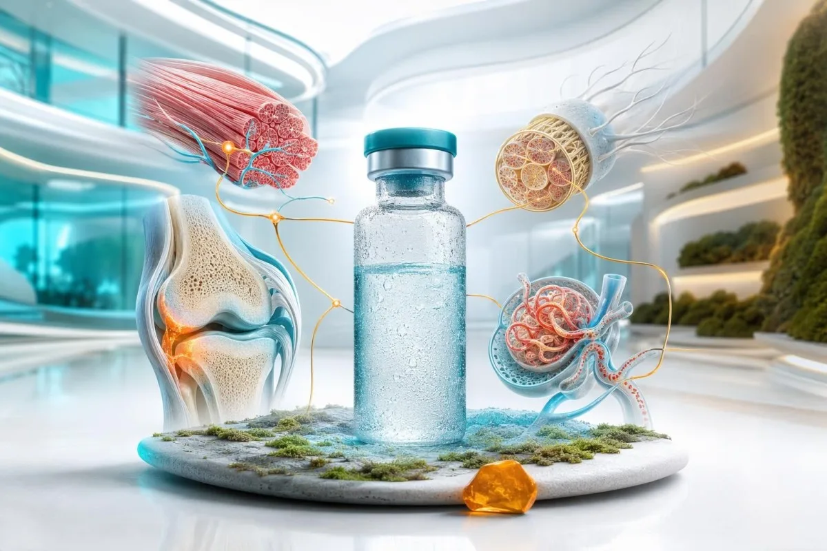
A useful first conversation usually covers CBC, ferritin when iron risk exists, renal and liver profile, calcium, glucose or HbA1c, TSH, and CRP or ESR when systemic symptoms are present. Add CK for muscle weakness or severe myalgia, B12 for neuropathic symptoms or metformin exposure, and urinalysis for kidney, infection, or autoimmune clues. This is a framework, not a self-ordering checklist.
If the first screen is normal and pain is widespread, chronic, and paired with poor sleep or fatigue, the next therapeutic step may be pain rehabilitation, graded activity, sleep support, mental-health care, or physiotherapy—not more antibodies. Most patients find this reframing relieving once it is explained properly. It acknowledges both the biology of persistent pain and the reassuring absence of dangerous systemic findings.
Kantesti provides AI-assisted result interpretation across major laboratory panels in more than 75 languages, but any worsening pain, new red flag, or result suggesting severe abnormality should be discussed directly with a clinician. Dr. Thomas Klein and our clinical team view this as the safest use of health technology: clearer questions, better follow-up, and no false reassurance.
Frequently Asked Questions
What blood tests are best for unexplained body pain?
The most useful blood tests for unexplained body pain are usually selected from a CBC, ferritin or iron studies, kidney and liver profile, calcium, glucose or HbA1c, TSH, and CRP or ESR when systemic symptoms are present. CK is appropriate for significant muscle pain or weakness, while vitamin B12 is useful for burning, tingling, or numbness. A ferritin below 15 ng/mL supports iron deficiency, and a TSH above 10 mIU/L with low free T4 supports overt hypothyroidism. The correct panel depends on the pain pattern, medicines, diet, and examination findings.
Can blood tests show why my whole body hurts?
Blood tests can identify systemic contributors to widespread pain, including anaemia, iron deficiency, thyroid disease, vitamin D deficiency, mineral disorders, kidney disease, diabetes, and inflammatory illness. They cannot diagnose fibromyalgia, most osteoarthritis, migraine, nerve compression, or many back-pain conditions. A CRP below 5 mg/L reduces the likelihood of major systemic inflammation but does not prove that pain has no physical basis. Persistent widespread pain with normal blood work still deserves a structured clinical assessment.
What does a normal CRP mean when I have joint pain?
A normal CRP means there is no clear laboratory evidence of substantial systemic inflammatory activity at the time of testing, but it does not rule out every joint condition. Osteoarthritis, tendon disorders, early inflammatory arthritis, gout between attacks, and localized joint infection can occur with a normal CRP. CRP commonly falls below 5 mg/L in healthy adults, although each laboratory sets its own reference interval. A hot swollen joint, fever, or inability to bear weight needs urgent assessment even when CRP is normal.
Can low vitamin D cause pain all over the body?
Severe vitamin D deficiency can contribute to diffuse bone discomfort, muscle aching, and proximal weakness, especially when 25-hydroxyvitamin D is below 12 ng/mL or 30 nmol/L. Vitamin D deficiency does not explain every type of chronic pain, and a low result should be interpreted with calcium, phosphate, alkaline phosphatase, kidney function, and sometimes parathyroid hormone. Symptoms usually improve gradually after appropriate correction rather than overnight. Focal severe pain, swelling, fever, or neurological symptoms require assessment for other causes.
When is a high CK level dangerous?
A CK level above 1,000 IU/L is concerning when it occurs with severe muscle pain, true weakness, dark urine, reduced urine output, fever, or a rising creatinine level. CK can rise modestly after unusual exercise, and the exact upper limit varies by laboratory, sex, muscle mass, and recent activity. Values more than 5 times the laboratory upper limit usually warrant prompt clinical interpretation and repeat kidney and electrolyte testing. Severe symptoms or brown urine require same-day urgent care because substantial muscle injury can affect kidney function.
Should I request an autoimmune panel for chronic pain?
An autoimmune panel is most useful when chronic pain is accompanied by objective swollen joints, morning stiffness lasting more than 30 to 60 minutes, rashes, mouth ulcers, Raynaud-type symptoms, eye inflammation, low blood counts, or abnormal urine. ANA and rheumatoid factor can be positive in people without autoimmune disease, particularly at low titres or with increasing age. A broad panel for non-specific pain therefore creates a real risk of false-positive results and unnecessary worry. A clinician should choose targeted antibodies after taking a detailed history and examining the joints, skin, nerves, and circulation.
Get AI-Powered Blood Test Analysis Today
Join over 2 million users worldwide who trust Kantesti for instant, accurate lab test analysis. Upload your blood test results and receive comprehensive interpretation of 15,000+ biomarkers in seconds.
📚 Referenced Research Publications
Klein, T., Mitchell, S., & Weber, H. (2026). Kantesti LTD. (2026). A Pre-Registered, Rubric-Based Automated Technical Benchmark of the Kantesti Blood-Test Interpretation Engine on 100,000 Synthetic Test Cases. Figshare.. Kantesti AI Medical Research.
Klein, T., Mitchell, S., & Weber, H. (2026). Kantesti LTD. (2026). Clinical Validation Framework v2.0 (Medical Validation Page). Zenodo.. Kantesti AI Medical Research.
📖 External Medical References
National Institute for Health and Care Excellence (2021). Chronic pain (primary and secondary) in over 16s: assessment and management. NICE Guideline NG193.
⚕️ Medical Disclaimer
This article is for educational purposes only and does not constitute medical advice. Always consult a qualified healthcare provider for diagnosis and treatment decisions.
E-E-A-T Trust Signals
Experience
Physician-led clinical review of lab interpretation workflows.
Expertise
Laboratory medicine focus on how biomarkers behave in clinical context.
Authoritativeness
Written by Dr. Thomas Klein with review by Dr. Sarah Mitchell and Prof. Dr. Hans Weber.
Trustworthiness
Evidence-based interpretation with clear follow-up pathways to reduce alarm.