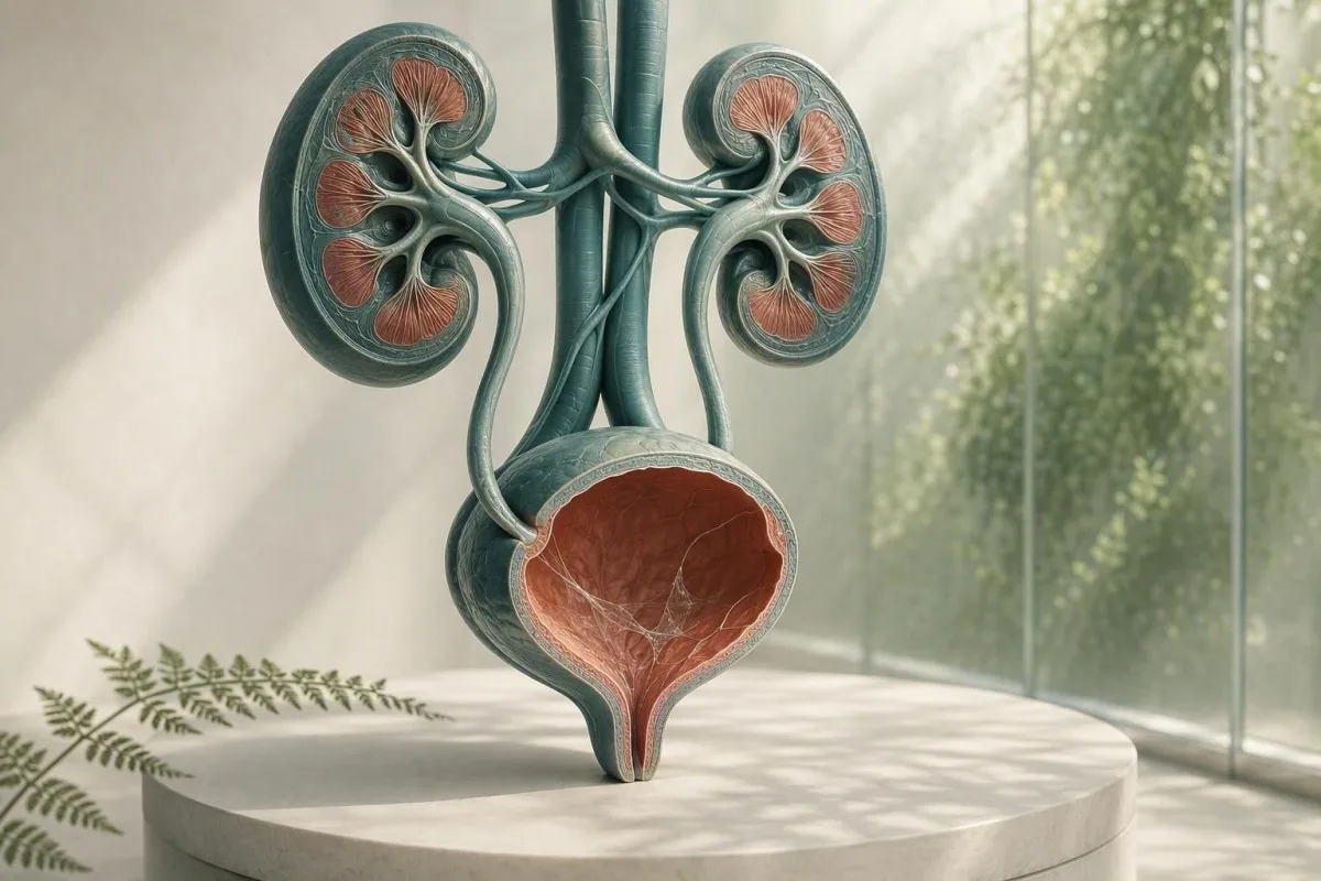
İdrar mikroskopisi raporundaki mukus genellikle bir toplama sorunudur, tanı değildir. İdrar tahlili bulguları, semptomlar ve örneğin kalitesi, takip gerektirip gerektirmediğini belirler.


- Mukus iplikçikleri tek başına, özellikle idrar semptomları olmayan tek bir temiz temizleme örneğinde yaygın olarak zararsızdır.
- Piyüri eşiği mikroskobik sahada 5'ten fazla beyaz küre, idrar yolu iltihabını destekler ancak bakteriyel İYE'yi kanıtlamaz.
- Kültür tetikleyicisi mukus, yanma, sık idrara çıkma, ateş, yan ağrısı, nitritler veya belirgin beyaz kürelerdir—mukus tek başına değildir.
- Yassı hücreler mikroskobik sahada kabaca 10'un üzeri sıklıkla genital deri kontaminasyonunu gösterir ve idrar sonucunu daha az güvenilir hale getirir.
- Görünür kan kolik yan ağrısı olan bir durum, mukus da rapor edilmiş olsa bile taş için acil değerlendirme gerektirir.
- Gebelik idrar kültürü ile, yalnızca mikroskopi ile değil, asemptomatik bakteriyüriye müdahale eşiğini değiştirir.
- Bulutlu idrar kristal, konsantre idrar, vajinal akıntı, meni veya hücreleri yansıtabilir; görünümüne göre enfeksiyon teşhisi koyamaz.
- Tekrar örnek mukus tek olağandışı bulgu olduğunda ve örneklemesi dikkatli bir orta akım temiz yakalama olmadığında mantıklıdır.
İdrar tahlilinde mukus iplikçikleri genellikle ne anlama gelir?
İdrardaki mukus iplikçikleri genellikle normal koruyucu salgının veya genital kontaminasyonun iplikçikleridir ve izole bir sonuç nadiren böbrek hastalığını işaret eder. Bulgu, idrar semptomları, beyaz kan hücreleri, bakteri, kan veya protein ile birlikte göründüğünde anlamlı hale gelir.
Laboratuvarlar, mikroskop altında görülen gevşek, yarı saydam iplikçikler olarak mukusu tanımlar; birçok rapor, sayısal bir referans aralığı atamak yerine sadece nadir, az, orta veya çok olarak derecelendirir. İdrar doğal olarak idrar yollarından gelen az miktarda glikoprotein açısından zengin malzeme içer ve toplama sırasında vajinal veya üretra salgıları daha fazlasını ekleyebilir.
Raporun ifadesi kulağa geldiğinden daha az endişe vericidir. 26 Ağustos 2026 itibarıyla, İYE, taş, kanser veya böbrek yetmezliğini teşhis eden doğrulanmış bir mukus iplikçik sayısı yoktur.; bu bağlamsal bir mikroskobik gözlemdir. Bir çubuk test ve mikroskop raporundaki diğer öğeler için daha geniş bir harita için bkz. idrar tahlili kılavuzunun tamamı.
Klinik deneyimimde, “orta” olarak işaretlenmiş mukusa sahip, sahada 0-2 beyaz küre, negatif nitrit ve kanı olmayan, semptomsuz bir yetişkin, genellikle güvence veya usulüne uygun toplanmış bir tekrar örnek ile en iyi şekilde hizmet edilir. Dr. Thomas Klein, bir laboratuvar bayrağının bir hastalık etiketiyle karıştırılması nedeniyle gereksiz antibiyotik kaygısının ortaya çıktığını sıkça görmektedir.
Kantesti, kreatinin ve eGFR gibi böbrekle ilgili kan belirteçlerini bir idrar tahlili bulgusunun yanına yerleştirebilen bir AI kan tahlili yorumlama platformudur, ancak yalnızca mukustan İYE teşhisi koyamaz. Bir idrar mikroskopisi sonucu, onunla birlikte gelen klinik hikayeyi gerektirir.
Bir laboratuvar neden bunu işaretler
Laboratuvar bilgi sistemleri, mukusu görsel olarak mevcut olduğu için işaretler, laboratuvar tehlikeli bir kesinti belirlemiş olduğu için değil. Bu nedenle bir işaret, “gözlemlendi” anlamına gelir, “anormal hastalık belirteci” anlamına gelmez, tıpkı izole bir iz bulgusunun tedavi gerektirmemesi gibi.
İdrar örneğindeki mukus nereden gelir?
İdrardaki mukus üretra, mesane astarı, rahim ağzı veya vajina, meni veya toplama sırasında dış genital deriden kaynaklanabilir. Kaynağı, iplikçiklerin kendisinden ziyade genellikle mikroskobik desenin geri kalanından çıkarılır.
Mesane ve üretra, sürtünmeyi ve mikrobiyal yapışmayı azaltan, üroplakin ile ilişkili malzeme dahil olmak üzere mukus içeren koruyucu bir yüzey tabakasına sahiptir. Dehidrasyon, yakın zamanda cinsel aktivite, hafif tahriş veya sadece konsantre bir ilk sabah örneğinin iplikçiklerin daha kolay görülmesini sağlaması nedeniyle küçük miktarlar idrara dökülebilir.
Adet gören veya vajinal akıntısı olan kişilerde, mesaneden gelmeden lökositler ve mukus kaba girebilir. Sahada 10'dan fazla yassı epitel hücresi kontaminasyonu daha olası hale getirir, ancak laboratuvarlar farklı raporlama eşikleri kullanır. Rehberimiz idrarda epitel hücreleri bunun bir sonucun güvenilirliğini neden değiştirdiğini açıklar.
Meni de boşalma sonrası birkaç saat boyunca iplikli malzeme veya bulanıklık oluşturabilir. Bu idrarı güvensiz yapmaz, ancak mikroskobiyi engelleyebilir; kültür düşünülüyorsa, genellikle mümkün olduğunda en az 24 saat sonra yeni bir örnek toplamanızı tavsiye ederim.
Cinsiyetin ve anatomisinin raporu neden etkilediği
Bir idrar kabı mesaneyi yakındaki dokulardan izole etmez. Bu nedenle, dış salgıların olduğu örneklerde “mukus var” daha sık rapor edilirken, klinisyenlerin daha temiz bir yanıt istediği durumlarda bazen sonda ile alınmış örnek kullanılır.
Mukus iplikçikleri ne zaman idrar yolu enfeksiyonunu işaret eder?
Mukus iplikçikleri, yalnızca semptomlar ve özellikle piyüri, nitrit pozitifliği veya ikna edici bir kültür ile birlikte bulunduğunda olası bir İYE'yi destekler. Mukusun tek başına bir İYE testi değildir.
Semptomatik alt idrar yolu enfeksiyonu (İYE) genellikle yanma, sıkışma, sık idrara çıkma, suprapubik rahatsızlık ve bazen de yeni idrar kokusuna neden olur. Düzgün toplanmış bir örnekte, yüksek güçlü bir sahada 5'ten fazla beyaz kan hücresi sıklıkla piyüri olarak adlandırılır; idrar yolu iltihabını destekler ancak taşlar, cinsel yolla bulaşan enfeksiyonlar ve kontaminasyon ile de ortaya çıkabilir.
Nitrit pozitif olduğunda oldukça spesifiktir ancak nitratı azaltmayan organizmalardan kaynaklanan enfeksiyonları kaçırır ve negatif bir nitrit sonucu İYE'yi dışlamaz. Lökosit esteraz, beyaz hücre enzim aktivitesini tespit eder ve vajinal salgıların örneği kirletmesi durumunda yanlış pozitif olabilir; lökosit esteraz sonuçları altına düşmesi görülebilir; bu tuzakları.
IDSA kılavuzu, antibiyotiklerin fayda sağlamadan zarar vermesi nedeniyle sağlıklı, gebe olmayan yetişkinlerde asemptomatik bakteriüri taraması veya tedavisini önermez (Nicolle ve ark., 2019). Bu prensip özellikle mukus için geçerlidir: yanma, ateş ve planlanmış bir ürolojik işlem yoksa, mikroskopi alanının dağınık görünmesi nedeniyle genellikle basitçe antibiyotik verilmez.
Kültürün daha iyi bir test olduğu durumlar
Semptomlar devam ederse, 4 hafta içinde semptomlar tekrarlarsa, gebelik varsa, piyelonefrit şüphesi varsa veya önceki antibiyotikler sonucu etkilemiş olabileceğinden, idrar kültürü tekrarlanan dipstik testinden daha faydalıdır. Yaygın bakteriyel üreme genellikle tek bir idrar patojeninden ziyade toplama kontaminasyonunu gösterir; idrar kültürü yorumlamamız.
Mukus, kristaller, taşlar ve mekanik irritasyon
Şiddetli dalgalı yan ağrısı veya idrarda kan ile birlikte mukus, idrar taşı ile oluşabilir, ancak mukus taşı ne doğrular ne de dışlar. Kan, kristaller, ağrı paterni, görüntüleme ve böbrek fonksiyonu daha önemlidir.
Bir taş idrar astarını sıyırabilir veya tıkayabilir, kırmızı kan hücreleri, beyaz kan hücreleri ve ek mukus üretebilir. Klasik semptom, sıklıkla bulantı eşliğinde kasığa doğru yayılan ani kramp tarzı ağrıdır; görünür kırmızı veya çay renkli idrar aciliyeti artırır. Hakkında daha fazla bilgi edinin idrarda kan kırmızı bayrakları her pembe örneğin basit bir enfeksiyon olduğunu varsaymak yerine.
Kalsiyum oksalat kristalleri, özellikle konsantre asidik idrarda sağlıklı insanlarda görülebilir, bu nedenle tek bir kristal türü aktif bir taşı kanıtlamaz. Ağrısı olan, tekrarlayan taşları olan veya sürekli hematürisi olan kişilerde, klinisyenler sedimantasyona güvenmek yerine ultrason veya düşük doz kontrastlı olmayan BT kullanabilirler. kalsiyum oksalat kristalleri kristal raporlarının sınırlarını ayrıntılı olarak açıklar.
Önemli bir istisna enfeksiyonla birlikte tıkanmadır: 38.0°C veya daha yüksek ateş, yan ağrısı, kusma ve idrar yapamama acil değerlendirmeyi gerektirir. Endişe mukus değil; enfekte olmuş, tıkanmış bir idrar sistemidir ve bu hızla kötüleşebilir.
Taşsız tahriş
Son kateter kullanımı, mesane prosedürleri, yoğun bisiklete binme ve pelvik radyasyon alt idrar yolunu tahriş edebilir ve mukus veya beyaz hücreleri artırabilir. Bir klinisyen, bu sonuçları zamanlamaya karşı yorumlamalıdır, çünkü bir işlemeden sonraki 48 saat içinde toplanan bir örnek, rutin bir tarama örneğiyle eşdeğer değildir.
Bulanık idrar anlamı: görünüm size ne söyleyebilir ve ne söyleyemez
Bulanık idrar, konsantre tuzlar, kristaller, mukus, hücreler, genital akıntı veya bakterilerden kaynaklanabilir, bu nedenle görünüm tek başına enfeksiyonu teşhis edemez. Bulanık ancak ağrısız ve kısa süreli idrar genellikle acil değildir.
Fosfat kristalleri, soğuduktan sonra alkali idrarı bulutlu gösterebilirken, ürik asit kristalleri asidik ve konsantre idrarı bulutlandırabilir. Oda sıcaklığında 2 saatten fazla bekletilmiş bir örnek, hücrelerin bozulması ve bakterilerin çoğalmasıyla giderek daha bulanık hale gelebilir, bu nedenle taze işleme veya soğutma önemlidir.
Bulanıklık ile dizüri, sık idrara çıkma ve piyüri, test gerektirir; egzersiz sonrası veya yetersiz sıvı alımı sonrası bulanıklık genellikle normal hidrasyonla düzelir. İdrar yolu enfeksiyonunu “temizlemek” anlamına gelmediği için aşırı su alımını zorlamak yerine açık sarı renkte idrar hedefleyin. Detaylı incelememiz bulanık idrar nedenleri görsel ipuçlarını güvenilir testlerden ayırır.
Kantesti bir AI kan testi analizörü kan belirteçlerini klinik bağlamda yorumlamak için tasarlanmıştır; yüksek kreatinin veya azalmış eGFR, idrar anormalliklerinin yanında tıbbi incelemeyi haklı çıkarabilirken, tek başına bulanık idrar, böbrek filtrasyonunun bozulduğunu göstermez. Dehidrasyon veya tıkanıklık belirtileri olduğunda temel metabolik panel faydalı bir bağlam ekleyebilir.
Koku bir kültür değildir
Güçlü kokulu idrar genellikle konsantrasyonu, kuşkonmaz metabolitlerini, B vitaminlerini veya çok uzun süre bekletilmiş bir kabı yansıtır. Ateş veya idrar semptomları ile birlikte yeni, kötü bir koku değerlendirme sebebidir, ancak koku mikroskopi ve kültürün yerini tutmaz.
Genital akıntı ve cinsel yolla bulaşan enfeksiyonlar
Üretral veya vajinal akıntı, idrardaki mukusa benzeyebilir ve idrar yanması ile birlikte yeni akıntı cinsel sağlık testi ile idrar değerlendirmesini gerektirir. Rutin idrar kültürü klamidya ve gonore'yi gözden kaçırabilir.
Klamidya ve gonore, dizüriye ve steril piyüriye neden olabilir - rutin bakteri üremesi olmayan beyaz küreler - özellikle yeni bir cinsel temastan sonra. Nükleik asit amplifikasyon testi, genellikle orta akım örneği yerine ilk yakalama idrarını kullanarak, standart kültür farklı organizmaları hedeflediği için uygun testtir.
Vajinası olan kişilerde bakteriyel vajinozis, kandidiyazis ve serviks iltihabı orta akım kabına akıntı ekleyebilir; vajinal sürüntü veya muayene, idrar mikroskobisini tekrarlamaktan daha bilgilendirici olabilir. Penisi olan kişilerde, görünen üretral akıntı “mukus iplikçikleri” olarak göz ardı edilmemelidir. Pratik ayrım, idrara çıkma dışında fark edilen akıntı ile yalnızca laboratuvar tarafından bildirilen iplikçikler arasındadır.
2021 CDC Cinsel Yolla Bulaşan Hastalıklar tedavi kılavuzu, yalnızca semptomlara değil, maruz kalma geçmişine göre ilgili anatomik bölgelerde NAAT tabanlı test önerir (Workowski et al., 2021). Pelvik ağrı, testis ağrısı, ateş, gebelik veya olası saldırı söz konusuysa, evde bırakılmış antibiyotiklerle kendi kendine tedavi yerine aynı gün tıbbi yardım alın.
Antibiyotikler neden resmi karmaşıklaştırabilir
Kültürden önce sadece 1 veya 2 doz antibiyotik almak, idrar semptomları ve beyaz küreler devam ederken normal bakteri üremesini baskılayabilir. Klinisyene tam olarak hangi ilacı, dozu ve son doz zamanını kullandığınızı bildirin; bu ayrıntı negatif bir kültürün nasıl yorumlandığını değiştirir.
Soruyu yanıtlayan bir idrar örneği nasıl toplanır?
Orta akım temiz yakalama örneği, akımın ilk veya son kısmını toplamaktan daha iyi mukus, yassı hücre ve karışık bakteri üremesini azaltır. Teknik basittir, ancak ilk 2 saniye orantısız bir fark yaratır.
Ellerinizi yıkayın, genital deriyi ayırın veya rahatsanız sünnet derisini geri çekin, kitin talimatlarına göre temizleyin, idrar yapmaya tuvalete başlayın, ardından kabın deriye temas etmemesi için orta kısmı toplayın. Genellikle 20-30 mL'lik bir örnek yeterlidir; büyük bir kabı ağzına kadar doldurmak testin sonucunu iyileştirmez.
Kabı derhal, tercihen 1 saat içinde teslim edin; gecikme kaçınılmazsa, laboratuvarın talimatlarına göre soğutun ve 24 saat içinde iade edin. Soğutma, bakteri üremesini yavaşlatır ancak zaten kontamine olmuş bir örneği geri kazandırmaz. Bu, özellikle talep yalnızca dipstick yerine kültür içerdiğinde önemlidir.
Kantesti bir Yapay zekâ destekli kan testi analiz aracı ülke genelinde kullanılır ve klinisyen tarafından incelenen iş akışlarımız, numune kalitesini bir dipnot yerine yorumun bir parçası olarak ele alır. Aynı prensip herhangi bir laboratuvar değeri için de geçerlidir: teknik olarak zayıf bir örnek, tıbbi olarak ikna edici ancak yanıltıcı bir sonuç yaratabilir. İnceleyin laboratuvar doğruluğu kontrol listesi tek başına bir uyarıya dayanarak hareket etmeden önce sorulacak sorular için.
Bekleyebilirseniz bu durumlarda numune almayın
Ağır adet kanaması sırasında, cinsel ilişkiden hemen sonra veya vajinal kremler kullandıktan sonra, klinisyen özellikle istemedikçe rutin testlerden kaçının. Testin ertelenemeyeceği durumlarda, laboratuvara veya klinisyene bildirin, çünkü bu bağlam mukus, kırmızı kan hücreleri veya dış hücreleri açıklayabilir.
Klinisyenler mukusu idrar tahlilinin geri kalanıyla nasıl okur?
En yararlı idrar tahlili paterni, semptomları beyaz küreler, kırmızı küreler, nitrit, lökosit esteraz, protein, glikoz, özgül ağırlık ve epitel hücreleri ile birleştirir. Mukoza bu desende küçük bir destekleyici özelliktir.
Mukoza, alan başına 0-2 beyaz küre, negatif nitrit ve kan içermeyen temiz bir örnek genellikle tedavi gerektirmez. Alan başına 5 beyaz küreden fazla mukoza ve pozitif lökosit esterazı, inflamasyon olasılığını artırır; nitrit veya tek organizmalı kültür eklenmesi, bakterilerin sorumlu olduğuna dair güveni artırır.
Protein kendi yoluna ihtiyaç duyar. Ateş, egzersiz veya konsantre idrardan sonra iz protein geçici olabilir, ancak kalıcı protein mukoza ile ilişkilendirilmek yerine bir albümin-kreatinin oranı ile ölçülmelidir. Rehberimiz idrarda protein çubuk proteinin ve böbrek riskinin birbirinin yerine kullanılamayacağını açıklar.
Özgül ağırlığı 1.003 ila 1.030 yetişkinlerde yaygındır, ancak referans aralıkları laboratuvarlara göre değişiklik gösterir. Yüksek özgül ağırlık, mukusu konsantre edebilir ve daha dramatik görünümlü bir sediman oluşturabilirken, düşük özgül ağırlık hücreleri lize edebilir ve mikroskopiyi aldatıcı derecede yumuşak hale getirebilir; özgül yoğunluk kılavuzumuzda hidrasyonun yorumlamayı nasıl etkilediğini gösterir.
Negatif bulguların değeri
Belirtisi olmayan bir kişide negatif kan, protein, nitrit ve lökosit esterazı, mukoza orta olarak rapor edilse bile endişeyi anlamlı derecede azaltır. Tek bir negatif test mükemmel değildir, ancak bu küme mukus derecesi kadar endişe verici değildir.
Test ne zaman tekrarlanmalı ve kültür ne zaman istenmeli?
Mukoza izole olduğunda, skuamöz hücreler kontaminasyonu düşündürdüğünde veya toplama aceleye getirildiğinde idrar örneğini tekrarlayın; belirtiler kalıcı, tekrarlayan, şiddetli veya yüksek riskli olduğunda kültür isteyin. Tekrar test “hiçbir şey yapmamak” değildir - genellikle en tanısal sonraki adımdır.
Belirtisi olmayan ve sadece mukus işareti olan gebe olmayan bir yetişkin için, güvenceye ihtiyaç duyuluyorsa 1-2 hafta içinde tekrarlanan temiz-yakalama idrar analizi makuldür; birçok klinisyen hiç tekrarlamaz. Bir reçete yazan klinik bir endikasyon belirlemedikçe, laboratuvar bayrağını bekleme süresince antibiyotiklerle tedavi etmeyin.
Ateş, yan ağrısı, gebelik, immünosupresyon, böbrek nakli, idrar kateteri kullanımı, erkek idrar semptomları veya 4 hafta içinde geri dönen semptomlar için antibiyotiklerden önce kültür özellikle faydalıdır. Gupta ve ark. (2011) tarafından yapılan kılavuz, şüpheli piyelonefrit ve direncin daha olası olduğu veya alternatif bir teşhisin daha olası olduğu durumlarda kültürü desteklemektedir.
Kültür sayımları toplama kalitesiyle okunmalıdır. Tek bir organizma mililitre başına 10^5 koloni oluşturan birim temiz orta akım idrarında geleneksel olarak bakteriüriyi desteklemiştir, ancak semptomatik hastalarda klinik olarak anlamlı düşük sayılar olabilir; “karışık flora” genellikle geniş spektrumlu bir antibiyotik yerine yeni bir örnek alınmasını gerektirir. Amacı karşılaştırın idrar tahlili ve kültür ikisinden birini istemeden önce.
Gebelik farklı bir eşik gerektirir
Gebelik bir istisnadır çünkü asemptomatik bakteriüri, piyelonefrit ve olumsuz gebelik sonuçları riskini artırabilir. Gebelik öncesi bakım, gebeliğin erken dönemlerinde yaygın olarak bir tarama kültürü kullanır; mikroskopide mukus bu kültürü değiştiremez.
Kan testleri ne zaman yararlı böbrek ve enfeksiyon bağlamı ekler?
Mukus idrarla birlikte ateş, yan ağrısı, tekrarlayan enfeksiyonlar, şişlik, idrar çıkışının azalması veya sürekli protein veya kan ile birlikte olduğunda kan testleri faydalıdır. Kreatinin ve eGFR filtrasyonu değerlendirir, tam kan sayımı (CBC) ve C-reaktif protein (CRP) sistemik hastalığın değerlendirilmesine yardımcı olabilir.
eGFR değeri En az 3 ay boyunca 60 mL/min/1.73 m² kronik böbrek hastalığı için bir kriteri karşılar, ancak dehidratasyon veya akut hastalık sırasında tek bir düşük sonuç bu tanıyı koymak için yeterli değildir. Kreatinin kas kütlesinden, diyetten ve bazı ilaçlardan etkilenir, bu nedenle tek bir sayıdan ziyade eğilimler ve idrar albümini genellikle daha bilgilendiricidir.
Yüksek beyaz kan hücresi sayımı veya CRP, inflamatuar bir süreci destekleyebilir ancak idrar yollarının kaynak olarak belirlenmesini sağlayamaz. Yan ağrısı olan ateşli bir kişide, özellikle kusma veya tıkanıklık böbrek fonksiyonunu bozabilecekse, klinisyenler görüntüleme veya ilaç seçimi yapmadan önce kreatinin düzeyini kontrol edebilir. Gözden geçirin kronik böbrek hastalığı evreleri klinisyenlerin kullandığı eGFR ve albümin kategorileri için.
Kantesti bir AI laboratuvar testi yorumlama hizmetinde yayımlanır. tek bir mukus notasyonunu böbrek tanısı olarak ele almak yerine kreatinin, eGFR, elektrolitler, tam kan sayımı (CBC) ve inflamatuar belirteçlerdeki kalıpları vurgulayan. Metodolojimiz klinik doğrulama denetimine tabidir, ancak acil semptomlar hala doğrudan tıbbi bakım gerektirir, uygulama tabanlı yorumlama değil.
Pratik bir dehidratasyon ayrımı
Dehidratasyon, özellikle ishal, ısı maruziyeti veya yoğun egzersizden sonra idrar spesifik gravitesini yükseltebilir ve kreatinini geçici olarak artırabilir. Tekrarlayan evde test yerine, yeniden hidrasyondan sonra kalıcı düşük idrar çıkışı, baş dönmesi veya eGFR düşüşü klinisyen değerlendirmesini gerektirir.
Tekrar testi beklemeden acil dikkat edilmesi gereken uyarı işaretleri
38.0°C veya daha yüksek ateş, yan ağrısı, kusma, görünen kan, idrar yapamama, kafa karışıklığı veya hamilelikle ilişkili idrar semptomları ile idrarda mukus için aynı gün acil değerlendirme isteyin. Bu kombinasyonlar önemlidir çünkü üst yol enfeksiyonu, tıkanıklık veya başka bir akut durumu işaret edebilirler.
Ateş artı tek taraflı sırt veya yan ağrısı, özellikle titreme veya kusma ile birlikte basit sistitten daha fazla piyelonefrit için endişe vericidir. Gecikmiş tedavi dehidrasyon, sepsis veya böbrek stresine yol açabilir; evde normal görünen bir idrar örneği bunu güvenli bir şekilde dışlamaz.
Görünür kan, özellikle 35 yaşından sonra, sigara içenlerde veya enfeksiyon semptomları dindiğinde kanama devam ederse, bir İYE makul görünse bile değerlendirilmelidir. AUA mikrohematüri kılavuzu, benign açıklamalar ele alındıktan sonra riske dayalı değerlendirme önerir (Barocas ve ark., 2020); kalıcı kan, mukus ile açıklanamaz.
Çocuklar, 65 yaşın üzerindeki yetişkinler, diyabet hastaları, bağışıklık sistemini baskılayan ilaçlar kullananlar ve tek böbreği olanlar değerlendirme için daha düşük bir eşiği hak eder. Dr. Thomas Klein'ın pratik kuralı basittir: semptomlar 6-12 saat içinde artıyorsa, güvence sağlamak için ikinci bir bardak için beklemeyin.
Şiddetli hastalık için şimdi acil servisi arayın
Yeni kafa karışıklığı, bayılma, şiddetli halsizlik, mavi veya gri dudaklar, şiddetli nefes darlığı veya idrar semptomları ile sıvıları tutamama durumunda acil servisi arayın. Bunlar rutin İYE semptomları değil, sistemik tehlike işaretleridir.
Hamilelik, çocukluk ve ileri yaşlarda idrarda mukus
Gebelik, çocuklar ve yaşlı yetişkinler daha dikkatli yorumlama gerektirir çünkü kontaminasyon yaygındır ancak enfeksiyonun gözden kaçırılmasının sonuçları daha büyük olabilir. Her yaş grubunda balgam derecesinden daha değerli olan belirtiler ve kültür kalitesidir.
Gebelik sırasında, sık idrara çıkma normal olabilir, bu da belirtileri daha az spesifik hale getirir; ateş, dizüri veya sırt ağrısı, acil obstetrik veya klinik değerlendirmeyi gerektirmelidir. Tarama kültürü tipik olarak prenatal bakımın erken dönemlerinde alınır ve tekrarlayan kültür, klinisyenin planına bağlı olarak tedavi sonrası kullanılabilir.
Çocuklarda torba ile toplanan idrar kontaminasyona yatkındır ve genellikle tek başına kültür ile İYE tanısı koymak için kullanılmamalıdır. Belirgin bir nedeni olmayan ateşi olan küçük bir çocukta kateterle alınmış veya dikkatlice alınmış temiz orta akım idrar örneği gerekebilir; torba örneğindeki balgam özellikle spesifik değildir.
İlerleyen yaşlarda, belirti vermeden bakteriüri ve piyüri daha yaygın hale gelir. Tek başına yeni gelişen deliryum, otomatik olarak İYE tedavisi yerine dehidrasyon, ilaçlar, ağrı, kabızlık ve enfeksiyon kaynakları için geniş bir tıbbi değerlendirmeyi tetiklemelidir; Nicolle ve ark. (2019) tarafından önerilen aynı kısıtlama geçerlidir.
Adet ve hormonla ilgili değişiklikler
Adet kanı ve servikal mukus, birkaç gün boyunca idrar tahlilini etkileyebilir ve menopoz sonrası vajinal kuruluk, idrar yanmasına benzeyen lokal tahrişe neden olabilir. Sonuçlar ve belirtiler uyumlu değilse, bir klinisyen tekrarlayan İYE tedavisi reçete etmek yerine genital nedenleri değerlendirebilir.
Laboratuvar raporunda mukus görüldükten sonraki yaygın hatalar
Bir anormallik gösteren idrar tahlilinden sonra artan antibiyotikleri kullanmayın, içten temizlemeyin veya aşırı su alımıyla balgamı “temizlemeye” çalışmayın. Bu eylemler kültürü gizleyebilir, normal florayı bozabilir veya doğru tanıyı geciktirebilir.
Kültür olmadan alınan antibiyotikler, sonraki bir testi yanlış negatif yapabilir ve ishal, döküntü, maya belirtileri veya direnç neden olabilir. Belirtiler hafif ancak kalıcıysa, mümkün olduğunca önce örneği alın, ardından tüm resmi göz önünde bulundurarak bir klinisyenin tedavi planını izleyin.
Kızılcık ürünleri bazı kişilerde tekrarlayan komplikasyonsuz İYE riskini mütevazı bir şekilde azaltabilir, ancak ateş, yan ağrısı veya doğrulanmış üst yol enfeksiyonunu tedavi etmezler. Kendi kendine tedavi olarak yüksek doz C vitaminden kaçının: bazı dipstick reaksiyonlarını değiştirebilir ve kalsiyum oksalat taşına yatkın kişilerde oksalat yükünü artırabilir.
Sıklıkla tuvalet kasesini lifler için kontrol etmeyin. Tuvalet kağıdı lifleri, temizlik kalıntıları, genital salgılar ve su türbülansı balgamı taklit edebilir; steril bir kapta toplanan laboratuvar örneği, onu değerlendirmek için uygun yerdir. İlgili renk değişiklikleri için, bizim idrar renk rehberimiz daha güvenilir görsel bağlam sunmaktadır.
İlaç listesi önemlidir
Fenazopiridin idrarı turuncuya çevirebilir ve görsel yorumlamayı etkileyebilir, diüretikler ise zamanlamaya bağlı olarak idrarı konsantre edebilir veya seyreltebilir. Randevuya reçeteli ilaçların, reçetesiz satılan ürünlerin ve takviyelerin bir listesini, mg cinsinden dozları dahil olmak üzere getirin.
İdrardaki mukus iplikçikleri için pratik bir takip planı
Balgam iplikçikleri olan ve belirti göstermeyen çoğu kişiye tedavi gerekmez; örnek şüpheliyse dikkatli bir tekrarlayan idrar tahlili makuldür. Belirtiler veya eşlik eden anormallikler, kültür, CYBE testi, görüntüleme veya kan testlerinin uygun olup olmadığını belirler.
Adım 1: yanma, aciliyet, ateş, yan ağrısı, görünen kan, akıntı, gebelik, son idrar yolu prosedürleri ve yeni cinsel maruziyeti kontrol edin. Adım 2: raporu beyaz küreler, nitrit, lökosit esteraz, kırmızı küreler, protein, bakteri ve skuamöz hücreler için okuyun — sadece balgam için değil.
Adım 3: kendinizi iyi hissediyorsanız ve balgam izole ise, yalnızca klinisyeniniz onay önereirse 1-2 hafta içinde bir orta akım temiz yakalama örneği toplayın. Adım 4: belirtiler varsa, tedavi öncesinde idrar kültürü alınması gerekip gerekmediğini ve CYBE testinin ilgili olup olmadığını sorun; tekrarlayan ataklar, üçüncü bir ampirik antibiyotikten daha dikkatli bir gözden geçirmeyi hak eder.
Kantesti, tıbbi bir randevu için ilgili kan sonucu eğilimlerini ve soruları organize edebilir, oysa bizim Tıbbi Danışma Kurulu klinisyen liderliğindeki güvenlik standartlarını desteklemektedir. Doğru sonuç, mükemmel derecede “temiz” bir mikroskopi raporu değildir - bu, belirtilerinize, örnek kalitenize ve risk faktörlerinize uyan bir açıklamadır.
Randevunuza götürmeniz gereken sorular
Numunede skuamöz hücreler olup olmadığını, kültürün bir organizma mı yoksa karışık flora mı ürediğini ve belirtiler düzeldikten sonra kan veya proteinin devam edip etmediğini sorun. Bu 3 soru, görülen balgam miktarı sormaktan genellikle daha faydalı cevaplar üretir.
Araştırma bağlamı ve mukus raporlamanın sınırları
Laboratuvarlar arasında mukus derecelendirmesi standardize edilmemiştir, bu nedenle klinisyenler “az”, “orta” veya “çok” ifadelerini hastalık şiddeti kategorileri olarak kullanmaktan kaçınmalıdır. Mikroskopi tekniği, örnek yaşı ve raporlama yazılımı, kelimelerin ifade ediliş biçimini değiştirebilir.
Bazı laboratuvarlar yaklaşık 10-15 mL idrarı santrifüj ettikten sonra sedimenti manuel olarak incelerken, diğerleri seçilen bayrakların manuel olarak gözden geçirilmesiyle otomatik partikül analizi kullanır. Bu varyasyon, bir laboratuvardan gelen “orta mukus” sonucunun, kişinin sağlığı değişmemiş olsa bile başka bir laboratuvarda tam olarak tekrarlanamayacağı anlamına gelir.
Faydalı bir laboratuvar anlayışı, bir sonucun analitik olarak gerçek mi, klinik olarak anlamlı mı ve tekrarlanabilir mi olduğunu sorgulamaktır. Bu yaklaşım kan testlerinden de tanıdıktır: kan biyobelirteç rehberimiz referans aralıklarının ve pre-analitik koşulların işaretlenmiş bir belirtecin eylemi hak edip etmediğini belirlediğini açıklar.
Kantesti Yapay Zekası, büyük panellerdeki sonuç desenlerini açıklamak için yapılandırılmış laboratuvar bağlamını kullanır, ancak idrar mikroskopi hala kaynağa özgü klinik muhakeme gerektirir. Teknik performansımızı ve klinik sınırları nasıl değerlendirdiğimizle ilgilenen okuyucular için, Yapay zeka teknolojisi rehberi yorumlayıcı yaklaşımımızın arkasındaki güvenlik önlemlerini açıklar.
İyi bir raporun netleştirmesi gerekenler
Faydalı bir rapor, kültür yapıldığında örnek türünü, toplama tarihini, mikroskobik elementleri ve herhangi bir kültür organizmasını ve duyarlılık sonuçlarını belirtir. Rapor yalnızca “mukus iplikçikleri mevcut” diyorsa, tanı için eksiktir ancak sıklıkla düşük riskli rastlantısal bir bulgu için tamamen yeterlidir.
Sıkça Sorulan Sorular
İdrarda mukus iplikçikleri normal midir?
İdrardaki mukus iplikçikleri genellikle normaldir veya genital sekresyonlardan kaynaklanan kontaminasyon nedeniyle oluşur, özellikle de kişide idrar yolu semptomları yoksa. 0-2 beyaz küre/yüksek güç alanı, negatif nitrit, kan veya protein olmamasıyla birlikte izole bir bulgu genellikle düşük risklidir. Laboratuvarlar, idrar yolu enfeksiyonu (İYE) veya böbrek hastalığını teşhis etmek için doğrulanmış bir mukus iplikçiği sayısı kullanmaz. Orijinal numune alımı aceleye getirilmişse veya gözle görülür şekilde kontamine olmuşsa tekrarlanan orta akım temiz idrar örneği makuldür.
İdrardaki mukus iplikçikleri idrar yolu enfeksiyonum olduğu anlamına mı geliyor?
İdrardaki mukus iplikçikleri tek başlarına idrar yolu enfeksiyonu (İYE) anlamına gelmez. Yanma, sıkışma, sık idrara çıkma, ateş veya suprapubik ağrı ile birlikte yüksek güç alanında 5'ten fazla beyaz hücre, pozitif lökosit esteraz, nitrit veya pozitif kültür olduğunda İYE olasılığı artar. Nitrit üretmeyen bakteriler olabildiği için negatif nitrit, İYE'yi tam olarak dışlamaz. Semptomlar devam ederse veya tekrarlarsa kültür genellikle mukus derecelendirmesinden daha bilgilendiricidir.
Dehidrasyon idrarınızda mukusa neden olabilir mi?
Dehidrasyon, mukusun daha kolay görülmesine neden olabilir çünkü konsantre idrarın normal salgılar ve tortu etrafında daha az suyu vardır. Genellikle 1,025-1,030 civarında olan yüksek bir idrar özgül ağırlığı bu etkiye eşlik edebilir, ancak tek başına dehidrasyonu kanıtlamaz. Normal şekilde yeniden hidrasyon yapmak ve uygun şekilde toplanmış bir örneği tekrarlamak bulguyu netleştirebilir. Aşırı su alımı gereksizdir ve kendi elektrolit risklerini oluşturabilir.
İdrarda mukus ve beyaz kan hücreleri ne anlama gelir?
İdrardaki mukus ve beyaz kan hücreleri idrar yolu veya genital sistem iltihabını düşündürür, ancak bakteriyel enfeksiyonu kanıtlamaz. Yüksek güç alanında (high-power field) ml'de 5'ten fazla beyaz küre, idrar yolu enfeksiyonu (ÜYE), taşlar, cinsel yolla bulaşan enfeksiyonlar, vajinal kontaminasyon veya yakın zamanda uygulanan idrar yolu girişimleri ile ortaya çıkabilir. Semptomlar, nitrit, epitel hücreleri ve kültür sonuçları bir sonraki adımı belirler. Ateşin 38.0°C veya daha yüksek olması ve yan ağrısı acil değerlendirme gerektirir.
Vajinal akıntı idrar testinde mukusa neden olabilir mi?
Vajinal akıntı, orta akım idrar örneğine girebilir ve mikroskopide yaygın olarak mukus, yassı epitel hücreleri ve bazen beyaz kan hücrelerinin görülmesine neden olur. Yüksek güçlü sahada 10'dan fazla yassı hücre bulunması, laboratuvarların raporlama yöntemleri farklılık gösterse de, kontaminasyon olasılığını artırır. Yoğun adet akışı dışında dikkatlice temizlenerek alınmış orta akım örnek, bu sorunu azaltabilir. Akıntı, kaşıntı, koku veya pelvik ağrı varsa, tekrarlayan idrar kültürlerinden daha faydalı olabilecek bir vajinal muayene veya sürüntü alınması gerekebilir.
İdrarda mukus iplikçikleri ve kan olduğunda ne zaman endişelenmeliyim?
İdrarda belirgin kan içeren mukus iplikçikleri, özellikle kan devam ediyorsa, yan ağrısı, pıhtı, ateş veya idrar yapmada zorluk varsa derhal değerlendirilmelidir. Taş, idrar yolu enfeksiyonu, yaralanma, böbrek hastalığı veya daha az yaygın olarak idrar yollarında büyüme kanamaya neden olabilir; mukus hangi nedenin mevcut olduğunu belirlemez. AUA, geçici nedenler giderildikten sonra kalıcı mikroskobik hematüri için risk bazlı değerlendirme önerir. Ateş, kusma, şiddetli ağrı veya idrar retansiyonu için acil değerlendirme uygundur.
Bugün Yapay Zekâ Destekli Kan Tahlili Analizini Alın
Anlık ve doğru laboratuvar testi analizi için Kantesti’ye güvenen dünya genelindeki 2 milyondan fazla kullanıcıya katılın. Kan testi sonuçlarınızı yükleyin ve saniyeler içinde 15,000+ biyobelirteçlerinin kapsamlı yorumunu alın.
📚 Kaynak Gösterilen Araştırma Yayınları
Klein, T., Mitchell, S., & Weber, H. (2026). aPTT Normal Aralığı: D-Dimer, Protein C Kan Pıhtılaşma Kılavuzu. Kantesti Yapay Zeka Tıbbi Araştırma.
Klein, T., Mitchell, S., & Weber, H. (2026). Serum Proteinleri Rehberi: Globulinler, Albumin ve A/G Oranı Kan Testi. Kantesti Yapay Zeka Tıbbi Araştırma.
📖 Harici Tıbbi Kaynaklar
⚕️ Tıbbi Uyarı
Bu makale yalnızca eğitim amaçlıdır ve tıbbi tavsiye niteliği taşımaz. Tanı ve tedavi kararları için her zaman yetkin bir sağlık hizmeti sağlayıcısına danışın.
E-E-A-T Güven Sinyalleri
Deneyim
Hekim liderliğinde laboratuvar yorumlama iş akışlarının klinik incelemesi.
Uzmanlık
Klinik bağlamda biyobelirteçlerin nasıl davrandığına odaklanan laboratuvar tıbbı.
Otorite
Dr. Thomas Klein tarafından yazılmış; Dr. Sarah Mitchell ve Prof. Dr. Hans Weber tarafından gözden geçirilmiştir.
Güvenilirlik
Alarmı azaltmaya yönelik net takip yollarıyla kanıta dayalı yorumlama.