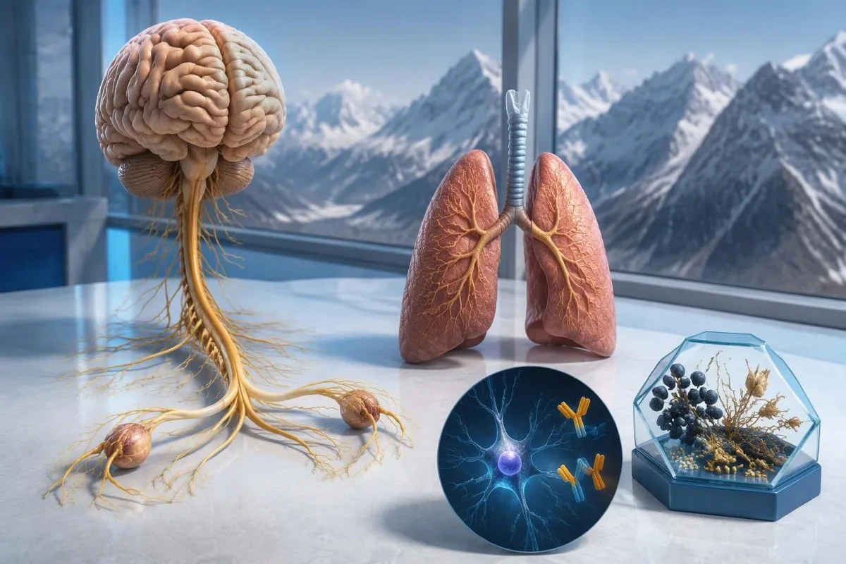
Pozitiven izvid protiteles anti-Hu vzbuja sum na nevrološki imunski odziv, povezan z rakom, zlasti drobnoceličnim pljučnim rakom, vendar ne pomeni diagnoze raka. Zdravniki ocenijo simptome, potrdijo laboratorijski izvid in uredijo preiskave za odkrivanje raka; tudi če so prvi slikovni izvidi negativni, bodo morda potrebni ponovni pregledi.



- Anti-Hu / ANNA-1 je visoko tvegano paraneoplastično protitelo, kategorija, povezana z rakom pri več kot 70% uveljavljenih kliničnih kohort – vendar to ne velja za vsak nepotrjen presejalni izvid.
- Diagnoza raka zahteva dokaze o osnovni maligni bolezni, običajno tudi pregled tkiva; en sam pozitiven izvid protiteles ni tak dokaz.
- Potrditev s testom pogosto združuje dva dopolnjujoča se pristopa: značilen vzorec obarvanja tkiva in test, specifičen za antigen.
- Nevrološki simptomi kot so napredujoča izguba občutka, motnje ravnotežja, epileptični napadi ali avtonomna disfunkcija, vplivajo na pomen in nujnost izvida.
- Negativen slikovni izvid pomeni, da s to preiskavo takrat niso odkrili raka; ne izključuje pa možnosti majhnega raka, ki ga sprva ni mogoče zaznati.
- Ponovitev presejalnega testiranja je v skladu z merili PNS iz leta 2021 priporočena na 4–6 mesecev v obdobju 2 let, kadar visoko tvegan nevrološki fenotip spremlja visoko tvegano protitelo.
- Daljše spremljanje je lahko primerno za izbrane bolnike; kohorta bolnikov s protitelesi anti-Hu iz leta 2023 je podprla razmislek o presejalnem testiranju, daljšem od 2 let, včasih do 5 let.
- Nujni simptomi vključujejo nove težave z dihanjem, nezmožnost varnega požiranja, hitro slabšanje šibkosti, prvi epileptični napad ali nenadne nevrološke izpade.
Kaj pomeni pozitiven izvid protiteles anti-Hu?
En pozitiven test na protitelesa anti-Hu pomeni, da je laboratorij zaznal reaktivnost proti nevronskim proteinom Hu; sam po sebi ne potrjuje raka ali nevrološkega sindroma. Interpretacija ima 3 dele: ali je rezultat resničen, ali se ujema z nevrološko klinično sliko in ali je mogoče odkriti pridruženi rak.
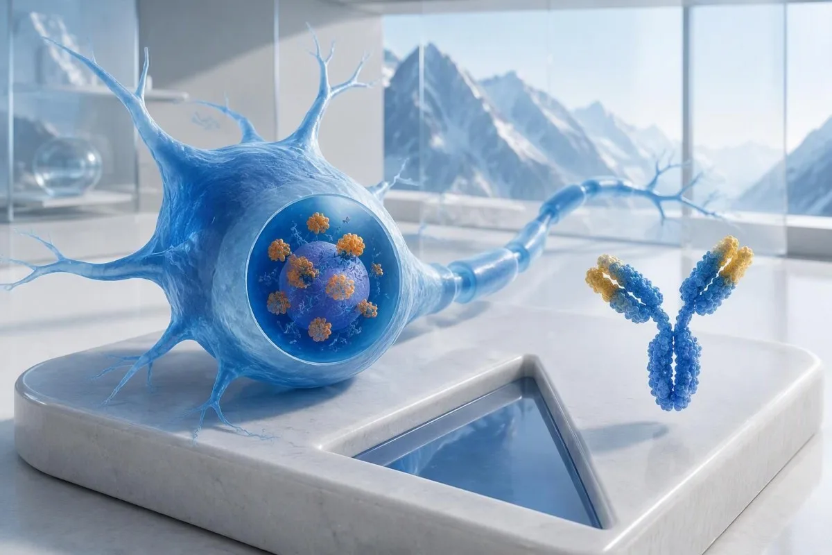
Protitelesa anti-Hu so uvrščena med protitelesa z visokim tveganjem, povezana z rakom, pri čemer je kategorija opredeljena s povezavo z rakom, ki v ustreznih kliničnih populacijah presega 70% (Graus et al., 2021). Tega odstotka ni mogoče preprosto pripisati sicer zdravi osebi s šibkim, nepotrjenim rezultatom panela: na verjetnost vplivajo preučevani bolniki, metoda testiranja in nevrološka klinična slika.
Pri hipotetični 62-letni osebi z 8 tedni vse hujše motnje ravnotežja in asimetrične izgube občutka je potrebna drugačna ocena kot pri osebi, pri kateri so protitelesa odkrili naključno med obsežnim panelom za utrujenost. Nobeni osebi ne bi smeli samo na podlagi laboratorijskega izvida reči: “Imate raka.” Naša razlaga pozitivnih ugotovitvah protiteles pomaga ločiti imunski označevalec od zaključene diagnoze.
Kantesti je AI analizator krvi ki lahko pomaga pojasniti besedilo izvida anti-Hu, vendar sta laboratorijska potrditev in ocena raka v domeni vaših lečečih zdravnikov. Kot Thomas Klein, MD, glavni zdravstveni direktor družbe Kantesti LTD, bi najprej vprašal, kaj je bil razlog za naročilo tega konkretnega testa — ne pa, kako zastrašujoča je rdeča zastavica; naš organizacija in klinična misija pojasnjujeta to razliko.
Ali je ANNA-1 isto protitelo kot anti-Hu?
Protitelo ANNA-1 in protitelo anti-Hu se na splošno nanašata na isto družino protiteles. ANNA-1 pomeni antinevronsko jedrno protitelo tipa 1; oznaka Hu se nanaša na znotrajcelične nevronske proteine, ki jih prepozna imunski odziv, ne pa na ločeno bolezen ali stadij raka.
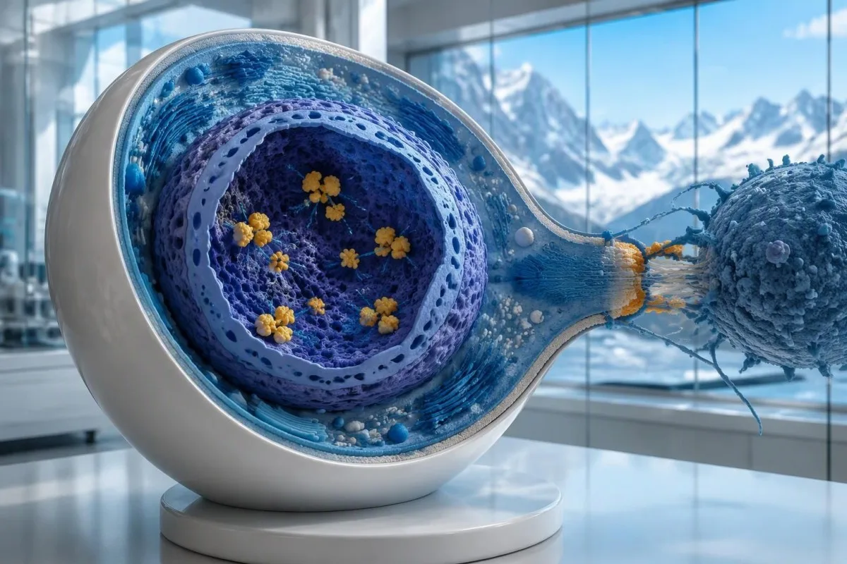
Proteini Hu so znotrajcelični proteini, ki vežejo RNK, protitelesa anti-Hu pa je najbolje razumeti kot označevalec imunskega odziva, ki pogosto vključuje citotoksične celice T. To se razlikuje od bolezni, pri katerih se protitelesa vežejo na dostopne proteine na površini nevronov. Ta razlika pojasnjuje, zakaj že samo znižanje ravni protiteles zanesljivo ne odpravi že nastale nevrološke okvare zaradi anti-Hu, tudi kadar 2 izvida pokažeta izrazito različne titre.
Izvid z zapisom “zaznano ANNA-1” ni enak običajnemu pozitivnemu testu ANA, kljub podobnim črkam. ANA običajno pomeni protijedrna protitelesa, ki se uporabljajo pri ocenjevanju sistemskih avtoimunskih bolezni, medtem ko ANNA-1 označuje vzorec nevronskih protiteles. Ko se pogovarjate o svojih izvidih, teh 2 kratic ne zamenjujte; naš vodnik po terminologiji ANA pojasnjuje bolj znani test.
Protitelesa anti-Hu je mogoče zaznati pri nekaterih ljudeh z drobnoceličnim pljučnim rakom, ki nimajo prepoznavnega nevrološkega sindroma. Nasprotno pa lahko nevrološki simptomi vodijo do zaznave protiteles, še preden rak postane viden. To sta 2 različni klinični poti: protitelo zdravniku samo po sebi ne pove, ali so živci trenutno poškodovani ali kje bi se lahko nahajal osnovni rak.
Kako je treba razlagati izvid testa protiteles anti-Hu?
An anti-Hu antibody test has no universal numerical cutoff that can be applied across laboratories. Read the specimen type, assay method, result category, laboratory reference interval and confirmation comments together; a dilution titer, band intensity and numerical index are 3 different ways of reporting reactivity.
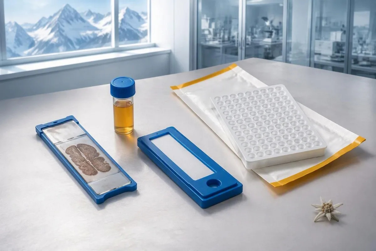
A hypothetical titer of 1:240 describes detectable reactivity after a particular dilution; it is not a cancer probability of 240, nor does it establish cancer stage. A numerical index from another method may use entirely different calibration. Our guide to kvalitativni v primerjavi s kvantitativnimi rezultati explains why “positive” and “how much” answer different laboratory questions.
Weak or borderline reactivity deserves method-specific interpretation, particularly when a broad panel was ordered without a convincing neurological indication. Ask whether the laboratory observed the characteristic neuronal staining pattern and whether an antigen-specific confirmation was performed. Two tests using the same underlying strip technology may reproduce the same analytical problem; agreement is more informative when the methods are genuinely complementary.
Bring the complete report, not just a screenshot of the word positive. The 5 most useful details are the specimen, collection date, assay name, reported strength or titer and laboratory interpretation. Also list recent intravenous immunoglobulin and other immune treatments, because passive antibodies or treatment effects can complicate some serological results; your laboratory can advise whether that issue plausibly affects this particular finding.
Kdaj je treba potrditi pozitiven izvid anti-Hu?
Unexpected, weak or clinically discordant anti-Hu results should be reviewed for confirmation, preferably with an experienced neuroimmunology laboratory. The 2021 PNS criteria emphasize interpreting neuronal antibody testing alongside the phenotype and using appropriate confirmatory methods rather than accepting an isolated commercial-panel result as a diagnosis.
Tissue-based immunohistochemistry or immunofluorescence and an antigen-specific assay provide different information. The first identifies a characteristic distribution of neuronal staining; the second tests reactivity against the relevant antigen. Graus et al. (2021) describe these complementary approaches, with specialist reassessment particularly useful when 1 method gives a result that does not fit the clinical picture.
Repeating a sample can address collection errors or a transient analytical concern, but repeating the same kit is not always enough. In a hypothetical 45-year-old with no objective neurological findings and a faint isolated band, I would want the laboratory’s explanation before treating the report as established anti-Hu autoimmunity. Our GAD65 interpretation guide illustrates another antibody for which context and assay characteristics change meaning.
Confirmation and urgent assessment can happen in parallel. A person who develops seizures, substantial walking difficulty or progressive swallowing impairment should not wait several weeks for a reference-laboratory answer before receiving neurological care. The practical question is not “repeat or investigate?” but which of those 2 tasks can safely proceed together, given the symptoms and the initial laboratory evidence.
Kateri nevrološki simptomi vzbujajo večji sum na anti-Hu?
Progressive sensory neuronopathy, encephalomyelitis, limbic encephalitis and marked autonomic dysfunction are recognized anti-Hu presentations. Symptoms can involve more than 1 part of the nervous system, so the pattern and speed of change are usually more informative than a single symptom such as tingling or fatigue.

Sensory neuronopathy can cause asymmetric numbness, impaired position sense and sensory ataxia, sometimes involving the hands or trunk early rather than following a simple feet-first pattern. A patient may lose balance in the dark because vision had been compensating for reduced position feedback. That is different from the focal finger distribution discussed in our carpal tunnel symptom guide, although neither pattern can be diagnosed from description alone.
Anti-Hu autonomic involvement can affect gut movement, blood-pressure regulation and bladder function. Severe constipation, unexplained intestinal pseudo-obstruction or dizziness on standing becomes more relevant when accompanied by objective neurological abnormalities. Recording symptoms over 7 days can reveal combinations that otherwise get presented as separate stomach, balance and urinary complaints; sudden severe abdominal symptoms still require assessment for non-neurological emergencies.
New memory impairment, behavioral change or seizures may indicate central nervous system involvement, while rapid weakness has a broader emergency differential. The opozorilnih znakih nujne obravnave zaradi šibkosti in our neurological safety guide apply regardless of the antibody label. Call emergency services for new breathing difficulty, unsafe swallowing, a first seizure or sudden stroke-like symptoms rather than waiting for a scheduled follow-up.
Katere nevrološke preiskave lahko sledijo pozitivnemu izvidu?
Neurological follow-up may include examination, nerve conduction studies, MRI and cerebrospinal fluid testing, selected according to the symptoms. There is no mandatory battery for every anti-Hu-positive person; the purpose is to document which structures are affected and exclude other causes, not merely to collect additional positive tests.
Nerve conduction studies can support a sensory neuronopathy pattern by showing reduced sensory responses across distributions that do not fit a single compressed nerve. Brain or spinal MRI addresses different questions, including structural disease and some inflammatory changes. A normal MRI does not exclude peripheral sensory ganglion involvement; these are 2 distinct anatomical compartments with different diagnostic tools.
Cerebrospinal fluid may show increased cells, protein or evidence of intrathecal immune activity, but none of those findings is specific to anti-Hu. A lumbar puncture is most useful when the neurological syndrome warrants it, rather than as an automatic response to 1 weak serum band. Our discussion of razlago oligoklonalnih trakov v cerebrospinalni tekočini explains why immune activity in spinal fluid does not identify a single disease.
Basic investigations still matter: glucose control, vitamin status, renal function and serum protein assessment can uncover treatable contributors to neuropathy. A clinician may request 2 or more of these alongside specialist testing, depending on the examination. The vodnik po preiskavi serumskih beljakovin provides background, but normal routine blood work neither confirms nor excludes an anti-Hu-associated neurological process.
Ali pozitivnost anti-Hu potrjuje paraneoplastični sindrom?
A positive antibody alone does not confirm anti-Hu paraneoplastic syndrome. The PNS-Care approach combines the neurological phenotype, antibody risk category, cancer findings and follow-up; clinicians also check for direct cancer effects, treatment toxicity and unrelated neurological conditions before attributing symptoms to a paraneoplastic immune response.
Definite, probable and possible PNS are diagnostic categories, not cancer stages. The 2021 criteria use a structured scoring system rather than allowing 1 antibody to determine the whole diagnosis (Graus et al., 2021). A confirmed anti-Hu result with a strongly compatible syndrome remains clinically significant even before a cancer is identified, but clinicians should state clearly which parts of the assessment remain unresolved.
An older patient can have anti-Hu reactivity and a separate cause of numbness, such as diabetes, nutritional deficiency or a monoclonal protein disorder. Coexistence is possible; either-or reasoning can miss treatable disease. If serum protein studies reveal a band, our immunofixation result guide explains why that finding requires its own interpretation rather than being automatically folded into the anti-Hu diagnosis.
Kantesti je platforma za razlaga krvne slike z AI that can help organize these distinctions in a laboratory explanation, but it cannot assign a reliable PNS-Care diagnosis from a report alone. Our klinični validacijski okvir separates technical evaluation from clinical conclusions. In practice, I would document 3 separate judgments: confidence in the antibody, confidence in the neurological syndrome and the current evidence for cancer.
Kako zdravniki iščejo z njim povezani rak?
Cancer evaluation usually prioritizes the chest because small-cell lung cancer is the strongest adult anti-Hu association. Doctors commonly begin with appropriate chest CT and may add FDG-PET/CT or targeted investigations if suspicion remains; the exact plan depends on age, symptoms, examination, smoking history and previous imaging.
The cancer association is substantial in established anti-Hu clinical cohorts. Villagrán-García et al. (2023) reported identified cancer in 349 of 466 patients, approximately 75%, in a specialist anti-Hu cohort. This is valuable evidence for taking confirmed, clinically relevant anti-Hu seriously; it is not a personalized prediction for an asymptomatic person with an unconfirmed positive screening strip.
A chest X-ray is not equivalent to a chest CT, and routine low-dose screening is not necessarily the correct examination for a suspected paraneoplastic syndrome. The clinician chooses the protocol and body regions based on the diagnostic question. Likewise, neurological symptoms do not automatically mean cancer has spread to the brain; our brain imaging warning-sign guide distinguishes structural investigation from immune-mediated disease.
A suspicious scan often leads to tissue sampling through a method chosen by respiratory or oncology specialists, such as bronchoscopic assessment of an accessible area. Imaging suspicion and cancer confirmation are 2 separate steps. Never-smoking status lowers the baseline probability of small-cell lung cancer but does not cancel a well-confirmed anti-Hu finding with a convincing neurological presentation; other cancers and non-cancer explanations still need consideration.
Zakaj so lahko prvi slikovni izvidi za odkrivanje raka negativni?
Initial imaging can be negative because an associated cancer is still too small to detect, lies outside the examined area or is not readily visible with that technique. Neurological autoimmunity may precede cancer detection, so a negative scan reduces concern without necessarily ending follow-up in a high-risk clinical presentation.
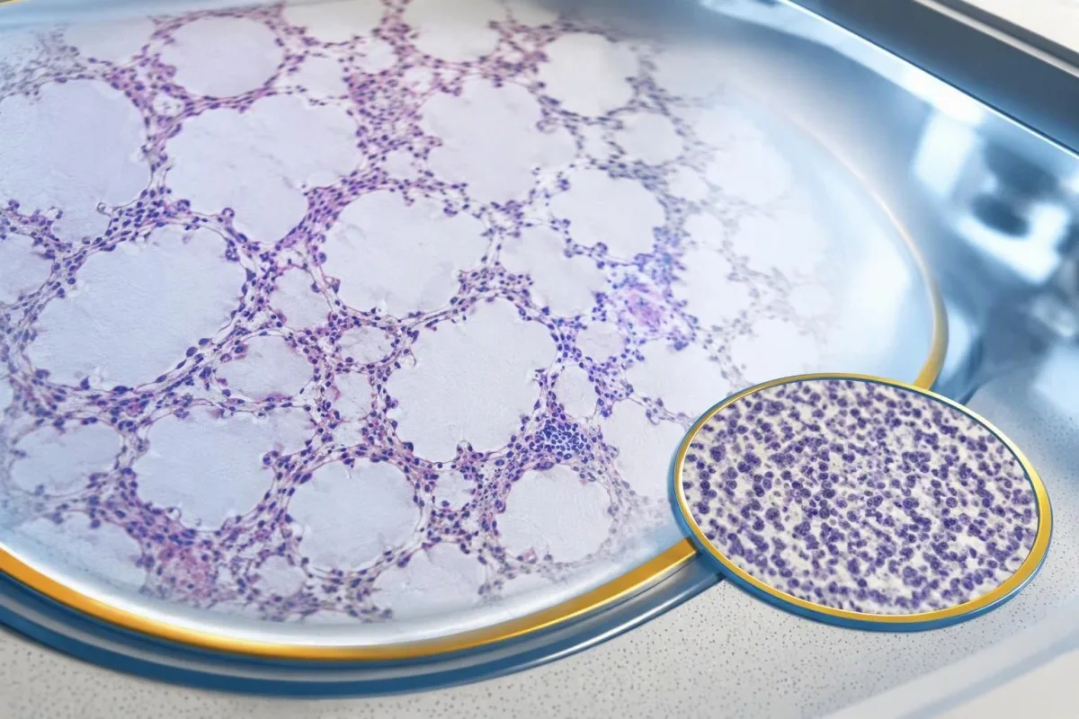
A negative scan is time-specific and method-specific. A normal chest CT performed on 1 date answers whether that examination identified a suspicious chest finding then; it does not guarantee that every possible associated cancer is absent indefinitely. Ask what was actually imaged, whether the protocol was suitable and whether the specialist recommends PET/CT or targeted investigation despite the initial result.
Villagrán-García et al. (2023) described cancers detected beyond the first 2 years in a minority of their anti-Hu cohort, supporting individualized longer surveillance. Their findings also support considering PET-based investigation when initial CT is unrevealing in an appropriate clinical context. This is an escalation decision for the treating team, not a reason to independently purchase repeated whole-body scans without weighing radiation, incidental findings and cost.
Neurological deterioration after negative imaging still needs reassessment. Cancer surveillance and neurological monitoring are 2 parallel tracks; one reassuring scan does not explain new loss of function. A marker such as neurofilament light chain may indicate neuronal injury in selected settings, but our NfL interpretation guide explains why it cannot establish anti-Hu disease, locate cancer or replace examination.
Kako dolgo je treba nadaljevati presejalne preglede za raka?
The 2021 PNS criteria recommend repeat cancer screening every 4–6 months for 2 years when a high-risk neurological phenotype accompanies a high-risk antibody. Longer surveillance may be appropriate for selected anti-Hu patients; the interval and duration should reflect confirmation, clinical evolution, initial investigations and individual cancer risk.

The 2-year schedule is not an instruction to scan every positive antibody result repeatedly. Graus et al. (2021) link that recommendation to the combination of high-risk phenotype and high-risk antibody. An incidental, weak result without a matching syndrome needs confirmation and individualized assessment first; repeating imaging mechanically can create incidental findings and unnecessary procedures without resolving the original uncertainty.
Older European guidance recommended longer surveillance. The EFNS task force advised repeating screening after 3–6 months and then every 6 months for up to 4 years in relevant antibody-positive paraneoplastic presentations (Titulaer et al., 2011). The 2023 anti-Hu cohort supported considering screening up to 5 years in selected patients, so a specialist may reasonably extend follow-up beyond the newer general 2-year framework.
Keep a dated plan stating the next examination, the clinician responsible and what symptoms should trigger earlier contact. Kantesti explanations can help you understand terminology, but the platform should not be treated as a scan-reminder service or substitute for that plan. Our guide to risk-based cancer screening addresses a different setting: routine prevention is not the same as investigating a possible paraneoplastic syndrome.
Kakšno zdravljenje pomaga pri nevrološki bolezni anti-Hu?
Treatment focuses on an identified cancer, selected immune therapy and symptom-directed rehabilitation. Anti-Hu-associated neurological disease often responds less predictably to immunotherapy than neuronal surface-antibody disorders because intracellular targets and T-cell-mediated injury are involved; treatment may aim to stabilize function rather than promise complete recovery.

Treating an associated cancer is a central part of care, but established neurological injury may persist even when cancer treatment succeeds. Neurology and oncology teams may consider corticosteroids, intravenous immunoglobulin, plasma exchange or other immune treatments in selected situations. There is no single evidence-based dose or 1 universal regimen appropriate for everyone with anti-Hu positivity, especially without a confirmed clinical syndrome.
An antibody titer is not a dependable treatment target on its own. A lower number does not necessarily mean restored nerve function, and a persistent positive result does not by itself prove cancer progression. This differs from some surface-antibody disorders; our MOG antibody diagnosis guide explains another immune neurological condition, but its diagnostic rules and treatment expectations should not be transferred to anti-Hu.
Immune checkpoint cancer therapies deserve an explicit discussion when anti-Hu neurological disease is present, because they can provoke or worsen paraneoplastic neurological autoimmunity. This is a specialist risk-benefit decision, not an automatic prohibition based on 1 laboratory result. Tell the oncologist about previous neurological symptoms and confirmed neuronal antibodies before treatment decisions are finalized; coordinating the teams early can prevent avoidable misunderstanding.
Kaj lahko varno storite med nadaljevanjem preiskav?
While investigations continue, protect mobility, swallowing and medication safety rather than trying to lower anti-Hu antibodies with supplements. Use the neurological symptoms—not the appearance of the laboratory flag—to decide whether ordinary follow-up is sufficient or urgent care is needed; a new functional loss warrants contact before the next planned scan.

Position-sense loss increases fall risk, particularly in poor lighting or on uneven ground. A physiotherapist may recommend a suitable walking aid and individualized balance strategies; occupational therapy can address hand coordination and home hazards. A short 7-day record of falls, near-falls and walking changes is often more useful at review than the vague statement “my balance is worse,” but stop activities that feel unsafe.
Excess vitamin B6 can itself cause sensory neuropathy, making unsupervised high-dose nerve supplements particularly unhelpful during this assessment. Review every supplement and its daily dose in mg, including overlapping multivitamins and combination products. Our vodila o varnosti B-kompleksa explains that problem; correcting a documented deficiency is different from taking progressively larger doses in the hope of treating anti-Hu autoimmunity.
Vitamin B12 deficiency can contribute to sensory and gait problems and may need investigation even when anti-Hu is confirmed. There is no universally interchangeable B12 cutoff: results in pg/mL in pmol/L require their own laboratory context, sometimes with functional testing. Our vodilo za spremljanje mejnega B12 explains those next steps; new choking, breathing difficulty or rapid weakness should be assessed urgently instead of managed with vitamins.
Kako lahko UI varno pomaga pri razlagi izvida anti-Hu?
AI can help explain report terminology and organize questions, but it cannot confirm anti-Hu specificity, diagnose paraneoplastic syndrome or rule out cancer. Safe interpretation preserves the original assay details and separates 3 issues: what the laboratory reported, what the result may mean and what a clinician has actually established.

Kantesti je Orodje za analizo krvnih testov z umetno inteligenco that can provide a plain-language explanation of uploaded laboratory information, including the distinction between an antibody screen and a confirmed clinical diagnosis. A rapid explanation—often around 60 seconds for report processing—is not a reference-laboratory assay. Our Vodnik po tehnologiji AI describes the interpretation workflow without making anti-Hu-specific diagnostic guarantees.
The complete source report should remain available for verification. An explanation should not silently replace “weakly reactive” with “confirmed,” drop the specimen type or invent a universal reference interval. Our guide to preverjanje pojasnil o medicini, ki jih ustvari UI describes practical source checks; for this antibody, compare at least the assay wording, confirmation comment and collection date against the original document.
Kantesti cannot examine sensation, review tissue staining directly through a report explanation or establish that negative imaging has excluded a tiny cancer. Those are 3 different clinical capabilities, not features that follow from understanding laboratory language. Share the explanation with the treating team as a discussion aid, and do not delay urgent assessment while uploading documents or waiting for a second automated interpretation.
Vaš naslednji pregled in preglednost raziskav
Bring the complete antibody report, a dated symptom history, previous imaging reports and a current medication list to your next appointment. Ask the clinician to clarify antibody confirmation, the neurological diagnosis and cancer surveillance separately; those 3 answers may have different levels of certainty and different follow-up dates.
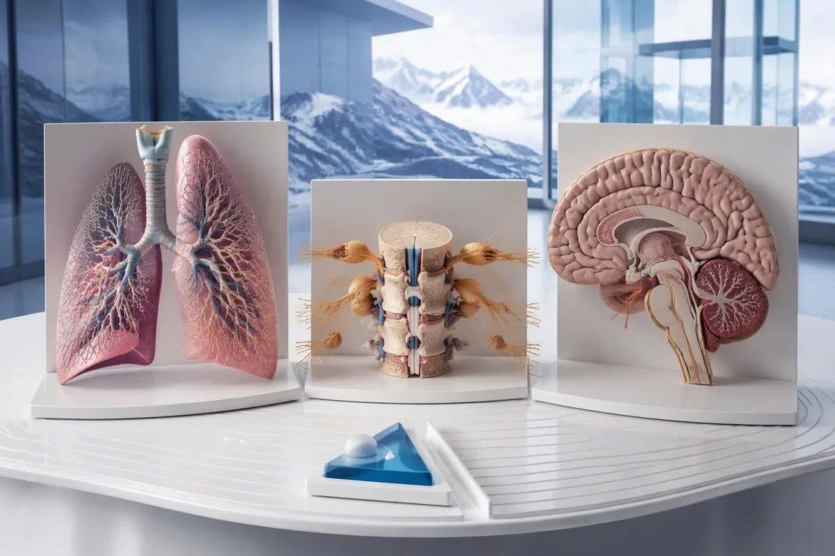
A useful 1-page symptom history records onset, progression, sensory distribution, walking changes, memory problems and autonomic symptoms. Include prior cancer treatment and any recent immunoglobulin therapy, then ask who will coordinate neurology and oncology if both are involved. As Thomas Klein, MD, I would rather see a clear timeline than 20 disconnected screenshots; it makes discrepancies between the laboratory result and clinical course easier to spot.
Medical authorship is not a substitute for an individual neurological examination. Naš informacije zdravniškega sveta describes the clinicians associated with our work, while your own specialist determines the appropriate next investigation. As of 25. oktober 2026, the recommendations discussed here draw on the 2021 international PNS criteria, the 2011 EFNS screening guidance and the 2023 anti-Hu cohort—not an invented 2026 guideline.
The 2 Figshare publications below are platform-related research references, not evidence validating anti-Hu diagnosis or cancer exclusion. Their supplied titles describe hantavirus triage deployment and a synthetic-case technical benchmark; neither title establishes anti-Hu-specific clinical performance. Because publication authors and dates were not supplied for verification, the citations use title-first APA formatting with “n.d.”; the ResearchGate and Academia.edu links are searches, not claims of verified publication copies.
Pogosto zastavljena vprašanja
Ali pozitiven rezultat protiteles anti-Hu pomeni, da imam raka?
0 Rezultat pozitiven na protitelesa anti-Hu sam po sebi ne pomeni diagnoze raka. Anti-Hu je protitelo, povezano z velikim tveganjem za raka, pri 349 od 466 bolnikov v specialistični kohorti anti-Hu iz leta 2023 pa so odkrili raka, vendar ta podatek ne določa tveganja pri naključnem, nepotrjenem presejalnem rezultatu. Zdravniki ocenijo nevrološko klinično sliko, potrdijo nepričakovane laboratorijske izvide in preverijo morebitno osnovno rakavo obolenje; pri odraslih običajno najprej pregledajo prsni koš. Diagnoza raka praviloma zahteva ločene klinične, slikovne in tkivne dokaze.
Ali sta anti-Hu in ANNA-1 ista stvar?
Anti-Hu in ANNA-1 na splošno označujeta isto družino nevronskih protiteles. ANNA-1 pomeni antinevronsko jedrno protitelo tipa 1, medtem ko se Hu nanaša na znotrajcelične nevronske beljakovine, ki jih protitelo prepozna. ANNA-1 ni isto kot rutinsko ANA, ki se uporablja pri testiranju za sistemske avtoimunske bolezni. Preverite natančno preiskavo in besedilo potrditve, saj lahko laboratoriji isto družino protiteles navajajo v različnih oblikah.
Ali je lahko test na protitelesa anti-Hu lažno pozitiven?
Testiranje protiteles anti-Hu lahko povzroči zavajajočo ali nespecifično reaktivnost, zlasti kadar se šibek presejalni rezultat ne ujema z nevrološko klinično sliko. Potrditev običajno temelji na dveh dopolnjujočih se pristopih: značilnem vzorcu obarvanja tkiva in za antigen specifičnem testu, ki ga interpretira izkušen laboratorij. Ponovitev iste komercialne metode lahko ponovi isto analitično težavo, namesto da bi jo odpravila. Hitro napredujoči nevrološki simptomi še vedno zahtevajo pravočasno oceno, medtem ko čakate na potrditev.
Kako pogosto je treba ponavljati slikovne preiskave, če je test na anti-Hu pozitiven?
Merila PNS iz leta 2021 priporočajo ponovitev presejanja za raka vsakih 4–6 mesecev v obdobju 2 let, kadar visoko tvegani nevrološki fenotip spremlja visoko tvegano protitelo. Ta urnik ne velja samodejno za vsak naključno odkrit pozitiven izvid anti-Hu. Starejše smernice EFNS so priporočale spremljanje do 4 let, kohortna raziskava bolnikov s protitelesi anti-Hu iz leta 2023 pa je podprla razmislek o spremljanju do 5 let pri izbranih bolnikih. Vaš specialist naj določi slikovno metodo, časovne razmake, trajanje spremljanja in simptome, ki zahtevajo zgodnejši pregled.
Ali je lahko anti-Hu pozitiven, če sta izvida CT in PET normalna?
0 Anti-Hu lahko ostane pozitiven tudi, kadar s CT ali PET-slikanjem niso odkrili raka. Nevrološka avtoimunost se lahko pojavi še pred odkritjem raka, negativen izvid slikanja pa odraža pregledana področja, uporabljeno tehniko in datum preiskave, ne pa trajnega zagotovila, da raka ni. Pri skladni klinični sliki z visokim tveganjem merila iz leta 2021 priporočajo ponovni pregled vsakih 4–6 mesecev v obdobju 2 let, pri izbranih bolnikih z anti-Hu pa individualno prilagojeno daljše spremljanje. Prav tako je treba ponovno preučiti nepotrjeno reaktivnost protiteles in razlage, ki niso povezane z rakom, namesto da bi predpostavljali, da je nevidni rak zagotovo prisoten.
Ali znižanje ravni protiteles anti-Hu pomeni, da zdravljenje deluje?
Zniževanje ravni protiteles anti-Hu ne dokazuje zanesljivo nevrološkega okrevanja ali uspešnega zdravljenja raka. Protitelesa anti-Hu prepoznajo znotrajcelične beljakovine, povezana nevrološka okvara pa pogosto vključuje mehanizme, ki jih posredujejo celice T, in ne zgolj učinke protiteles. Tudi titra iz različnih laboratorijev morda nista primerljiva zaradi različnih metod. Zdravniki namesto uporabe vrednosti protiteles kot samostojnega cilja zdravljenja ocenijo funkcionalnost, izsledke pregleda, ustrezne preiskave in odziv raka na zdravljenje.
Danes pridobite analizo krvnih preiskav z umetno inteligenco
Pridružite se več kot 2 milijonoma uporabnikov po vsem svetu, ki zaupajo Kantesti za takojšnjo, natančno analizo laboratorijskih preiskav. Naložite svoje rezultate krvnih preiskav in v nekaj sekundah prejmite celovito razlago biomarkerjev 15,000+.
📚 Citirane raziskovalne publikacije
Klein, T., Mitchell, S., & Weber, H. (2026). Vejezično klinično podporo odločanju s pomočjo umetne inteligence za zgodnje triaže hantavirusov: zasnova, inženirsko vrednotenje in uvedba v resničnem svetu za 50.000 interpretiranih poročil o krvnih preiskavah. Kantesti AI Medical Research.
Klein, T., Mitchell, S., & Weber, H. (2026). Predregistrirana, na rubrikah temelječa avtomatizirana tehnična primerjava interpretacijskega stroja za krvne teste Kantesti na 100.000 sintetičnih testnih primerih. Kantesti AI Medical Research.
📖 Zunanje medicinske reference
Graus F et al. (2021). Updated Diagnostic Criteria for Paraneoplastic Neurologic Syndromes. Neurology: Neuroimmunology & Neuroinflammation.
Titulaer MJ et al. (2011). Screening for tumours in paraneoplastic syndromes: report of an EFNS Task Force. European Journal of Neurology.
Villagrán-García D et al. (2023). Revisiting anti-Hu paraneoplastic autoimmunity: phenotypic characterization and cancer diagnosis. Brain Communications.
⚕️ Medicinska izjava o omejitvi odgovornosti
Ta članek je namenjen izobraževalnim namenom in ne predstavlja medicinskega nasveta. Za odločitve o diagnozi in zdravljenju se vedno posvetujte z usposobljenim izvajalcem zdravstvenih storitev.
E-E-A-T zaupanja vredni signali
Izkušnje
Zdravniški klinični pregled delovnih postopkov za interpretacijo laboratorijskih izvidov.
Strokovno znanje
Laboratorijska medicina s poudarkom na tem, kako se biomarkerji obnašajo v kliničnem kontekstu.
Avtoritativnost
Napisal dr. Thomas Klein, pregledala dr. Sarah Mitchell in prof. dr. Hans Weber.
Zanesljivost
Interpretacija na podlagi dokazov z jasnimi nadaljnjimi potmi za zmanjšanje alarmiranja.
