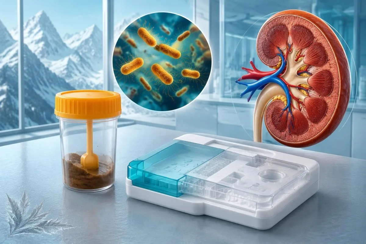
A positive stool result usually indicates Shiga toxin-producing E. coli, but it does not automatically mean kidney damage. Contact your clinician the same day; reduced urination, unusual bruising, marked pallor, or confusion require urgent assessment.

Key takeaways
- Shiga toxin positive means a stool assay detected Shiga toxin or its genes, usually indicating Shiga toxin-producing E. coli, also called STEC.
- Same-day advice is appropriate after a positive result, even if diarrhea is improving; the clinician can assess hydration and arrange kidney and platelet monitoring.
- Antibiotics are generally avoided for STEC O157, Shiga toxin 2-producing STEC, or STEC with an unknown toxin type because treatment may increase HUS risk.
- Anti-diarrheal medicines such as loperamide and diphenoxylate should be avoided in suspected or confirmed STEC infection unless a specialist specifically directs otherwise.
- HUS timing is commonly around days 5–10 after diarrhea begins, and the complication can emerge as bowel symptoms improve.
- Urgent warning signs include substantially less urine, unusual bruising, marked pallor, severe fatigue, swelling, confusion, or seizures; do not wait for another stool result.
- Kidney injury thresholds include a creatinine rise of at least 0.3 mg/dL, approximately 26.5 µmol/L, within 48 hours; a result still inside the laboratory range can therefore be concerning.
- Hydration usually means oral rehydration solution while urine output remains adequate; reduced urination or swelling needs assessment rather than forced fluids at home.
What does a shiga toxin positive stool result mean?
A shiga toxin positive stool result means the laboratory detected Shiga toxin or genes that encode it, usually indicating STEC infection. Contact your clinician the same day, and seek urgent assessment for reduced urination, unusual bruising, marked pallor, severe weakness, or confusion; these can signal hemolytic uremic syndrome, or HUS.

A positive stool test does not establish HUS, and it does not measure how much toxin has reached the circulation. HUS involves 3 linked problems—damage to circulating red cellular elements, a falling platelet count, and acute kidney injury—which require clinical assessment and blood testing rather than interpretation of the stool result alone.
The wording on the report matters: an assay may list Shiga toxin, stx1, stx2, STEC, or an E. coli O157 target, and these are not interchangeable descriptions. A toxin-positive result with O157 not detected can still represent clinically significant non-O157 STEC; our qualitative result guide explains why detected does not mean a measured concentration.
Kantesti is an AI blood test analyzer that can help explain the kidney and platelet results requested after a positive stool test, but our interpretation cannot diagnose or exclude evolving HUS. Information about our organization identifies Kantesti LTD and its service scope; urgent symptoms should go directly to a healthcare professional, not through an interpretation workflow.
How do Shiga toxin stool tests, PCR and culture differ?
A Shiga toxin stool test may detect toxin protein, while a molecular assay detects toxin-producing genes; culture attempts to recover the organism itself. These methods answer different questions, and a positive molecular result does not prove that living bacteria remain in the sample or specify an individual's HUS risk.

PCR panels commonly target 2 gene families, stx1 and stx2, although some reports combine them into one STEC result. A toxin immunoassay can be performed directly on stool or after laboratory enrichment; neither a simple positive result nor an unreported PCR cycle threshold should be interpreted as a reliable toxin dose or severity score.
The 2017 Infectious Diseases Society of America guideline recommends testing that detects Shiga toxin or its genes and distinguishes E. coli O157 from other STEC when this infection is suspected (Shane et al., 2017). Culture and public-health characterization can help investigate outbreaks and identify strains, so a laboratory may request additional material even after a rapid panel is already positive.
Discordant tests deserve explanation, not automatic dismissal: a gene-positive, culture-negative sample can reflect collection timing, transport conditions, or reduced organism viability, while an unexpected isolated toxin result may need confirmation. In contrast, stool inflammatory markers detect intestinal immune activity rather than STEC specifically; a calprotectin result cannot confirm or rule out Shiga toxin production.
Which symptoms and timing fit STEC infection?
STEC infection commonly causes abdominal cramps and diarrhea that may become bloody; fever can be absent or relatively mild. Symptoms usually start about 3–4 days after exposure, although a roughly 1–10-day incubation range means the most recent meal is not necessarily the source.
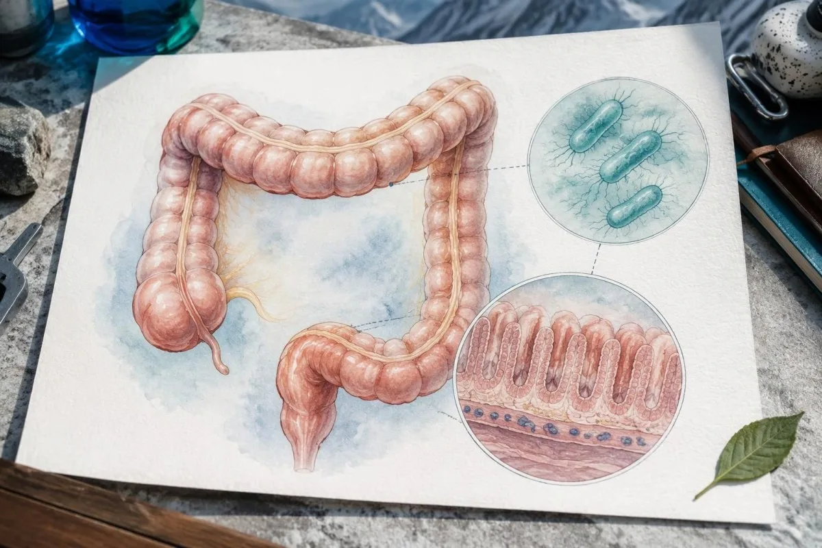
Bloody diarrhea increases concern for STEC, but its absence does not exclude the infection or later HUS. The first watery stool is generally the useful starting point for tracking illness day; for example, a laboratory result received on Thursday after diarrhea began Monday represents approximately day 4, not day 1 of the complication-monitoring period.
Severe abdominal pain warrants examination even when a stool panel has identified a pathogen, because a positive result does not exclude another abdominal emergency. The amount and appearance of stool blood also matter: our stool blood warning guide explains why black tarry stool, heavy bleeding, or faintness should not simply be attributed to infectious diarrhea.
Fever does not reliably separate different causes of diarrhea, and broad panels sometimes detect more than 1 organism. A second result may alter treatment decisions—Campylobacter treatment decisions differ from STEC decisions—so clinicians should interpret co-detections against symptom severity, exposure history, and the specific toxin finding rather than treating every positive target identically.
Who has a higher risk of hemolytic uremic syndrome?
Children younger than 5 years, older adults, and people with more severe STEC illness deserve particular attention for HUS. Strains producing Shiga toxin 2 are more strongly associated with HUS than strains producing Shiga toxin 1 alone, but toxin type cannot predict an individual outcome.

There is no single HUS percentage that applies to every Shiga toxin-positive report: published estimates depend on age, strain, outbreak setting, and how cases were identified. A child with O157 and bloody diarrhea belongs to a different risk group from an otherwise well adult with an incidentally detected non-O157 strain; quoting one pooled percentage can create misleading reassurance or alarm.
Dehydration, persistent vomiting, substantial intestinal symptoms, and concerning platelet or creatinine trends can increase the need for observation, but none is a stand-alone prediction tool. In a hypothetical 4-year-old who is drinking less and has fewer wet diapers, the immediate question is whether hydration and kidney function are deteriorating—not whether the child has completed the usual diarrhea timeline.
Pediatric laboratory interpretation requires age-specific reference intervals, especially for creatinine, because children normally have lower values than many adults. Our children's interpretation safety guide describes these limitations; Kantesti AI does not provide a validated HUS prediction score, consistent with our responsible AI boundaries.
Why are antibiotics often avoided in STEC infection treatment?
STEC infection treatment usually avoids antibiotics because some antibiotics may increase toxin production or release and may increase the risk of HUS. IDSA recommends avoiding antimicrobial treatment for STEC O157, Shiga toxin 2-producing STEC, and STEC when the toxin genotype is unknown (Shane et al., 2017).
The evidence is genuinely mixed across antibiotic classes and study designs, but that uncertainty does not establish a safe routine antibiotic regimen. Freedman et al.'s 2016 meta-analysis included 17 reports and 1,896 patients; the pooled association was inconclusive overall, while the subset with lower risk of bias and accepted HUS definitions found higher HUS odds with antibiotics, OR 2.24, 95% CI 1.45–3.46.
Antibiotics can stress bacteria and activate toxin-related pathways, while bacterial breakdown can release toxin already present; the effect depends partly on the drug and organism. This explains why treatment is not simply a choice between stronger and weaker antibiotics, and why decisions for a Salmonella-positive stool result cannot be transferred directly to STEC.
Already taking an antibiotic? Contact the prescriber promptly, ideally before the next dose, and explain the Shiga toxin result, the medicine, and the reason it was prescribed. A separate serious infection may still require treatment, and sepsis must not be undertreated; infectious-disease advice can help balance competing risks rather than having the patient independently stop, substitute, or restart medication.
Which anti-diarrheal and pain medicines should be avoided?
Loperamide and diphenoxylate, which slow bowel movement, should be avoided in suspected or confirmed STEC infection because they may increase complication risk. Bloody diarrhea is not a situation for self-directed bowel-slowing treatment, and medicines that could worsen kidney perfusion also need review when dehydration or HUS is possible.
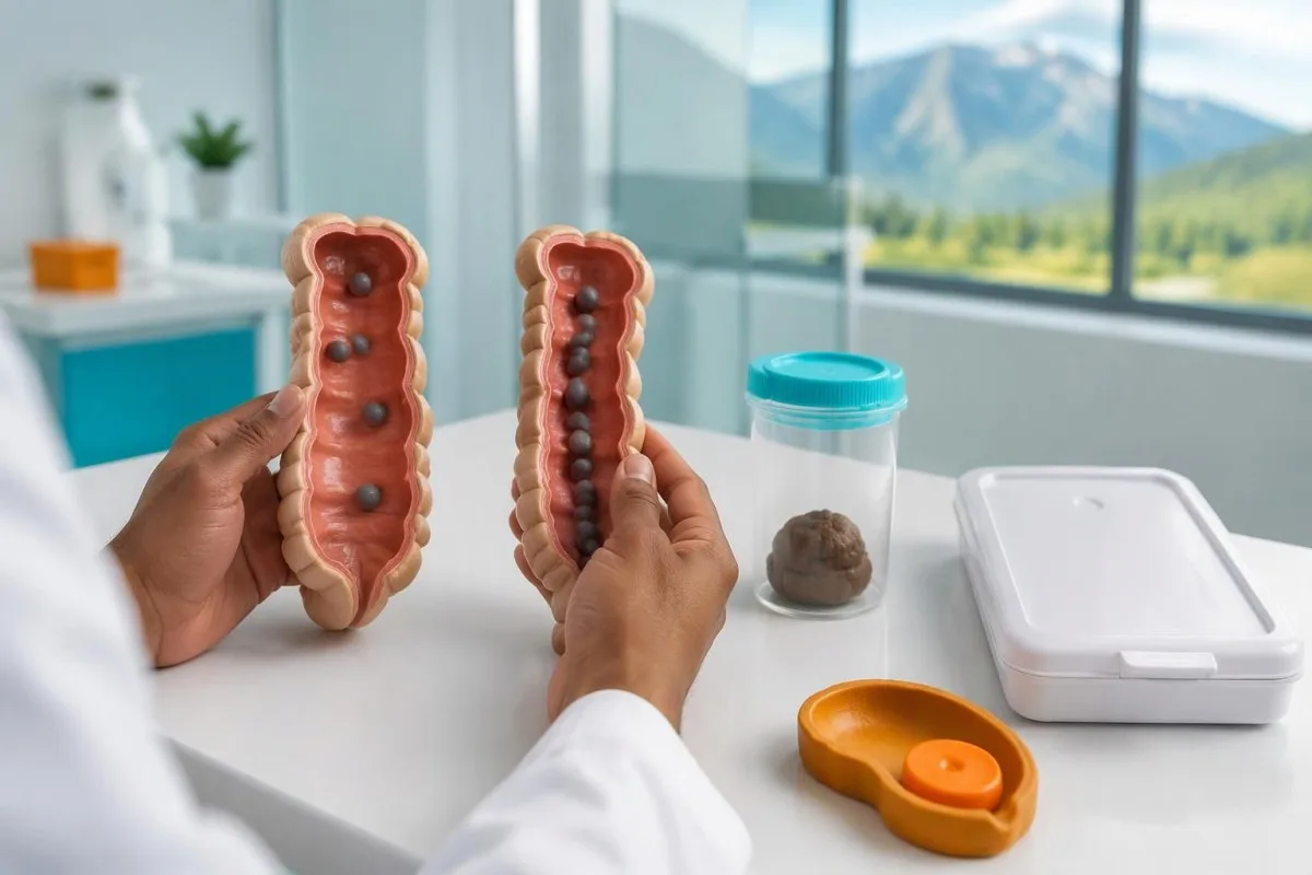
Reducing the number of stools does not prove that toxin exposure is ending; slowing intestinal transit may prolong contact with intestinal contents. These medicines also do not prevent dehydration unless fluid losses are adequately replaced, so a patient with fewer stools but worsening urine output still needs assessment rather than an assumption that the illness is resolving.
Ibuprofen, naproxen, and other nonsteroidal anti-inflammatory drugs can worsen kidney function during volume depletion, particularly in someone taking a diuretic or certain blood-pressure medicines. Paracetamol, also called acetaminophen, is generally the more suitable fever or pain option when otherwise appropriate, but dosing must follow the patient's age, weight, liver status, and the product label rather than a generic adult recommendation.
Bismuth products, herbal diarrhea remedies, and probiotics are not established treatments for preventing STEC-associated HUS; some bismuth products contain salicylate and are unsuitable for particular patients. Medication recommendations also vary by the cause of diarrhea—our C. difficile result explanation shows a different toxin-testing framework—so bring the exact stool report and every medicine name to the clinical review.
How should you hydrate safely after a positive result?
Oral rehydration solution is usually preferred for mild or moderate dehydration when a person can drink and urine output remains adequate. Persistent vomiting, substantial dehydration, or reduced urination may require supervised intravenous fluids; suspected kidney injury changes the fluid plan and makes forced drinking potentially unsafe.

Commercial oral rehydration solution uses glucose and sodium together to improve intestinal water absorption; plain water does not replace all the salts lost in diarrhea. Mix sachets with exactly the stated water volume, because concentrating the solution can create an excessive sodium load; small frequent sips are often better tolerated than drinking a large glass quickly.
Early fluid support may influence kidney outcomes, but the evidence does not justify a fixed home fluid target for every patient. Hickey et al. (2011) reported an observational association between intravenous volume expansion during the first 4 illness days and less oligoanuric renal failure among children who subsequently developed HUS; this is not proof that unrestricted fluids prevent HUS in all patients.
At Kantesti, we distinguish dehydration-related kidney changes from evidence that needs an urgent in-person assessment, without assuming that a higher creatinine is harmless. Our dehydration and GFR guide explains the overlap; reduced urine, swelling, or breathlessness after STEC requires examination because additional fluids may accumulate when the kidneys cannot clear them.
Which HUS warning signs require urgent assessment?
Substantially reduced urine, marked pallor, unusual bruising, severe fatigue, swelling, confusion, or seizures after STEC infection require urgent assessment for HUS. The complication commonly appears around days 5–10 after diarrhea begins, and improving bowel symptoms do not make these warning signs safe to observe at home.
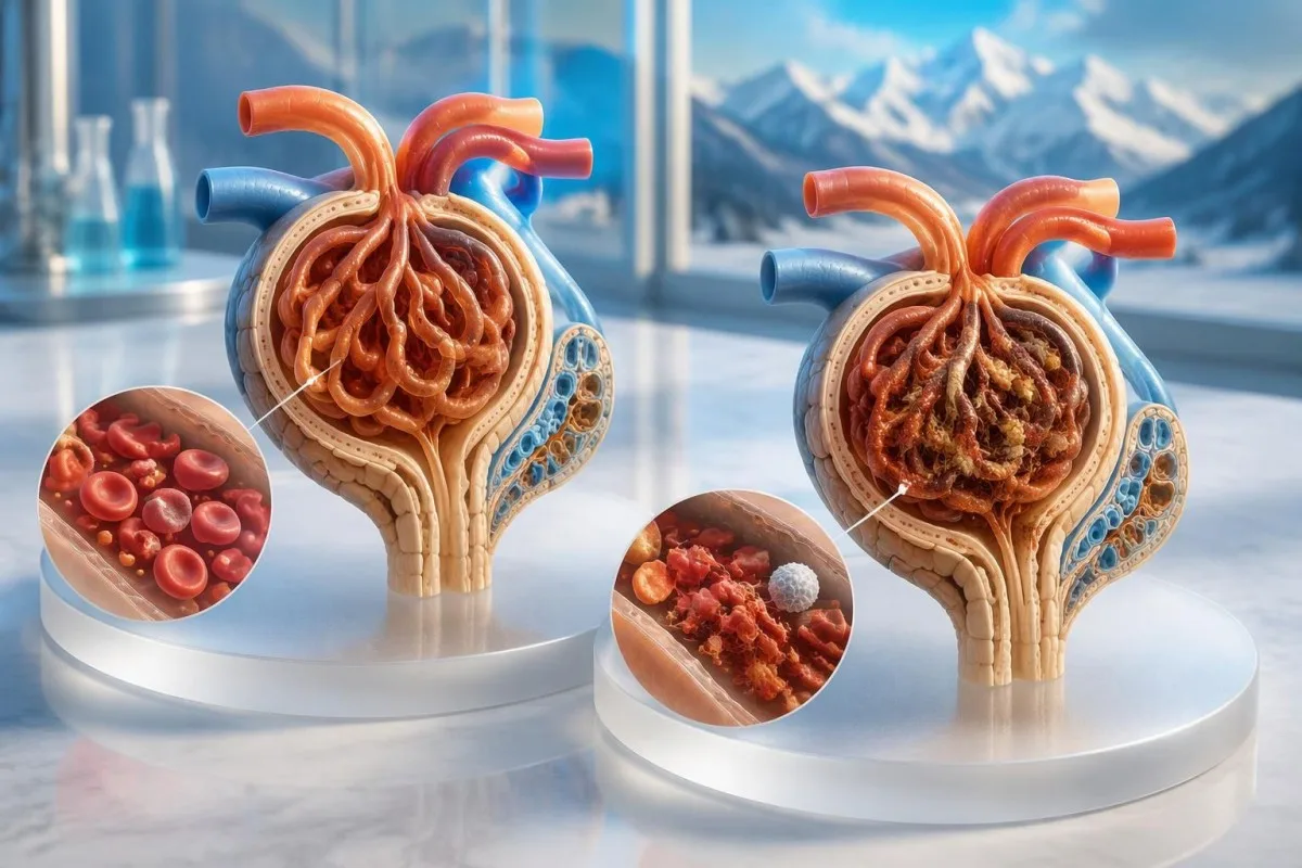
Reduced urine can reflect dehydration or kidney injury, and families cannot reliably distinguish these at home. A clinician may use urine output below 0.5 mL/kg/hour for at least 6 hours as an acute kidney injury criterion, but patients should not wait to measure that threshold when urine is clearly decreasing, a child has notably fewer wet diapers, or swelling appears.
Pallor and exhaustion can reflect falling hemoglobin, while new pinpoint spots or unexplained bruises may accompany a falling platelet count; the combination is more concerning than either symptom in isolation. Seek emergency help for seizures, confusion, difficulty waking, severe breathlessness, collapse, or rapidly worsening symptoms, and tell the receiving team that a Shiga toxin stool test was positive.
Severe abdominal pain, inability to retain fluids, or ongoing substantial bloody diarrhea also needs prompt assessment, even without the full HUS pattern. Our urgent kidney warning guide provides supporting context, but a numerical eGFR is not required before seeking care; waiting for the next planned test can lose valuable time.
Which blood and urine tests check for developing HUS?
Clinicians assess possible HUS using a complete blood count, platelets, creatinine, urea, electrolytes, and tests for red cellular element breakdown. Urine testing and urine-output measurements add kidney information; a single normal panel early in STEC illness cannot exclude a complication that develops later.
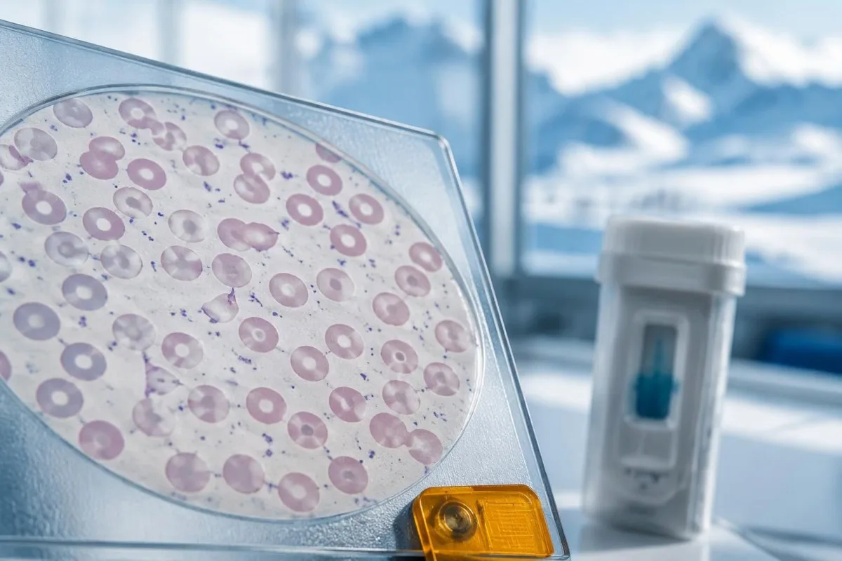
A falling platelet count together with rising creatinine and evidence of hemolysis raises concern for a thrombotic microangiopathy, including HUS. Adult platelet reference intervals commonly span 150–450 × 10^9/L, but a rapid decline within that interval can matter; the smear finding discussed in our schistocyte assessment guide must be interpreted alongside the overall pattern.
LDH, haptoglobin, bilirubin, and reticulocytes help evaluate hemolysis, but each has limitations: LDH can rise from several tissues, inflammation can affect haptoglobin, and marrow compensation may take time. Our LDH result explanation explains why elevated LDH alone does not establish HUS; clinicians may also request a direct antiglobulin test when the anemia mechanism is uncertain.
Creatinine may lag behind the start of kidney injury, and calculated eGFR becomes less reliable when creatinine is changing rapidly. A rise of at least 0.3 mg/dL, approximately 26.5 µmol/L, within 48 hours can meet an acute kidney injury criterion even if the latest value remains inside the printed range; urgent treatment decisions use trends, urine output, examination, and electrolytes together.
| Level | Range | What it means |
|---|---|---|
| Typical adult platelet interval | 150–450 × 10^9/L | Use the laboratory's interval; a substantial downward trend can be concerning before the count becomes low. |
| Creatinine change suggesting acute kidney injury | Rise ≥0.3 mg/dL within 48 hours, or ≥1.5 times baseline within 7 days | Equivalent to a rise of approximately 26.5 µmol/L for the first criterion; the cause requires clinical assessment. |
| Schistocytes supporting microangiopathy | >1% in an adult peripheral sample | Suspicious for thrombotic microangiopathy in the appropriate clinical context, but not diagnostic of HUS by itself. |
| Potassium requiring urgent clinical review | ≥6.0 mmol/L | May require urgent ECG assessment and treatment; sample-related false elevation must be considered without delaying necessary care. |
Why do creatinine and platelet trends matter more than flags?
Serial creatinine and platelet measurements can reveal developing HUS before a laboratory report shows several abnormal flags. The direction and speed of change matter, particularly during the first 1–2 weeks after diarrhea begins; repeat-testing intervals should be set by the treating clinician rather than an automated reminder alone.
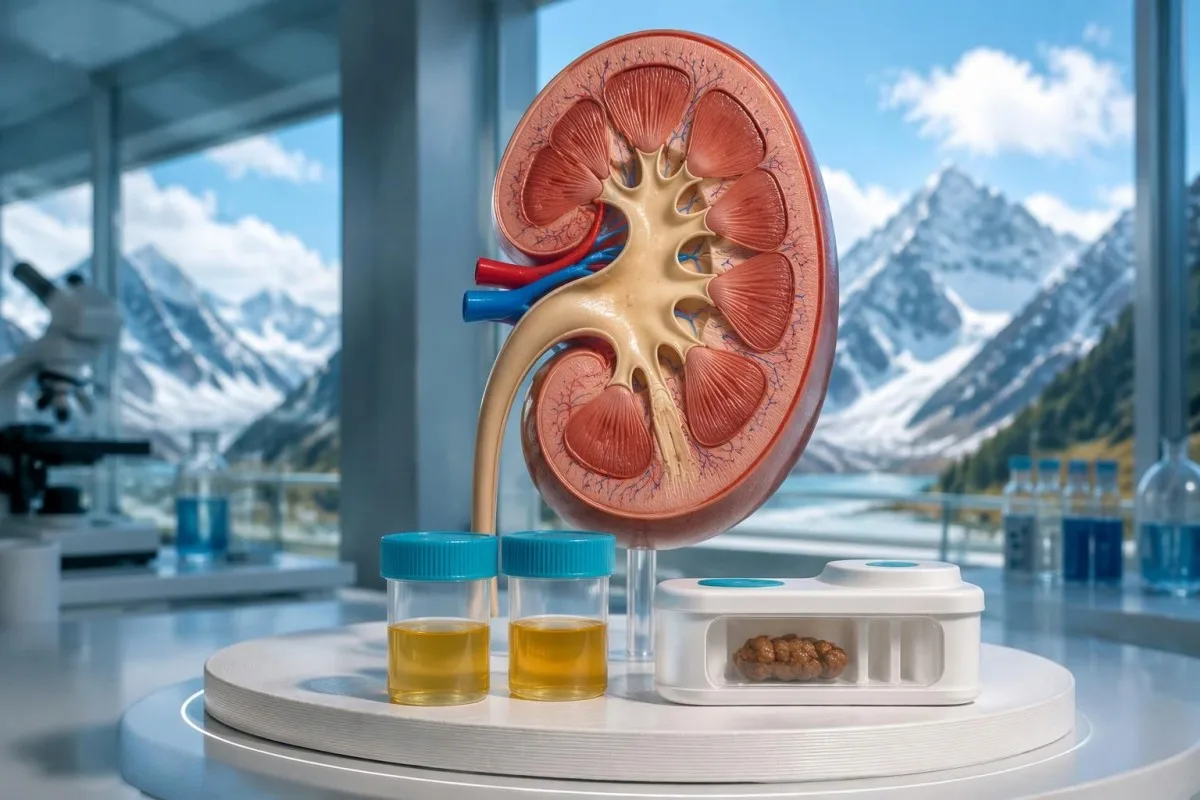
Consider a clearly hypothetical adult whose creatinine rises from 0.60 to 0.95 mg/dL over 48 hours while platelets fall from 310 to 165 × 10^9/L. Both final values might appear within that laboratory's reference intervals, yet the 0.35 mg/dL creatinine rise and substantial platelet decline warrant clinical review after STEC; they do not prove HUS, but they should not be labeled reassuringly normal.
Unexpected platelet results also require attention to sample quality: EDTA-related clumping can produce a falsely low count, which laboratories may investigate using a cell sample slide or an alternative collection tube. Our platelet clumping explanation describes that artifact, while the urea and creatinine guide explains why dehydration can alter kidney markers without identifying the whole cause.
Kantesti is an AI blood test interpretation platform that can place recorded platelet and creatinine values in chronological context, but it cannot replace the clinician's decisions about daily testing or hospital observation. Our validation and clinical oversight describes interpretation methodology; checking dates, units, baseline values, and whether results came from different laboratories is necessary before comparing any 2 reports.
What happens if HUS is suspected or confirmed?
Suspected HUS usually requires hospital assessment, where clinicians monitor kidney function, electrolytes, blood pressure, fluid balance, and the blood count. Treatment is mainly supportive and may include carefully managed fluids, red cellular element transfusion, and dialysis when kidney failure causes complications; a positive stool result alone does not mean dialysis is needed.
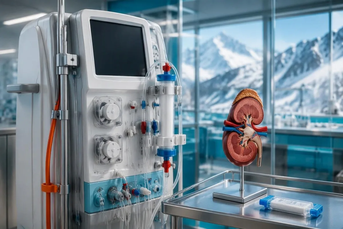
Dialysis decisions depend on the clinical picture—such as dangerous potassium accumulation, fluid overload, severe acidosis, or uremic complications—not a single universal creatinine cutoff. A person with creatinine of 3 mg/dL may have very different treatment needs from another person with the same result, depending on urine output, body size, baseline kidney function, and the speed of deterioration.
Platelet transfusion is not routinely given simply because the platelet count is low in a thrombotic microangiopathy; significant bleeding or a necessary procedure may create specific indications. Plasma exchange and complement-blocking treatment are not routine therapy for uncomplicated STEC-associated HUS, and any proposed use requires specialist assessment because alternative diagnoses and selected severe presentations may change the approach.
Anemia, low platelets, and kidney injury also occur in other emergencies, so the team must consider diagnoses beyond STEC-associated HUS when the pattern is atypical. In particular, ADAMTS13 testing for TTP can be relevant, because activity below 10% strongly supports thrombotic thrombocytopenic purpura in the appropriate context; suspected TTP may need treatment before confirmation returns.
How can you reduce household spread of STEC?
STEC spreads through fecal contamination, including contaminated food or water, animal contact, and person-to-person transmission. Wash hands with soap and water after toileting or diaper changes and before food handling; people with diarrhea should not prepare food for others, and certain jobs or childcare settings require public-health clearance.

Soap-and-water washing is especially useful after contact with stool because it physically removes contamination; alcohol hand sanitizer should not replace washing when hands are visibly soiled. Clean frequently touched bathroom surfaces using an appropriate household product according to its contact-time instructions, avoid sharing towels, and wash contaminated laundry without shaking it around the room.
Food precautions include avoiding unpasteurized dairy or juice, preventing raw-food cross-contamination, and cooking foods thoroughly with a food thermometer rather than judging doneness by color. STEC can survive when contamination is carried into the center of minced meat, which is one reason surface browning alone is insufficient; washing produce reduces surface contamination but cannot guarantee removal of every organism.
A person may continue shedding STEC after diarrhea stops, so 48 symptom-free hours is not a universal clearance rule for this organism. Our post-diarrhea contagiousness guide illustrates why recovery and transmission are separate questions; for STEC, childcare attendance, food-handling work, and repeat stool testing should follow the local public-health team's specific requirements.
When are recovery, repeat stool tests and kidney follow-up needed?
Many people recover from STEC diarrhea within about 5–7 days, but symptom improvement does not establish that HUS risk has passed. Repeat stool testing is usually driven by public-health requirements or a specific clinical question, while people who developed HUS need individualized follow-up of kidney function, urine protein, and blood pressure.
A negative repeat stool result cannot confirm that kidney function or platelet counts are safe, because stool clearance and systemic complications are different processes. Likewise, persistent gene detection does not automatically justify antibiotics; if diarrhea continues beyond approximately 14 days, the clinician may review ongoing infection, another detected pathogen, medication effects, or a post-infectious bowel disorder rather than repeatedly treating a test.
After HUS, apparently recovered creatinine does not rule out later hypertension or proteinuria, which is why renal follow-up can extend well beyond the initial hospital stay. New or persistent abnormalities may prompt a broader evaluation; complement testing context is relevant to selected alternative diagnoses, but normal C3 and C4 do not exclude complement-mediated HUS and these tests do not routinely confirm STEC-HUS.
We at Kantesti encourage readers to keep the first diarrhea date, hospitalization details, discharge plan, and prior kidney results together, because those details make later interpretation more meaningful. Returning to ordinary meals as tolerated is generally reasonable, but a person with ongoing kidney injury may need individualized sodium, potassium, protein, or fluid advice; a general wellness nutrition plan is not a substitute for renal dietetic guidance.
How can blood test interpretation support safe follow-up?
Blood test interpretation can help organize creatinine, platelets, hemoglobin, and electrolytes after STEC, but it cannot determine whether a symptomatic person is safe to stay home. Kantesti is an AI lab test interpretation service that explains recorded laboratory findings; reduced urination, unusual bruising, or neurological symptoms require direct medical assessment.

A useful same-day call includes 4 essentials: the exact positive test wording, the first diarrhea date, current urine output and fluid intake, and all medicines taken since symptoms began. Ask whether the report distinguishes stx1 from stx2, whether culture or public-health confirmation is pending, and whether blood testing is needed now or again during the next several days.
Interpretation should preserve uncertainty rather than convert every abnormal value into a diagnosis; a hemolyzed laboratory sample, for example, can distort potassium or LDH without proving hemolysis inside the body. Kantesti LTD, UK Company No. 17090423, describes its approach to sourcing and scope in our medical content standards; the treating clinician remains responsible for urgent assessment and treatment decisions.
The 2 Zenodo publications listed below are broader educational resources on serum proteins and complement testing, not clinical trials of STEC treatment or evidence that our service detects HUS. Direct support for the infection-management recommendations comes from the separately listed IDSA guideline and peer-reviewed studies; keeping those evidence categories distinct helps readers judge what each reference can—and cannot—support.
Frequently asked questions
Does shiga toxin positive mean I have E. coli?
A shiga toxin positive stool result usually indicates Shiga toxin-producing E. coli, or STEC, but the precise meaning depends on whether the assay detected toxin protein or toxin genes. Some tests report stx1 and stx2 separately, while others combine them. Rarely, another organism such as Shigella dysenteriae type 1 can produce Shiga toxin, so culture and the laboratory's assay details may clarify the source. A positive stool result does not by itself diagnose hemolytic uremic syndrome or measure kidney damage.
Should I take antibiotics for a Shiga toxin-positive stool test?
Antibiotics are generally avoided for STEC O157, Shiga toxin 2-producing STEC, or STEC when the toxin type is unknown because they may increase the risk of hemolytic uremic syndrome. In a 2016 meta-analysis, the lower-bias study subset found an association with higher HUS odds, OR 2.24, although the overall analysis was inconclusive. If an antibiotic was already prescribed, contact the prescriber promptly rather than independently changing treatment. A separate serious infection or suspected sepsis can require a different, specialist-guided decision.
Can I take Imodium or loperamide with STEC?
Loperamide and other bowel-slowing medicines should be avoided in suspected or confirmed STEC infection because they may increase complication risk. Having fewer stools after a medicine does not establish that the infection is resolving or that the kidneys are safe. HUS commonly appears around days 5–10 after diarrhea begins, sometimes as diarrhea improves. Contact a clinician about symptom relief and seek urgent assessment for reduced urine, unusual bruising, marked pallor, or severe weakness.
When should I go to hospital after a Shiga toxin-positive result?
Seek urgent assessment after a Shiga toxin-positive result if urine output falls substantially, unexplained bruising appears, or you develop marked pallor, severe fatigue, swelling, or worsening illness. Confusion, seizures, collapse, difficulty waking, or severe breathlessness warrant emergency help. HUS often emerges around days 5–10 after diarrhea starts, so improving bowel symptoms should not override these warning signs. Severe abdominal pain, persistent vomiting, or substantial bloody diarrhea also needs prompt clinical evaluation.
Can normal blood tests rule out HUS?
One normal blood panel cannot rule out HUS that is still developing after STEC infection. Clinicians use repeat platelet counts, hemoglobin, creatinine, electrolytes, and hemolysis tests when the illness pattern warrants monitoring. A creatinine rise of at least 0.3 mg/dL, approximately 26.5 µmol/L, within 48 hours can indicate acute kidney injury even if the final value remains within the laboratory's reference interval. Symptoms and urine output matter alongside the numerical results.
How long am I contagious after STEC diarrhea stops?
STEC can continue to be shed after diarrhea resolves, so feeling well does not guarantee that transmission risk has ended. A 48-hour symptom-free interval is not a universal STEC clearance rule, particularly for young children, food handlers, or people providing close personal care. Local public-health authorities may require repeat stool samples or specific exclusion periods based on the person's setting and strain. Continue soap-and-water handwashing and follow the clearance instructions provided for your situation.
Get your blood test results explained
Upload your lab report and Kantesti's AI explains each biomarker in plain language within seconds. Used by more than 2 million people worldwide.
References
- Klein, T., Mitchell, S., & Weber, H. (2026). Serum Proteins Guide: Globulins, Albumin & A/G Ratio Blood Test. Kantesti AI Medical Research. DOI
- Klein, T., Mitchell, S., & Weber, H. (2026). C3 C4 Complement Blood Test & ANA Titer Guide. Kantesti AI Medical Research. DOI
- Shane AL et al. (2017). 2017 Infectious Diseases Society of America Clinical Practice Guidelines for the Diagnosis and Management of Infectious Diarrhea. Clinical Infectious Diseases. DOI
- Freedman SB et al. (2016). Shiga Toxin-Producing Escherichia coli Infection, Antibiotics, and Risk of Developing Hemolytic Uremic Syndrome: A Meta-analysis. Clinical Infectious Diseases. DOI
- Hickey CA et al. (2011). Early Volume Expansion During Diarrhea and Relative Nephroprotection During Subsequent Hemolytic Uremic Syndrome. Archives of Pediatrics & Adolescent Medicine.
- 2M+Tests analysed
- 127+Countries
- 100+Languages
About the medical team


E-E-A-T Trust Signals
Experience
Physician-led clinical review of lab interpretation workflows.
Expertise
Laboratory medicine focus on how biomarkers behave in clinical context.
Authoritativeness
Medically reviewed by Dr. Thomas Klein, with contributions from Dr. Sarah Mitchell and Prof. Dr. Hans Weber.
Trustworthiness
Evidence-based interpretation with clear follow-up pathways to reduce alarm.


Medical disclaimer
This article is for educational purposes only and does not constitute medical advice. Always consult a qualified healthcare provider for diagnosis and treatment decisions.
Published · Contact the editorial team
Published by Kantesti Ltd, registered in England & Wales, company no. 17090423, London, United Kingdom.
