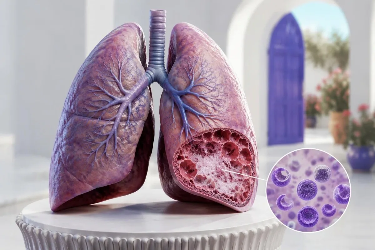
A pneumocystis PCR positive result detects fungal DNA, not automatically pneumonia. Active PJP is more likely with immune suppression, compatible breathing symptoms, low oxygen and imaging changes; severe or rapidly worsening breathlessness needs emergency evaluation.

Key takeaways
- Positive PCR means Pneumocystis jirovecii DNA was detected; 1 positive result cannot independently distinguish pneumonia from colonization.
- Urgent breathing signs include severe breathlessness, confusion or a repeated resting oxygen saturation of 92% or lower when that is not your established baseline.
- Specimen type matters: bronchoalveolar lavage samples the lower respiratory tract more directly than a throat or nasal swab.
- PCR quantity has no universal pneumonia cutoff; a Ct of 30 from 1 laboratory cannot be interpreted using another laboratory's threshold.
- Immune risk changes interpretation: most HIV-associated PJP occurs with CD4 counts below 200 cells/µL, but non-HIV PJP does not follow one CD4 cutoff.
- Beta-D-glucan supports the overall assessment but is not Pneumocystis-specific; a 2020 meta-analysis reported approximately 91% sensitivity and 79% specificity.
- LDH and routine blood tests cannot confirm or exclude PJP; even an LDH result twice a laboratory's upper limit needs another explanation or corroborating evidence.
- Treatment and prevention are separate decisions: prolonged prednisone-equivalent exposure around 20 mg/day for 4 weeks is a commonly used risk marker, not an automatic treatment rule.
What a positive Pneumocystis PCR actually proves
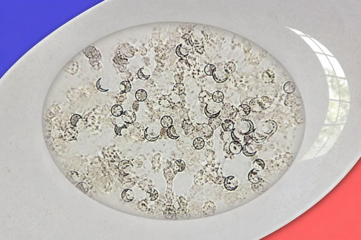
A pneumocystis PCR positive result establishes that the tested sample contained detectable Pneumocystis jirovecii DNA—not that the organism is causing pneumonia. Interpretation requires at least 3 separate questions: where the sample came from, how vulnerable the person is, and whether the lungs show a compatible illness.
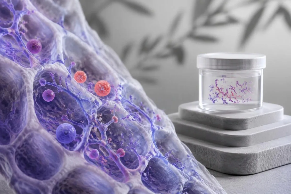
Colonization means the organism is present without clinical evidence that it is causing PJP pneumonia. A highly sensitive assay may detect a small amount of DNA in someone with chronic lung disease or immune suppression, so a genuine analytical positive can still have a different clinical meaning from active disease.
PCR amplifies genetic material rather than measuring whether organisms are alive or damaging lung tissue. There are at least 3 explanations for detection: active pneumonia, colonization, or residual DNA following treatment; uncommon contamination or technical problems become another consideration when an unexpected low-level result conflicts with everything else.
Kantesti is an AI blood test analyzer that can help explain accompanying laboratory findings, but it cannot establish PJP from a respiratory PCR result alone. Our organization and clinical scope distinguish educational interpretation from diagnosis, which requires symptoms, oxygen assessment and imaging that a laboratory upload cannot supply.
When breathing symptoms need urgent evaluation
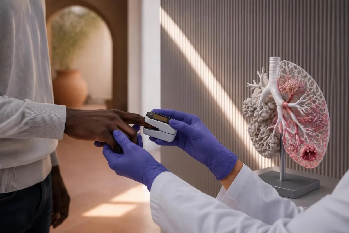
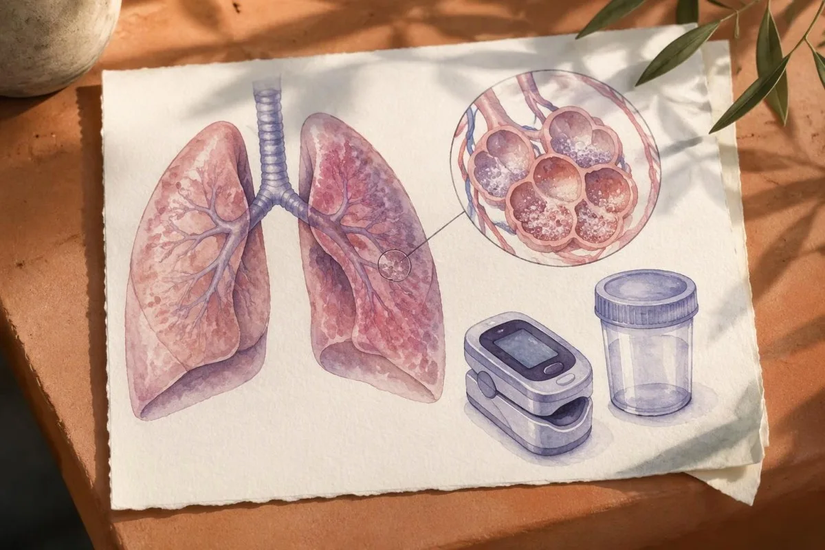
Severe or rapidly worsening breathlessness requires emergency assessment, regardless of whether PCR quantity is high or low. A repeated resting oxygen saturation of 92% or lower, when this is not the person's established baseline or prescribed target, is a reason to seek urgent help rather than await another laboratory result.
Inability to speak full sentences, blue or gray lips, confusion, collapse, or significant chest pain are emergency signs even without an oxygen reading. Call the local emergency number—999 or 112 in the UK—and do not drive yourself if breathing is severely impaired; our shortness-of-breath test guide explains why outpatient blood testing is not the first step.
An oxygen saturation of 93–94%, a sustained fall below your usual reading, or new breathlessness during ordinary activities deserves same-day clinical advice, especially with immune suppression. People whose usual saturation is lower need an individualized plan; a change from baseline and increasing oxygen requirements can matter more than one general threshold.
Home pulse oximeters can misread with movement, cold fingers or poor circulation, and some devices overestimate oxygen saturation more often in people with darker skin. A reassuring 96% reading must not override obvious breathing difficulty; clinicians may use an arterial blood gas assessment when oxygen delivery and gas exchange need more precise evaluation.
Who is most at risk of active PJP pneumonia?
PJP risk rises when cell-mediated immunity is impaired, including advanced HIV, transplantation, some cancers and immunosuppressive treatment. Most HIV-associated PJP occurs at a CD4 count below 200 cells/µL, but a person without HIV can develop PJP without crossing that same threshold.
HIV viral suppression and the CD4 count answer different questions: viral load measures replication, while CD4 cells help describe immune vulnerability. A person with a CD4 count of 80 cells/µL and new breathlessness needs prompt assessment even if a recent viral load was low; our HIV viral load explanation helps separate these measurements.
Prednisone-equivalent exposure around 20 mg/day for 4 weeks is a commonly used warning marker for PJP risk, especially alongside another immunosuppressant. It is not a universal boundary: combined treatments, lymphocyte depletion, underlying disease and recent dose changes can create substantial risk below that exposure.
Transplant recipients should report the full immunosuppressive regimen, recent rejection treatment and missed preventive medication—not just one drug concentration. A tacrolimus trough within its prescribed range does not certify protection from PJP; the tacrolimus monitoring guide explains why a medication level measures exposure rather than the entire immune response.
Why BAL, induced sputum and swabs differ
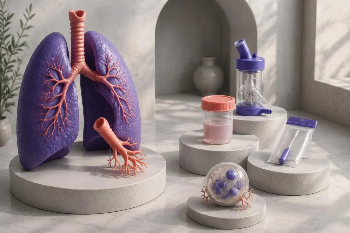
Bronchoalveolar lavage, or BAL, samples the lower respiratory tract more directly than an upper-airway swab, making it particularly useful when PJP is suspected. Pneumocystis jirovecii PCR interpretation must identify the exact specimen; the word positive carries different evidential weight in these 3 settings.
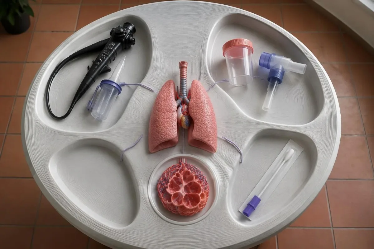
Induced sputum is collected after inhaling a solution that helps mobilize lower-airway secretions; it is not equivalent to saliva or casually coughed material. The 2016 ECIL diagnostic guideline supports real-time PCR and identifies BAL as the preferred specimen in hematological patients, while emphasizing limitations of less invasive samples (Alanio et al., 2016).
A positive BAL PCR still does not independently separate colonization from pneumonia, but a negative, technically adequate BAL PCR usually reduces the likelihood substantially. A negative upper-airway result is less reassuring because the relevant organisms may be concentrated farther down; specimen quality, assay inhibition and previous treatment also affect what a negative means.
BAL fluid is diluted by instilled saline, so copies/mL are not a direct measurement of organisms in undiluted lung tissue. The sampled segment, recovered volume and processing method can change the reported quantity; our serum versus BAL antigen guide illustrates why specimen compartments also matter in a different fungal test, without making the two assays interchangeable.
How clinical findings support a PJP pneumonia diagnosis
PJP pneumonia diagnosis combines immune risk, a compatible respiratory illness, imaging and microbiological evidence. The combination of new breathlessness, reduced oxygen and bilateral ground-glass changes is more persuasive than any 1 finding, but none of these features is unique to Pneumocystis.
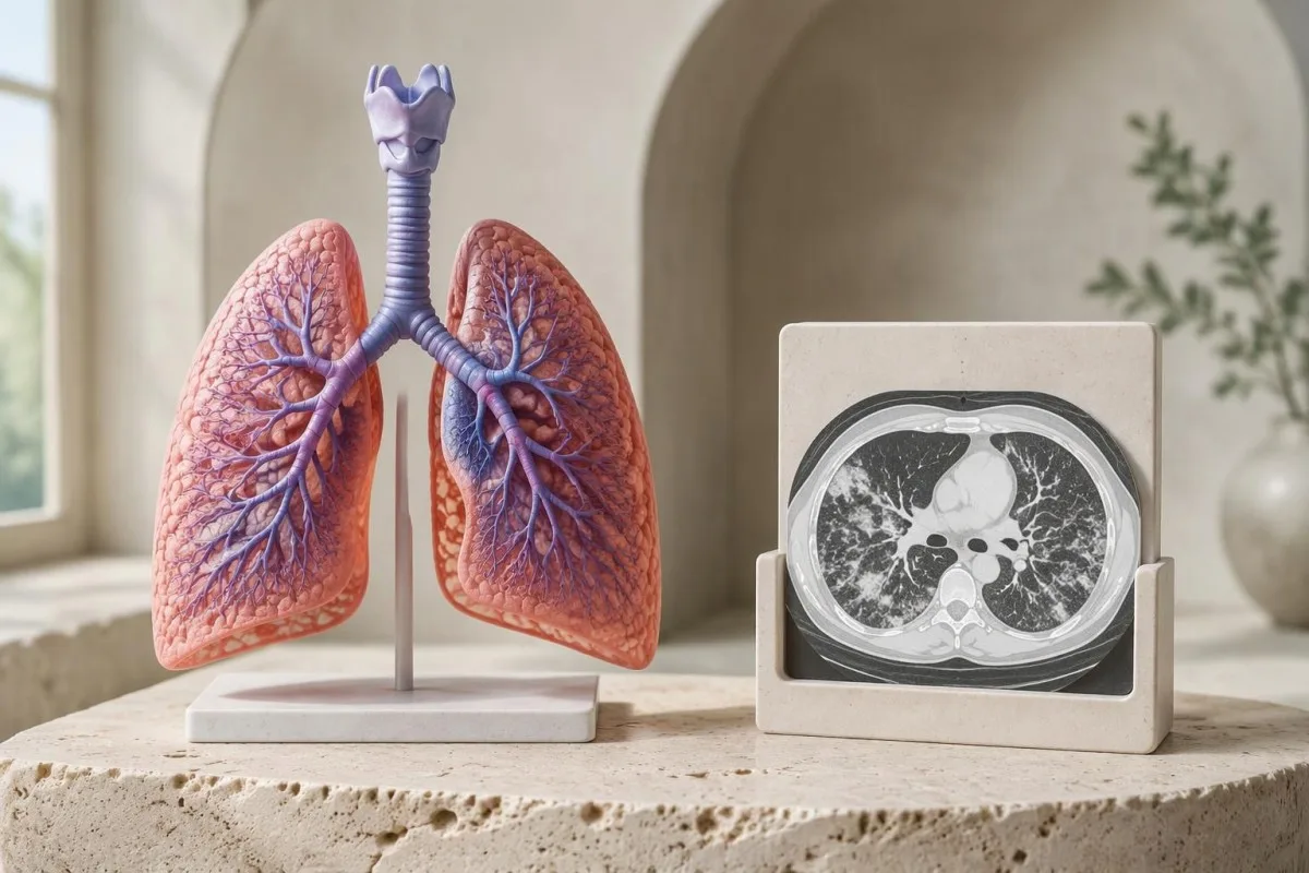
HIV-associated PJP often develops over days to weeks, with dry cough, fever and increasing breathlessness; non-HIV PJP can deteriorate over a shorter period. Fever may be absent, especially during immunosuppressive treatment, and listening to the chest can reveal surprisingly little despite impaired oxygen transfer.
A chest radiograph can appear normal early, while CT may reveal ground-glass opacity that the radiograph misses. However, the same pattern can occur with viral pneumonia, medication-related pneumonitis, edema or other lung conditions; an alternative test such as Mycoplasma IgM also needs its own timing and confirmation checks.
Finding another respiratory pathogen does not eliminate PJP because 2 conditions can coexist, particularly in immunocompromised people. Kantesti's laboratory marker guide can help readers identify accompanying tests, but a plausible diagnosis still needs an explanation for the person's oxygen level, imaging distribution and rate of deterioration.
Why Ct values and PCR quantity cutoffs vary
There is no universal Pneumocystis PCR quantity or cycle-threshold cutoff that proves pneumonia. Within the same validated assay and specimen type, a lower Ct generally indicates more starting DNA, but a Ct of 30 cannot be translated safely into another laboratory's disease threshold.
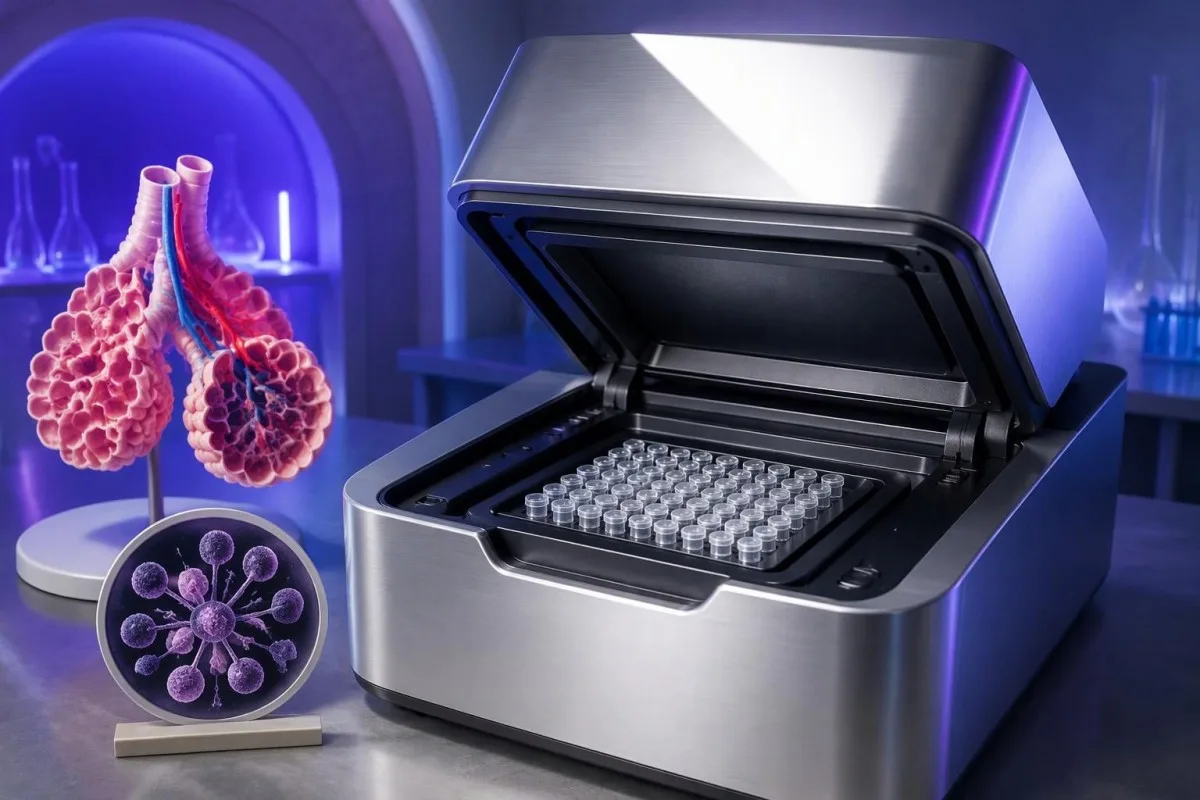
Gene target, extraction volume, amplification chemistry and calibration standards all influence the result. Some targets occur in multiple copies per organism, so 1,000 target copies/mL is not necessarily 1,000 organisms/mL; laboratories may report copies per reaction, copies per volume, Ct, or simply detected.
Under ideal doubling efficiency, a difference of 3 PCR cycles corresponds to approximately an eightfold difference in starting target material. That mathematical relationship does not establish an eightfold difference in pneumonia severity, particularly when BAL dilution or sample quality differs; our qualitative and quantitative test guide explains the distinction between detecting and measuring.
Fauchier and colleagues found that quantitative PCR helped distinguish PJP from colonization in their 2016 study, but interpretation differed between HIV-positive and HIV-negative groups (Fauchier et al., 2016). Ask whether your laboratory has validated lower and upper decision limits with a gray zone between them; never substitute an internet cutoff for that local validation.
What beta-D-glucan adds—and what it cannot prove
Serum beta-D-glucan can support suspected PJP, but it is not specific for Pneumocystis and cannot diagnose pneumonia alone. Del Corpo and colleagues' 2020 meta-analysis reported pooled sensitivity of approximately 91% and specificity of 79%, meaning both false negatives and false positives remain clinically relevant.
The same meta-analysis reported sensitivity around 94% in people with HIV and 86% in people without HIV (Del Corpo et al., 2020). A negative result therefore cannot safely settle a high-suspicion case, especially outside HIV; the consequences of missing rapidly progressive pneumonia are different from dismissing incidental low-level detection.
Some assays classify less than 60 pg/mL as negative, 60–79 pg/mL as indeterminate and 80 pg/mL or higher as positive, but other methods use different thresholds. These are assay-specific detection categories, not universal PJP severity bands; always read the method and reference information printed on the report.
Other fungal conditions, some infusions such as immunoglobulin products, and particular medical materials or procedures can raise beta-D-glucan without PJP. The timing of these exposures matters; a separate Histoplasma antigen interpretation may be relevant when geography and clinical findings suggest another fungus, rather than assuming every fungal marker confirms the same diagnosis.
Can LDH, CRP or lymphocytes settle the diagnosis?
LDH, CRP and routine lymphocyte counts cannot independently confirm or exclude PJP. An LDH value of 600 U/L, for example, may indicate tissue injury but does not identify its location or cause; the laboratory's upper limit and sample quality are needed before even calling the elevation meaningful.
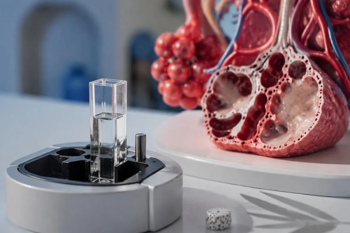
LDH may rise with lung injury, hemolysis, liver problems or other conditions, and a hemolyzed sample can raise it artificially. If a laboratory's upper limit is 250 U/L, an LDH of 600 U/L is 2.4 times that limit—not a PJP diagnostic threshold; our LDH test explanation separates enzyme release from disease identification.
A normal total white-cell count or modest CRP result does not reliably exclude serious respiratory illness during immune suppression. Steroids can also change white-cell distributions, while the absolute lymphocyte count is not the same measurement as a CD4 count in cells/µL; replacing one with the other can misclassify immune risk.
Trends can reveal change, but a fall from 600 to 400 U/L cannot by itself prove treatment success or organism clearance. Our LDH and reticulocyte guide discusses other reasons for LDH elevation, which can help clinicians decide whether a respiratory process or a separate cellular turnover problem better explains the result.
How colonization and pneumonia look in worked examples
Pneumocystis colonization vs infection is a probability assessment, not a binary reading of the PCR line. These 2 hypothetical examples show why the same detected result can prompt routine clarification in one setting and immediate hospital evaluation in another; they are illustrations, not accounts of actual patients.
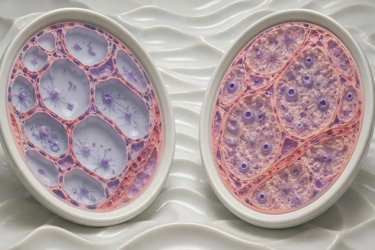
Imagine a 67-year-old with stable chronic lung disease, no new respiratory symptoms, usual resting saturation of 96%, and low-level PCR detection during an investigation for another reason. If imaging is unchanged and there is no major new immune suppression, colonization becomes more plausible—but the treating team still checks why the sample was obtained and whether preventive treatment is indicated.
Now imagine a 45-year-old receiving substantial immunosuppressive treatment, with worsening dry cough, saturation of 89% and new bilateral ground-glass opacity. Even a low-level positive could be clinically significant, particularly in non-HIV PJP; the same detection-versus-disease principle appears in our adenovirus detection discussion, although its shedding biology and treatment are different.
Kantesti is an AI blood test interpretation platform that can help organize accompanying results, but these examples require clinical information beyond a laboratory PDF. Our AI technology explanation describes the interpretation workflow; no numerical model should override an oxygen saturation of 89% or obvious respiratory deterioration.
What changes after prophylaxis or PJP treatment?
Previous PJP treatment or preventive medication can change organism burden and the meaning of PCR detection. A positive result after 7 days of treatment does not automatically mean treatment failure, while a negative or low-level result during prophylaxis does not automatically exclude breakthrough pneumonia.
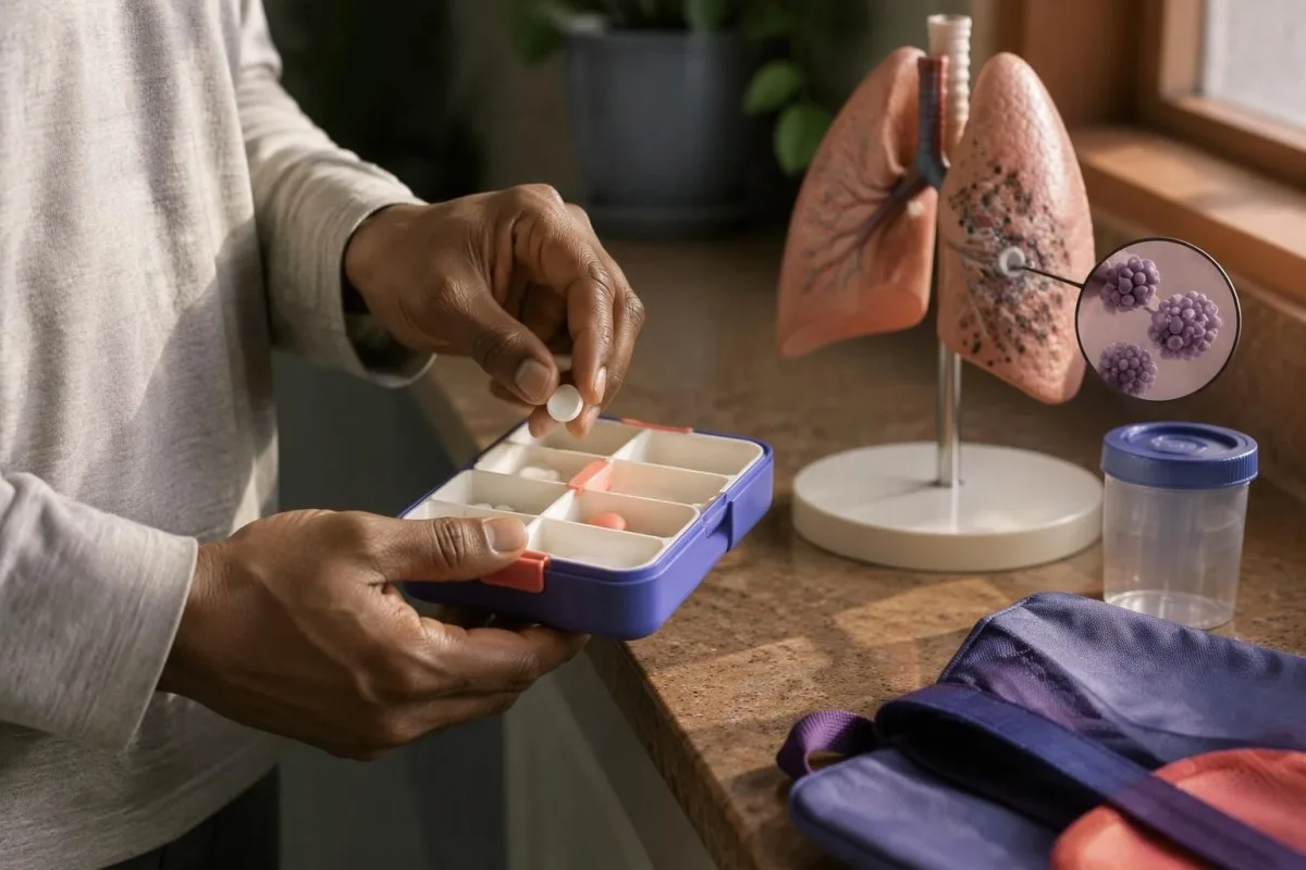
Preventive treatment lowers risk but does not reduce it to 0%, especially when doses are missed, absorption is impaired or immune suppression intensifies. The clinician needs the actual drug, prescribed schedule, recent adherence and any vomiting—not simply the statement that you take prophylaxis.
PCR can remain positive because genetic material persists after organisms have been damaged or cleared, so routine repeat PCR is not a reliable stand-alone test of cure. A repeat at 1 week cannot be interpreted without the original specimen type, assay and clinical response; our PCR timing guide explains a similar timing principle in a different respiratory condition, not a shared clearance schedule.
Worsening after treatment starts can reflect ongoing PJP, an inflammatory response, another infection, medication toxicity or a complication such as pneumothorax. Kantesti cannot distinguish these from a laboratory trend alone; new breathlessness or falling oxygen requires reassessment rather than waiting for the next planned weekly test.
When clinicians treat before every result returns
Clinicians may start PJP treatment before full confirmation when the illness and immune risk make delay dangerous. Trimethoprim-sulfamethoxazole is usually first-line therapy; standard treatment commonly uses 15–20 mg/kg/day of the trimethoprim component, with renal adjustment and specialist selection of dose and duration.
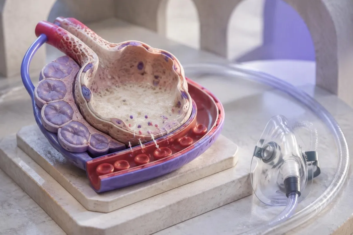
For a hypothetical 60 kg adult, that standard dosing range corresponds to 900–1,200 mg/day of trimethoprim, divided into prescribed doses—not that amount of the combined tablet product. This is an explanation of guideline dosing, not a self-treatment instruction; toxicity risk, kidney function and alternative dosing approaches need clinician assessment.
The US NIH opportunistic-infection guideline recommends adjunctive corticosteroids for HIV-associated moderate-to-severe PJP when room-air PaO2 is below 70 mmHg, or the alveolar–arterial oxygen gradient is 35 mmHg or higher. These correspond to approximately 9.3 kPa and 4.7 kPa; evidence for adjunctive steroids in non-HIV PJP is less settled, and an oxygen saturation percentage is not interchangeable with either criterion.
Treatment may require potassium, creatinine and complete blood count monitoring because drug effects and illness can produce overlapping abnormalities. Trimethoprim can raise creatinine without a matching fall in filtration, but true kidney injury remains possible; our kidney marker guide provides context, while G6PD testing considerations matter before certain alternatives such as dapsone or primaquine.
What to ask the laboratory and treating team
Ask for the specimen, assay interpretation and clinical plan—not merely whether PCR is positive. A useful conversation covers 5 practical details: collection site, quantitative result, laboratory decision limits, previous treatment and the findings that support or argue against pneumonia.
Start with the report wording: was the result simply detected, quantified, or detected below the limit of quantification? That last category means the assay found its target but cannot measure it with the validated precision of a quantified result; it does not mean the signal is false or clinically irrelevant.
Next ask whether the laboratory's pneumonia-versus-colonization cutoffs apply to your exact specimen and patient group. A threshold validated in BAL from HIV-positive adults may not work for induced sputum from a non-HIV transplant recipient; request the laboratory comment rather than comparing your Ct with an unrelated online example.
Kantesti can help organize these questions and accompanying laboratory terminology, but our responsible AI approach does not replace respiratory triage. Before using any automated explanation, verify the source report and units with our medical report safety checks, and ask explicitly what to do if oxygen falls to 92% or below or symptoms worsen.
What follow-up matters after a positive PCR?
Follow-up should track symptoms, oxygen needs, medication tolerance and immune risk—not aim only for a negative PCR. Someone without pneumonia still needs a clear plan for reassessment and prevention, while a treated patient may need laboratory monitoring during a course commonly lasting 21 days in HIV-associated PJP.
An asymptomatic person with likely colonization should clarify whether the immune-suppressing regimen warrants PJP prophylaxis and what symptoms trigger same-day review. There is no universally accepted rule to treat every colonized person or repeat PCR after 2 weeks; a scheduled check should answer a specific clinical question.
For treated PJP, improving breathlessness and decreasing supplemental oxygen requirements are more clinically informative than a small Ct change. A comparison between 2 laboratory visits also needs consistent methods and awareness of medication effects; our blood-test timeline guide explains why a numerical change may reflect timing rather than recovery.
Do not stop preventive medication, reduce immune-suppressing treatment or declare yourself recovered because one PCR becomes negative. Agree on 3 items before discharge or follow-up: the monitoring schedule, who will review abnormal results, and the emergency plan; treatment duration and future prophylaxis depend on the underlying reason for immune vulnerability.
Our interpretation limits and research publications
This article supports understanding of laboratory results; it does not diagnose PJP or provide a personal prescription. The 3 external medical references below directly support the discussion of respiratory sampling, quantitative PCR and beta-D-glucan, whereas the listed repository publications have narrower or unrelated subject matter.
Kantesti is an AI-powered blood test analysis tool that can help explain accompanying LDH, blood count and kidney results while recognizing that respiratory diagnosis needs information beyond those tests. Our medical content standards set out the distinction between evidence-based education and individualized clinical decisions, including why 1 positive PCR cannot settle pneumonia versus colonization.
The Figshare publication titled B Negative Blood Type, LDH Blood Test & Reticulocyte Count Guide is listed for its discussion of LDH and hematology, not as validation of a PJP diagnostic algorithm. A repository DOI makes a document persistently identifiable; it does not, by itself, establish peer review, diagnostic accuracy or the validity of any Ct threshold.
The second Figshare publication addresses gastrointestinal symptoms rather than Pneumocystis testing and does not support any PJP-specific claim in this article. Its gastrointestinal companion guide belongs to a separate symptom discussion; neither of these 2 repository items should be substituted for the directly relevant diagnostic guideline and peer-reviewed studies below.
Frequently asked questions
Does a positive Pneumocystis PCR mean I have pneumonia?
A positive Pneumocystis PCR means fungal DNA was detected, but it does not independently establish pneumonia. Active PJP becomes more likely when immune suppression, compatible symptoms, low oxygen and imaging changes occur together. A single low-level positive without those findings may represent colonization or residual DNA after treatment. Severe breathlessness or a repeated resting oxygen saturation of 92% or lower, outside an established lower baseline, needs urgent evaluation.
What Ct value confirms Pneumocystis pneumonia?
No universal Ct value confirms Pneumocystis pneumonia across laboratories. A Ct of 30 from one assay cannot be interpreted using another assay's cutoff because gene targets, extraction methods and specimen processing differ. Lower Ct generally means more starting DNA within the same assay, but organism burden is not identical to disease severity. Ask the laboratory whether its decision limits were validated for your specimen and patient group.
Can a negative beta-D-glucan rule out PJP?
A negative serum beta-D-glucan reduces the likelihood of PJP but cannot reliably exclude it when clinical suspicion is high. A 2020 meta-analysis reported overall sensitivity around 91%, with approximately 94% sensitivity in people with HIV and 86% in people without HIV. These figures mean some genuine PJP cases have negative results. Interpretation must also account for the assay threshold, respiratory specimen results and the person's oxygen and imaging findings.
Is a positive BAL PCR more meaningful than a positive throat swab?
A positive bronchoalveolar lavage PCR generally provides more direct evidence of lower-respiratory Pneumocystis detection than a positive throat swab. However, even 1 positive BAL result cannot independently distinguish colonization from pneumonia. BAL dilution, the sampled lung segment and assay methodology affect quantitative results. A negative upper-airway swab is less reliable for excluding PJP than a technically adequate negative BAL PCR.
Do I need treatment if Pneumocystis colonization is suspected?
Suspected Pneumocystis colonization does not automatically require a full pneumonia treatment course. The clinician separately assesses whether immune risk warrants preventive medication, including factors such as prolonged prednisone-equivalent exposure around 20 mg/day for 4 weeks and other immunosuppressants. That exposure is a risk marker, not a universal prescribing rule. New breathlessness, falling oxygen or compatible imaging changes require reassessment because low-level detection can still accompany active PJP.
Should Pneumocystis PCR become negative after treatment?
Pneumocystis PCR does not have to become negative immediately for treatment to be working. Genetic material can persist, so a repeat positive after 7 days does not independently establish treatment failure. Follow-up focuses on breathing, oxygen requirements, medication tolerance and relevant laboratory monitoring. HIV-associated PJP treatment commonly lasts 21 days, but duration and dosing must be determined by the treating team.
Get your blood test results explained
Upload your lab report and Kantesti's AI explains each biomarker in plain language within seconds. Used by more than 2 million people worldwide.
References
- Klein, T., Mitchell, S., & Weber, H. (2026). B Negative Blood Type, LDH Blood Test & Reticulocyte Count Guide. Kantesti AI Medical Research. DOI
- Klein, T., Mitchell, S., & Weber, H. (2026). Diarrhea After Fasting, Black Specks in Stool & GI Guide 2026. Kantesti AI Medical Research. DOI
- Alanio A et al. (2016). ECIL guidelines for the diagnosis of Pneumocystis jirovecii pneumonia in patients with haematological malignancies and stem cell transplant recipients. Journal of Antimicrobial Chemotherapy.
- Fauchier T et al. (2016). Detection of Pneumocystis jirovecii by Quantitative PCR To Differentiate Colonization and Pneumonia in Immunocompromised HIV-Positive and HIV-Negative Patients. Journal of Clinical Microbiology. DOI
- Del Corpo O et al. (2020). Diagnostic accuracy of serum (1-3)-β-D-glucan for Pneumocystis jirovecii pneumonia: a systematic review and meta-analysis. Clinical Microbiology and Infection.
- 2M+Tests analysed
- 127+Countries
- 100+Languages
About the medical team


E-E-A-T Trust Signals
Experience
Physician-led clinical review of lab interpretation workflows.
Expertise
Laboratory medicine focus on how biomarkers behave in clinical context.
Authoritativeness
Medically reviewed by Dr. Thomas Klein, with contributions from Dr. Sarah Mitchell and Prof. Dr. Hans Weber.
Trustworthiness
Evidence-based interpretation with clear follow-up pathways to reduce alarm.
Medical disclaimer
This article is for educational purposes only and does not constitute medical advice. Always consult a qualified healthcare provider for diagnosis and treatment decisions.
Published · Contact the editorial team
Published by Kantesti Ltd, registered in England & Wales, company no. 17090423, London, United Kingdom.
