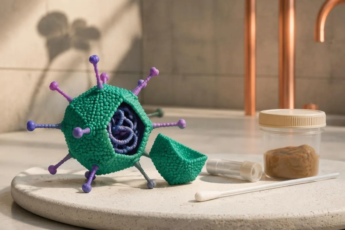
An adenovirus PCR detected result identifies viral DNA—not necessarily a new infection or ongoing contagiousness. DNA can remain detectable for weeks or months after recovery; interpretation depends on the sample, symptoms, and immune status. Immunocompromised patients need earlier assessment because persistent detection can also represent continuing replication.

Key takeaways
- Detected means DNA found, not proof that adenovirus is currently causing symptoms or that infectious virus is present.
- Prolonged detection can last weeks to months, particularly in children; there is no universal day when every adenovirus PCR becomes negative.
- Respiratory sampling should be interpreted against cough, fever, breathing changes, and the timing of the swab—not against stool findings alone.
- Stool panels often specifically detect enteric adenovirus types 40 and 41; a broader adenovirus assay can have a different clinical meaning.
- Contagiousness cannot be calculated from one positive PCR or a fixed cycle-threshold value; symptoms, exposure setting, and immune status matter.
- Routine recovery usually does not require repeat PCR testing or antiviral treatment in an otherwise healthy person whose symptoms are improving.
- Immunocompromised patients should contact their treating team promptly about a new positive result, especially with fever, diarrhea, respiratory symptoms, or adenovirus DNA detected in blood.
- Urgent warning signs include difficult breathing, confusion, significant dehydration, or an infant younger than 3 months with a temperature of 38°C or higher.
What does an adenovirus PCR detected result mean?
Adenovirus PCR detected means the laboratory found adenovirus DNA in the submitted specimen. The result can reflect a current infection, detection after recovery, or persistent virus that is not causing the present illness; one qualitative result cannot distinguish these possibilities.
A positive molecular result answers a narrow question: was the assay's target DNA present above its detection limit? It does not answer three separate clinical questions—whether the virus is replicating, whether it explains the symptoms, and whether the person can transmit it to someone else.
For example, imagine a 7-year-old whose fever and sore throat resolved 3 weeks before a respiratory panel was collected for a new cough. Adenovirus detection could belong to the earlier illness, the new illness, or both; another detected pathogen and the examination may change which explanation is most plausible.
The laboratory wording also matters. “Detected,” “positive,” and “adenovirus DNA present” usually describe the same qualitative finding, whereas “inconclusive” or “below the quantification limit” describe different analytical situations; ask for the specimen source, collection date, and exact assay name before interpreting a screenshot.
Kantesti is an AI blood test analyzer that helps explain accompanying laboratory findings without treating a single adenovirus result as a diagnosis. Our company background explains who provides the service; infection assessment and decisions about isolation remain the responsibility of the treating clinical team.
Why can adenovirus DNA persist after recovery?
Adenovirus prolonged shedding occurs because symptom resolution and viral clearance are different biological events. PCR may detect remaining DNA, low-level virus production, or persistent adenovirus in lymphoid tissue; a positive result after recovery is not automatically harmless debris.
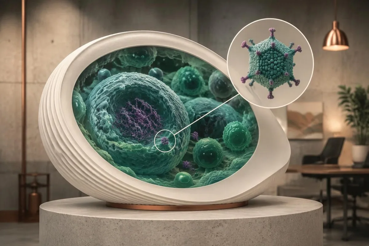
Adenovirus is a non-enveloped virus with a double-stranded DNA genome, unlike the RNA viruses detected on many respiratory panels. Certain species C adenoviruses can persist in tonsillar and adenoidal lymphoid tissue, providing a biological explanation for repeated detection that does not require a brand-new exposure every time.
Kalu et al. described persistent and repeated adenovirus nucleic-acid detection in children's nasopharyngeal secretions in the Pediatric Infectious Disease Journal in 2010. Their work supports caution about calling every positive swab a fresh illness; prolonged detection can occur across separate respiratory episodes rather than following one tidy, continuously positive timeline.
There is no reliable universal maximum shedding period. Age, adenovirus type, specimen, assay sensitivity, and immune function all affect duration; a result at 4 weeks cannot be classified as active or inactive from the calendar alone, and prolonged stool detection need not match the duration of respiratory detection.
Symptoms and laboratory detectability also recover on different schedules. Our explanation of the illness and testing timeline shows why a collection date matters: a specimen obtained after someone feels better still reflects the biology at that particular sampling time, not a retrospective measure of how unwell they were.
How should a positive respiratory swab be interpreted?
A respiratory adenovirus PCR positive result is more convincing evidence of current adenovirus illness when it accompanies a compatible new syndrome. Fever, sore throat, cough, conjunctivitis, or pneumonia strengthen the association, but an upper-airway swab alone cannot establish the cause or severity of lung disease.

In their 2016 study, Song et al. examined whether positive pediatric adenovirus PCR results were sufficient to diagnose acute infection. Viral culture, species information, and the amount of detected DNA helped distinguish likely acute illness from incidental detection; the practical lesson is that the assay's sensitivity can exceed its ability to explain symptoms.
A multiplex panel may detect 2 or more pathogens in the same specimen. That can represent true coinfection, residual adenovirus plus a new virus, or incidental detection of one target; it does not prove that both organisms need treatment or that adenovirus is responsible for every symptom.
A nasal or nasopharyngeal result samples the upper respiratory tract, not the lung tissue itself. A person with worsening breathlessness may need oxygen assessment and chest imaging despite a familiar positive panel; our adult RSV symptom guide illustrates why respiratory severity is judged clinically rather than from pathogen detection alone.
Collection quality can change what a swab detects, and different panels do not cover identical organisms. With a prolonged cough, a clinician may consider a separate test whose timing and sampling requirements differ; our discussion of whooping cough PCR timing explains that distinction without assuming adenovirus accounts for the entire illness.
Why does stool adenovirus PCR have a different meaning?
Stool adenovirus PCR must be interpreted according to the assay's targets and whether diarrhea is present. Many gastrointestinal panels specifically detect enteric adenovirus types 40 and 41, while broader adenovirus assays may also detect types associated with respiratory illness or persistent intestinal carriage.
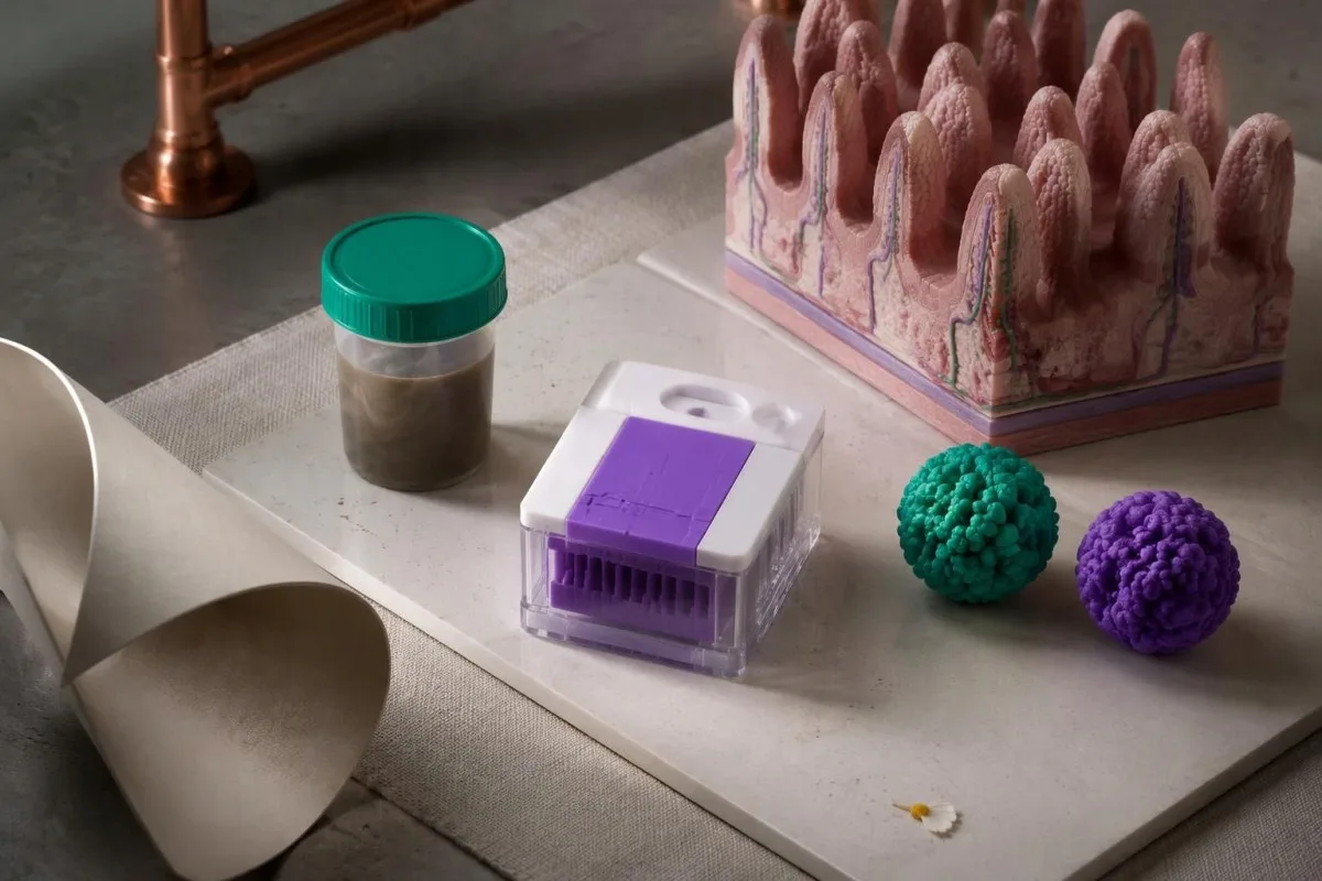
A report reading “adenovirus F40/41 detected” is not interchangeable with a report reading simply “adenovirus detected.” Types 40 and 41 are established causes of gastroenteritis, especially in children; a compatible episode of watery diarrhea makes that result more explanatory than a broad positive test in someone with normal bowel movements.
Stool positivity does not prove that a current cough is caused by adenovirus, and a positive respiratory swab does not prove that diarrhea has the same cause. The 2 specimens sample different compartments; paired positivity can occur without bloodstream spread and should not automatically be described as disseminated disease.
Persistent diarrhea needs a fresh clinical assessment rather than automatic attribution to the first positive target. Medicines, another infection, postinfectious changes, and intestinal disease can coexist with adenovirus DNA; the reasoning in our Campylobacter PCR interpretation guide shows why stool detection and treatment decisions are separate steps.
For a hypothetical child with watery diarrhea for 4 days and F40/41 detected, adenovirus gastroenteritis is plausible, but hydration status determines the immediate priority. By contrast, a broad stool PCR collected 6 weeks after recovery offers much less evidence about the cause of a new abdominal complaint.
Is adenovirus contagious after recovery?
Some people can remain adenovirus contagious after recovery, but PCR cannot determine an individual's remaining transmission risk. Detectable DNA may come from infectious virus or noninfectious material, and the relationship between symptoms, shedding, and transmission varies by adenovirus type and specimen.

Adenovirus can spread through respiratory secretions, contaminated hands and surfaces, stool-associated exposure, and eye secretions. Becoming fever-free does not switch all these routes off simultaneously; a child who feels better but still needs help with toileting may create a different household exposure pattern from an adult with a fading cough.
For ordinary respiratory illness, many general return-to-activity recommendations use improving symptoms and at least 24 hours without fever-reducing medication. This is a practical risk-management rule, not an adenovirus clearance test; healthcare, childcare, transplant units, and outbreak settings may require different precautions or occupational-health approval.
In UK settings, vomiting and diarrhea commonly require exclusion until 48 hours after the last episode, subject to local policy. The rule does not guarantee that stool PCR has become negative; our norovirus recovery and contagiousness guide explains the broader distinction between a workable exclusion interval and complete molecular clearance.
Wash hands with soap and water for at least 20 seconds, particularly after toileting, changing nappies, and handling eye secretions. Because adenovirus lacks an envelope, alcohol-only products are not reliably effective against every type; use surface products with a specific adenovirus effectiveness claim and follow their stated wet-contact time.
Can Ct values or viral load reveal infectiousness?
Cycle-threshold values and viral loads cannot independently establish adenovirus contagiousness. A lower Ct generally indicates more target DNA within the same assay, but specimen quality, processing, platform, and biological context prevent a universal infectious-versus-noninfectious cutoff.

Under ideal amplification conditions, a difference of roughly 3.3 PCR cycles corresponds to a 10-fold difference in starting target quantity. Real clinical assays depart from that ideal, however; the calculation cannot turn a nasal Ct into a reliable count of infectious particles or justify comparing results from unrelated laboratories.
A Ct of 35 on one platform cannot be assumed equivalent to a Ct of 35 on another. Swab depth, mucus, extraction volume, inhibitors, and the assay's target all affect the value; even repeat specimens from the same person may vary because the collector sampled a different amount of material.
“Detected below the quantification limit” means DNA was identified but could not be measured with the laboratory's validated quantitative precision. It is not the same as “not detected,” and it is not proof of contagiousness; our qualitative versus quantitative results guide explains why these report categories should remain separate.
Viral culture can demonstrate recoverable infectious virus under laboratory conditions, but it is not a routine clearance test and does not reproduce every real-world exposure. A negative culture can also reflect handling or culture sensitivity; clinicians therefore avoid using either PCR or culture as an absolute certificate that transmission is impossible.
What can accompanying blood tests tell you?
CBC, CRP, kidney tests, and liver tests assess the body's response and possible complications; they do not determine adenovirus contagiousness. Adenovirus can produce substantial inflammatory responses, so raised CRP or white-cell counts do not automatically prove a bacterial infection.
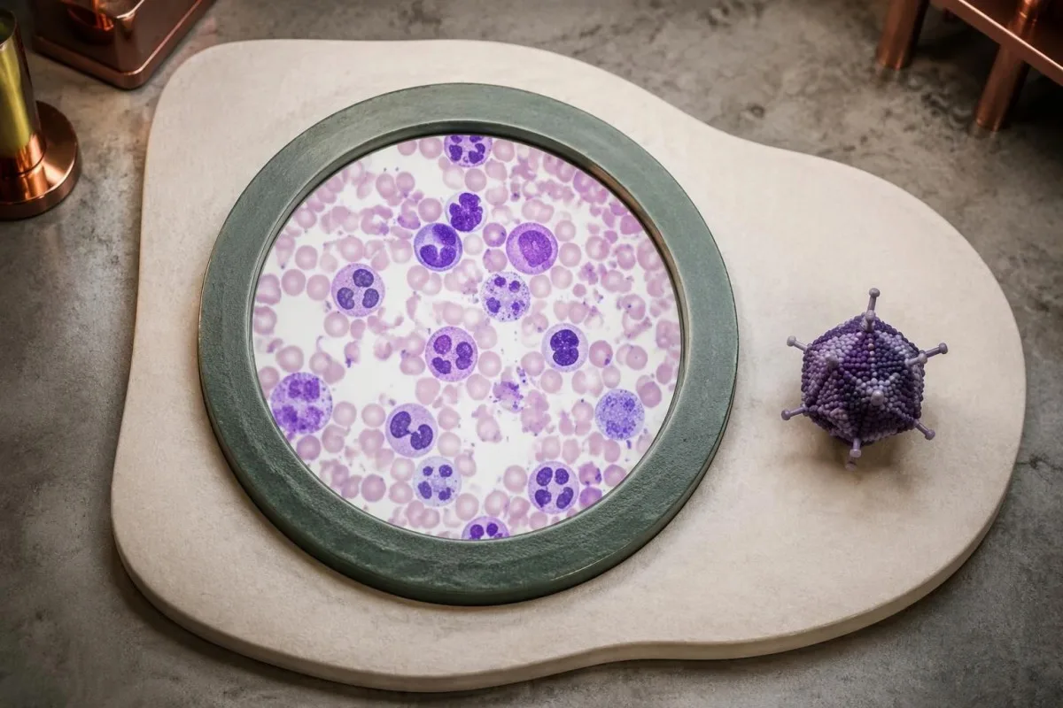
A hypothetical CRP of 80 mg/L with adenovirus detected should prompt interpretation of the whole illness, not an automatic antibiotic prescription. The same value can occur in different infectious and inflammatory conditions; new focal findings, circulatory instability, cultures, and the trajectory may carry more diagnostic weight than the isolated number.
Absolute cell counts are usually more useful than percentages alone when assessing immune vulnerability. For example, lymphocytes making up 15% of a CBC cannot be interpreted without the total white-cell count and an age-appropriate range; a single CBC also cannot establish whether someone's antiviral T-cell function is adequate.
Kantesti is an AI biomarker interpretation platform that can place CBC, CRP, creatinine, and liver results alongside the report's stated ranges and previous measurements. Our laboratory marker guide explains this context, but neither our AI nor a normal inflammatory marker can certify that a PCR-positive person is noncontagious.
Trends may clarify whether the response is settling, but inflammation can lag behind symptom improvement. If CRP falls from 80 to 30 mg/L over several days, that is supportive rather than definitive evidence of recovery; our WBC and CRP comparison explains why these markers sometimes move in different directions.
When is adenovirus detection more concerning?
Adenovirus detection deserves earlier assessment in immunocompromised patients, particularly after hematopoietic stem-cell transplantation or with marked T-cell impairment. A clinically well person may still need specialist monitoring, because ongoing replication can precede obvious organ symptoms.

Risk is not identical across everyone taking an immunosuppressive medicine. Recent transplantation, T-cell-depleting treatment, graft-versus-host disease, intensive chemotherapy, and profound immune deficiency can create different risks; the dose, combination of medicines, time since treatment, and immune recovery matter more than the broad label “immunocompromised.”
An immunocompromised patient with a new positive result should notify their transplant, oncology, or immunology team promptly, even if symptoms are mild. Fever, worsening diarrhea, urinary symptoms, or breathlessness increases urgency; do not wait for a second positive specimen or a large viral-load value before making contact.
Blood detection changes the concern because it indicates DNAemia rather than detection confined to a swab or stool sample. It does not by itself establish organ-invasive disease, but it often warrants a more structured assessment; our BK virus compartment guide illustrates why blood and nonblood viral results cannot share the same interpretation.
Kantesti AI can help explain a reported lymphocyte count, but cannot measure antiviral immune competence or replace a transplant protocol. A result of 0.4 × 10^9/L needs clinical context rather than an automatic prediction of adenovirus progression; our low lymphocyte interpretation guide describes the limitations of that number.
How is adenovirus blood PCR used in specialist care?
Quantitative adenovirus blood PCR helps specialists monitor DNAemia and its trajectory in selected high-risk patients. Treatment decisions depend on immune status, organ involvement, viral-load kinetics, and the transplant protocol—not on a single universal copies/mL threshold.

Lindemans, Leen, and Boelens described risk-adapted monitoring and management in their 2010 Blood review of hematopoietic stem-cell transplant recipients. The review includes weekly PCR surveillance for selected high-risk patients; this specialist strategy should not be extrapolated into weekly retesting of otherwise healthy children with recovering respiratory illness.
Consider a hypothetical plasma viral load increasing from 1,000 to 10,000 copies/mL in 7 days. That is a 10-fold, or 1-log10, rise and may be concerning in a high-risk recipient, but the numbers are not universal treatment triggers; symptoms, immune recovery, analytical variation, and subsequent results still influence the decision.
Whole blood and plasma are not interchangeable matrices, and copies/mL values from different assays are not necessarily harmonized. For follow-up, ask whether the same laboratory, specimen type, and measurement method were used; switching any of these can create an apparent change that does not accurately represent a biological rise or fall.
Reducing immunosuppression can help immune control in some cases, but can also threaten a graft or worsen other conditions. Never adjust tacrolimus or another immune-modifying medicine independently; our tacrolimus monitoring explanation shows why dose timing, measured concentrations, and specialist instructions must be considered together.
Which symptoms require prompt or emergency assessment?
Difficult breathing, confusion, severe dehydration, or rapidly worsening illness require urgent assessment, regardless of an adenovirus result. A positive PCR must not distract from bacterial coinfection, a noninfectious condition, or a complication that needs separate evaluation.

An infant younger than 3 months with a temperature of 38°C or higher needs urgent medical assessment, even when a viral panel is positive. Adults with severe breathing difficulty, blue or grey lips, or altered consciousness need emergency help; a persistent resting oxygen reading of 92% or lower is also an urgent warning unless a clinician has specified a different baseline.
Fever during chemotherapy or significant immune suppression should follow the person's emergency plan, not a routine home-recovery schedule. Traditional febrile-neutropenia definitions include one temperature of 38.3°C or 38°C sustained for 1 hour, but many treatment teams instruct patients to call at 38°C immediately—do not wait if your plan uses the lower trigger.
Children with fever lasting 5 days or longer need reassessment, particularly with red eyes, rash, mouth changes, swollen hands or feet, or unusual irritability. Adenovirus detection does not exclude Kawasaki disease; our Kawasaki diagnostic explanation explains why overlapping symptoms must not be resolved by the PCR result alone.
Painful light sensitivity or reduced vision with a red eye needs prompt eye assessment rather than being dismissed as ordinary conjunctivitis. Significant blood in stool, severe localized abdominal pain, or inability to retain fluids also changes the plan; our stool blood warning signs explain why these findings need their own evaluation.
What does routine adenovirus recovery care involve?
Routine recovery usually involves fluids, symptom relief, and watching for deterioration, not treatment aimed at making PCR negative. Otherwise healthy people whose symptoms are improving generally do not need an adenovirus-specific antiviral or antibiotics solely because DNA remains detectable.

With diarrhea or vomiting, oral rehydration solution replaces water and salts more appropriately than plain water alone when losses are substantial. For a child who tolerates fluids, small amounts such as 5–10 mL every 1–2 minutes may be easier to retain; persistent vomiting, unusual sleepiness, or reduced urination warrants clinical advice.
Continue breastfeeding and resume usual age-appropriate foods as tolerated rather than using prolonged fasting to stop diarrhea. Postinfectious bowel changes can persist after the acute episode, but ongoing weight loss or worsening symptoms deserves reassessment; our gastrointestinal recovery guide explains why symptom persistence does not automatically equal persistent infection.
A cough or reduced energy can outlast fever without implying worsening viral replication. Track practical recovery measures—fluid intake, breathing effort, urine output, and the ability to resume normal activity—rather than repeatedly measuring temperature or seeking a new panel every few days to obtain reassurance.
For suspected adenoviral conjunctivitis, avoid sharing towels and touching or rubbing the eyes; follow professional advice about contact lenses and cleaning. Do not self-start steroid eye drops, which can complicate some eye conditions; eye pain, light sensitivity, or vision change should override an otherwise reassuring recovery pattern.
Do you need another PCR to prove recovery?
Repeat adenovirus PCR is usually unnecessary after uncomplicated recovery in an immunocompetent person. A test of cure is more likely to be useful when a specialist is monitoring DNAemia, investigating persistent or worsening symptoms, or following an institution-specific outbreak or infection-control protocol.
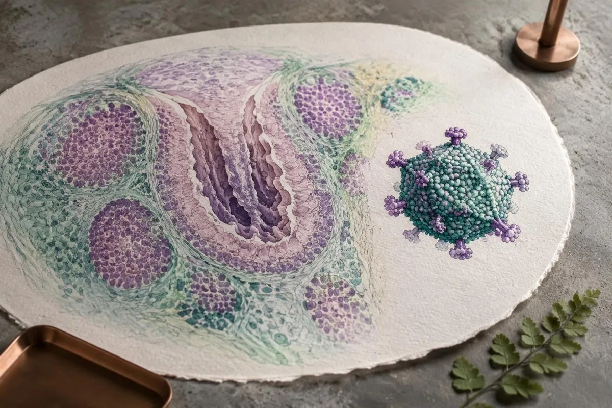
A repeat positive result can prolong worry without changing treatment, particularly when the original specimen came from the nose or stool. A repeat negative result is not an absolute guarantee either: it describes one collection from one compartment, while exposure risk and detection elsewhere may follow a different course.
For example, a well 32-year-old with a residual cough and a positive swab 21 days after initial illness usually gains less from another identical panel than from reassessment if the cough worsens. New chest pain, breathlessness, recurrent fever, or a focal examination finding creates a different question that may justify different testing.
Kantesti is an AI blood test interpretation platform that can help explain accompanying laboratory trends while keeping PCR clearance separate from clinical recovery. Our validation and oversight approach describes methodological safeguards; it should not be interpreted as validation of AI-based decisions about whether an individual remains contagious.
Before repeating a test, ask what action would follow each possible result. If neither a positive nor a negative would change care, the test may have limited value; our AI report safety guide also explains why an automated report should preserve specimen details and uncertainty rather than manufacture certainty.
When are antivirals considered, and why monitor kidneys?
Adenovirus antivirals are generally reserved for selected severe or high-risk cases under specialist care. Cidofovir is used off-label in some settings and can injure the kidneys; persistent nasal or stool PCR positivity alone is not a reason to start it.

Cidofovir decisions are particularly complex because antiviral benefit depends partly on immune recovery, while the medicine introduces its own risks. Monitoring commonly includes creatinine, urine protein, and electrolytes; protective hydration and other measures follow specialist protocols rather than a self-directed regimen derived from an online article.
Brincidofovir and virus-specific T-cell approaches may be considered in selected settings, depending on access, approvals, and clinical circumstances. Availability is not the same as proven suitability for every patient; clinicians must weigh evidence, organ involvement, interactions, and whether symptoms might instead reflect another process such as graft-versus-host disease.
Kidney results can also change from diarrhea and poor intake before any antiviral is given. A hypothetical creatinine increase from 0.8 to 1.3 mg/dL needs comparison with baseline, hydration, medicines, and urine findings; our BUN and creatinine guide explains why one ratio cannot identify the cause of a renal change.
Kantesti AI can help explain reported kidney measurements, but cannot decide whether a transplant recipient should receive or stop an antiviral. The treating team needs the actual PCR trend, collection matrix, medication record, and clinical assessment; an AI summary or isolated laboratory value should never delay a same-day specialist call.
Research publications: what the evidence can and cannot establish
Adenovirus research supports contextual interpretation, not a universal contagiousness cutoff. Pediatric respiratory studies address incidental or persistent detection, while transplant literature addresses progression risk and specialist monitoring; those populations answer different clinical questions and should not be combined into one clearance rule.

The 2010 Kalu paper and 2016 Song paper are directly relevant to interpreting pediatric respiratory detection, but they cannot determine one adult's household transmission risk. Culture associations and repeated PCR findings provide evidence about likely infection and persistence; they do not establish a validated number of safe days after recovery.
The 2010 Blood review by Lindemans and colleagues is relevant to hematopoietic stem-cell transplantation, where immune impairment changes the consequences of viral replication. It does not justify applying transplant treatment thresholds to a healthy person with one positive stool result; this difference in population is central to avoiding both undertreatment and unnecessary intervention.
Kantesti's medical content standards distinguish directly relevant external evidence from educational resources. The 2 Zenodo publications listed below concern complement/ANA testing and Nipah diagnosis, not adenovirus shedding; they are background reading rather than evidence for this article's adenovirus conclusions, and a repository DOI does not establish peer review.
For a separate example of specimen- and timing-dependent viral diagnosis, our Nipah diagnostic testing guide discusses a different virus with different clinical implications. Do not transfer its testing pathways to adenovirus; the practical adenovirus questions remain the specimen source, symptom trajectory, immune status, and what further testing would change.
Frequently asked questions
Does adenovirus PCR detected mean I have an active infection?
Adenovirus PCR detected means viral DNA was found, but it does not prove that adenovirus is currently causing an illness. In a hypothetical person who recovered 3 weeks earlier, a positive swab could reflect prolonged detection rather than a new infection. New compatible symptoms increase the likelihood of current illness, while the specimen source and immune status change interpretation. A clinician should assess worsening symptoms rather than relying on PCR alone.
How long can adenovirus PCR stay positive after recovery?
Adenovirus PCR can remain positive for weeks to months after clinical recovery, particularly in children and in some immunocompromised patients. A result at 4 weeks does not by itself distinguish lingering DNA from continuing viral replication. Duration varies with adenovirus type, specimen, immune function, and assay sensitivity. There is no universal maximum duration or standard day when every patient must test negative.
Am I contagious if my adenovirus PCR is still positive?
A positive adenovirus PCR cannot determine whether an individual is still contagious because it detects DNA rather than directly measuring transmission. Some people continue to shed infectious virus after symptoms improve, while other positive results may reflect noninfectious material. General respiratory return-to-activity rules often include at least 24 hours without fever-reducing medicine and improving symptoms, but these are not adenovirus clearance guarantees. Follow setting-specific advice and continue handwashing, particularly around vulnerable contacts.
What does adenovirus 40/41 detected in stool mean?
Adenovirus 40/41 detected in stool identifies enteric adenovirus types 40 or 41, which are established causes of gastroenteritis. The result is more supportive of current illness when it accompanies compatible watery diarrhea, especially in a child. A broad adenovirus stool assay may detect additional types and is not interchangeable with an F40/41-specific panel. Persistent detection after recovery does not automatically require treatment or repeat testing.
Should I repeat adenovirus PCR until it becomes negative?
Routine repeat adenovirus PCR is generally unnecessary after uncomplicated recovery in an otherwise healthy person. A positive result 21 days after illness may not change care, while one negative specimen does not certify that no virus remains elsewhere. Repeat quantitative blood PCR can be useful under transplant or other specialist protocols. Ask what clinical decision the repeat result would change before arranging another test.
Is adenovirus PCR positive dangerous after a transplant?
Adenovirus PCR positivity after a transplant requires prompt discussion with the treating team because immune suppression can permit ongoing replication and severe disease. Blood DNAemia, rising viral loads, fever, diarrhea, urinary symptoms, or respiratory deterioration increase concern. A hypothetical rise from 1,000 to 10,000 copies/mL is a 10-fold increase, but no single threshold applies to every transplant recipient or assay. Do not adjust immunosuppressive medicines or wait for a second positive result before contacting the team.
Get your blood test results explained
Upload your lab report and Kantesti's AI explains each biomarker in plain language within seconds. Used by more than 2 million people worldwide.
References
- Klein, T., Mitchell, S., & Weber, H. (2026). C3 C4 Complement Blood Test & ANA Titer Guide. Kantesti AI Medical Research. DOI
- Klein, T., Mitchell, S., & Weber, H. (2026). Nipah Virus Blood Test: Early Detection & Diagnosis Guide 2026. Kantesti AI Medical Research. DOI
- Kalu SU et al. (2010). Persistence of adenovirus nucleic acids in nasopharyngeal secretions: a diagnostic conundrum. The Pediatric Infectious Disease Journal.
- Song E et al. (2016). Diagnosis of Pediatric Acute Adenovirus Infections: Is a Positive PCR Sufficient? The Pediatric Infectious Disease Journal.
- Lindemans CA, Leen AM, Boelens JJ (2010). How I treat adenovirus in hematopoietic stem cell transplant recipients. Blood. DOI
- 2M+Tests analysed
- 127+Countries
- 100+Languages
About the medical team


E-E-A-T Trust Signals
Experience
Physician-led clinical review of lab interpretation workflows.
Expertise
Laboratory medicine focus on how biomarkers behave in clinical context.
Authoritativeness
Medically reviewed by Dr. Thomas Klein, with contributions from Dr. Sarah Mitchell and Prof. Dr. Hans Weber.
Trustworthiness
Evidence-based interpretation with clear follow-up pathways to reduce alarm.





Medical disclaimer
This article is for educational purposes only and does not constitute medical advice. Always consult a qualified healthcare provider for diagnosis and treatment decisions.
Published · Contact the editorial team
Published by Kantesti Ltd, registered in England & Wales, company no. 17090423, London, United Kingdom.
