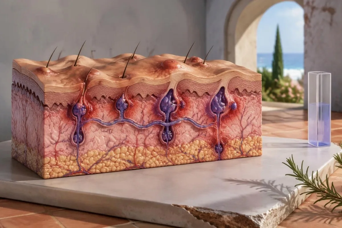
No hidradenitis suppurativa blood test can confirm or exclude HS. Diagnosis depends on characteristic skin changes, their location and recurrence; laboratory testing answers different questions about associated conditions and treatment safety.

Key takeaways
- Diagnosis depends on 3 features: characteristic lesions, typical locations and a recurrent or chronic pattern—not a positive blood test.
- Recurrence of at least 2 episodes in 6 months is a commonly used diagnostic clue, not a reason to delay assessment.
- Normal blood work does not rule out HS, including when CRP is below a laboratory’s usual upper limit of 5 mg/L.
- Boils versus HS cannot be distinguished by 1 photograph or a bacterial culture alone; tunnels, scarring and repeated fold-area flares favor HS.
- Severity can be measured with IHS4: nodules count as 1, abscesses as 2 and draining tunnels as 4 points each.
- Associated conditions may justify HbA1c, glucose or lipid testing; HbA1c of 6.5% or higher requires diabetes assessment, not HS confirmation.
- Treatment safety may require tuberculosis and hepatitis screening before biologics, with additional tests chosen for the specific medication.
- Urgent assessment is needed for rapidly spreading redness, severe deterioration or fever of 38°C or higher with a worsening painful lump.
Can blood work detect HS or rule it out?
Blood work cannot confirm or exclude hidradenitis suppurativa. There is no validated diagnostic blood marker or laboratory cutoff for HS; clinicians diagnose the condition from the skin findings and their history, while tests such as CRP or a complete blood count investigate inflammation, associated illness or medication safety.
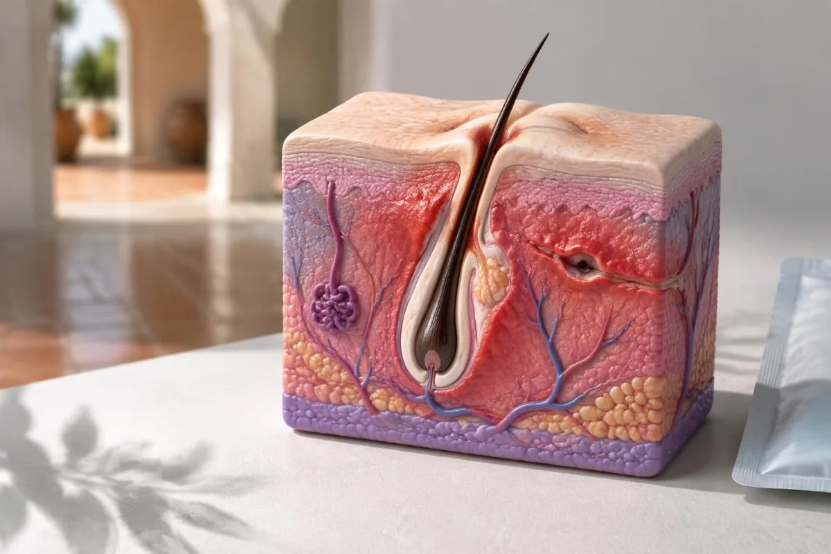
A normal laboratory report does not cancel out a convincing skin pattern. For example, a hypothetical adult with 3 recurrent armpit nodules and CRP of 2 mg/L may still have HS, whereas another person with CRP of 25 mg/L may have an unrelated infection or inflammatory condition.
The North American clinical management guideline bases diagnosis on 3 elements: typical lesions, typical distribution and recurrence (Alikhan et al., 2019). It does not recommend routine biomarker testing to establish HS, because systemic inflammatory markers cannot reliably identify the cause of a particular lump.
Kantesti is an AI blood test analyzer that helps explain findings such as CRP of 12 mg/L without treating them as proof of HS. Our organization and clinical purpose are separate from the role of a dermatologist, who can examine tunnels, scars and lesion distribution directly.
Ordering 10 additional inflammatory or antibody tests is not a substitute for examining recurrent lumps. Ask the clinician what each proposed test will change: the diagnosis of another condition, the urgency of treatment, or the safety of a medication—those are useful laboratory questions.
How is hidradenitis suppurativa diagnosed clinically?
Hidradenitis suppurativa diagnostic criteria combine characteristic lesions, typical locations and a recurrent course. At least 2 episodes in 6 months is a commonly used recurrence threshold, but clinicians interpret it alongside the examination rather than treating it as an inflexible rule or a requirement to wait.
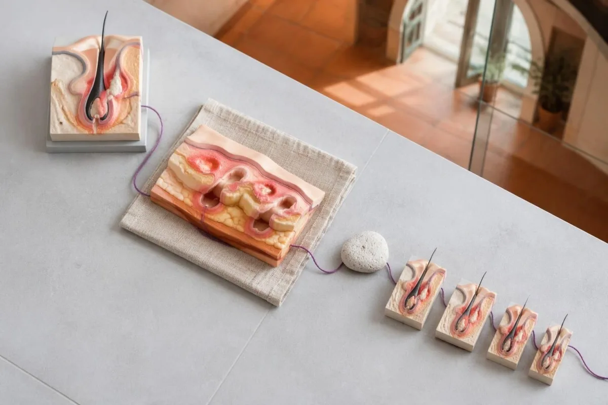
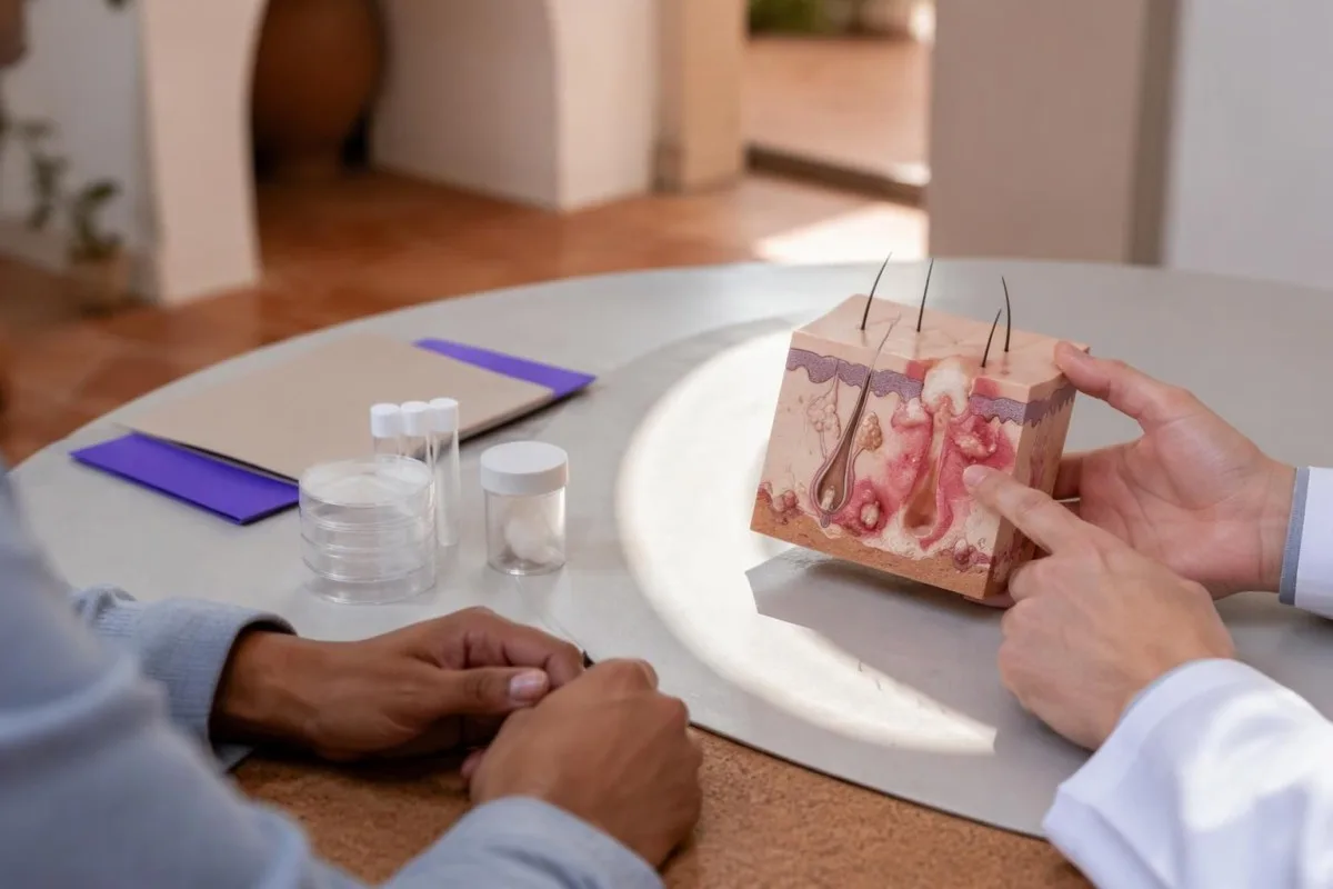
Characteristic lesions include deep inflammatory nodules, abscesses, draining tunnels and distinctive scars; paired or double-ended comedones can also support the diagnosis. One superficial spot after shaving is much less specific than 3 repeated deep lumps appearing in the same fold with persistent openings between flares.
Typical sites include the armpits, groin, inner thighs, buttocks and folds beneath the breasts. HS does not have to affect 2 different body regions, and lesions need not be symmetrical; a repeatedly affected single armpit can still fit the clinical pattern.
Recurrence can be undercounted when someone calls every flare a separate boil. Bring dates or photographs from the preceding 6 months, including episodes treated elsewhere; a clinician may recognize chronic disease from existing tunnels and scars even when the earlier calendar is incomplete.
Early disease may lack scars, so a first visit can produce a working diagnosis and follow-up plan rather than immediate certainty. We describe the distinction between supportive interpretation and diagnostic validation in our clinical validation framework; no claimed laboratory accuracy percentage substitutes for the 3 clinical features of HS.
How do recurrent HS lumps differ from ordinary boils?
HS typically causes repeated deep lumps in characteristic fold areas, sometimes with tunnels and scarring; an ordinary boil is usually a localized follicular infection. Neither appearance nor a positive bacterial culture settles the distinction by itself, because recurrent boils can occur and HS lesions can develop secondary infection.
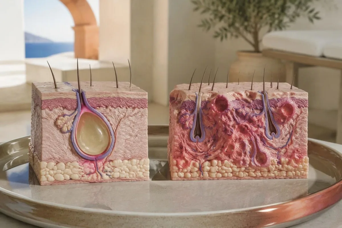
A boil may have a central pustule and resolve after drainage or treatment, whereas HS can leave persistent openings or firm scar tissue. In a hypothetical 6-month history, 4 flares in one groin fold plus a tunnel are more suggestive of HS than 4 unrelated small pustules on different limbs.
Odor and drainage do not mean HS is caused by poor hygiene. HS involves follicular occlusion and an abnormal inflammatory response; bacteria can contribute to the local environment, but finding 1 bacterial species does not prove that infection is the underlying disease mechanism.
Recurrent bacterial boils deserve their own assessment, particularly when several household members have similar lesions or infections occur at unrelated sites. Our guide to testing frequent infections explains when glucose, a blood count or targeted immune evaluation may answer a different question from HS diagnosis.
Repeated incision and drainage may relieve an acutely tense lump but does not address an established HS tunnel. If the same area returns 2 or more times, ask whether the management plan should move beyond treating each episode as a new boil; do not squeeze or puncture it yourself.
How is HS severity measured without a blood test?
HS severity is measured from lesions and tissue damage, not from a CRP cutoff. Hurley staging describes structural disease, while IHS4 measures current inflammatory activity by assigning 1 point per nodule, 2 per abscess and 4 per draining tunnel.
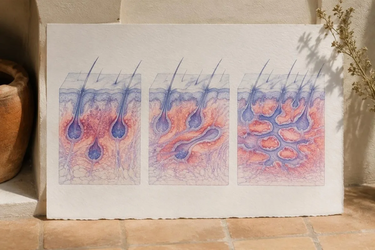
IHS4 scores of 3 or lower indicate mild disease, 4–10 moderate disease and 11 or higher severe disease. The original validation paper established this weighted scoring approach to capture active disease more dynamically than structural staging alone (Zouboulis et al., 2017).
Hurley stage I has no established tunnels or scarring; stage II includes recurrent lesions with tunnels and scarring separated by unaffected areas. Stage III describes diffuse involvement with interconnected tunnels across an affected region, so 1 region can be advanced even when the overall body distribution is limited.
Consider a hypothetical count of 3 nodules, 1 abscess and 1 draining tunnel: IHS4 equals 3 + 2 + 4, or 9. That moderate activity score can coexist with substantial scarring; counting lesions does not erase the separate effects on movement, sleep or quality of life.
Kantesti AI can support medication safety trend review, but it cannot calculate a reliable skin severity score from a CBC. A falling CRP from 20 to 6 mg/L may be encouraging in context, yet clinicians still need lesion counts, pain assessment and examination of persistent tunnels.
What do CRP, ESR and a CBC tell you about HS?
CRP, ESR and a CBC can show systemic inflammation or its consequences, but none diagnoses HS. Many laboratories use CRP below 5 mg/L and an adult white blood cell count around 4.0–11.0 × 10⁹/L as reference values; the range printed on your own report takes priority.
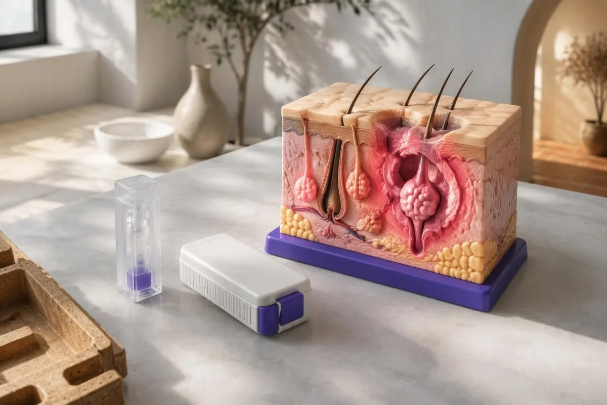
CRP may rise with active HS, bacterial infection, obesity or many other inflammatory conditions. A hypothetical CRP of 18 mg/L with stable chronic lesions means something different from the same value with new fever and rapidly expanding redness; our WBC and CRP comparison explains why mismatched results need clinical context.
ESR changes more slowly and is influenced by age, anemia and plasma proteins. Common adult reference limits include approximately 15 mm/hour for younger men and 20 mm/hour for younger women, but laboratory methods and age-adjusted reporting differ enough that a single universal HS threshold would be misleading.
A CBC may identify anemia, raised platelets or leukocytosis associated with inflammation, but each finding has alternative explanations. A platelet count of 480 × 10⁹/L, for example, could reflect iron deficiency or a reactive inflammatory response; persistence and the rest of the blood count determine the next step.
Medication and sampling context matter when comparing 2 reports. Estrogen-containing contraception can alter CRP, as discussed in our guide to contraception and CRP changes; standard CRP and hs-CRP measure the same protein, but cardiovascular hs-CRP categories should not be used to stage an active HS flare.
| Level | Range | What it means |
|---|---|---|
| CRP within a common reference limit | <5 mg/L | Does not exclude HS; use the reporting laboratory’s reference interval. |
| CRP above that reference limit | ≥5 mg/L | Nonspecific inflammation; no concentration confirms HS or distinguishes it from infection. |
| Common adult WBC interval | 4.0–11.0 × 10⁹/L | An illustrative adult range; normal counts do not exclude HS or serious infection. |
| WBC above that interval | >11.0 × 10⁹/L | May reflect infection, inflammation, medicines or other causes; assess the differential count and symptoms. |
When do anemia and iron studies help in HS?
Iron studies help when someone with HS has anemia, fatigue or other reasons to suspect iron deficiency. Ferritin below 15 ng/mL strongly supports depleted iron stores in adults, but a normal or elevated ferritin cannot reliably exclude deficiency during inflammation.
Inflammation increases hepcidin, which reduces iron release from storage and intestinal absorption. A hypothetical ferritin of 90 ng/mL with transferrin saturation of 12% and low hemoglobin can therefore represent iron restriction, mixed iron deficiency and inflammation, or another process—not automatically adequate usable iron.
Transferrin saturation below 20% suggests reduced circulating iron availability, but it does not by itself establish the cause. The combination of hemoglobin, MCV, ferritin, transferrin saturation and CRP is more informative than ferritin alone; our complete iron studies guide explains these interactions.
Low transferrin or TIBC can point toward an inflammatory pattern, while uncomplicated iron deficiency more often raises iron-binding capacity. Our explanation of inflammation lowering transferrin helps distinguish these patterns, particularly when 2 mechanisms coexist and the usual textbook picture becomes blurred.
Anemia should not simply be attributed to HS. Heavy menstrual bleeding, gastrointestinal blood loss, nutritional deficiencies and kidney disease may coexist; for example, hemoglobin falling from 125 to 103 g/L deserves an explanation rather than an assumption that treating the skin alone will correct it.
Which metabolic blood tests may be useful with HS?
HbA1c, glucose and a lipid panel may help assess metabolic conditions associated with HS. HbA1c of 6.5% or higher and fasting plasma glucose of 126 mg/dL or higher—7.0 mmol/L—are diabetes thresholds, not hidradenitis suppurativa diagnostic criteria.

The US and Canadian HS Foundations recommend screening for relevant comorbidities, including diabetes and metabolic syndrome (Garg et al., 2022). The practical plan combines blood pressure, weight-related measures, smoking history and selected laboratory tests; there is no requirement for every person with 1 suspected HS lesion to receive an extensive metabolic panel.
A standard metabolic panel usually measures glucose but not cholesterol. Our guide to metabolic panels versus cholesterol explains why a separate lipid panel may be needed; triglycerides of 150 mg/dL or higher, about 1.7 mmol/L, are one metabolic risk signal rather than evidence of HS.
Kantesti is an AI blood test interpretation platform that can explain an HbA1c of 6.1% alongside the reported units and other available results. That value may identify increased diabetes risk, but the risk-category boundary differs between some health systems, and it cannot explain the cause of recurrent nodules.
Diabetes-range results usually need confirmation unless there is unequivocal hyperglycemia with a compatible clinical presentation. Iron deficiency, hemolysis or recent transfusion can distort HbA1c; our guide to alternatives when A1c misleads explains why a plasma glucose-based assessment may be more reliable in those circumstances.
When should HS prompt hormone, bowel or joint assessment?
HS warrants targeted assessment for associated conditions when symptoms point toward PCOS, inflammatory bowel disease or inflammatory arthritis. These are separate diagnostic pathways: neither 1 abnormal hormone result nor a positive inflammatory marker proves that the skin lumps are HS.
Irregular menstrual cycles, excess facial or body hair and persistent acne may justify PCOS assessment. Adult PCOS commonly requires 2 of 3 diagnostic features after alternative causes are excluded, as explained in our PCOS diagnostic criteria guide; a broad hormone panel is not routinely needed solely because HS flares around menstruation.
Persistent diarrhea, nocturnal bowel symptoms, unexplained weight loss or rectal bleeding deserve assessment rather than being blamed on stress or antibiotics. A hypothetical 4-week history of diarrhea with HS may justify stool testing and gastrointestinal review; our stool inflammation test comparison explains the role of fecal calprotectin and lactoferrin.
Back pain lasting more than 3 months, marked morning stiffness or swollen joints can suggest inflammatory musculoskeletal disease. ESR and CRP may support that assessment, but normal values do not reliably exclude it, and HLA-B27 is not a universal screening test for everyone with HS.
Perianal drainage requires particular care because HS tunnels and Crohn-related fistulas may need different evaluation and treatment. The comorbidity recommendations favor symptom-based assessment rather than indiscriminate testing (Garg et al., 2022); record at least 3 details before the visit: duration, associated bowel symptoms and whether the opening repeatedly returns.
Which tests are needed before HS biologic treatment?
Before an HS biologic, clinicians assess infection risks and order tests required by the specific drug and local protocol. Tuberculosis testing and hepatitis B assessment are commonly relevant; baseline CBC and liver tests may also be requested, but there is no single pretreatment panel suitable for every biologic.
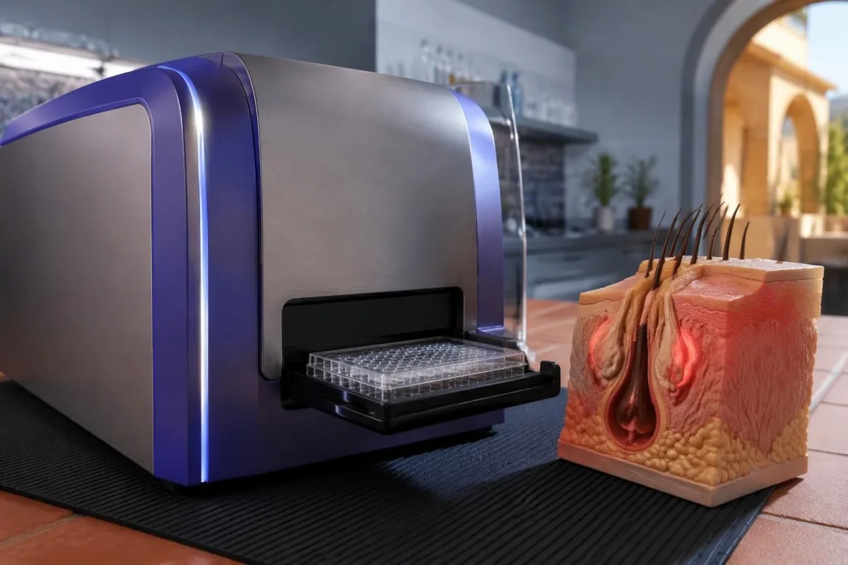
An IGRA checks immune recognition of tuberculosis antigens; it does not determine whether a person has active tuberculosis by itself. Our IGRA result interpretation guide explains why 1 positive or indeterminate result may require symptom review, imaging or repeat testing before treatment proceeds.
Hepatitis B screening commonly includes 3 markers: HBsAg, total anti-HBc and anti-HBs. Anti-HBs alone is insufficient because it cannot distinguish every pattern of previous infection and reactivation risk; our explanation of hepatitis B immunity results helps clarify why a protective antibody result is only part of the assessment.
Kantesti can explain the reported hepatitis marker pattern, but a prescriber must decide whether monitoring, specialist review or antiviral prevention is required. For example, 1 positive anti-HBc result should not be translated into either automatic treatment refusal or reassurance that reactivation is impossible.
Screening is not limited to blood work: vaccination history, current symptoms, past serious infections and relevant bowel disease also influence the choice of medication. Repeat testing intervals differ by drug, exposure risk and jurisdiction; a 12-month automatic schedule should not be assumed to fit every patient or biologic.
How do laboratory tests support HS medication safety?
HS medication monitoring is drug-specific rather than a way to confirm the diagnosis. Potassium and kidney function may matter with spironolactone, liver tests with rifampicin-containing regimens, and G6PD assessment with dapsone; a result such as potassium of 5.5 mmol/L answers a safety question, not an HS question.
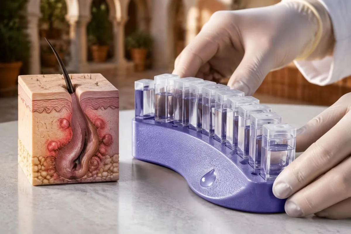
Spironolactone monitoring depends on age, kidney function, other medicines and hyperkalemia risk. A hypothetical patient taking an ACE inhibitor with eGFR of 45 mL/min/1.73 m² needs a different plan from a young otherwise healthy adult; monitoring should not be copied from someone else’s prescription.
Rifampicin can affect liver tests and interact with many medicines, including reducing the effectiveness of some hormonal contraception. If ALT rises from 22 to 110 IU/L, the prescriber considers the laboratory upper limit, bilirubin, symptoms and timing rather than assuming every increase has the same significance.
Dapsone can cause hemolysis and methemoglobinemia, and G6PD deficiency increases certain risks. Our guide to G6PD testing and safety explains why 1 apparently normal enzyme result can be misleading during or shortly after hemolysis; normal G6PD activity also does not remove every dapsone-related risk.
Routine laboratory schedules are not identical for short antibiotic courses, prolonged combination therapy, hormonal treatment and biologics. Before starting treatment, obtain 3 practical answers: which baseline results matter, when the first recheck is due and which symptoms require contacting the prescriber before the next planned appointment.
When are cultures, skin biopsy or genetic tests appropriate?
Cultures and skin biopsy are selective tests in suspected HS, not routine confirmation methods. A culture may help when secondary infection is suspected, and biopsy may assess an atypical growth or competing diagnosis; no routine genetic panel is recommended to diagnose typical HS.
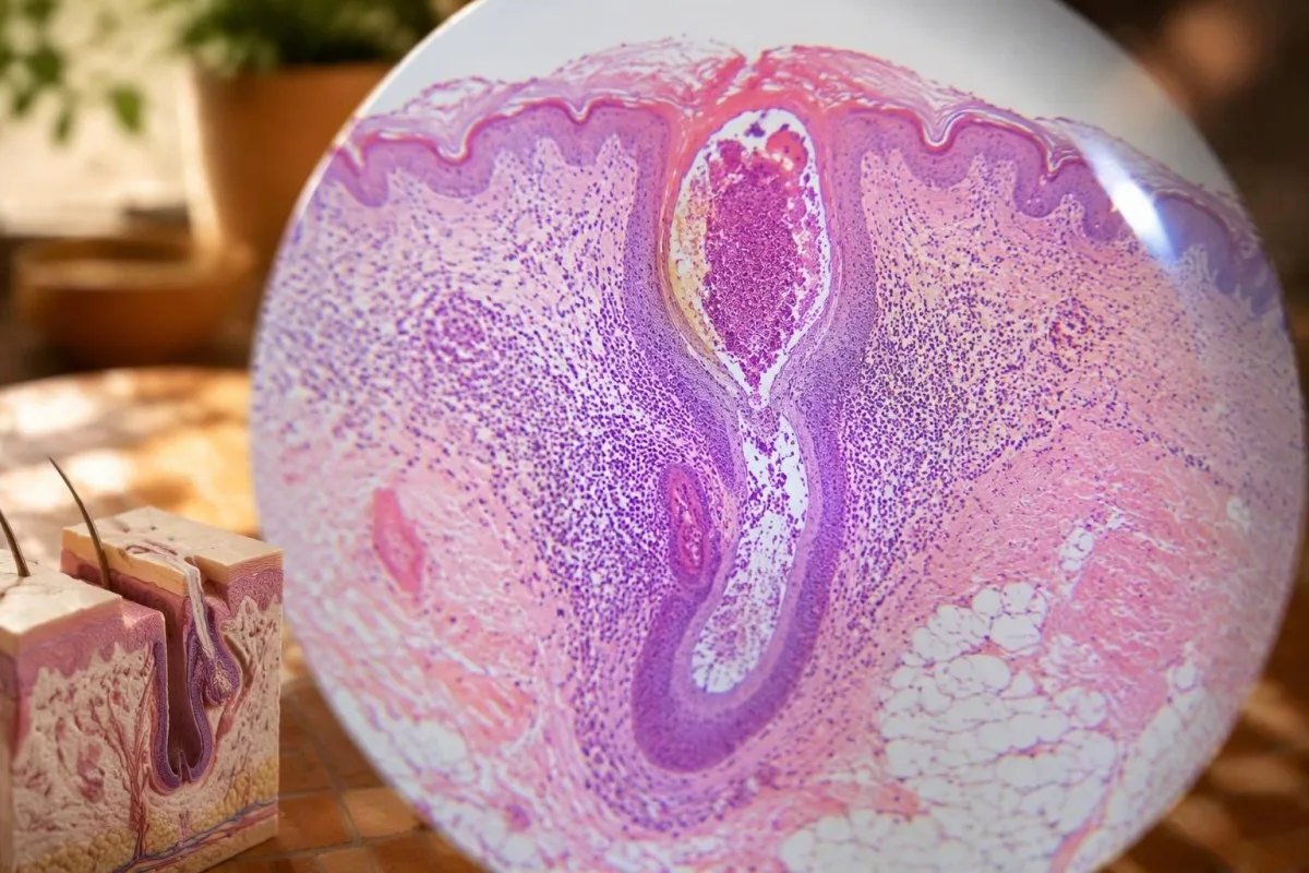
Routine swabbing of every draining HS lesion can produce bacteria that are colonizing the site rather than driving the whole condition. The North American guideline reserves microbiological testing for situations such as suspected secondary infection (Alikhan et al., 2019); 1 positive swab must be interpreted with the examination and clinical course.
Biopsy is more useful when a persistent area behaves differently from the usual flares—such as a new firm growth, unexplained bleeding or a site that does not heal. Rare squamous cell carcinoma can develop in longstanding HS, particularly anogenital disease; a change lasting several weeks deserves examination rather than another automatic antibiotic course.
Immune-function testing is not standard for adult-onset HS. Repeated unusual infections beginning in childhood may justify a separate immunology pathway, sometimes including a targeted oxidative burst test; the reason is the infection history, not the presence of 2 ordinary HS nodules.
Rare familial forms can involve variants in gamma-secretase pathway genes, but most HS is not established through a single-gene test. Even a strong family history involving 3 relatives does not replace clinical assessment, and a negative genetic result cannot rule out the much more common multifactorial form.
Can vitamin D, zinc or experimental biomarkers diagnose HS?
Vitamin D, zinc, cytokines and inflammatory ratios cannot diagnose HS. Low nutrient concentrations may justify assessment for deficiency, but no validated HS diagnostic cutoff exists for vitamin D, zinc, the neutrophil-to-lymphocyte ratio or a commercial inflammatory biomarker panel.

Plasma zinc can fall during inflammation because zinc is redistributed, not simply because dietary intake is inadequate. Our guide to zinc results during inflammation explains why a hypothetical result of 58 µg/dL should be read with the local range, sampling conditions and inflammatory state rather than automatically triggering high-dose supplementation.
Vitamin D associations do not establish that deficiency causes HS or that replacing it reliably controls the condition. A 25-hydroxyvitamin D result of 12 ng/mL, equivalent to 30 nmol/L, may warrant deficiency management for general health, but improvement in HS cannot be promised from that number.
Nutritional treatment evidence for HS is limited and heterogeneous; the uncertainty concerns disease control, not whether genuine deficiency should be addressed. The 2019 North American guideline found insufficient evidence for routine vitamin D supplementation as an HS treatment and only limited support for zinc-based approaches (Alikhan et al., 2019).
The neutrophil-to-lymphocyte ratio can be calculated by dividing the 2 absolute counts, but an NLR of 4 has no accepted diagnostic meaning for HS. Group-level differences reported in research do not establish the sensitivity and specificity needed to decide whether an individual person’s recurrent lump is HS.
What should you bring to an HS appointment—and when is it urgent?
Bring a recurrence timeline, photographs and a medication list; seek urgent care for systemic illness or rapidly worsening local symptoms. Fever of 38°C or higher with spreading redness, marked deterioration or a worsening painful lump needs prompt assessment, but serious infection can also occur without fever.
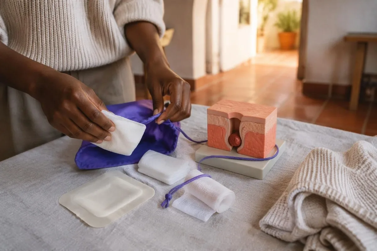
A useful timeline records the date, site, drainage, treatment and recovery of each flare over the last 6 months. Include areas that seemed to heal but left openings or scars, and take photographs only if you are comfortable doing so; recurrence patterns often provide more diagnostic information than another isolated laboratory result.
CBC, CRP and HbA1c do not usually require fasting, although other ordered tests may have preparation instructions. Our baseline test preparation guide explains why recording recent illness, supplements and medication timing is useful when comparing 2 reports; do not stop prescribed treatment simply to obtain a baseline.
Pain out of proportion to visible changes, rapidly spreading swelling, confusion, faintness or severe deterioration requires emergency assessment. A previous CRP of 3 mg/L or a normal white cell count should not delay care for new symptoms, especially during immune-modifying treatment or in someone with diabetes.
Between visits, avoid squeezing lesions and use non-adherent dressings when needed; comfortable clothing can reduce friction without implying that clothing caused HS. Ask for an agreed flare plan with 3 parts: symptom relief, when to contact the treating team and which changes mean the usual home plan is no longer appropriate.
AI interpretation and research: what cannot establish an HS diagnosis
AI blood test interpretation can support understanding of laboratory results, but it cannot establish HS from those results. The 2 Figshare publications listed below concern an interpretation-engine benchmark and hantavirus triage, not an HS diagnostic-accuracy study or validation of a definitive hidradenitis suppurativa blood test.
Kantesti is an AI-powered blood test analysis tool that can help organize abnormal results and changes between reports for discussion with your clinician. Our AI interpretation technology guide explains the workflow; 1 uploaded report does not provide the skin examination or recurrence history needed to diagnose HS.
Keep 3 questions separate: whether the skin pattern fits HS, whether another condition needs investigation and whether treatment is safe. This separation prevents an elevated CRP from being treated as diagnostic proof and prevents normal blood work from incorrectly ending the assessment of recurrent painful lumps.
A benchmark using 100,000 synthetic cases evaluates technical performance within the chosen test design, not outcomes in 100,000 real patients with HS. Similarly, a report concerning 50,000 interpreted laboratory reports for hantavirus triage addresses a different clinical task; neither title supports a claim that AI can distinguish HS from boils.
The publications are listed in the DOI references for transparency, not as evidence for the 3 HS diagnostic criteria. Our responsible AI commitments describe why interpretation must remain bounded by the available information; the practical next step for recurring fold-area lumps is clinical assessment, even when every laboratory result is within range.
Frequently asked questions
Is there a blood test for hidradenitis suppurativa?
There is no validated blood test that confirms or excludes hidradenitis suppurativa. Clinicians diagnose HS using 3 features: characteristic lesions, typical locations and recurrence or chronicity. CBC, CRP, glucose and other tests may assess associated conditions or treatment safety, but an abnormal result is not proof of HS. Normal laboratory results should not prevent assessment of recurrent painful lumps.
Can you have HS with normal blood work?
Yes, hidradenitis suppurativa can occur with a normal CBC and normal inflammatory markers. A CRP of 2 mg/L, for example, does not rule out HS even if the laboratory uses an upper reference limit of 5 mg/L. Local skin activity and structural tunnels are not reliably captured by systemic blood markers. The diagnosis still depends on lesion appearance, location and recurrence.
How many boils mean you might have hidradenitis suppurativa?
At least 2 episodes in 6 months in a characteristic area is a commonly used clue for hidradenitis suppurativa, but counting lumps alone cannot diagnose it. Deep recurrent nodules, tunnels, paired comedones and characteristic scars make HS more likely. Repeated bacterial boils can also occur and may need a different assessment. Do not wait for another flare if current lesions are severe or the pattern is already concerning.
Does a high CRP mean that HS is infected?
A high CRP does not prove that an HS lesion has a bacterial infection. CRP of 20 mg/L could reflect HS activity, an unrelated inflammatory condition or infection, depending on the symptoms and examination. Fever of 38°C or higher with worsening pain or spreading redness warrants prompt assessment, although serious infection can occur without fever. Cultures are selected when clinically useful rather than taken routinely from every draining HS site.
What blood tests are needed before HS biologics?
Pretreatment testing for HS biologics depends on the medication, local protocol and individual infection risks. Tuberculosis assessment and hepatitis B screening are commonly relevant, and hepatitis B screening often uses 3 markers: HBsAg, total anti-HBc and anti-HBs. CBC and liver tests may also be requested. A positive screening result needs clinical interpretation and does not automatically mean every biologic is unsuitable.
Can a skin biopsy or bacterial swab confirm HS?
Typical hidradenitis suppurativa is usually diagnosed clinically without a skin biopsy or routine bacterial swab. A positive culture identifying 1 organism may represent secondary infection or colonization rather than establish the underlying skin condition. Biopsy may help assess an atypical persistent growth or another diagnosis. The 3 clinical features—lesion type, distribution and recurrence—remain the main basis for recognizing HS.
Can an AI blood test analyzer diagnose hidradenitis suppurativa?
An AI blood test analyzer cannot diagnose hidradenitis suppurativa from laboratory values alone. It can help explain a result such as CRP of 12 mg/L or HbA1c of 6.1%, but neither result identifies the cause of recurring skin lumps. HS assessment requires the lesion pattern, affected locations and recurrence history, usually supported by examination. AI interpretation should support discussion with a clinician rather than replace that assessment.
Get your blood test results explained
Upload your lab report and Kantesti's AI explains each biomarker in plain language within seconds. Used by more than 2 million people worldwide.
References
- Klein, T., Mitchell, S., & Weber, H. (2026). Multilingual AI Assisted Clinical Decision Support for Early Hantavirus Triage: Design, Engineering Validation, and Real-World Deployment Across 50,000 Interpreted Blood Test Reports. Kantesti AI Medical Research. DOI
- Klein, T., Mitchell, S., & Weber, H. (2026). A Pre-Registered, Rubric-Based Automated Technical Benchmark of the Kantesti Blood-Test Interpretation Engine on 100,000 Synthetic Test Cases. Kantesti AI Medical Research. DOI
- Alikhan A et al. (2019). North American clinical management guidelines for hidradenitis suppurativa: A publication from the United States and Canadian Hidradenitis Suppurativa Foundations: Part I: Diagnosis, evaluation, and the use of complementary and procedural management. Journal of the American Academy of Dermatology. DOI
- Garg A et al. (2022). Comorbidity screening in hidradenitis suppurativa: Evidence-based recommendations from the US and Canadian Hidradenitis Suppurativa Foundations. Journal of the American Academy of Dermatology.
- Zouboulis CC et al. (2017). Development and validation of the International Hidradenitis Suppurativa Severity Score System (IHS4), a novel dynamic scoring system to assess HS severity. British Journal of Dermatology. DOI
- 2M+Tests analysed
- 127+Countries
- 100+Languages
About the medical team


E-E-A-T Trust Signals
Experience
Physician-led clinical review of lab interpretation workflows.
Expertise
Laboratory medicine focus on how biomarkers behave in clinical context.
Authoritativeness
Medically reviewed by Dr. Thomas Klein, with contributions from Dr. Sarah Mitchell and Prof. Dr. Hans Weber.
Trustworthiness
Evidence-based interpretation with clear follow-up pathways to reduce alarm.





Medical disclaimer
This article is for educational purposes only and does not constitute medical advice. Always consult a qualified healthcare provider for diagnosis and treatment decisions.
Published · Contact the editorial team
Published by Kantesti Ltd, registered in England & Wales, company no. 17090423, London, United Kingdom.
