Fish odor syndrome is investigated by measuring urinary trimethylamine and trimethylamine N-oxide, sometimes on repeated samples. Selected FMO3 genetic testing can confirm inherited trimethylaminuria when the biochemical findings fit; odor alone cannot diagnose it.

Key takeaways
- Diagnostic starting point: A urine trimethylamine test measures 2 compounds—trimethylamine (TMA) and trimethylamine N-oxide (TMAO)—rather than simply checking whether urine smells fish-like.
- Oxidation percentage: Calculate TMAO ÷ (TMA + TMAO) × 100 using compatible molar units; a lower percentage can indicate reduced conversion of odor-producing TMA.
- Published interpretation bands: A clinical review describes oxidation above 92% as typical of unaffected individuals and below 84% as suggestive of impairment, but the testing laboratory's protocol takes priority.
- Intermittent symptoms: A single normal sample collected during a symptom-free period or after dietary restriction may not exclude trimethylaminuria.
- Collection protocol: Laboratories may request a spot specimen, a timed collection, or a 24-hour collection; do not substitute one method for another.
- Genetic confirmation: Inherited FMO3-related trimethylaminuria generally requires 2 disease-causing variants on opposite gene copies, interpreted alongside urinary findings.
- Dietary safety: Do not perform a food-loading challenge or start severe choline restriction yourself; adult US adequate intakes are 425 mg/day for women and 550 mg/day for men.
- Testing limits: Routine urine dipsticks, standard metabolic panels, and cardiovascular TMAO measurements do not replace specialist urinary TMA/TMAO testing.
How is fish odor syndrome investigated and confirmed?
Fish odor syndrome diagnosis starts with specialist urine testing for TMA and TMAO, interpreted with symptoms, diet, and collection conditions. Selected FMO3 genetic testing can establish an inherited cause, but neither a fish-like smell nor a routine blood panel can confirm trimethylaminuria.

The diagnostic pathway answers 3 different questions: is excess odor-producing TMA being excreted, is its conversion to TMAO reduced, and is an inherited FMO3 defect responsible? Keeping these questions separate prevents an abnormal urine result from being automatically labeled a lifelong genetic condition.
A GP or primary-care clinician can first establish whether the odor is generalized or comes from 1 site, then arrange referral to a metabolic service or biochemical genetics laboratory when appropriate. The specialist laboratory should provide collection instructions before the specimen is obtained—not after it reaches reception.
Kantesti is an AI blood test analyzer that can explain accompanying liver and kidney panels, but it cannot confirm trimethylaminuria from routine blood results. Our organization and clinical scope are separate from the specialist laboratory work needed to measure these 2 urinary compounds.
For example, a hypothetical adult with episodic odor and 2 normal metabolic panels still needs the correct urine assay if trimethylaminuria remains plausible. Conversely, an abnormal oxidation result needs context before FMO3 testing is ordered; a biochemical finding and a genetic explanation are related, not interchangeable.
Why does a fish-like odor not prove trimethylaminuria?
A fish-like odor is a symptom, not a diagnostic test, because several local and systemic processes can produce amine-like smells. Trimethylaminuria becomes more plausible when odor affects multiple routes, such as sweat and urine, but even 2 affected routes do not establish the diagnosis.

TMA is a volatile compound that can contribute to odor in sweat, breath, and urine; TMAO is its much less odorous oxidation product. Mitchell and Smith's 2001 review explains this biochemical distinction, which is why measuring 2 compounds is more informative than asking someone to judge odor intensity.
An examination on 1 symptom-free day cannot reliably exclude an intermittent condition. Recent washing, antiperspirant use, clothing changes, and ordinary variation in sweating can alter what another person notices without showing whether urinary TMA metabolism is normal.
Localized odor needs localized assessment. Persistent odor from an underarm with nodules or drainage warrants examination for skin disease rather than an automatic metabolic diagnosis; our guide to skin diagnosis and lab limits explains why a normal blood result cannot replace that examination.
Odor concerns deserve respectful evaluation even when 1 clinician does not notice a smell. If repeated, appropriately collected testing is reassuring, clinicians can reassess other explanations—including altered smell perception or persistent odor-related anxiety—without assuming the original complaint was imagined or dismissing its effect on daily life.
What does a urine trimethylamine test actually measure?
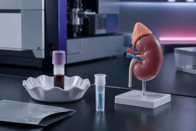
A urine trimethylamine test quantifies TMA and TMAO, allowing the laboratory to assess both total exposure and the proportion converted to TMAO. These 2 measurements are specialist biochemical assays; they are not included automatically in a routine urinalysis.
Laboratories may use methods such as nuclear magnetic resonance spectroscopy or validated chromatographic techniques to distinguish the 2 compounds. The request should explicitly name urinary TMA and TMAO; sending an ordinary specimen labeled only 'body odor' may result in the wrong investigation.
Reports can express results as concentrations, creatinine-normalized quantities, or amounts excreted during a timed collection. A value in µmol/L cannot be compared directly with a value in µmol/mmol creatinine, and a 24-hour amount cannot be treated as a spot concentration.
A normal routine urine dipstick does not exclude trimethylaminuria. Dipsticks assess other features, such as glucose, protein, and selected markers of urinary tract problems; they do not measure the 2 amines needed for this diagnosis.
Blood TMAO testing used in cardiovascular research answers a different question from urinary TMA oxidation testing. A raised blood TMAO result does not diagnose fish odor syndrome, and a urine report containing only TMAO cannot establish whether conversion of TMA was adequate without the companion TMA measurement.
When should urine be collected if the odor comes and goes?
Collecting urine during a typical symptomatic period can improve the usefulness of testing, provided the laboratory's dietary and timing instructions are followed. There is no single clock time that suits every trimethylaminuria test; a spot specimen and a 24-hour collection answer somewhat different sampling questions.

A practical 7–14-day diary can record odor episodes, meals, supplements, exercise, and menstrual timing before an appointment. This is an organizational aid, not a validated diagnostic score; its purpose is to identify a realistic sampling window and explain why a previous specimen may have missed symptoms.
Some people report stronger symptoms around menstruation, while exercise may make odor more noticeable through increased sweating. These are 2 different mechanisms: altered metabolic capacity or substrate handling may affect urinary chemistry, whereas sweating alone can amplify odor without proving reduced FMO3 activity.
Symptom-linked urine sampling is also relevant in some other metabolic investigations, although the substances and protocols differ completely. Our explanation of urine testing during metabolic symptoms illustrates this timing principle; porphyria testing is not a substitute for TMA/TMAO measurement.
For a hypothetical person whose odor appears after particular evening meals, an unrelated symptom-free morning specimen may be less informative than a laboratory-approved collection covering the relevant period. Record the last meal and collection time; do not assume that fasting, which suits some blood tests, improves this urine test.
How do food and supplements affect trimethylaminuria testing?
Diet changes the supply of compounds that can become TMA, so severe restriction before collection can produce an uninformative or falsely reassuring result. Marine fish contains TMAO, while choline and carnitine can contribute to intestinal TMA production; these are 3 related but different dietary influences.

Eggs, beans, and other nutritious foods contain choline, while some supplements provide concentrated choline or carnitine. The effect of a meal depends on portion size, intestinal metabolism, and individual physiology; eating 1 implicated food does not prove that it caused an abnormal oxidation result.
A specialist may arrange a standardized substrate or food-loading protocol when ordinary intake produces too little exposure for interpretation. Mackay and colleagues' 2011 review discusses loading in diagnostic assessment, but protocols differ; copying a challenge dose from an online account can create symptoms without producing a valid specimen.
Bring the name and actual daily dose of every supplement, including products measured in mg or g. Our free and total carnitine guide explains a different test: a blood carnitine result does not tell the laboratory how much urinary TMA you produced after a supplement.
Recent antibiotics, activated charcoal, restrictive diets, and other odor-directed measures may change substrate availability or intestinal production. Discuss these before testing rather than stopping prescribed treatment yourself; the laboratory may want a baseline sample, a standardized challenge, or 2 differently prepared samples depending on the clinical question.
How should the urine sample be collected and transported?
Follow the receiving laboratory's exact container, storage, and transport instructions, because TMA is volatile and specimen conditions can affect measurement. A spot sample, a timed sample, and a 24-hour collection are 3 distinct protocols; the correct one must be confirmed before collection begins.
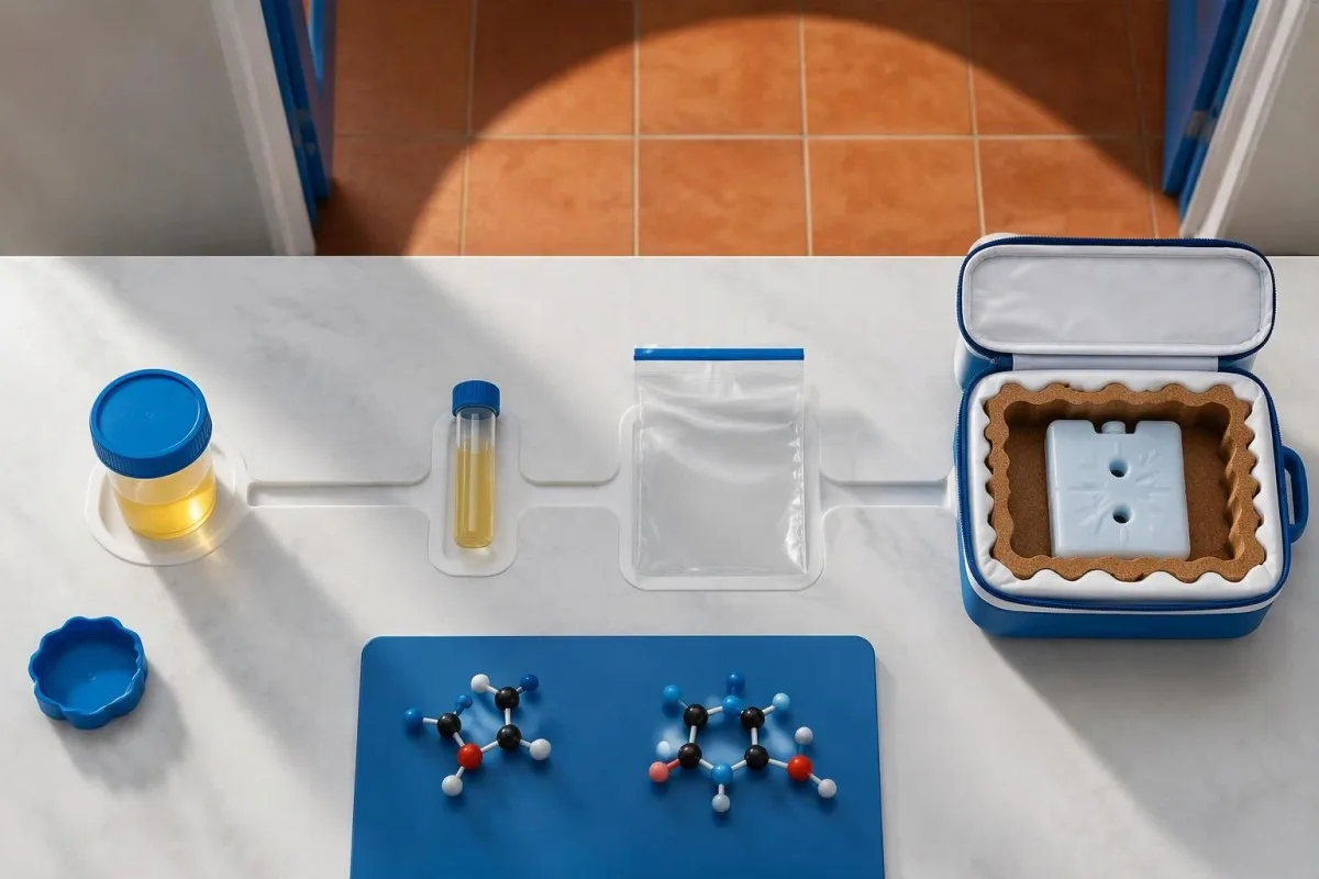
Ask whether the laboratory supplies a container or preservative and whether the specimen needs refrigeration, freezing, or another handling method. There is no universal home-storage rule for every TMA assay; adding even 1 unrequested chemical can invalidate the sample or interfere with the laboratory's preparation.
For a 24-hour collection, the laboratory's written instructions should explain the starting void, finishing void, and handling of each intervening specimen. Missing a void can compromise the calculated daily amount, even if a ratio within the submitted portion still appears mathematically plausible.
Collection rules from another urine assay should not be borrowed. Our guide to timed urine collection procedures shows why collection details matter, but catecholamine preservatives and dietary restrictions are not automatically appropriate for the 2 compounds measured in trimethylaminuria testing.
A useful handover includes collection start and finish times, relevant meals, symptoms, and recent treatment. If 1 specimen was left warm or its container leaked, tell the laboratory before interpreting the number; repeating an unsuitable collection is safer than building a genetic diagnosis around a compromised result.
How is the TMA oxidation percentage interpreted?
The TMA oxidation percentage is TMAO ÷ (TMA + TMAO) × 100, calculated with compatible molar quantities. Mackay et al. describe values above 92% in unaffected individuals and below 84% as suggestive of impairment, with an intermediate band; these are published interpretation bands, not universal diagnostic cutoffs.
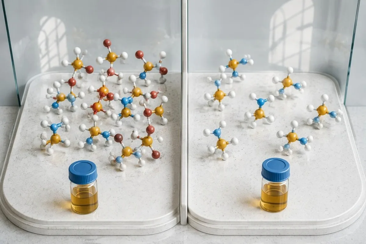
A hypothetical result of TMA 12 µmol/mmol creatinine and TMAO 48 µmol/mmol creatinine gives 48 ÷ 60 × 100 = 80% oxidation. That pattern suggests reduced conversion under the collection conditions, but it does not independently establish whether the cause is inherited, secondary, or influenced by preparation.
Another hypothetical sample containing TMA 5 µmol/mmol creatinine and TMAO 95 µmol/mmol creatinine gives 95% oxidation. Its free TMA fraction is 5%; oxidation percentage and free fraction add to 100% when both use the same 2-analyte denominator.
Some reports instead provide a TMA-to-TMAO ratio, which is not numerically interchangeable with oxidation percentage. For example, 12 ÷ 48 = 0.25, not 80%; read the formula and units before comparing laboratories, particularly when urine creatinine normalization is used.
No single absolute urinary TMA concentration provides a universally accepted diagnosis across all protocols. Hydration changes µmol/L values, creatinine excretion affects normalized results, and dietary substrate affects both compounds; the laboratory's reference interval and assessment of total exposure therefore matter alongside the percentage.
| Level | Range | What it means |
|---|---|---|
| Published unaffected pattern | >92% oxidation | Usually efficient conversion under adequate testing conditions; a low-exposure sample may still be uninformative. |
| Published intermediate band | Approximately 84–92% oxidation | Requires the laboratory's own interpretation, review of preparation, and sometimes repeat testing. |
| Published impaired-conversion pattern | <84% oxidation | Supports impaired TMA oxidation under the test conditions; does not by itself prove inherited FMO3-related disease. |
Can a normal or borderline trimethylaminuria test miss it?
A normal or borderline result may be inconclusive when dietary exposure was low, symptoms were absent, or collection was unsuitable. One reassuring specimen is more persuasive when the laboratory confirms adequate total TMA-related exposure and the sample reflects the person's usual symptomatic conditions.

Consider a hypothetical sample with TMA 0.1 and TMAO 0.4 µmol/mmol creatinine: the calculated oxidation is 80%, but the total is only 0.5 µmol/mmol. The percentage alone cannot show whether exposure was diagnostically adequate, and measurement uncertainty becomes more influential near an assay's quantification limits.
A repeat test should fix a specific limitation rather than simply produce another number. If the first sample followed restriction, the second may use a laboratory-approved dietary protocol; if symptoms were absent, collection during a representative episode may be preferable to 2 identically uninformative specimens.
Kantesti is an AI blood test interpretation platform that organizes routine laboratory trends; even 3 normal metabolic panels cannot exclude a condition requiring a different assay. Our guide to meaningful changes between lab results explains why preparation and sampling conditions belong beside the numerical trend.
When properly prepared results repeatedly show efficient oxidation, the clinician should reconsider the working diagnosis instead of ordering increasingly broad genetic panels automatically. Document what was tested, under which conditions, and what uncertainty remains; 'not supported by these 2 samples' can be more accurate than an unconditional exclusion.
When does FMO3 genetic testing help confirm the cause?
FMO3 genetic testing is most useful when urinary findings support impaired TMA oxidation and an inherited explanation is clinically plausible. Primary FMO3-related trimethylaminuria is usually autosomal recessive, so confirmation generally involves 2 disease-causing variants on opposite copies of the gene.
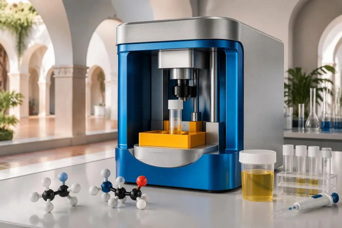
FMO3 encodes an enzyme involved in converting TMA to TMAO, rather than a protein whose quantity is routinely measured in a standard blood panel. Mitchell and Smith's 2001 review connects deficient oxidation with this pathway; measuring liver enzymes is not the same as measuring FMO3 function.
The laboratory may offer sequence analysis and, when relevant, additional methods for deletions or duplications. Ask what the assay covers and what it can miss: a negative result from 1 limited method does not necessarily exclude every possible genetic mechanism.
Biochemistry followed by targeted genetics is a familiar strategy in inherited metabolic assessment, but the ordering and evidence differ by disease. Our explanation of enzyme and genetic confirmation illustrates the distinction without implying that a Gaucher enzyme assay or its thresholds apply to these 2 urinary amines.
Genetic testing may also be considered earlier in selected family situations, such as a known familial FMO3 diagnosis. Nevertheless, finding 1 common variant on a consumer report cannot replace a diagnostic laboratory's classification, clinical history, and assessment of whether the urinary phenotype fits.
What do FMO3 variants mean for you and your family?
Two pathogenic FMO3 variants in trans can support inherited trimethylaminuria when the clinical and biochemical findings fit. One pathogenic variant usually indicates carrier status rather than a complete recessive diagnosis; a variant of uncertain significance, or VUS, is not diagnostic proof.

Two variants are not automatically on 2 different gene copies. If both are on the same copy, called 'in cis,' the other copy may remain functional; parental or other family testing can sometimes establish phase and change the meaning of what initially looked like a diagnostic result.
When both parents carry a disease-causing variant for the same autosomal recessive condition, each pregnancy has a 25% chance of an affected child, a 50% chance of a carrier child, and a 25% chance of a child inheriting neither familial variant. These probabilities restart for each pregnancy rather than predicting a sequence of outcomes.
A molecular diagnosis does not determine odor intensity on every day of life. Some FMO3 variants retain partial activity, and developmental stage, diet, and hormonal circumstances can influence expression; 2 people with similar genetic findings may therefore have different symptom patterns.
Family testing should target a clinically established finding, not an unexplained list of variants. Our guide to genetic results and family risk explains transferable counseling principles, while the genes and inheritance patterns discussed there are different from FMO3-related trimethylaminuria.
How do clinicians distinguish inherited and secondary causes?
Inherited trimethylaminuria involves reduced FMO3 function, while secondary or transient patterns can reflect other physiological or medical circumstances. Urinary TMA/TMAO testing identifies a biochemical pattern; distinguishing at least 2 categories of cause requires history, examination, and selected additional investigations.
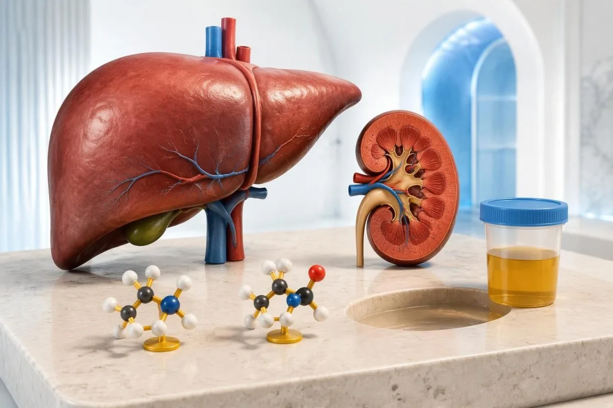
Advanced liver dysfunction and certain portosystemic shunts can disrupt normal handling of compounds produced in the intestine. A mild ALT rise alone does not establish this mechanism; our liver panel interpretation guide explains why ALT, bilirubin, albumin, and clinical findings answer different questions.
Kidney impairment can alter circulating TMAO and urinary excretion, but it does not automatically demonstrate defective FMO3 oxidation. A creatinine result of 1.4 mg/dL, for example, has different implications depending on age, muscle mass, and eGFR; interpret it as renal context, not a trimethylaminuria cutoff.
Kantesti AI can help explain reported liver and kidney markers, but those results cannot identify the cause of a low urinary oxidation percentage by themselves. Our guide to BUN and creatinine differences helps distinguish clearance concerns from other influences on these 2 routine markers.
Transient reduced oxidation can occur in childhood as enzyme activity develops, and symptom changes around menstruation have been described. Neither onset before age 18 nor adult onset alone proves a genetic or acquired cause; the urinary pattern, persistence, and selected genetic results need to tell a consistent story.
What other causes of fish-like odor should be checked?
The location and accompanying symptoms determine which alternative causes deserve testing. Odor confined to 1 area—such as vaginal discharge, urine, the mouth, or a skin fold—often calls for targeted examination before a generalized metabolic diagnosis is assumed.
Bacterial vaginosis can produce a fish-like vaginal odor through local amine production, but that does not demonstrate systemic FMO3 deficiency. When discharge, irritation, or exposure history warrants it, clinicians may also assess other causes; our guide to testing for trichomoniasis covers 1 distinct diagnostic pathway.
Urine odor alone is not a reliable reason to diagnose or treat a urinary infection. Dysuria, frequency, fever, and relevant clinical risk help determine whether urinalysis or culture is needed; bacteria without urinary symptoms often does not require antibiotics, apart from specific situations.
Dental disease, retained moisture in clothing, and local skin conditions can create or concentrate odors without altering urinary TMA oxidation. If 1 site consistently accounts for the smell, a focused examination may answer more than a large blood panel, particularly when there are visible local changes.
Seek prompt assessment if odor appears with fever, jaundice, confusion, or markedly reduced urine output; these signs raise questions beyond isolated trimethylaminuria. Conversely, odor without those features is not automatically an emergency, and there is no urinary oxidation percentage that serves as a universal emergency threshold.
What should you do while waiting for results?
While awaiting results, follow the testing plan and avoid unsupervised food challenges, antibiotics, or severe dietary restriction. A useful follow-up visit reviews 3 items together: sample conditions, the urinary biochemical pattern, and whether selected FMO3 testing would change diagnosis or family advice.
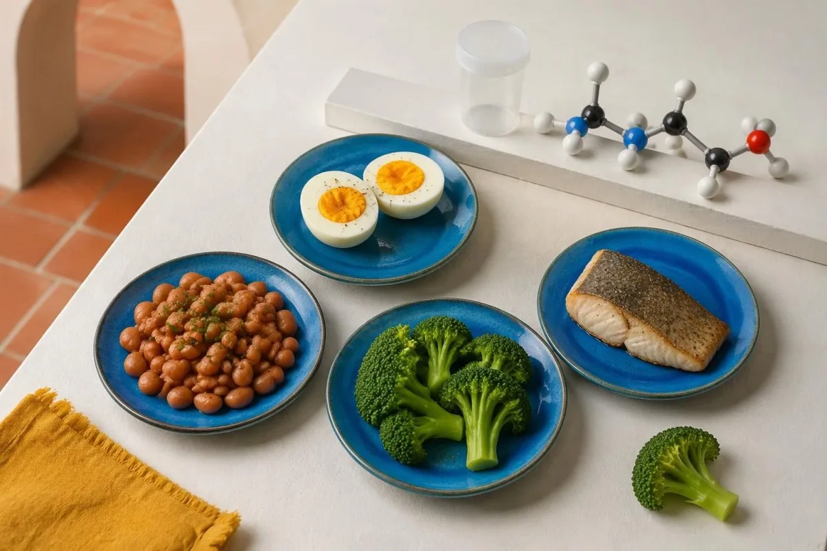
Choline is an essential nutrient, not simply an odor trigger to eliminate. US adequate intake values are 425 mg/day for adult women and 550 mg/day for adult men; pregnancy and lactation values are 450 and 550 mg/day, respectively, and these nutritional targets are not trimethylaminuria challenge doses.
An experienced dietitian can help adjust suspected triggers without removing entire food groups unnecessarily. Our guide to nutrient needs and supplement risks explains why restrictive plans need nutritional review; a generic supplement list cannot establish which intervention will improve a person's TMA handling.
Kantesti is an AI-powered blood test analysis tool, not a replacement for the metabolic laboratory that performs and validates urinary TMA/TMAO testing. Our validation and clinical limitations matter here: performance on routine blood reports does not establish diagnostic accuracy for this specialist urine assay.
Genetic and odor-related information can be sensitive, so share only what is necessary for the task and understand who will access it. Kantesti's privacy and data practices describe our approach; do not assume a family member's consent covers another person's FMO3 report or that 1 shared account replaces individual permission.
Research sources and related laboratory education
The diagnostic explanation here draws on published trimethylaminuria reviews, not on routine kidney or urinalysis guides. The 2 related Zenodo publications listed below provide background education only; they do not validate a trimethylaminuria test or establish FMO3 diagnostic accuracy.
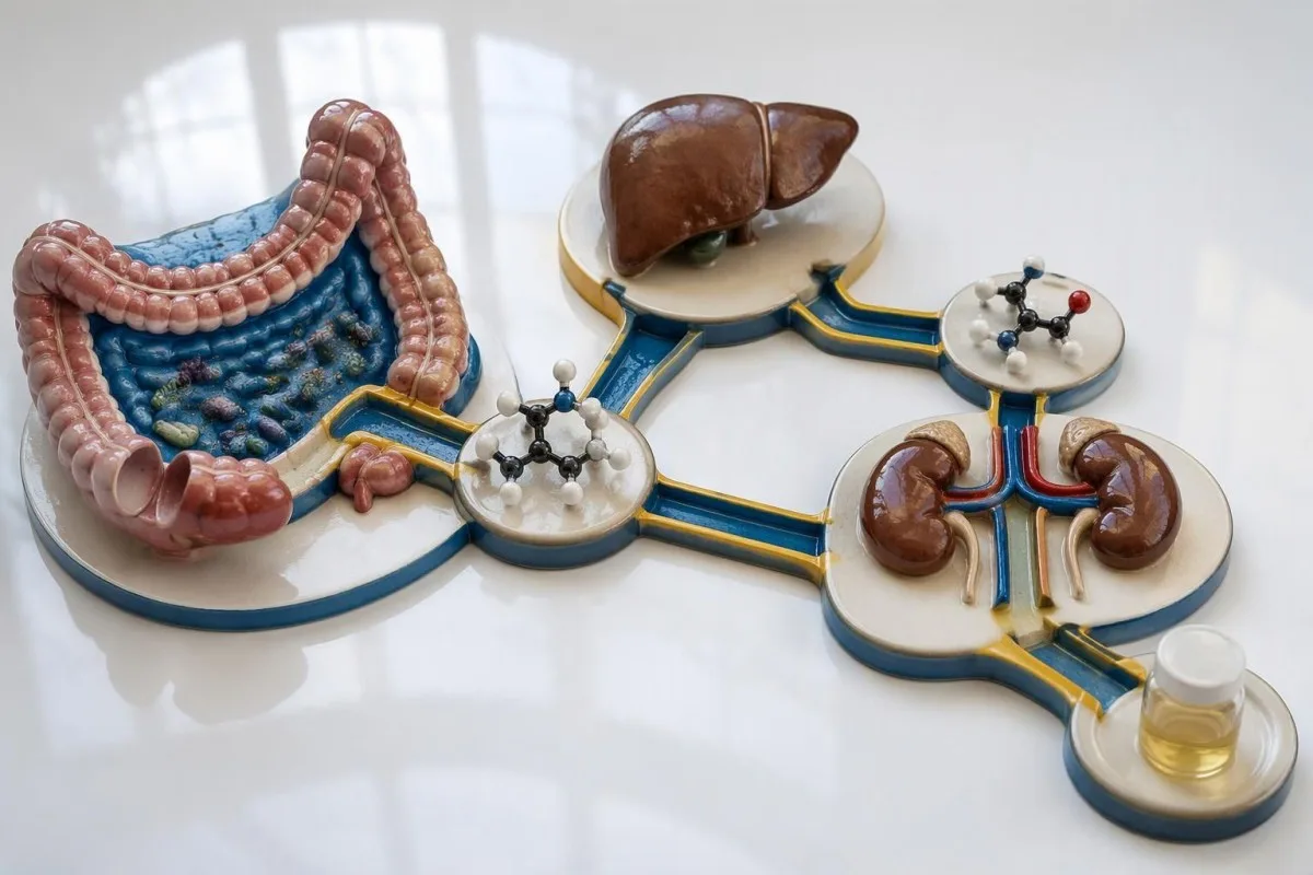
Mitchell and Smith's 2001 review, 'Trimethylaminuria: the fish malodor syndrome,' explains the biochemical basis and clinical presentation. Mackay et al.'s 2011 review, 'Trimethylaminuria: Causes and Diagnosis of a Socially Distressing Condition,' discusses testing and interpretation; neither supplies 1 universal collection protocol for every current laboratory.
Kantesti's related kidney function education helps explain why renal handling may matter when reviewing laboratory context. Its Zenodo record is an archived educational publication, not a trimethylaminuria clinical trial; a BUN/creatinine ratio cannot replace measurement of the 2 urinary amines.
Our general urinalysis background covers urine findings such as urobilinogen, which answer different questions from TMA oxidation. The associated Zenodo DOI provides a persistent publication link, but the presence of 1 DOI does not itself demonstrate independent peer review or clinical validation.
For an individual diagnosis, the receiving laboratory's method, reference interval, and written preparation instructions remain decisive alongside clinical assessment. Our medical content standards distinguish educational explanations from individualized diagnosis; the practical next step is to request the correct assay and its protocol before collecting a specimen.
Frequently asked questions
What is the test for fish odor syndrome?
The main biochemical test for fish odor syndrome measures urinary trimethylamine (TMA) and trimethylamine N-oxide (TMAO). The laboratory interprets these 2 compounds together, often calculating the percentage converted to TMAO. Selected FMO3 genetic testing can confirm an inherited cause when the biochemical and clinical findings fit. A routine urine dipstick or standard blood panel does not perform this assessment.
Can a normal urine trimethylamine test rule out trimethylaminuria?
One normal urine trimethylamine test may not exclude trimethylaminuria if dietary exposure was inadequate, symptoms were absent, or collection was unsuitable. A clinical review describes oxidation above 92% as typical of unaffected individuals, but that percentage is meaningful only within the laboratory's testing conditions. A specialist may repeat testing during symptoms or use an approved dietary protocol. Repeating a sample should address the original limitation rather than simply repeat the same preparation.
Should I eat fish before a trimethylaminuria test?
Eat fish before a trimethylaminuria test only if the receiving laboratory or specialist specifically instructs you to do so. Marine fish contains TMAO, and its consumption can change the amounts of both measured compounds. Different laboratories may use an ordinary diet, a standardized challenge, a timed specimen, or a 24-hour collection. An improvised fish meal does not guarantee a valid diagnostic test.
Is there a blood test for fish odor syndrome?
Routine blood tests do not diagnose fish odor syndrome; the principal biochemical investigation measures 2 urinary compounds, TMA and TMAO. Blood-based FMO3 genetic testing may help identify an inherited cause, but it answers a different question from urine chemistry. Liver and kidney panels can investigate relevant alternative or secondary causes. A cardiovascular blood TMAO result is not a substitute for urinary oxidation testing.
Does one FMO3 mutation mean I have trimethylaminuria?
One pathogenic FMO3 variant usually indicates carrier status rather than confirming recessive FMO3-related trimethylaminuria. Inherited disease generally requires 2 disease-causing variants on opposite gene copies, interpreted alongside urinary and clinical findings. Two variants on the same copy do not have the same meaning as variants on opposite copies. A variant of uncertain significance is not diagnostic proof.
Can fish odor syndrome happen without a genetic cause?
An abnormal TMA-related odor or urinary pattern can occur without a confirmed inherited FMO3 diagnosis. Secondary or transient explanations include selected liver-related conditions, altered substrate exposure, and developmental or hormonal circumstances; local odor sources can also mimic a systemic problem. Measuring the 2 urinary compounds helps characterize the pattern but does not identify the cause by itself. A negative genetic result must be interpreted according to the assay's coverage and the biochemical evidence.
Should I cut out choline while waiting for a diagnosis?
Do not start severe choline restriction while waiting for a trimethylaminuria diagnosis, particularly before a planned urine test. Choline is essential, with US adequate intake values of 425 mg/day for adult women and 550 mg/day for adult men. Restriction can compromise nutrition and reduce substrate exposure enough to weaken test interpretation. A specialist and dietitian can agree on a safer plan after reviewing the diagnostic question.
Get your blood test results explained
Upload your lab report and Kantesti's AI explains each biomarker in plain language within seconds. Used by more than 2 million people worldwide.
References
- Klein, T., Mitchell, S., & Weber, H. (2026). BUN/Creatinine Ratio Explained: Kidney Function Test Guide. Kantesti AI Medical Research. DOI
- Klein, T., Mitchell, S., & Weber, H. (2026). Urobilinogen in Urine Test: Complete Urinalysis Guide 2026. Kantesti AI Medical Research. DOI
- Mitchell SC and Smith RL (2001). Trimethylaminuria: the fish malodor syndrome. Drug Metabolism and Disposition.
- Mackay RJ et al. (2011). Trimethylaminuria: Causes and Diagnosis of a Socially Distressing Condition. Clinical Biochemist Reviews.
- 2M+Tests analysed
- 127+Countries
- 100+Languages
About the medical team


E-E-A-T Trust Signals
Experience
Physician-led clinical review of lab interpretation workflows.
Expertise
Laboratory medicine focus on how biomarkers behave in clinical context.
Authoritativeness
Medically reviewed by Dr. Thomas Klein, with contributions from Dr. Sarah Mitchell and Prof. Dr. Hans Weber.
Trustworthiness
Evidence-based interpretation with clear follow-up pathways to reduce alarm.
Medical disclaimer
This article is for educational purposes only and does not constitute medical advice. Always consult a qualified healthcare provider for diagnosis and treatment decisions.
Published · Contact the editorial team
Published by Kantesti Ltd, registered in England & Wales, company no. 17090423, London, United Kingdom.
