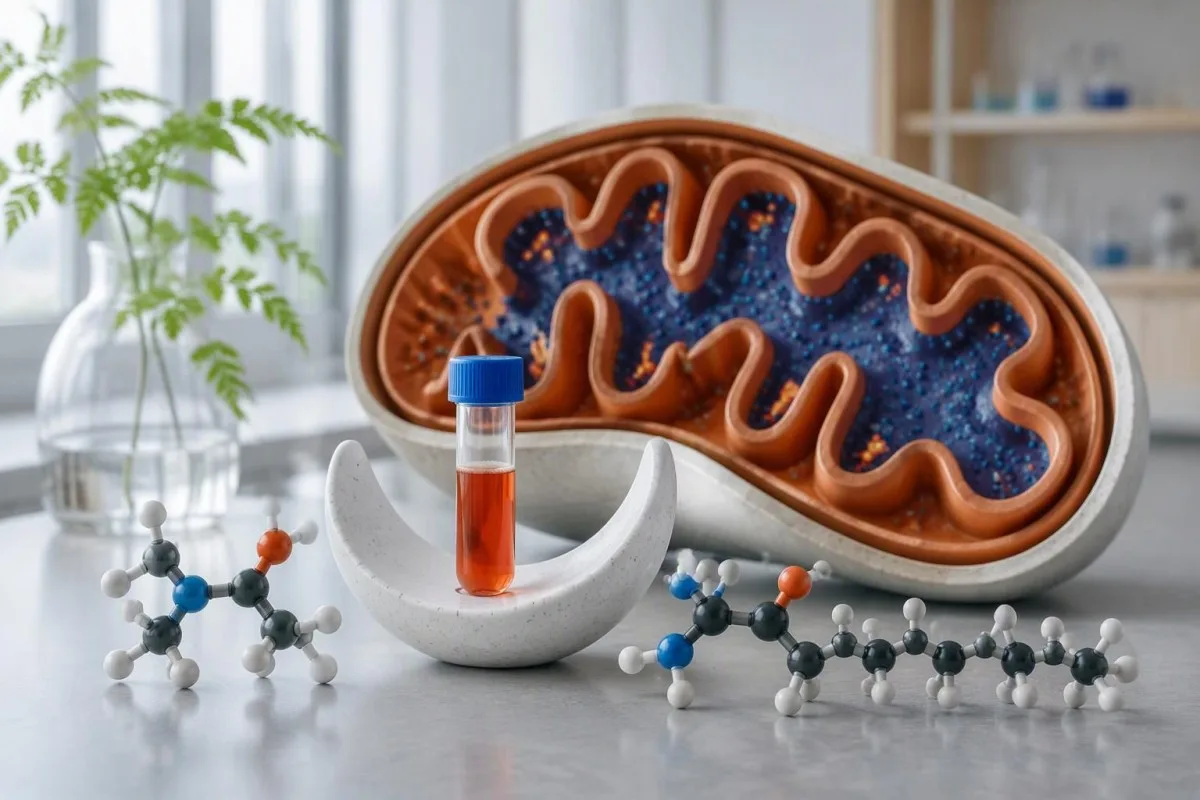
Free carnitine measures the unesterified pool; total carnitine includes free and acyl-bound forms. Their ratio helps distinguish depletion from a shifted balance—but cannot identify the cause by itself.


- Free carnitine is the unesterified fraction; one commonly used adult plasma reference interval is 25–54 µmol/L, but your laboratory’s interval takes priority.
- Total carnitine levels include free plus esterified carnitine; a normal total result can conceal low free carnitine.
- Acyl-to-free ratio is calculated as (total − free) ÷ free when both measurements use the same specimen and units.
- Different laboratory cutoffs matter: a ratio of 0.60 may be flagged by a laboratory using 0.40 as its upper limit but not one using 0.80.
- Marked depletion with persistent free carnitine below approximately 5 µmol/L raises concern for primary carnitine deficiency, although secondary causes must also be assessed.
- Medication review should include valproate and pivalate-containing medicines; do not stop prescribed treatment without clinical advice.
- Specialist testing may include individual acylcarnitines, urine organic acids and genetic analysis rather than another total carnitine measurement alone.
- Urgent symptoms such as confusion, repeated vomiting, collapse or severe muscle weakness need prompt assessment; glucose below 54 mg/dL, or 3.0 mmol/L, is a serious accompanying finding.
What does a carnitine blood test actually measure?
A carnitine blood test measures free carnitine and, usually, total carnitine; total includes free plus esterified forms called acylcarnitines. The acyl-to-free ratio describes their balance, so normal total carnitine can coexist with low free carnitine; an abnormal pattern warrants a cause-based review, not automatic supplementation.
Free carnitine is not the same concept as free thyroid hormone. Here, “free” means carnitine without an attached acyl group—not simply carnitine unbound from a circulating protein; confusing those meanings can derail interpretation before the numbers are even considered.
A total of 40 µmol/L with free carnitine of 15 µmol/L illustrates the problem: the total may sit within an adult interval while the free fraction is clearly below it. The calculated esterified fraction is 25 µmol/L, so the question becomes why so much of the circulating pool is esterified.
Kantesti is an AI blood test analyzer that can help organize free, total and calculated carnitine results alongside the laboratory’s own reference intervals. Our organization and clinical approach provide background; I would still want the original report, medication list and symptom history before treating any 1 result as a diagnosis.
Why do free and total carnitine tell different stories?
Free carnitine and total carnitine answer different questions: free measures the unesterified circulating pool, while total measures that pool plus esterified forms. Carnitine helps transfer long-chain fatty-acid groups into mitochondria, but a plasma concentration is not a direct measurement of mitochondrial performance or muscle energy production.
The carnitine shuttle involves CPT I, the carnitine-acylcarnitine translocase and CPT II. CPT I forms an acylcarnitine at the outer mitochondrial membrane, the translocase moves it across the inner membrane, and CPT II transfers the acyl group back to coenzyme A inside the mitochondrion; that sequence explains why measuring 1 circulating pool cannot locate every possible defect.
Most body carnitine is stored in tissues, particularly skeletal and cardiac muscle, rather than plasma. Longo, Amat di San Filippo and Pasquali described how transport defects and shuttle defects create distinct biochemical patterns in their 2006 review; a low plasma value therefore needs interpretation as a circulating clue, not a complete tissue inventory (Longo et al., 2006).
Carnitine is neither creatine nor creatinine, despite the similar spelling. It is also not ordinarily included in a routine basic or comprehensive metabolic panel; our explanation of what metabolic panels include helps clarify why a separate carnitine request may be needed even after 2 routine chemistry panels.
What are normal free and total carnitine levels?
Carnitine reference intervals depend on age, specimen and laboratory method. One commonly used adult plasma set is free carnitine 25–54 µmol/L, total carnitine 34–78 µmol/L, esterified carnitine 5–30 µmol/L and an esterified-to-free ratio of 0.10–0.80; these are examples, not universal diagnostic cutoffs.
A laboratory reference interval describes a comparison population, not a treatment boundary. Free carnitine of 24 µmol/L against a lower limit of 25 µmol/L is a different clinical proposition from 4 µmol/L, even though both carry the same low flag; symptoms, reproducibility and the other fractions determine how quickly to investigate.
Infant and child results require age-specific interpretation. A newborn screening value from a dried laboratory spot cannot simply be compared with an adult plasma interval of 25–54 µmol/L; specimen composition, screening methodology and maternal carnitine status can all affect the apparent discrepancy between 2 reports.
Reference-range disagreement is not necessarily laboratory error. Some laboratories use broader free-carnitine intervals or a different ratio upper limit; our guide to out-of-range laboratory results explains why I prefer the printed interval over a search-engine cutoff, especially when a result is only 1–2 µmol/L outside range.
How is the acyl-to-free carnitine ratio calculated?
The acyl to free carnitine ratio is esterified carnitine divided by free carnitine. When esterified carnitine is calculated from the same sample, the formula is (total carnitine − free carnitine) ÷ free carnitine; the result is unitless, and it is not the same as total divided by free.
Total 50 µmol/L and free 40 µmol/L produce a ratio of 0.25. First subtract 40 from 50 to obtain esterified carnitine of 10 µmol/L, then divide 10 by 40; dividing total directly by free would instead give 1.25, an entirely different quantity that could falsely alarm a reader.
A high ratio can reflect a small denominator rather than a large esterified concentration. Total 20 µmol/L and free 5 µmol/L yield esterified carnitine of 15 µmol/L and a ratio of 3.0; the striking feature is severe free-pool depletion, even though 15 µmol/L may fall within the laboratory’s esterified interval.
Calculated ratios inherit measurement and transcription errors. If rounding gives total slightly below free, the negative difference is not a real biological acyl pool; check units, dates and source values using our PDF extraction error checklist before comparing 2 results, and ask the laboratory about unexplained inconsistencies.
Can total carnitine be normal when free carnitine is low?
Normal total carnitine does not exclude low free carnitine. The esterified fraction can keep the combined total within range while the free fraction falls; for example, total 40 µmol/L and free 15 µmol/L produce esterified carnitine of 25 µmol/L and an acyl-to-free ratio of approximately 1.67.
Proportionate depletion can produce a reassuring-looking ratio. Total 28 µmol/L and free 22 µmol/L give esterified carnitine of 6 µmol/L and a ratio of 0.27; both total and free may be low against the example adult intervals, so a ratio within range must not cancel those concentration findings.
A ratio of 0.50 is not automatically evidence of a metabolic disorder. Total 60 µmol/L and free 40 µmol/L generate esterified carnitine of 20 µmol/L; that balance may be acceptable under a laboratory interval extending to 0.80, whereas a laboratory using 0.40 would flag it and require contextual review.
Pattern changes are more useful when sampling conditions remain comparable. A fall in free carnitine from 35 to 15 µmol/L after a medication change carries a different meaning from 2 measurements collected at different points in an acute illness; our guide to meaningful changes between visits explains that distinction without assuming every numerical shift is disease progression.
What causes low free carnitine besides inherited disease?
Low free carnitine can result from reduced intake or synthesis, urinary losses, dialysis, medication effects or secondary depletion during other metabolic disorders. An adult free-carnitine result of 20 µmol/L therefore does not, by itself, distinguish nutritional depletion from renal loss or increased formation and elimination of acylcarnitines.
Plant-based diets generally supply less preformed carnitine than diets containing animal foods. Healthy adults can synthesize carnitine from amino-acid precursors and conserve it through the kidneys; a free value of 23 µmol/L in a well vegan adult should not automatically prompt a high-dose prescription or a conclusion that the diet is inadequate.
Prematurity, prolonged nutrition support and substantial malnutrition change the interpretation. In a patient receiving long-term parenteral nutrition, 2 questions matter separately: whether the formulation provides carnitine and whether illness, organ function or losses have changed requirements; simply adding dietary meat is neither a feasible nor a sufficient answer in that setting.
Renal tubular dysfunction can cause carnitine wasting even when filtration looks relatively preserved. A low result accompanied by urinary glucose without hyperglycemia, phosphate loss or unexplained acidosis needs a renal assessment; our plant-based nutrient assessment guide addresses the separate nutritional question, including why 1 low marker does not prove a generalized deficiency state.
Which medications warrant a carnitine-focused review?
Valproate and pivalate-containing medicines are particularly relevant when free carnitine is low. Medication review should consider exposure, dose changes, nutrition and symptoms rather than the carnitine value alone; free carnitine of 18 µmol/L in a person taking valproate warrants a different discussion from the same result without that exposure.
Valproate can contribute to carnitine depletion and hyperammonemic toxicity, but routine supplementation is not appropriate for every user. Lheureux and colleagues’ 2005 Critical Care review discusses the biochemical rationale and limitations of the evidence; overdose, encephalopathy and selected high-risk situations are different from an otherwise well adult with 1 mildly low value (Lheureux et al., 2005).
New confusion, vomiting or unusual drowsiness during valproate treatment needs prompt assessment. Clinicians may check ammonia, liver chemistry and valproate exposure even if free carnitine is above 25 µmol/L, because a normal carnitine concentration does not rule out toxicity; patients should not independently stop an antiseizure medicine while waiting for an explanation.
Pivalate can increase urinary carnitine loss through pivaloylcarnitine formation. Some medicines contain pivoxil or related pivalate-generating groups, so the exact generic drug name matters; our medication trend monitoring guide can help organize 2 dated reports around an exposure, but a prescriber or pharmacist must decide whether treatment should change.
How do kidney disease and dialysis change carnitine results?
Kidney dysfunction can alter carnitine clearance, while hemodialysis can remove circulating free carnitine. The resulting pattern depends on residual kidney function, dialysis timing and supplementation; a free value of 20 µmol/L before a session is not directly interchangeable with 20 µmol/L immediately after one.
Reduced clearance can increase some acylcarnitines without proving an inherited oxidation defect. A ratio of 0.90 in advanced kidney disease needs comparison with the laboratory interval and the wider renal picture; otherwise, a retention-related pattern could be mistaken for a primary metabolic condition and trigger unnecessary genetic testing.
Dialysis-related carnitine treatment is a nephrology decision, not a general supplement recommendation. Symptoms such as cramps and fatigue have multiple competing causes, and a numerical rise after supplementation does not establish clinical benefit; our kidney disease staging guide explains why eGFR and urine ACR answer different questions from 1 carnitine result.
A useful dialysis comparison records the session and treatment schedule. Ask whether each of 2 samples was collected before or after dialysis and before or after prescribed carnitine; the BUN and creatinine interpretation guide supplies complementary renal context, but neither the BUN/creatinine ratio nor eGFR can quantify carnitine stores.
When does low carnitine suggest a primary transport disorder?
Persistent, markedly low free carnitine—often below approximately 5 µmol/L—raises concern for primary carnitine deficiency, especially with cardiomyopathy, hypoglycemia or suggestive family history. Primary carnitine deficiency involves impaired OCTN2 transport associated with SLC22A5 variants; a mildly low value alone is not enough to diagnose it.
Primary carnitine deficiency can present beyond infancy, including in adults with few symptoms. El-Hattab and Scaglia’s 2015 review describes the clinical spectrum and the role of biochemical and genetic assessment; 2 low results plus a credible renal-wasting or family pattern are more informative than assuming every adult case must resemble a newborn crisis (El-Hattab and Scaglia, 2015).
A low newborn screening carnitine result can reflect maternal deficiency as well as infant disease. That is why the follow-up pathway may assess 2 people—the infant and mother—rather than treating the screening result as a diagnosis; supplementation before confirmatory sampling should be coordinated with the metabolic team, without delaying treatment when the infant is unwell.
Specialists may assess urinary loss, SLC22A5 variants and, in selected unresolved cases, transport function. A family history of unexplained cardiomyopathy or sudden death increases concern even if the current patient feels well; our discussion of children’s interpretation safety limits explains why adult intervals such as 25–54 µmol/L must not guide pediatric decisions.
When is an acylcarnitine profile more useful than the ratio?
An acylcarnitine profile is more useful when the question is which metabolic pathway may be affected. Free and total carnitine summarize pools, whereas a profile measures individual species such as C8, C14:1 or C16; a ratio of 1.20 cannot reveal which species accounts for the imbalance.
Specific acylcarnitine patterns guide, but do not independently establish, metabolic diagnoses. C8 enrichment can support investigation for medium-chain acyl-CoA dehydrogenase deficiency, while C14:1 can support investigation for very-long-chain acyl-CoA dehydrogenase deficiency; the full pattern, ratios, clinical setting and confirmatory tests matter more than 1 isolated species (Longo et al., 2006).
Normal results between episodes do not exclude every fatty-acid oxidation disorder. Recurrent illness-associated hypoglycemia with unexpectedly low ketones is a stronger referral signal than a modest ratio elevation alone; our beta-hydroxybutyrate interpretation guide explains why a ketone result of 0.2 mmol/L must be interpreted against the simultaneous glucose and feeding state.
Specialist evaluation may combine acylcarnitines, urine organic acids, glucose, ketones, ammonia, lactate and genetics. Repeated exercise-associated episodes with creatine kinase above 1,000 IU/L also change the differential, although exercise itself can elevate CK; our high creatine kinase guide explains the muscle assessment, and clinicians should never provoke an episode through unsupervised fasting.
How should you prepare for a repeat carnitine test?
A repeat carnitine test is most useful when specimen type, laboratory and sampling conditions are comparable. Record meals, supplements, illness and medication timing; there is no universal fasting or supplement washout rule, and a result of 22 µmol/L should not prompt a self-directed prolonged fast to “confirm” deficiency.
Recent L-carnitine supplementation can raise the circulating value and obscure the untreated baseline. Give the clinician the product, dose in mg and last-dose time, including acetyl-L-carnitine products; our explanation of supplements before laboratory testing helps separate ordinary preparation advice from a medication interruption that requires approval.
Repeat timing depends on the reason for testing, not a fixed calendar rule. An otherwise well adult with free carnitine of 24 µmol/L may have a planned outpatient repeat after a reversible issue is addressed, whereas 4 µmol/L with weakness should not wait weeks for a routine redraw; prescribed carnitine should not be withheld simply to produce an untreated number.
Acute illness and treatment can make 2 samples biologically different even with perfect laboratory technique. Dextrose, feeding and recovery may change a metabolic profile, so emergency teams may collect diagnostic specimens early when feasible without delaying care; our illness and medication timeline guide explains why the timestamp belongs beside the concentration.
Why not treat every abnormal result with carnitine supplements?
An abnormal carnitine result does not establish that an over-the-counter supplement will help. Treatment depends on the cause: confirmed primary deficiency, selected secondary deficiencies and certain toxicology situations differ from unexplained fatigue with free carnitine of 23 µmol/L; raising a plasma concentration is not the same as improving health.
Treatment for confirmed primary carnitine deficiency often uses weight-based prescription levocarnitine. El-Hattab and Scaglia describe doses around 100–200 mg/kg/day, adjusted to clinical response and laboratory monitoring; those specialist doses are not a self-treatment template, and a consumer serving of 500 mg or 1,000 mg does not address every reason a free value may be low.
Carnitine is not automatically beneficial in every fatty-acid oxidation disorder. In some long-chain disorders, clinicians are cautious about increasing potentially harmful long-chain acylcarnitine pools; our lab-guided fatigue supplement guide addresses the broader distinction between correcting a demonstrated need and trying 2 products while the actual diagnosis remains unresolved.
Levocarnitine can cause nausea, diarrhea or a fishy odor, and drug interactions require review. INR monitoring may need closer attention in people taking warfarin, and reported seizure events warrant discussion in susceptible patients; before prescribing 1 course, I would want a treatment goal, a monitoring plan and a clear reason that diet or medication review alone is insufficient.
Which symptoms make an abnormal carnitine result urgent?
Urgency depends on symptoms and accompanying chemistry, not the carnitine flag alone. Confusion, collapse, repeated vomiting, inability to maintain intake, new cardiac symptoms or severe muscle weakness need prompt assessment; glucose below 54 mg/dL, or 3.0 mmol/L, is a serious finding whether carnitine is low or normal.
Symptomatic hypoglycemia should be treated without waiting for a carnitine assay. A conscious person able to swallow can follow their established rapid-carbohydrate plan, but confusion, seizures or inability to swallow require emergency help; our low glucose causes guide explains why medication exposure and illness remain common explanations even when glucose is below 70 mg/dL.
Severe muscle weakness with dark urine needs assessment for muscle breakdown and kidney complications. Free carnitine of 18 µmol/L does not explain away that presentation or establish the cause; our dark urine warning signs help distinguish a color observation from the need to check CK, kidney function, potassium and hydration.
A known or suspected metabolic disorder changes the advice during vomiting or poor intake. Follow the individual emergency regimen and contact the metabolic team early, particularly for children; conversely, 1 borderline result in an asymptomatic adult is usually an outpatient investigation, and a ratio of 0.45 without other abnormalities should not be presented as an impending crisis.
What should you bring to follow-up, and what can AI help explain?
Bring the complete report, laboratory intervals, medication and supplement list, symptoms and collection conditions to follow-up. As of October 3, 2026, there is no universally accepted acyl-to-free cutoff that diagnoses a metabolic disorder; a ratio of 0.60 still needs interpretation alongside concentrations, age, kidney function and the clinical question.
Kantesti is an AI blood test interpretation platform that can help explain why free carnitine of 15 µmol/L deserves attention even when total carnitine is 40 µmol/L. Our technology and interpretation guide describes the workflow; checking units, calculations and source values remains necessary before any explanation is used in care.
AI-generated context is not confirmation of an inherited disorder or a prescribing instruction. Our clinical standards and limitations describe the boundaries of interpretation; I’m Thomas Klein, Kantesti’s Chief Medical Officer, and my editorial approach is to separate 3 questions: whether the result is reliable, whether depletion is present and what evidence identifies its cause.
A useful appointment should end with an agreed next step, not merely a renamed abnormality. Ask whether the plan is repeat testing, medication review, renal assessment or metabolic referral, and what would change that plan; Kantesti’s medical advisory board information describes our physician advisory roles, but this article does not document an individual consultation or completed patient-specific review.
Keep the original laboratory terminology
Kantesti is an AI-powered blood test analysis tool that can help organize related measurements without treating every ratio as interchangeable. Our biomarker terminology guide supports that distinction; as Thomas Klein, I would want all 3 reported quantities—free, total and ratio—preserved exactly, including whether the laboratory called the numerator “acyl” or “esterified.”
Research publications and source boundaries
The 2 Zenodo publications below provide background on other laboratory measurements, not direct evidence for carnitine treatment. The RDW and red-cell guide may help when fatigue also prompts a CBC review; carnitine-specific reasoning in this article instead draws on the separately listed peer-reviewed references, and discovery links do not verify indexing or peer review.
Frequently Asked Questions
What does a carnitine blood test show?
A carnitine blood test usually measures free and total carnitine, with an esterified concentration or acyl-to-free ratio reported or calculated. Free carnitine is unesterified, while total includes free plus esterified forms. One commonly used adult plasma interval is 25–54 µmol/L for free carnitine and 34–78 µmol/L for total, but laboratory-specific intervals take priority. The test can identify depletion or an altered balance, but it cannot establish the cause without clinical context.
Can free carnitine be low if total carnitine is normal?
Free carnitine can be low even when total carnitine falls within the laboratory reference interval. Total 40 µmol/L with free 15 µmol/L leaves an esterified fraction of 25 µmol/L and an acyl-to-free ratio of approximately 1.67. The combined total conceals the reduced free pool because esterified forms contribute to the total. Medication effects, kidney function and metabolic causes should be reviewed rather than assuming a normal total excludes a problem.
How do you calculate the acyl-to-free carnitine ratio?
The acyl-to-free carnitine ratio is esterified carnitine divided by free carnitine. If esterified carnitine is calculated from total and free, use (total − free) ÷ free, with both concentrations from the same specimen in the same units. Total 50 µmol/L and free 40 µmol/L produce a ratio of 0.25. Total divided by free would give 1.25 and is not the same ratio.
Is an acyl-to-free carnitine ratio above 0.4 abnormal?
An acyl-to-free carnitine ratio above 0.4 is abnormal only relative to a laboratory or clinical framework using that cutoff. Some adult laboratory intervals extend to approximately 0.8, so a ratio of 0.6 may be flagged on one report and not another. A high ratio may reflect reduced free carnitine, increased esterified carnitine or both. No single ratio cutoff independently diagnoses an inherited metabolic disorder.
Does low free carnitine mean I should take a supplement?
Low free carnitine does not automatically mean an over-the-counter supplement is appropriate. A mildly low result such as 23 µmol/L should be assessed against the laboratory interval, medication exposure, nutrition, kidney function and symptoms. Persistent free carnitine below approximately 5 µmol/L warrants prompt clinical investigation, but it still does not identify the cause by itself. Prescription levocarnitine is appropriate in defined situations, while some long-chain fatty-acid oxidation disorders require specialist caution.
When should abnormal carnitine results lead to metabolic specialist testing?
Metabolic specialist testing is appropriate when carnitine abnormalities are marked, persistent or accompanied by recurrent hypoglycemia, cardiomyopathy, exercise-associated muscle breakdown or a suggestive family history. Persistent free carnitine below approximately 5 µmol/L is a particularly concerning clue for a transport disorder after secondary causes are considered. Follow-up may include individual acylcarnitines, urine organic acids and genetic testing rather than free and total carnitine alone. Confusion, collapse or glucose below 54 mg/dL requires urgent assessment rather than waiting for an outpatient referral.
Get AI-Powered Blood Test Analysis Today
Join over 2 million users worldwide who trust Kantesti for instant, accurate lab test analysis. Upload your blood test results and receive comprehensive interpretation of 15,000+ biomarkers in seconds.
📚 Referenced Research Publications
Klein, T., Mitchell, S., & Weber, H. (2026). RDW Blood Test: Complete Guide to RDW-CV, MCV & MCHC. Kantesti AI Medical Research.
Klein, T., Mitchell, S., & Weber, H. (2026). BUN/Creatinine Ratio Explained: Kidney Function Test Guide. Kantesti AI Medical Research.
📖 External Medical References
Longo N, Amat di San Filippo C, Pasquali M. (2006). Disorders of carnitine transport and the carnitine cycle. American Journal of Medical Genetics Part C: Seminars in Medical Genetics.
Lheureux PER et al. (2005). Science review: Carnitine in the treatment of valproic acid-induced toxicity—what is the evidence?. Critical Care.
El-Hattab AW, Scaglia F. (2015). Carnitine Deficiency Disorders: Clinical Diagnosis and Management. Molecules.
⚕️ Medical Disclaimer
This article is for educational purposes only and does not constitute medical advice. Always consult a qualified healthcare provider for diagnosis and treatment decisions.
E-E-A-T Trust Signals
Experience
Physician-led clinical review of lab interpretation workflows.
Expertise
Laboratory medicine focus on how biomarkers behave in clinical context.
Authoritativeness
Written by Dr. Thomas Klein with review by Dr. Sarah Mitchell and Prof. Dr. Hans Weber.
Trustworthiness
Evidence-based interpretation with clear follow-up pathways to reduce alarm.