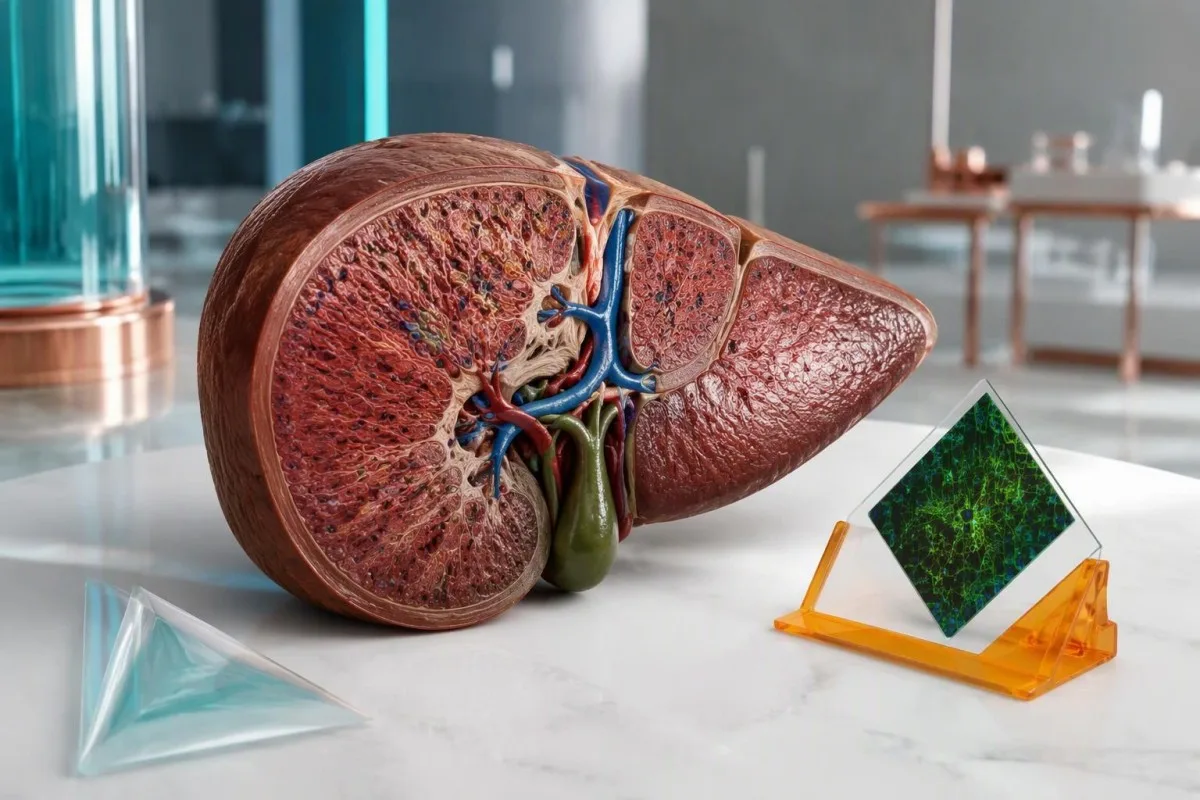
Een positieve antistofuitslag is een aanwijzing, geen diagnose. De relevante vraag is of deze past bij uw leverenzymen, IgG-gehalte, medicatiegeschiedenis en weefselbevindingen.


- Positieve gladde-spierantistoffen alleen stellen geen diagnose auto-immuunhepatitis; een ASMA-titer van 1:40 bij een volwassene levert slechts 1 punt op voor de vereenvoudigde diagnostische score.
- Hogere ASMA-titers versterken de diagnostische aanwijzingen, maar titers van 1:80 en 1:320 leveren volgens de vereenvoudigde criteria hetzelfde maximum van 2 punten voor antistoffen op.
- Antistoffen tegen F-actine wijzen op een specifieker antistofdoelwit, maar voor een positieve uitslag zijn nog steeds de uitslagen van levertests en de klinische context nodig; assay-eenheden kunnen niet worden omgerekend naar ASMA-titers.
- Totaal IgG boven de bovengrens van het laboratorium ondersteunt de diagnose auto-immuunhepatitis; een gehalte boven 1,1 keer die grens levert 2 punten op voor de vereenvoudigde score.
- Normale ALT maakt toevallige positiviteit minder zorgwekkend, hoewel normale enzymen of IgG auto-immuunhepatitis niet altijd uitsluiten.
- Medicatiegeschiedenis moet ook recepten en supplementen omvatten die maanden of jaren eerder zijn gestart, met name nitrofurantoïne en minocycline.
- Leverbiopsie helpt doorgaans bij het vaststellen van auto-immuunhepatitis en het beoordelen van fibrose; urgente behandelbeslissingen mogen bij een ernstig zieke patiënt niet wachten op niet-spoedeisend onderzoek.
- Spoedbeoordeling is nodig bij geelzucht met verwardheid, ernstige sufheid of een stijgende INR; een INR van ten minste 1,5 met encefalopathie kan wijzen op acuut leverfalen.
Betekenen positieve gladde-spierantistoffen dat ik auto-immuunhepatitis heb?
Positieve antistoffen tegen glad spierweefsel stellen op zichzelf geen diagnose van auto-immuunhepatitis. Een uitslag met een lage titer, zoals 1:40, kan aspecifiek zijn; artsen kijken naar een overeenkomstig patroon van verhoogde ALT of AST, verhoogd IgG, uitsluiting van andere leveraandoeningen en doorgaans ondersteunende bevindingen in leverweefsel.
ASMA, ook SMA genoemd, detecteert antistoffen die reageren met componenten van glad spierweefsel en verwante celstructuren; dit betekent niet dat uw spieren worden afgebroken. ASMA wordt het sterkst in verband gebracht met auto-immuunhepatitis type 1, maar kan ook voorkomen bij andere immuunreacties of leveraandoeningen. Daarom is het laboratoriumwoord ‘positief’ geen ziektelabel.
Bekijk 2 illustratieve uitslagen: ASMA 1:40 met ALT 24 IU/L en normaal IgG, tegenover ASMA 1:160 met ALT 286 IU/L en IgG boven de laboratoriumgrens. Het tweede patroon verdient aanzienlijk meer onderzoek, hoewel geen van beide de oorzaak vaststelt zonder klinische evaluatie. Onze uitleg van positief antilichaamresultaat betekenis maakt onderscheid tussen de detectie van antistoffen en aanwijzingen voor actieve ziekte.
Kantesti is een AI-bloedtestanalysator die kunnen helpen ASMA-uitslagen samen met leverenzymen en IgG te beoordelen, in plaats van een markering bij 1:40 als diagnose te behandelen. Als Thomas Klein, Chief Medical Officer, wil ik in dit artikel mijn redenering openlijk uitleggen, ook waar de cijfers geen uitsluitsel kunnen geven. Onze organisatie en klinische focus bieden context bij deze educatieve rol; de diagnose blijft de verantwoordelijkheid van een arts.
Interpretatie van de ASMA-titer: wat betekenen 1:40 en 1:80?
ASMA-titers geven aan hoe sterk een monster kan worden verdund terwijl de antistofkleuring detecteerbaar blijft. Bij volwassenen kennen de vereenvoudigde criteria voor auto-immuunhepatitis 1 punt toe voor ANA of SMA van ten minste 1:40 en 2 punten voor ten minste 1:80; geen van beide drempelwaarden stelt op zichzelf de diagnose hepatitis.
Een titer van 1:160 betekent dat kleuring na een grotere verdunning detecteerbaar blijft dan bij 1:40; het betekent niet dat er 4 keer zoveel leverschade is. In de vereenvoudigde criteria van Hennes et al. stopt de puntentoekenning voor antistoffen bij 2, dus 1:80 en 1:640 leveren niet steeds meer diagnostische punten op. Antistofconcentratie, testgevoeligheid en weefselschade hangen slechts onvolmaakt samen—en dat onderscheid voorkomt onnodige ongerustheid.
Laboratoriummethoden beïnvloeden de vergelijkbaarheid. Indirecte immunofluorescentie op weefselsubstraten, antigeenspecifieke tests, startverdunningen en rapportageconventies verschillen per laboratorium; een verandering van 1:40 naar 1:80 kan één verdunningsstap vertegenwoordigen, en geen klinisch betekenisvolle verandering. Dezelfde voorzichtigheid geldt voor de interpretatie van de ANA-test, met name wanneer meerdere zwak positieve antistoffen tegelijk worden aangetroffen.
Bij kinderen is een andere interpretatie nodig: SMA-titers vanaf 1:20 kunnen relevant zijn bij een passend klinisch beeld bij kinderen. Drempelwaarden voor volwassenenscores mogen niet zonder specialistisch oordeel worden toegepast op de uitslag van een kind; bovendien kan een screening volgens de volwassenenmethode bevindingen met een lagere titer bij kinderen missen. Vraag het laboratorium welk substraat en welke startverdunning zijn gebruikt; die details zijn vaak nuttiger dan de test elders te herhalen.
Verduidelijkt een positieve uitslag van antistoffen tegen F-actine de diagnose?
An F-actin antibody positive result can strengthen suspicion of autoimmune hepatitis, but it is not a stand-alone confirmation. F-actin testing targets filamentous actin, one antigen recognized by some smooth muscle antibodies; its usefulness depends on the assay, strength of positivity, and accompanying liver abnormalities.
F-actin assays and ASMA titers are different measurements. Some commercial ELISA methods report less than 20 units as negative, 20–30 as weakly positive, and above 30 as positive, but other kits use different categories and units. A result of 25 units must therefore be read against that report's reference interval, not a cutoff copied from another laboratory or an online discussion.
A weak F-actin result with ASMA 1:40 and normal ALT may add little certainty; a clearly positive result alongside persistent enzyme elevation and increased IgG is more persuasive. F-actin antibodies can occur outside autoimmune hepatitis, including in other liver diseases, while some patients with autoimmune hepatitis lack detectable F-actin antibodies. This is a probability adjustment, not a yes-or-no confirmation switch.
Discordant tests deserve a method check before another diagnosis is added. ASMA may recognize targets other than F-actin, and antigen preparation can influence an ELISA result, so positive ASMA with negative F-actin is not automatically a laboratory error. Our guide to kwalitatieve en kwantitatieve resultaten explains why 1:80, 25 units, and a simple positive flag cannot be converted into one interchangeable number.
Waarom is totaal IgG belangrijker dan nog een positieve antistofuitslag?
Elevated total IgG supports autoimmune hepatitis because it reflects increased immunoglobulin production accompanying immune-mediated liver injury. The simplified criteria award 1 point above the laboratory upper limit and 2 points above 1.1 times that limit; normal IgG does not completely exclude the disease.
Adult IgG reference intervals commonly approximate 7–16 g/L, equivalent to 700–1,600 mg/dL, although laboratory and age-specific ranges take priority. If the upper limit is 16 g/L, an IgG of 17 g/L exceeds normal, while 18 g/L exceeds 1.1 times the limit of 17.6 g/L. Use the measured total IgG—not total protein—to make that comparison.
Globulin and the gamma gap cannot substitute for quantitative IgG. For example, total protein 84 g/L minus albumin 38 g/L produces a 46 g/L gap, but that gap contains proteins other than IgG and does not prove autoimmune hepatitis. Our gamma gap interpretation guide explains why an apparently useful shortcut can send the investigation down the wrong path.
Normal IgG should lower confidence in a classic presentation, not close the case when ALT is 500 IU/L or the patient is jaundiced. Acute presentations can lack the usual immunoglobulin pattern, and IgG can also rise with cirrhosis or other inflammatory conditions. Read IgG, IgA en IgM samen when the protein abnormality is broader than isolated IgG elevation.
Welk patroon in levertestuitslagen maakt ASMA-positiviteit zorgwekkender?
Persistent ALT and AST elevation makes positive ASMA more concerning, especially when IgG is also elevated. Autoimmune hepatitis usually produces a hepatocellular pattern—aminotransferases rising more prominently than alkaline phosphatase—but the enzyme magnitude alone cannot identify the cause or accurately measure liver function.
ALT reflects injury more than function. An ALT of 240 IU/L with an upper limit of 40 is 6 times the upper limit, but that ratio does not distinguish autoimmune hepatitis from viral hepatitis, drug injury, or several other causes. Bilirubin, INR, albumin, symptoms, and the direction of change tell a different part of the story; our ALT-resultaatverklaring separates these roles.
De R-ratio compares ALT divided by its upper limit with ALP divided by its upper limit; a ratio at least 5 supports a hepatocellular injury pattern, particularly in drug-injury assessment. ALT 240/40 and ALP 120/120 give an R ratio of 6. This calculation classifies the pattern, not the diagnosis, and should use values obtained at the same time.
Disproportionate ALP elevation changes the differential diagnosis. If ALP is 3 times its upper limit while ALT is only mildly raised, clinicians consider biliary obstruction, primary biliary cholangitis, primary sclerosing cholangitis, or an overlap presentation rather than assuming isolated autoimmune hepatitis. Our guide to cholestatic liver-test patterns shows why GGT and imaging may become more useful than another ASMA measurement.
Hoe geruststellend zijn normale leverenzymen bij positieve ASMA?
Positive ASMA with repeatedly normal liver enzymes is generally less concerning than ASMA with unexplained enzyme elevation. In a retrospective cohort reported by Healey et al. in 2016, only 1 of 202 people with SMA positivity and ALT below 55 IU/L subsequently received an autoimmune hepatitis diagnosis.
The observed proportion was approximately 0.5%, not zero. That study's ALT threshold of 55 IU/L was part of its design and should not become everyone's definition of normal, particularly when your laboratory uses a lower limit. The retrospective population also cannot provide an individualized probability for someone with jaundice, high IgG, prior abnormal tests, or established liver disease.
For an illustrative patient with ASMA 1:40, ALT 26 IU/L, normal bilirubin, and normal IgG, the next step is usually clinical review rather than immediate immunosuppression. A clinician may choose a repeat liver panel in roughly 6–12 weeks when a recent illness, medication exposure, or uncertain baseline needs clarification, but there is no universal ASMA-specific retesting interval. Review mild ALT elevation follow-up if the supposedly normal result is actually borderline.
Previous results can overturn a reassuring snapshot. An ALT of 30 IU/L today means something different after an unexplained value of 300 IU/L last month, especially if corticosteroids were started between samples. Bring at least 2 dated panels when possible; the important question is whether injury never occurred, resolved after a trigger disappeared, or was suppressed by treatment rather than truly excluded.
Kunnen geneesmiddelen positieve ASMA en auto-immuunachtige hepatitis veroorzaken?
Some medicines can cause liver injury with ASMA positivity, increased IgG, and tissue findings resembling autoimmune hepatitis. Nitrofurantoin and minocycline are classic examples; exposure may precede recognition by months or years, so a medication taken uneventfully for 2 years still belongs in the history.
The start date matters as much as the medicine name. Hydralazine, methyldopa, and some biologic medicines can also produce autoimmune-like liver injury, while herbal products may cause other injury patterns that coexist with incidental antibodies. Write down dose changes, intermittent courses, and supplements used during the preceding 12 months; include longer-standing drugs rather than limiting the list to something newly prescribed last week.
Drug-induced autoimmune-like hepatitis and idiopathic autoimmune hepatitis can look remarkably similar initially. Improvement after supervised withdrawal and the longer-term relapse pattern help distinguish them, but ALT may take weeks to fall and some patients still require corticosteroids. A fall from 400 to 200 IU/L is encouraging, not proof of the cause; persistent or recurrent injury needs reassessment rather than a premature label.
Kantesti is een AI-biomarkerinterpretatieplatform that can help arrange laboratory trends beside medication dates, but it cannot determine causality from an ASMA titer of 1:80. Our AI-interpretatietechnologiegids describes the supporting workflow rather than a replacement for hepatology assessment. Do not stop an essential prescription on your own; suspected liver injury requires prompt prescriber advice, with emergency assessment when symptoms are severe.
Wat moet nog meer worden uitgesloten voordat dit auto-immuunhepatitis wordt genoemd?
Viral hepatitis, drug injury, metabolic liver disease, and selected inherited conditions must be considered before attributing liver injury to positive ASMA. The AASLD practice guidance emphasizes compatible findings and exclusion of competing explanations; antibody positivity does not protect a patient from having a second, more common condition.
Acute hepatitis E can resemble autoimmune hepatitis, including marked aminotransferase elevation, and testing depends on exposure, geography, immune status, and clinical presentation. Hepatitis B and C assessment is also part of a standard liver work-up, with hepatitis A or other viruses considered when appropriate. Our hepatitis E testing explanation clarifies why IgM, IgG, and PCR answer different questions rather than supplying 3 interchangeable confirmations.
MASLD can coexist with positive autoantibodies. An illustrative person with metabolic risk factors, liver fat on imaging, ALT 65 IU/L, ASMA 1:40, and normal IgG may have steatotic liver disease rather than autoimmune hepatitis, although coexisting disease remains possible. Our guide to metabolic liver disease testing explains how fibrosis assessment differs from identifying an immune cause.
In younger patients, unexplained hepatitis may require consideration of Wilson disease; iron overload and alpha-1 antitrypsin deficiency enter the differential according to age and presentation. An ALP elevation of 3 times normal also raises questions about biliary disease or overlap syndromes rather than simply increasing the apparent strength of ASMA. Kantesti AI should support a discussion of missing context, not automatically order every available test because 1 antibody is positive.
Wanneer is een leverbiopsie nodig om positieve ASMA te onderzoeken?
Liver biopsy is usually needed to establish autoimmune hepatitis when the clinical and laboratory findings suggest it. Tissue assessment can identify hepatitis, evaluate fibrosis, and reveal competing explanations; the AASLD guidance advises against diagnosing autoimmune hepatitis without biopsy, while urgent or unsafe situations require specialist judgment.
Interface hepatitis describes immune-cell activity at the boundary between portal areas and liver-cell plates; it supports autoimmune hepatitis but is not unique to it. Plasma-cell-rich inflammation can be helpful, yet its absence does not exclude the diagnosis, and drug injury can produce similar appearances. A tissue report must be interpreted alongside ASMA 1:80, IgG, viral testing, and the medication timeline—not treated as another isolated positive test.
Fibrosis tests cannot replace diagnostic histology. Liver stiffness may rise during active hepatitis, so a FibroScan value obtained while ALT is 500 IU/L can overstate fibrosis relative to a measurement after the injury settles. Our FibroScan result guide explains the distinction between stiffness and cause; a scan cannot show the cellular pattern needed to evaluate autoimmune hepatitis.
Biopsy timing and route depend on safety. A patient with impaired clotting, fluid accumulation, or severe acute illness may need a transjugular approach or a different immediate plan rather than routine percutaneous sampling. Likewise, a FIB-4 risk calculation can be distorted by acute AST elevation or platelets falling for another reason; none of these tools should delay urgent care when INR is rising toward or above 1.5.
Hoe combineren artsen de score voor auto-immuunhepatitis?
The simplified autoimmune hepatitis score combines autoantibodies, IgG, liver histology, and exclusion of viral hepatitis. Hennes et al. developed an 8-point system in which at least 6 points supports probable and at least 7 supports definite autoimmune hepatitis; the score assists clinical judgment rather than replacing it.
Autoantibody points are capped at 2. ANA 1:160 plus ASMA 1:160 does not produce 4 points, and adding F-actin positivity is not a separate extra-point category. Anti-LKM1 at the qualifying threshold or positive anti-SLA can contribute within the same antibody category, but assay details matter. This prevents several related tests from being mistaken for several independent proofs of one diagnosis.
An illustrative adult with ASMA 1:80, IgG 1.2 times normal, typical histology, and excluded viral hepatitis reaches 8 points: 2 in each category. Another patient could mathematically reach 6 without histology, but that arithmetic does not justify bypassing the AASLD biopsy recommendation or ignoring medication-related injury. The original Hennes et al. criteria are a clinical framework, not a self-diagnosis calculator.
A low simplified score does not reliably dismiss every atypical presentation. Acute severe disease, normal IgG, or absent conventional antibodies can require a specialist assessment using additional evidence or a more detailed scoring system. Our klinische methodologische standaarden explain why interpretation tools need explicit limitations; neither a 5-point estimate nor an 8-point estimate should independently decide whether someone starts immunosuppression.
Welke bevindingen vereisen een dringende beoordeling in plaats van een routinecontrole?
Urgency depends on symptoms and liver dysfunction, not the ASMA titer. New jaundice with confusion, unusual drowsiness, significant bleeding, or rapidly worsening illness needs emergency assessment; INR at least 1.5 with hepatic encephalopathy can indicate acute liver failure in someone without pre-existing cirrhosis.
A rising INR can be more concerning than a rising antibody titer. Interpretation must account for warfarin, vitamin K deficiency, and other causes, but an unexplained INR of 1.6 during acute hepatitis deserves prompt clinical attention rather than waiting for repeat ASMA. Our uitleg van INR-uitslagen distinguishes a clotting-time measurement from a diagnosis and explains why medication context changes its meaning.
ALT at least 5 times the upper limit warrants timely investigation, particularly with symptoms or increasing bilirubin, although there is no universal ASMA-specific ALT threshold that automatically means admission. A patient with ALT 220 IU/L, rising bilirubin, and poor intake may need faster assessment than someone with ALT 400 IU/L who has stable function and an established treatment plan. Numbers need a trajectory and a bedside assessment.
Conjugated bilirubin elevation points toward hepatobiliary dysfunction, but it does not distinguish autoimmune hepatitis from obstruction or another hepatitis. New yellow eyes or dark urine should prompt assessment, even if ASMA is only 1:40; our directe bilirubinepatroonhandleiding explains the competing mechanisms. Do not wait for an AI interpretation, a second antibody result, or an elective appointment if confusion or significant drowsiness develops.
Moet positieve ASMA worden behandeld met steroïden of azathioprine?
Positive ASMA alone should not be treated with corticosteroids or azathioprine. Treatment targets confirmed or strongly supported active autoimmune hepatitis, not an antibody concentration; even a titer of 1:320 does not justify immunosuppression when the clinical evidence for liver disease is absent.
Confirmed autoimmune hepatitis is treatable, commonly with corticosteroid-based induction and a steroid-sparing medicine selected for the individual patient. Disease severity, cirrhosis, pregnancy considerations, infection risk, and medication tolerance affect the regimen, so a laboratory report cannot safely generate a dose. The initial aim is improvement and eventual normalization of ALT, AST, and IgG—not necessarily making an ASMA titer of 1:160 disappear.
Azathioprine requires safety assessment and monitoring. TPMT and, where appropriate, NUDT15 testing help identify susceptibility to severe thiopurine toxicity, but reassuring results do not remove the need for blood-count and liver-test monitoring. Our TPMT testing explanation describes these limits; early checks are often arranged within weeks, sometimes every 1–2 weeks during initiation or adjustment, according to the treating service's protocol.
Diet cannot erase positive smooth muscle antibodies or replace immunosuppression. Adequate protein and individualized calcium and vitamin D assessment may support patients receiving prolonged corticosteroids, whereas unregulated liver-detox supplements can complicate the investigation. Kantesti can help organize 2 or more dated panels for a treatment discussion, but treatment response, steroid tapering, and possible withdrawal must be managed by the clinician because relapse can occur despite previous normal results.
Wat moet u doen nadat u een positieve ASMA-uitslag hebt ontvangen?
After a positive ASMA result, obtain the exact titer or assay value and review it with liver enzymes, total IgG, symptoms, and medication history. A useful first appointment needs the original report and previous results; 2 dated panels often reveal more than repeating the antibody immediately.
Bring the report rather than only a screenshot of the positive flag. Confirm whether the result was ASMA by immunofluorescence, F-actin by ELISA, or both, and include the laboratory reference interval and units. Kantesti is an AI-aangedreven tool voor analyse van bloedtesten that can help organize this information, but an extracted value such as 25 units should always be checked against the original document before it influences care.
I would prioritize 3 questions: Is there persistent hepatocellular injury, is IgG genuinely elevated, and has a plausible competing cause been addressed? That is the reasoning Thomas Klein uses here—not a claim that one threshold can diagnose an individual reader. Our medisch adviespanel describes the clinical expertise behind our educational approach; a hepatologist must still decide whether additional serology, imaging, or tissue assessment is appropriate.
Share only the information needed for the task, and check consent before uploading a relative's results. Our privacy- en gegevenspraktijken describe the handling framework; laboratory identifiers and unrelated personal details deserve attention even when a result feels urgent. As of 20 oktober 2026, the practical message remains unchanged: use ASMA as 1 component of a liver assessment, not as a diagnosis or a reason to start treatment yourself.
Onderzoekpublicaties en het bewijs achter deze interpretatie
The clinical interpretation rests on autoimmune hepatitis guidance, diagnostic-criteria research, and outcome data—not on educational DOI registration alone. The 2008 Hennes criteria, 2019 AASLD guidance published in 2020, and Healey's 2016 cohort address different questions: diagnostic support, clinical management, and the significance of incidental SMA positivity.
The AASLD guidance supports a combined diagnostic assessment, including liver histology and exclusion of competing causes (Mack et al., 2020). Hennes et al. (2008) provide the 8-point simplified framework, while Healey et al. (2016) offer observational evidence about patients whose ALT was below 55 IU/L. These sources are not interchangeable: a retrospective follow-up study cannot replace a biopsy recommendation or determine an individual treatment plan.
Serum-protein education helps prevent misreading IgG-related clues. Ons interpretatiegids voor serumproteïnen explains albumin and globulin distinctions, including why albumin 38 g/L and a raised protein gap do not establish immune-mediated hepatitis. The related Zenodo publication is listed below as supporting educational material, not as primary evidence validating ASMA specificity, the 1:80 threshold, or autoimmune hepatitis treatment.
Coagulation education explains a different part of liver assessment: the significance of impaired protein synthesis and interpretation of clotting results. Our coagulation biomarker guide provides background, but an INR of 1.5 during acute hepatitis must be assessed using the patient's clinical situation rather than an aPTT reference range. The 2 Kantesti educational publications below use n.d. where publication dates were not supplied; discovery links are searches, not claims of verified hosting on ResearchGate or Academia.edu.
Veelgestelde vragen
Wat betekent een positieve test op gladde-spierantistoffen?
Een positieve test op gladde-spierantistoffen betekent dat er antistoffen zijn aangetroffen die reageren met cellulaire doelwitten die geassocieerd zijn met gladde spieren; de test stelt op zichzelf geen diagnose van auto-immuunhepatitis. Bij volwassenen levert ASMA bij 1:40 1 punt op in de vereenvoudigde diagnostische score, terwijl ten minste 1:80 2 punten oplevert. Artsen beoordelen deze bevinding in samenhang met ALT, AST, totaal IgG, medicatie, uitsluitingsonderzoek en doorgaans een leverbiopsie. Herhaaldelijk normale leverenzymwaarden maken een incidentele positieve uitslag over het algemeen minder zorgwekkend.
0 Is een ASMA-titer van 1:40 zorgwekkend?
Een ASMA-titer van 1:40 is een lage titer bij volwassenen die kan voorkomen zonder auto-immuunhepatitis. Deze levert 1 punt op volgens de vereenvoudigde criteria, maar is geen diagnose en geen maat voor leverschade. Een resultaat van 1:40 met ALT 25 IU/L en normale IgG wordt anders geïnterpreteerd dan dezelfde titer met aanhoudend verhoogde ALT en geelzucht. Bij kinderen kunnen lagere titers klinisch relevant zijn, waaronder 1:20; daarom moeten pediatrische resultaten op leeftijdsadequate wijze worden beoordeeld.
Bevestigt een positieve F-actine-antistof auto-immuunhepatitis?
Een positieve F-actine-antistofuitslag bevestigt op zichzelf geen auto-immuunhepatitis. Sommige assays classificeren minder dan 20 eenheden als negatief en 20–30 eenheden als zwak positief, maar deze afkapwaarden zijn kitspecifiek en het referentie-interval van het laboratorium dat de uitslag rapporteert, heeft voorrang. F-actine-uitslagen kunnen niet worden omgerekend naar ASMA-verdunningstiters zoals 1:80. Verhoogde leverenzymen, een verhoogd IgG, uitsluiting van andere oorzaken en doorgaans leverhistologie bepalen of de uitslag klinisch betekenisvol is.
Kunnen gladde-spierantistoffen positief zijn bij normale leverenzymwaarden?
0 Gladde spierantistoffen kunnen positief zijn ondanks normale leverenzymen. In een retrospectief cohort dat Healey et al. in 2016 publiceerden, kreeg 1 van de 202 mensen met SMA-positiviteit en een ALT lager dan 55 IU/L later de diagnose auto-immuunhepatitis, ongeveer 0.5%. De drempelwaarde van die studie is geen universele normale ALT-grenswaarde, en eerdere afwijkende testuitslagen, symptomen of een hoog IgG kunnen de beoordeling beïnvloeden. De follow-up moet worden afgestemd op de individuele situatie, in plaats van ervan uit te gaan dat de antistof behandeling of levenslang frequent testen vereist.
Heb ik een leverbiopsie nodig als mijn ASMA-test positief is?
Een geïsoleerd positieve ASMA-test vereist niet automatisch een leverbiopsie. Een biopsie is doorgaans nodig wanneer er op klinische gronden een vermoeden bestaat van auto-immuunhepatitis vanwege aanhoudende leverschade, een verhoogd IgG of andere ondersteunende bevindingen; volgens de richtlijnen van de AASLD mag auto-immuunhepatitis niet zonder biopsie worden vastgesteld. Een titer van 1:160 bij normale leverwaarden is op zichzelf geen indicatie voor een spoedbiopsie. Bij ernstige acute ziekte of een INR van ten minste 1,5 moet een specialist de urgentie, veiligheid en geschikte onderzoeksroute beoordelen.
Kunnen positieve gladde-spierantistoffen verdwijnen?
De resultaten van de antistoffen tegen glad spierweefsel kunnen na verloop van tijd zwakker of negatief worden, met name wanneer een voorbijgaande immuunreactie of een medicatiegerelateerde trigger verdwijnt. Een verandering van 1:80 naar 1:40 is slechts 1 verdunningsstap en kan ook wijzen op variatie in de test in plaats van op een betekenisvolle klinische verbetering. Bij vastgestelde auto-immuunhepatitis zijn ALT, AST en IgG over het algemeen nuttigere markers voor de behandelingsrespons dan herhaalde ASMA-titers. Aanhoudende positiviteit van antistoffen is op zichzelf geen reden om met immunosuppressie te beginnen of de dosis ervan te verhogen.
Ontvang vandaag nog AI-aangedreven bloedtestanalyse
Sluit je aan bij meer dan 2 miljoen gebruikers wereldwijd die Kantesti vertrouwen voor directe, nauwkeurige analyse van labtests. Upload je bloedwaarden resultaten en ontvang binnen enkele seconden een uitgebreide interpretatie van 15,000+-biomarkers.
📚 Geraadpleegde wetenschappelijke publicaties
Klein, T., Mitchell, S., & Weber, H. (2026). aPTT-normaalbereik: D-dimeer, proteïne C Bloedstollingsgids. Kantesti AI medisch onderzoek.
Klein, T., Mitchell, S., & Weber, H. (2026). Gids voor serumproteïnen: Globulinen, albumine en A/G-ratio bloedtest. Kantesti AI medisch onderzoek.
📖 Externe medische referenties
Mack CL et al. (2020). Diagnosis and Management of Autoimmune Hepatitis in Adults and Children: 2019 Practice Guidance and Guidelines From the American Association for the Study of Liver Diseases. Hepatology.
Hennes EM et al. (2008). Simplified criteria for the diagnosis of autoimmune hepatitis. Hepatology.
Healey R et al. (2016). Do anti-smooth muscle antibodies predict development of autoimmune hepatitis in patients with normal liver function? A retrospective cohort review. Autoimmunity Reviews.
⚕️ Medische disclaimer
Dit artikel is uitsluitend bedoeld voor educatieve doeleinden en vormt geen medisch advies. Raadpleeg altijd een gekwalificeerde zorgverlener voor beslissingen over diagnose en behandeling.
E-E-A-T Vertrouwenssignalen
Ervaring
Klinische beoordeling door artsen van lab-interpretatieworkflows.
Expertise
Laboratoriumgeneeskunde met focus op hoe biomarkers zich gedragen in een klinische context.
Gezag
Geschreven door Dr. Thomas Klein, met review door Dr. Sarah Mitchell en Prof. Dr. Hans Weber.
Betrouwbaarheid
Evidence-based interpretatie met duidelijke vervolgstappen om onrust te verminderen.