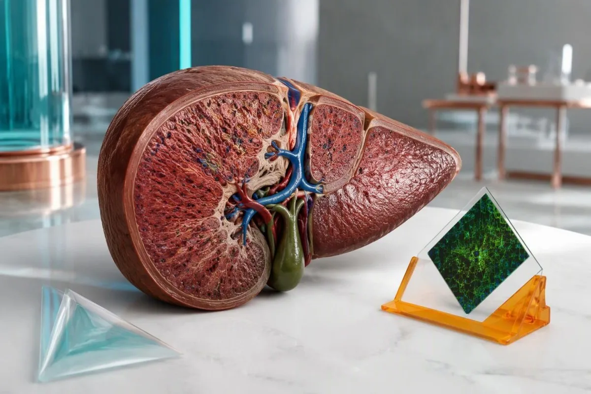
نتیجه مثبت آنتیبادی یک سرنخ است، نه تشخیص. پرسش مفید این است که آیا با آنزیمهای کبدی، سطح IgG، سابقه مصرف دارو و یافتههای بافتی شما همخوانی دارد.


- آنتیبادیهای عضله صاف مثبت بهتنهایی هپاتیت خودایمنی را تشخیص نمیدهند؛ تیتر ASMA برابر با 1:40 در بزرگسالان فقط 1 امتیاز به امتیاز تشخیصی سادهشده اضافه میکند.
- تیترهای بالاتر ASMA پشتوانه تشخیصی را افزایش میدهند، اما تیترهای 1:80 و 1:320 در معیارهای سادهشده حداکثر امتیاز 2 را برای آنتیبادی دریافت میکنند.
- آنتیبادیهای F-actin هدف آنتیبادی اختصاصیتری را شناسایی میکنند، اما نتیجه مثبت همچنان باید در بستر آزمایشهای کبدی و شرایط بالینی تفسیر شود؛ واحدهای سنجش را نمیتوان به تیترهای ASMA تبدیل کرد.
- IgG تام بالاتر از حد بالای آزمایشگاه، مؤید هپاتیت خودایمنی است؛ سطحی بالاتر از 1.1 برابر آن حد، 2 امتیاز در امتیازدهی سادهشده میگیرد.
- ALT طبیعی مثبت شدن اتفاقی را کمتر نگرانکننده میکند، هرچند طبیعی بودن آنزیمها یا IgG در همه موارد هپاتیت خودایمنی را رد نمیکند.
- سابقه مصرف دارو باید شامل داروهای تجویزی و مکملهایی باشد که ماهها یا سالها پیش مصرفشان آغاز شده است، بهویژه نیتروفورانتوئین و مینوسیکلین.
- نمونهبرداری از کبد معمولاً به تشخیص هپاتیت خودایمنی و ارزیابی فیبروز کمک میکند؛ در بیمار مبتلا به بیماری شدید، تصمیمهای فوری درمانی نباید تا انجام آزمایشهای غیراورژانسی به تأخیر بیفتند.
- ارزیابی فوری برای زردی همراه با گیجی، خوابآلودگی قابلتوجه یا افزایش INR لازم است؛ INR برابر یا بالاتر از 1.5 همراه با انسفالوپاتی میتواند نشاندهنده نارسایی حاد کبد باشد.
آیا مثبت بودن آنتیبادیهای عضله صاف به معنای هپاتیت خودایمنی است؟
مثبت بودن آنتیبادیهای عضله صاف بهتنهایی تشخیص هپاتیت خودایمنی نیست. نتیجهای با تیتر پایین، مانند 1:40، ممکن است غیراختصاصی باشد؛ پزشکان به دنبال الگویی همخوان شامل افزایش ALT یا AST، بالا بودن IgG، رد سایر بیماریهای کبدی و معمولاً یافتههای تأییدکننده در بافت کبد هستند.
ASMA, ، که SMA نیز نامیده میشود، آنتیبادیهایی را شناسایی میکند که با اجزای عضله صاف و ساختارهای سلولی مرتبط واکنش نشان میدهند؛ این به معنای تخریب عضلات شما نیست. ASMA بیش از همه با هپاتیت خودایمنی نوع ۱, مرتبط است، اما ممکن است در پاسخهای ایمنی دیگر یا اختلالات کبدی نیز دیده شود؛ به همین دلیل، واژه «مثبت» در گزارش آزمایشگاه نام یک بیماری نیست.
دو نتیجه نمونه را در نظر بگیرید: ASMA با تیتر 1:40، ALT برابر با 24 IU/L و IgG طبیعی، در مقایسه با ASMA با تیتر 1:160، ALT برابر با 286 IU/L و IgG بالاتر از حد آزمایشگاه. الگوی دوم بررسی بسیار گستردهتری را ایجاب میکند، هرچند هیچیک بدون ارزیابی بالینی علت را مشخص نمیکند. توضیح ما درباره معنای نتیجه مثبت آنتیبادی شناسایی آنتیبادی را از شواهد بیماری فعال جدا میکند.
کانتستی یک آنالایزر آزمایش خون هوش مصنوعی که میتواند به بررسی نتایج ASMA در کنار آنزیمهای کبدی و IgG کمک کند، بهجای آنکه نتیجه 1:40 را تشخیص تلقی کنیم. من، توماس کلاین، مدیر ارشد پزشکی، در این مقاله رویکردم توضیح شفاف این استدلال است، از جمله مواردی که اعداد نمیتوانند پاسخ را مشخص کنند. مطالب ما سازمان و تمرکز بالینی ما زمینهای برای این نقش آموزشی فراهم میکنند؛ تشخیص همچنان بر عهده پزشک است.
تفسیر تیتر ASMA: 1:40 و 1:80 به چه معنا هستند؟
تیترهای ASMA نشان میدهند که نمونه را تا چه اندازه میتوان رقیق کرد و همچنان رنگآمیزی آنتیبادی را تشخیص داد. در بزرگسالان، معیارهای سادهشده هپاتیت خودایمنی برای ANA یا SMA برابر یا بالاتر از 1:40، یک امتیاز و برای تیتر برابر یا بالاتر از 1:80، دو امتیاز در نظر میگیرند؛ هیچیک از این آستانهها بهتنهایی تشخیص هپاتیت نیست.
یک تیتر 1:160 یعنی رنگآمیزی پس از رقیقسازی بیشتر نسبت به 1:40 همچنان قابل تشخیص میماند؛ به این معنا نیست که آسیب کبدی ۴ برابر بیشتر است. در معیارهای سادهشده Hennes و همکاران، امتیاز آنتیبادی حداکثر ۲ است؛ بنابراین تیترهای 1:80 و 1:640 امتیاز تشخیصی بیشتری به دست نمیآورند. غلظت آنتیبادی، حساسیت آزمون و آسیب بافتی ارتباطی نهچندان دقیق با یکدیگر دارند—و توجه به این تمایز از نگرانی بیمورد جلوگیری میکند.
روشهای آزمایشگاهی بر قابلیت مقایسه نتایج اثر میگذارند. ایمونوفلورسانس غیرمستقیم روی بسترهای بافتی، آزمونهای اختصاصی آنتیژن، رقتهای آغازین و شیوههای گزارشدهی در آزمایشگاههای مختلف متفاوتاند؛ تغییر از 1:40 به 1:80 ممکن است نشاندهنده یک مرحله رقت باشد، نه تغییری با اهمیت بالینی. همین احتیاط درباره تفسیر آزمایش ANA, نیز صدق میکند، بهویژه وقتی چندین آنتیبادی با مثبت بودن ضعیف همزمان دیده شوند.
تفسیر در کودکان متفاوت است: تیترهای SMA به پایینیِ 1:20 نیز میتوانند در صورت وجود تابلوی بالینی مناسب در کودکان اهمیت داشته باشند. آستانههای امتیازدهی بزرگسالان را نباید بدون نظر متخصص بر گزارش آزمایش کودک اعمال کرد و غربالگری منفی با معیارهای بزرگسالان ممکن است یافتههای کودکان با تیتر پایینتر را از دست بدهد. از آزمایشگاه بپرسید از چه بستری و رقت آغازینی استفاده شده است؛ این جزئیات اغلب از تکرار آزمایش در جای دیگر مفیدترند.
آیا نتیجه مثبت آنتیبادی F-actin به روشن شدن تشخیص کمک میکند؟
An F-actin antibody positive result can strengthen suspicion of autoimmune hepatitis, but it is not a stand-alone confirmation. F-actin testing targets filamentous actin, one antigen recognized by some smooth muscle antibodies; its usefulness depends on the assay, strength of positivity, and accompanying liver abnormalities.
F-actin assays and ASMA titers are different measurements. Some commercial ELISA methods report less than 20 units as negative, 20–30 as weakly positive, and above 30 as positive, but other kits use different categories and units. A result of 25 units must therefore be read against that report's reference interval, not a cutoff copied from another laboratory or an online discussion.
A weak F-actin result with ASMA 1:40 and normal ALT may add little certainty; a clearly positive result alongside persistent enzyme elevation and increased IgG is more persuasive. F-actin antibodies can occur outside autoimmune hepatitis, including in other liver diseases, while some patients with autoimmune hepatitis lack detectable F-actin antibodies. This is a probability adjustment, not a yes-or-no confirmation switch.
Discordant tests deserve a method check before another diagnosis is added. ASMA may recognize targets other than F-actin, and antigen preparation can influence an ELISA result, so positive ASMA with negative F-actin is not automatically a laboratory error. Our guide to نتایج کیفی و کمی explains why 1:80, 25 units, and a simple positive flag cannot be converted into one interchangeable number.
چرا IgG تام از مثبت شدن یک آنتیبادی دیگر اهمیت بیشتری دارد؟
Elevated total IgG supports autoimmune hepatitis because it reflects increased immunoglobulin production accompanying immune-mediated liver injury. The simplified criteria award 1 point above the laboratory upper limit and 2 points above 1.1 times that limit; normal IgG does not completely exclude the disease.
Adult IgG reference intervals commonly approximate 7–16 g/L, equivalent to 700–1,600 mg/dL, although laboratory and age-specific ranges take priority. If the upper limit is 16 g/L, an IgG of 17 g/L exceeds normal, while 18 g/L exceeds 1.1 times the limit of 17.6 g/L. Use the measured total IgG—not total protein—to make that comparison.
Globulin and the gamma gap cannot substitute for quantitative IgG. For example, total protein 84 g/L minus albumin 38 g/L produces a 46 g/L gap, but that gap contains proteins other than IgG and does not prove autoimmune hepatitis. Our gamma gap interpretation guide explains why an apparently useful shortcut can send the investigation down the wrong path.
Normal IgG should lower confidence in a classic presentation, not close the case when ALT is 500 IU/L or the patient is jaundiced. Acute presentations can lack the usual immunoglobulin pattern, and IgG can also rise with cirrhosis or other inflammatory conditions. Read IgG، IgA و IgM با هم when the protein abnormality is broader than isolated IgG elevation.
کدام الگوی آزمایش کبد، مثبت بودن ASMA را نگرانکنندهتر میکند؟
Persistent ALT and AST elevation makes positive ASMA more concerning, especially when IgG is also elevated. Autoimmune hepatitis usually produces a hepatocellular pattern—aminotransferases rising more prominently than alkaline phosphatase—but the enzyme magnitude alone cannot identify the cause or accurately measure liver function.
ALT reflects injury more than function. An ALT of 240 IU/L with an upper limit of 40 is 6 times the upper limit, but that ratio does not distinguish autoimmune hepatitis from viral hepatitis, drug injury, or several other causes. Bilirubin, INR, albumin, symptoms, and the direction of change tell a different part of the story; our توضیح نتایج ALT separates these roles.
این نسبت R compares ALT divided by its upper limit with ALP divided by its upper limit; a ratio at least 5 supports a hepatocellular injury pattern, particularly in drug-injury assessment. ALT 240/40 and ALP 120/120 give an R ratio of 6. This calculation classifies the pattern, not the diagnosis, and should use values obtained at the same time.
Disproportionate ALP elevation changes the differential diagnosis. If ALP is 3 times its upper limit while ALT is only mildly raised, clinicians consider biliary obstruction, primary biliary cholangitis, primary sclerosing cholangitis, or an overlap presentation rather than assuming isolated autoimmune hepatitis. Our guide to cholestatic liver-test patterns shows why GGT and imaging may become more useful than another ASMA measurement.
طبیعی بودن آنزیمهای کبدی همراه با ASMA مثبت تا چه حد اطمینانبخش است؟
Positive ASMA with repeatedly normal liver enzymes is generally less concerning than ASMA with unexplained enzyme elevation. In a retrospective cohort reported by Healey et al. in 2016, only 1 of 202 people with SMA positivity and ALT below 55 IU/L subsequently received an autoimmune hepatitis diagnosis.
The observed proportion was approximately 0.5%, not zero. That study's ALT threshold of 55 IU/L was part of its design and should not become everyone's definition of normal, particularly when your laboratory uses a lower limit. The retrospective population also cannot provide an individualized probability for someone with jaundice, high IgG, prior abnormal tests, or established liver disease.
For an illustrative patient with ASMA 1:40, ALT 26 IU/L, normal bilirubin, and normal IgG, the next step is usually clinical review rather than immediate immunosuppression. A clinician may choose a repeat liver panel in roughly 6–12 weeks when a recent illness, medication exposure, or uncertain baseline needs clarification, but there is no universal ASMA-specific retesting interval. Review mild ALT elevation follow-up if the supposedly normal result is actually borderline.
Previous results can overturn a reassuring snapshot. An ALT of 30 IU/L today means something different after an unexplained value of 300 IU/L last month, especially if corticosteroids were started between samples. Bring at least 2 dated panels when possible; the important question is whether injury never occurred, resolved after a trigger disappeared, or was suppressed by treatment rather than truly excluded.
آیا داروها میتوانند باعث مثبت شدن ASMA و هپاتیت شبهخودایمنی شوند؟
Some medicines can cause liver injury with ASMA positivity, increased IgG, and tissue findings resembling autoimmune hepatitis. Nitrofurantoin and minocycline are classic examples; exposure may precede recognition by months or years, so a medication taken uneventfully for 2 years still belongs in the history.
The start date matters as much as the medicine name. Hydralazine, methyldopa, and some biologic medicines can also produce autoimmune-like liver injury, while herbal products may cause other injury patterns that coexist with incidental antibodies. Write down dose changes, intermittent courses, and supplements used during the preceding 12 months; include longer-standing drugs rather than limiting the list to something newly prescribed last week.
Drug-induced autoimmune-like hepatitis and idiopathic autoimmune hepatitis can look remarkably similar initially. Improvement after supervised withdrawal and the longer-term relapse pattern help distinguish them, but ALT may take weeks to fall and some patients still require corticosteroids. A fall from 400 to 200 IU/L is encouraging, not proof of the cause; persistent or recurrent injury needs reassessment rather than a premature label.
کانتستی یک پلتفرم تفسیر بیومارکرهای AI that can help arrange laboratory trends beside medication dates, but it cannot determine causality from an ASMA titer of 1:80. Our فناوری تفسیر AI describes the supporting workflow rather than a replacement for hepatology assessment. Do not stop an essential prescription on your own; suspected liver injury requires prompt prescriber advice, with emergency assessment when symptoms are severe.
پیش از تشخیص هپاتیت خودایمنی، باید چه موارد دیگری رد شوند؟
Viral hepatitis, drug injury, metabolic liver disease, and selected inherited conditions must be considered before attributing liver injury to positive ASMA. The AASLD practice guidance emphasizes compatible findings and exclusion of competing explanations; antibody positivity does not protect a patient from having a second, more common condition.
Acute hepatitis E can resemble autoimmune hepatitis, including marked aminotransferase elevation, and testing depends on exposure, geography, immune status, and clinical presentation. Hepatitis B and C assessment is also part of a standard liver work-up, with hepatitis A or other viruses considered when appropriate. Our hepatitis E testing explanation clarifies why IgM, IgG, and PCR answer different questions rather than supplying 3 interchangeable confirmations.
MASLD can coexist with positive autoantibodies. An illustrative person with metabolic risk factors, liver fat on imaging, ALT 65 IU/L, ASMA 1:40, and normal IgG may have steatotic liver disease rather than autoimmune hepatitis, although coexisting disease remains possible. Our guide to metabolic liver disease testing explains how fibrosis assessment differs from identifying an immune cause.
In younger patients, unexplained hepatitis may require consideration of Wilson disease; iron overload and alpha-1 antitrypsin deficiency enter the differential according to age and presentation. An ALP elevation of 3 times normal also raises questions about biliary disease or overlap syndromes rather than simply increasing the apparent strength of ASMA. Kantesti AI should support a discussion of missing context, not automatically order every available test because 1 antibody is positive.
چه زمانی برای بررسی ASMA مثبت به نمونهبرداری از کبد نیاز است؟
Liver biopsy is usually needed to establish autoimmune hepatitis when the clinical and laboratory findings suggest it. Tissue assessment can identify hepatitis, evaluate fibrosis, and reveal competing explanations; the AASLD guidance advises against diagnosing autoimmune hepatitis without biopsy, while urgent or unsafe situations require specialist judgment.
Interface hepatitis describes immune-cell activity at the boundary between portal areas and liver-cell plates; it supports autoimmune hepatitis but is not unique to it. Plasma-cell-rich inflammation can be helpful, yet its absence does not exclude the diagnosis, and drug injury can produce similar appearances. A tissue report must be interpreted alongside ASMA 1:80, IgG, viral testing, and the medication timeline—not treated as another isolated positive test.
Fibrosis tests cannot replace diagnostic histology. Liver stiffness may rise during active hepatitis, so a FibroScan value obtained while ALT is 500 IU/L can overstate fibrosis relative to a measurement after the injury settles. Our FibroScan result guide explains the distinction between stiffness and cause; a scan cannot show the cellular pattern needed to evaluate autoimmune hepatitis.
Biopsy timing and route depend on safety. A patient with impaired clotting, fluid accumulation, or severe acute illness may need a transjugular approach or a different immediate plan rather than routine percutaneous sampling. Likewise, a FIB-4 risk calculation can be distorted by acute AST elevation or platelets falling for another reason; none of these tools should delay urgent care when INR is rising toward or above 1.5.
پزشکان چگونه امتیاز هپاتیت خودایمنی را ترکیب میکنند؟
The simplified autoimmune hepatitis score combines autoantibodies, IgG, liver histology, and exclusion of viral hepatitis. Hennes et al. developed an 8-point system in which at least 6 points supports probable and at least 7 supports definite autoimmune hepatitis; the score assists clinical judgment rather than replacing it.
Autoantibody points are capped at 2. ANA 1:160 plus ASMA 1:160 does not produce 4 points, and adding F-actin positivity is not a separate extra-point category. Anti-LKM1 at the qualifying threshold or positive anti-SLA can contribute within the same antibody category, but assay details matter. This prevents several related tests from being mistaken for several independent proofs of one diagnosis.
An illustrative adult with ASMA 1:80, IgG 1.2 times normal, typical histology, and excluded viral hepatitis reaches 8 points: 2 in each category. Another patient could mathematically reach 6 without histology, but that arithmetic does not justify bypassing the AASLD biopsy recommendation or ignoring medication-related injury. The original Hennes et al. criteria are a clinical framework, not a self-diagnosis calculator.
A low simplified score does not reliably dismiss every atypical presentation. Acute severe disease, normal IgG, or absent conventional antibodies can require a specialist assessment using additional evidence or a more detailed scoring system. Our استانداردهای روششناسی بالینی ما explain why interpretation tools need explicit limitations; neither a 5-point estimate nor an 8-point estimate should independently decide whether someone starts immunosuppression.
کدام یافتهها به ارزیابی فوری نیاز دارند، نه پیگیری معمول؟
Urgency depends on symptoms and liver dysfunction, not the ASMA titer. New jaundice with confusion, unusual drowsiness, significant bleeding, or rapidly worsening illness needs emergency assessment; INR at least 1.5 with hepatic encephalopathy can indicate acute liver failure in someone without pre-existing cirrhosis.
A rising INR can be more concerning than a rising antibody titer. Interpretation must account for warfarin, vitamin K deficiency, and other causes, but an unexplained INR of 1.6 during acute hepatitis deserves prompt clinical attention rather than waiting for repeat ASMA. Our تفسیر نتیجه INR distinguishes a clotting-time measurement from a diagnosis and explains why medication context changes its meaning.
ALT at least 5 times the upper limit warrants timely investigation, particularly with symptoms or increasing bilirubin, although there is no universal ASMA-specific ALT threshold that automatically means admission. A patient with ALT 220 IU/L, rising bilirubin, and poor intake may need faster assessment than someone with ALT 400 IU/L who has stable function and an established treatment plan. Numbers need a trajectory and a bedside assessment.
Conjugated bilirubin elevation points toward hepatobiliary dysfunction, but it does not distinguish autoimmune hepatitis from obstruction or another hepatitis. New yellow eyes or dark urine should prompt assessment, even if ASMA is only 1:40; our الگوی بیلی روبین مستقیم explains the competing mechanisms. Do not wait for an AI interpretation, a second antibody result, or an elective appointment if confusion or significant drowsiness develops.
آیا ASMA مثبت باید با استروئید یا آزاتیوپرین درمان شود؟
Positive ASMA alone should not be treated with corticosteroids or azathioprine. Treatment targets confirmed or strongly supported active autoimmune hepatitis, not an antibody concentration; even a titer of 1:320 does not justify immunosuppression when the clinical evidence for liver disease is absent.
Confirmed autoimmune hepatitis is treatable, commonly with corticosteroid-based induction and a steroid-sparing medicine selected for the individual patient. Disease severity, cirrhosis, pregnancy considerations, infection risk, and medication tolerance affect the regimen, so a laboratory report cannot safely generate a dose. The initial aim is improvement and eventual normalization of ALT, AST, and IgG—not necessarily making an ASMA titer of 1:160 disappear.
Azathioprine requires safety assessment and monitoring. TPMT and, where appropriate, NUDT15 testing help identify susceptibility to severe thiopurine toxicity, but reassuring results do not remove the need for blood-count and liver-test monitoring. Our TPMT testing explanation describes these limits; early checks are often arranged within weeks, sometimes every 1–2 weeks during initiation or adjustment, according to the treating service's protocol.
Diet cannot erase positive smooth muscle antibodies or replace immunosuppression. Adequate protein and individualized calcium and vitamin D assessment may support patients receiving prolonged corticosteroids, whereas unregulated liver-detox supplements can complicate the investigation. Kantesti can help organize 2 or more dated panels for a treatment discussion, but treatment response, steroid tapering, and possible withdrawal must be managed by the clinician because relapse can occur despite previous normal results.
پس از دریافت گزارش ASMA مثبت چه باید بکنید؟
After a positive ASMA result, obtain the exact titer or assay value and review it with liver enzymes, total IgG, symptoms, and medication history. A useful first appointment needs the original report and previous results; 2 dated panels often reveal more than repeating the antibody immediately.
Bring the report rather than only a screenshot of the positive flag. Confirm whether the result was ASMA by immunofluorescence, F-actin by ELISA, or both, and include the laboratory reference interval and units. Kantesti is an ابزار تحلیل آزمایش خون مبتنی بر AI that can help organize this information, but an extracted value such as 25 units should always be checked against the original document before it influences care.
I would prioritize 3 questions: Is there persistent hepatocellular injury, is IgG genuinely elevated, and has a plausible competing cause been addressed? That is the reasoning Thomas Klein uses here—not a claim that one threshold can diagnose an individual reader. Our هیئت مشاوران پزشکی describes the clinical expertise behind our educational approach; a hepatologist must still decide whether additional serology, imaging, or tissue assessment is appropriate.
Share only the information needed for the task, and check consent before uploading a relative's results. Our حریم خصوصی و رویههای داده describe the handling framework; laboratory identifiers and unrelated personal details deserve attention even when a result feels urgent. As of ۲۰ اکتبر ۲۰۲۶, the practical message remains unchanged: use ASMA as 1 component of a liver assessment, not as a diagnosis or a reason to start treatment yourself.
مقالات پژوهشی و شواهد پشت این تفسیر
The clinical interpretation rests on autoimmune hepatitis guidance, diagnostic-criteria research, and outcome data—not on educational DOI registration alone. The 2008 Hennes criteria, 2019 AASLD guidance published in 2020, and Healey's 2016 cohort address different questions: diagnostic support, clinical management, and the significance of incidental SMA positivity.
The AASLD guidance supports a combined diagnostic assessment, including liver histology and exclusion of competing causes (Mack et al., 2020). Hennes et al. (2008) provide the 8-point simplified framework, while Healey et al. (2016) offer observational evidence about patients whose ALT was below 55 IU/L. These sources are not interchangeable: a retrospective follow-up study cannot replace a biopsy recommendation or determine an individual treatment plan.
Serum-protein education helps prevent misreading IgG-related clues. ما راهنمای تفسیر پروتئین سرم explains albumin and globulin distinctions, including why albumin 38 g/L and a raised protein gap do not establish immune-mediated hepatitis. The related Zenodo publication is listed below as supporting educational material, not as primary evidence validating ASMA specificity, the 1:80 threshold, or autoimmune hepatitis treatment.
Coagulation education explains a different part of liver assessment: the significance of impaired protein synthesis and interpretation of clotting results. Our coagulation biomarker guide provides background, but an INR of 1.5 during acute hepatitis must be assessed using the patient's clinical situation rather than an aPTT reference range. The 2 Kantesti educational publications below use n.d. where publication dates were not supplied; discovery links are searches, not claims of verified hosting on ResearchGate or Academia.edu.
سوالات متداول
مثبت بودن آزمایش آنتیبادی عضله صاف به چه معناست؟
نتیجه مثبت آزمایش آنتیبادی عضله صاف به این معناست که آنتیبادیهایی که با اهداف سلولی مرتبط با عضله صاف واکنش میدهند شناسایی شدهاند؛ این نتیجه بهتنهایی هپاتیت خودایمنی را تشخیص نمیدهد. در بزرگسالان، ASMA با تیتر 1:40 یک امتیاز به امتیاز تشخیصی سادهشده اضافه میکند، درحالیکه تیتر دستکم 1:80 دو امتیاز دارد. پزشکان این یافته را در کنار ALT، AST، IgG تام، داروها، آزمایشهای ردکننده و معمولاً نمونهبرداری از کبد تفسیر میکنند. طبیعیبودن مکرر آنزیمهای کبدی معمولاً نگرانی درباره مثبتشدن اتفاقی را کاهش میدهد.
آیا تیتر ASMA برابر با 1:40 نگرانکننده است؟
تیتر ASMA برابر با 1:40 در بزرگسالان تیتر پایینی است که میتواند بدون هپاتیت خودایمنی رخ دهد. این نتیجه در معیارهای سادهشده 1 امتیاز دارد، اما تشخیص یا معیاری برای آسیب کبدی نیست. نتیجه 1:40 همراه با ALT برابر با 25 IU/L و IgG طبیعی، با همین تیتر در صورت وجود افزایش پایدار ALT و زردی، متفاوت تفسیر میشود. در کودکان ممکن است تیترهای پایینتری، از جمله 1:20، از نظر بالینی اهمیت داشته باشند؛ بنابراین نتایج کودکان باید متناسب با سن ارزیابی شوند.
آیا مثبت بودن آنتیبادی ضد F-اکتین، هپاتیت خودایمنی را تأیید میکند؟
نتیجه مثبت آنتیبادی F-actin بهتنهایی هپاتیت خودایمنی را تأیید نمیکند. در برخی آزمونها، کمتر از ۲۰ واحد منفی و ۲۰ تا ۳۰ واحد بهعنوان مثبت ضعیف طبقهبندی میشود، اما این حدود مختص کیت هستند و بازه مرجع آزمایشگاه گزارشدهنده در اولویت است. نتایج F-actin را نمیتوان به تیترهای رقت ASMA مانند ۱:۸۰ تبدیل کرد. افزایش آنزیمهای کبدی، افزایش IgG، رد سایر علل و معمولاً بافتشناسی کبد تعیین میکنند که آیا این نتیجه از نظر بالینی معنادار است.
آیا ممکن است آنتیبادیهای ضد عضله صاف با آنزیمهای طبیعی کبد مثبت باشند؟
آنتیبادیهای عضله صاف ممکن است با وجود طبیعی بودن آنزیمهای کبدی مثبت باشند. در یک مطالعهٔ گروهی گذشتهنگر که Healey و همکاران در سال ۲۰۱۶ منتشر کردند، از میان ۲۰۲ فرد دارای SMA مثبت و ALT کمتر از ۵۵ IU/L، یک نفر بعداً به هپاتیت خودایمنی مبتلا تشخیص داده شد؛ یعنی تقریباً ۰.۵۱TP54T. آستانهٔ مورد استفاده در آن مطالعه، معیار جهانی برای ALT طبیعی نیست و آزمایشهای غیرطبیعی قبلی، علائم یا IgG بالا میتوانند ارزیابی را تغییر دهند. پیگیری باید برای هر فرد بهصورت جداگانه تعیین شود، نه با این فرض که آنتیبادی مستلزم درمان یا آزمایشهای مکرر مادامالعمر است.
اگر نتیجه آزمایش ASMA من مثبت باشد، آیا به نمونهبرداری از کبد نیاز دارم؟
مثبت بودنِ آزمایش ASMA بهتنهایی بهطور خودکار مستلزم نمونهبرداری از کبد نیست. نمونهبرداری معمولاً زمانی لازم است که بهدلیل آسیب پایدار کبدی، افزایش IgG یا یافتههای مؤید دیگر، از نظر بالینی به هپاتیت خودایمنی مشکوک باشند؛ راهنمای AASLD توصیه میکند بدون نمونهبرداری، هپاتیت خودایمنی تشخیص داده نشود. تیتر ۱:۱۶۰ همراه با طبیعی بودن آزمایشهای کبدی، بهتنهایی اندیکاسیون نمونهبرداری فوری نیست. بیماری حاد شدید یا INR برابر با ۱٫۵ یا بالاتر مستلزم ارزیابی تخصصی درباره فوریت، ایمنی و مسیر مناسب آزمایش است.
آیا آنتیبادیهای عضله صافِ مثبت میتوانند از بین بروند؟
0 آنتیبادیهای عضله صاف ممکن است بهمرور ضعیفتر شوند یا منفی شوند، بهویژه هنگامی که یک پاسخ ایمنی گذرا یا عامل محرک مرتبط با دارو برطرف شود. تغییر از 1:80 به 1:40 فقط یک مرحله رقت است و ممکن است بهجای بهبود بالینی معنادار، بازتابی از تغییرپذیری آزمایش باشد. در هپاتیت خودایمنی تثبیتشده، ALT، AST و IgG معمولاً شاخصهای مفیدتری برای پاسخ به درمان نسبت به اندازهگیری مکرر تیتر ASMA هستند. مثبتماندن مداوم آنتیبادی بهتنهایی دلیلی برای شروع یا افزایش سرکوب ایمنی نیست.
همین امروز آنالیز آزمایش خون با هوش مصنوعی را دریافت کنید
به بیش از 2 میلیون کاربر در سراسر جهان بپیوندید که Kantesti را برای تحلیل فوری و دقیق آزمایشهای آزمایشگاهی مورد اعتماد قرار میدهند. نتایج آزمایش خون خود را بارگذاری کنید و در عرض چند ثانیه، تفسیر جامع 15,000+ از نشانگرهای زیستی را دریافت کنید.
📚 انتشارات پژوهشی ارجاعشده
کلاین، تی.، میچل، اس.، و وبر، اچ. (۲۰۲۶). محدوده طبیعی aPTT: راهنمای لخته شدن خون D-Dimer، پروتئین C. پژوهش پزشکی مبتنی بر هوش مصنوعی Kantesti.
کلاین، تی.، میچل، اس.، و وبر، اچ. (۲۰۲۶). راهنمای پروتئینهای سرم: آزمایش خون گلوبولینها، آلبومین و نسبت A/G. پژوهش پزشکی مبتنی بر هوش مصنوعی Kantesti.
📖 منابع پزشکی خارجی
Mack CL et al. (2020). Diagnosis and Management of Autoimmune Hepatitis in Adults and Children: 2019 Practice Guidance and Guidelines From the American Association for the Study of Liver Diseases. کبدشناسی.
Hennes EM et al. (2008). Simplified criteria for the diagnosis of autoimmune hepatitis. کبدشناسی.
Healey R et al. (2016). Do anti-smooth muscle antibodies predict development of autoimmune hepatitis in patients with normal liver function? A retrospective cohort review. Autoimmunity Reviews.
⚕️ سلب مسئولیت پزشکی
این مقاله فقط برای اهداف آموزشی است و توصیه پزشکی محسوب نمیشود. برای تصمیمهای مربوط به تشخیص و درمان، همیشه با یک ارائهدهنده مراقبتهای بهداشتی واجد شرایط مشورت کنید.
سیگنالهای اعتماد E-E-A-T
تجربه
بازبینی بالینی مبتنی بر نظر پزشک از فرایندهای تفسیر آزمایشگاه.
تخصص
تمرکز بر پزشکی آزمایشگاهی و اینکه نشانگرهای زیستی در زمینه بالینی چگونه رفتار میکنند.
اقتدارگرایی
نوشتهشده توسط دکتر توماس کلاین، با بازبینی توسط دکتر سارا میچل و پروفسور دکتر هانس وبر.
قابل اعتماد بودن
تفسیر مبتنی بر شواهد با مسیرهای پیگیری روشن برای کاهش هشدارها.