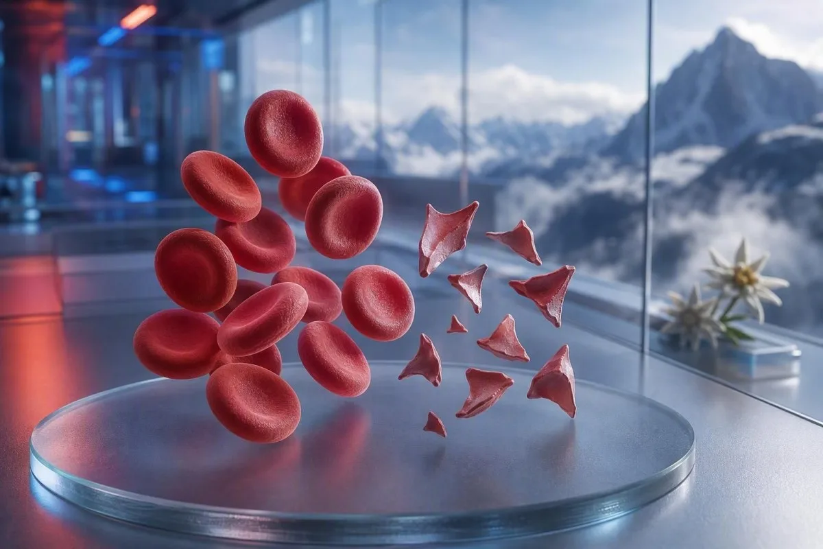
स्किस्टोसाइट्स हे तुकड्यात तुटलेले लाल रक्तपेशी असतात जे धोकादायक लहान-रक्तवाहिन्यांतील गुठळ्या होण्याचे संकेत देऊ शकतात, पण स्मिअरवरील एकच टिप्पणी निदान ठरत नाही. तातडीपणा हा तुकड्यांचे टक्केवारी, प्लेटलेट संख्या, हिमोग्लोबिनचा ट्रेंड, मूत्रपिंड कार्य, हेमोलिसिसचे मार्कर्स आणि लक्षणे यांवर अवलंबून असतो.


- स्किस्टोसाइट थ्रेशहोल्ड: चांगल्या प्रकारे तयार केलेल्या प्रौढ पेरिफेरल स्मिअरवर 1% पेक्षा जास्त स्किस्टोसाइट्स आढळल्यास, इतर लाल रक्तपेशींच्या आकारातील बदल नसतील तर थ्रोम्बोटिक मायक्रोअँजिओपॅथीबाबत संशय येतो.
- तातडीचा क्लस्टर: स्किस्टोसाइट्ससोबत प्लेटलेट्स 150 × 10^9/L पेक्षा कमी, हिमोग्लोबिन कमी होत जाणे, LDH वाढलेले, आणि हॅप्टोग्लोबिन कमी असणे यासाठी त्याच दिवशी चिकित्सकांकडून मूल्यांकन आवश्यक आहे.
- TTP संकेत: 10% पेक्षा कमी असलेली गंभीर ADAMTS13 क्रियाशीलता इम्यून थ्रोम्बोटिक थ्रोम्बोसाइटोपेनिक पर्प्युराला पाठिंबा देते; क्लिनिकल संशय जास्त असल्यास, पुष्टीकरणात्मक चाचण्या परत येण्याआधीच उपचार केले जातात.
- प्रत्येक तुकडा धोकादायक नसतो: यांत्रिक हार्ट व्हॉल्व्ह, गंभीर उच्च रक्तदाब, डायलिसिस सर्किट्स, भाजणे (बर्न्स), आणि नमुन्याचे नुकसान यामुळे तुकड्यात तुटलेल्या लाल रक्तपेशी तयार होऊ शकतात.
- स्मिअर हा मॅन्युअल पुरावा आहे: स्वयंचलित विश्लेषक शिस्टोसाइट्सचे अचूक निदान विश्वसनीयपणे करू शकत नाही; अनुभवी मॉर्फोलॉजिस्टने स्लाइड तपासावी.
- गर्भधारणेमुळे जोखीम वाढते: 20 आठवड्यांनंतर उच्च रक्तदाब, लघवीत प्रथिने, किंवा कमी प्लेटलेट्स असलेल्या शिस्टोसाइट्ससाठी तात्काळ प्रसूतीतज्ज्ञांकडून मूल्यांकन आवश्यक आहे.
- पुन्हा चाचणी केल्याने मदत होते: ताज्या गोळा केलेल्या EDTA नमुन्याचा आणि पुन्हा फिल्मचा वापर करून संग्रहणातील त्रुटी (artefact) आणि खऱ्या चालू असलेल्या लाल रक्तपेशींचे तुकडे होणे (fragmentation) वेगळे करता येते.
- स्वतःहून उपचार करू नका: आयर्न, फोलेट, किंवा सप्लिमेंट्स मायक्रोएंजिओपॅथिक हेमोलिसिसवर उपचार करत नाहीत आणि तातडीच्या मूल्यांकनापासून लक्ष विचलित करू शकतात.
तुकड्यात तुटलेल्या लाल रक्तपेशींना तातडीने वैद्यकीय तपासणी कधी आवश्यक असते
शिस्टोसाइट्स दिसल्यास, कमी प्लेटलेट्सची संख्या, अॅनिमिया, जैवरासायनिक हेमोलिसिस, मूत्रपिंडाचा त्रास, न्यूरोलॉजिकल लक्षणे, ताप, किंवा तीव्र उच्च रक्तदाब यांसोबत ते आढळल्यास त्यासाठी त्याच दिवशी तातडीने पुनरावलोकन आवश्यक आहे. फक्त स्मिअरवरील टिप्पणी (comment) एकतर निरुपद्रवी किंवा कृत्रिम (artifactual) असू शकते; धोकादायक निष्कर्ष हा पॅटर्न आहे. 7 ऑगस्ट 2026 पर्यंत, नवीन शिस्टोसाइट्स आणि प्लेटलेट्स 100 × 10^9/L पेक्षा कमी असल्यास मी याला “आता लगेच डॉक्टरांना कळवा” असा निकाल मानेल, नियमित फॉलो-अप नाही.
प्लेटलेट्स कमी होणे अनेकदा सर्वात जलद तातडीचा इशारा असतो. हिमोग्लोबिन 92 g/L आणि प्लेटलेट्स 68 × 10^9/L आणि नव्याने नोंदवलेले शिस्टोसाइट्स हे, हिमोग्लोबिन 145 g/L आणि स्थिर प्लेटलेट्स 260 × 10^9/L असलेल्या 1% तुकड्यांपेक्षा खूपच अधिक चिंताजनक आहे. Kantesti हा एक AI रक्त चाचणी विश्लेषक आहे जो या क्लस्टरला संपूर्ण रक्त गणना (complete blood count) मध्ये वाचतो, आणि स्वतंत्रपणे मॉर्फोलॉजी टिप्पणी वाढवत नाही.
माझ्या 15 वर्षांच्या क्लिनिकल प्रॅक्टिसमध्ये, मी पटकन जे निर्णय घेतो ते अशा रुग्णांसाठी असतात ज्यांचे निकाल काही दिवसांत बदलले आहेत: हिमोग्लोबिन कमी होणे, क्रिएटिनिन वाढणे, आणि लॅक्टेट डिहायड्रोजनेज वाढणे, म्हणजेच LDH. डॉ. थॉमस क्लाइनचा व्यावहारिक नियम सोपा आहे: या पॅटर्नसोबत गोंधळ, नवीन अशक्तपणा, छातीत दुखणे, श्वास लागणे, फारच कमी लघवी, किंवा तीव्र डोकेदुखी असल्यास आजच आपत्कालीन मूल्यांकन आवश्यक आहे.
शिस्टोसाइट्स हे व्यक्ती अस्वस्थ असल्यास पुन्हा चाचणीची वाट पाहण्याचे कारण नाही. सामान्य पोटॅशियम किंवा सामान्य पांढऱ्या पेशींची संख्या थ्रोम्बोटिक मायक्रोएंजिओपॅथीचा धोका रद्द करत नाही. कमी संख्या समजून घेण्याचा प्रयत्न करणारे रुग्ण आमच्या मार्गदर्शकाकडेही पाहू शकतात कमी लाल रक्तपेशी लक्षणे, पण तीव्र लक्षणांना ऑनलाइन केलेल्या व्याख्येपेक्षा नेहमीच प्राधान्य असते.
साठी आता तातडीच्या आपत्कालीन सेवा कॉल करा
नवीन एकतर्फी अशक्तपणा, बेशुद्ध पडणे, झटका (seizure), तीव्र श्वास लागणे, छातीत दाबून/कुचंबणारा त्रास, काळे शौच (black stools), किंवा गोंधळ झपाट्याने वाढणे—यासाठी क्लिनिकला संदेश देण्याऐवजी आपत्कालीन सेवांची गरज असते. ही लक्षणे अवयवांचा सहभाग दर्शवू शकतात, आणि शिस्टोसाइट्सचे टक्केवारी सुरक्षितपणे हे नाकारते असे नाही.
पेरिफेरल स्मिअरवर स्किस्टोसाइट्सचा प्रत्यक्ष अर्थ काय असतो
शिस्टोसाइट्स हे अनियमित लाल रक्तपेशींचे तुकडे असतात, जेव्हा रक्तप्रवाहातील पेशी शारीरिकरित्या कापल्या/कातरण्यात येतात; बहुतेक वेळा हे फायब्रिनच्या तंतूंनी किंवा लहान रक्तवाहिन्यांमधील असामान्य उच्च-शियर पृष्ठभागांमुळे होते. त्यांना साधारणपणे तीक्ष्ण कोन, हेल्मेटसारखे आकार, किंवा लहान त्रिकोणी आकृत्या असतात आणि अखंड लाल रक्तपेशीतील मध्यवर्ती फिकटपणा (central pallor) नसतो.
खरे शिस्टोसाइट्स हे मॉर्फोलॉजीचा निष्कर्ष आहे; तो मानक CBC विश्लेषकाने तयार केलेले मूल्य नाही. हेमॅटोलॉजीतील मानकीकरणासाठी आंतरराष्ट्रीय परिषद (International Council for Standardization in Haematology) शिफारस करते की, प्रमाण ठरवणे महत्त्वाचे असेल तेव्हा सर्वोत्तम स्मिअर झोनमध्ये किमान 1,000 लाल पेशी मोजाव्यात (Zini et al., 2021). हा तपशील स्पष्ट करतो की एक प्रयोगशाळा “दुर्मिळ तुकडे” नोंदवू शकते, तर दुसरी टक्केवारी नोंदवते.
पारंपरिक चिंता ही मायक्रोएंजिओपॅथिक हेमोलिटिक अॅनिमिया (microangiopathic haemolytic anaemia), जिथे रक्तपेशींची प्लेटलेट-समृद्ध मायक्रोथ्रोम्बी आणि परिसंचरणातील फायब्रिनशी भेट होते. तरीही गंभीर अऑर्टिक स्टेनोसिस, मेकॅनिकल व्हॉल्व्ह, एक्स्ट्राकॉर्पोरियल सर्किट्स, आणि मोठी थर्मल इजा वेगळ्या यंत्रणेद्वारे खरे तुकडे निर्माण करू शकतात. त्यामुळे प्रश्न “तुकडे खरे आहेत का?” असा नसून “तुकडे कोण निर्माण करत आहे?” असा आहे.”
Kantesti हे एक AI रक्त तपासणी अहवाल समजून घ्या प्लॅटफॉर्म आहे जे अपलोड केलेला पूर्ण रक्त गणना (CBC) अहवाल बाजूला ठेवू शकते आणि CBC मध्ये काय समाविष्ट असते बिलीरुबिन, रेटिक्युलोसाइट्स, किंवा हॅप्टोग्लोबिन यांसारखे हरवलेले साथीचे निकाल ठळकपणे दाखवू शकते. ते स्लाइड स्वतः तपासू शकत नाही; हे काम प्रशिक्षित प्रयोगशाळा व्यावसायिकाचेच असते.
प्रौढांच्या स्मिअरमध्ये स्किस्टोसाइटोसिस किती प्रमाणात चिंताजनक असते
प्रौढांच्या परिघीय स्मिअरमध्ये 1% पेक्षा जास्त स्किस्टोसाइट्सची संख्या, तुकडे हे प्रमुख लाल-कोशिकीय असामान्यता असतील तर, थ्रोम्बोटिक मायक्रोअँजिओपॅथीबाबत संशयास्पद ठरते. 1% पेक्षा कमी संख्या रोगाला नाकारत नाही, विशेषतः जर फिल्म द्रवपदार्थ, रक्त संक्रमण, किंवा विकसित होत असलेल्या आजारानंतर तयार केली असेल तर.
1% ही कटऑफ ही तीव्रतेची (severity) मापपट्टी नसून निदानासाठी सूचना (diagnostic prompt) आहे. ICSH मार्गदर्शक तत्त्वे निरोगी प्रौढ आणि टर्म नवजातांसाठी 1% पेक्षा कमी अशी संदर्भ श्रेणी वापरतात; प्रीटर्म शिशूंमध्ये त्याच अर्थाशिवाय 5% पर्यंत तुकडे असू शकतात (Zini et al., 2021). गंभीर थ्रोम्बोसाइटोपेनियासह 2% हा परिणाम, मेकॅनिकल सर्क्युलेटरी सपोर्ट डिव्हाइस बसवल्यानंतरचा 4% पेक्षा अधिक चिंताजनक आहे.
प्रयोगशाळा टक्केवारीऐवजी “occasional,” “few,” किंवा “1–2 per high-power field” असे अहवाल देऊ शकतात. ही वाक्ये एकमेकांच्या बदल्यात वापरता येत नाहीत, कारण फील्डचा आकार, स्मिअरची जाडी, आणि अहवाल देण्याची धोरणे बदलतात. प्रयोगशाळेने ICSH-शैलीतील टक्केवारी मोजणी केली का हे विचारणे, स्वतः वर्णनात्मक वाक्याचे रूपांतर करण्याचा प्रयत्न करण्यापेक्षा अनेकदा अधिक उपयुक्त ठरते.
उच्च RDW स्किस्टोसाइट्स मोजत नाही. RDW पेशींच्या आकारातील वैविध्य दर्शवते आणि रेटिक्युलोसाइटोसिस, लोहाची कमतरता, रक्त संक्रमण, किंवा तुकडीकरण (fragmentation) यांसह वाढू शकते. आमचे सविस्तर RDW आणि लाल-कोशिकीय निर्देशांक (red-cell index) मार्गदर्शक स्पष्ट करते की उच्च RDW स्वतः TTP ला किंवा केवळ आर्टिफॅक्टला (artefact) पाठिंबा देत नाही.
खऱ्या हेमोलिसिसला पाठिंबा देणारा रक्त तपासणीचा नमुना
खरे लाल-कोशिकीय तुकडीकरण सामान्यतः उच्च LDH, कमी हॅप्टोग्लोबिन, वाढलेला अप्रत्यक्ष बिलीरुबिन, आणि अॅनिमियासोबत रेटिक्युलोसाइट प्रतिसाद निर्माण करते. एकही एकच निर्देशक निश्चित नसतो, कारण यकृतविकार, दाह (inflammation), अलीकडील रक्त संक्रमण (transfusion), आणि अस्थिमज्जेचा आजार हे चित्र बिघडवू शकतात.
LDH संवेदनशील (sensitive) आहे, पण विशिष्ट (specific) नाही. 600 IU/L पेक्षा जास्त LDH अनेकदा योग्य परिस्थितीत सक्रिय हेमोलिसिसला पाठिंबा देते; मात्र स्नायू इजा, यकृत इजा, संसर्ग, आणि कर्करोगही ते वाढवू शकतात. प्रयोगशाळेच्या खालच्या मर्यादेपेक्षा कमी हॅप्टोग्लोबिन, साधारणपणे 0.3 g/L पेक्षा कमी, याला अधिक वजन मिळते कारण ते प्लाझ्मामध्ये मुक्त झालेल्या हेमोग्लोबिनला बांधते.
रेटिक्युलोसाइट (reticulocyte) मोजणी सामान्यतः परिघीय नाश (peripheral destruction) दरम्यान वाढायला हवी, पण फोलेट कमतरता, लोह कमतरता, मूत्रपिंडाचा आजार, किंवा तीव्र अस्थिमज्जा दमन (acute marrow suppression) यामध्ये ती कमीच राहू शकते. हा स्पष्ट विरोधाभास सहज दुर्लक्षित होतो: एखाद्या रुग्णाला खरेखुरे हेमोलिसिस असू शकते आणि तरीही रेटिक्युलोसाइट प्रतिसाद अयोग्यरीत्या कमी असू शकतो. ते हॅप्टोग्लोबिन निकाल मार्गदर्शक सामान्य अर्थ लावण्यातील सापळे (interpretive traps) कव्हर करते.
Kantesti AI त्याच वेळबिंदूपासून हेमोग्लोबिन, MCV, प्लेटलेट्स, LDH, बिलिरुबिन, क्रिएटिनिन, आणि रेटिक्युलोसाइट्स यांची तुलना करून तुकड्यात विभागलेल्या लाल रक्तपेशींचे नमुने (patterns) समजावते. 48 ते 72 तासांत 20 g/L पेक्षा जास्त घटलेले हेमोग्लोबिन हे, पूर्वीच्या तुलनेशिवाय असलेल्या सौम्य असामान्य मूल्यापेक्षा चिकित्सकीयदृष्ट्या अधिक माहितीपूर्ण असते.
बिलिरुबिन सामान्य का असू शकते
हेमोलिसिसच्या सुरुवातीला, अंतःशिरा द्रव (intravenous fluid) देऊन dilution झाल्यानंतर, किंवा यकृत बिलिरुबिन कार्यक्षमतेने साफ करत असताना अप्रत्यक्ष (indirect) बिलिरुबिन सामान्य असू शकते. त्यामुळे सामान्य बिलिरुबिनने खात्रीशीर तुकडे (convincing fragments), कमी हॅप्टोग्लोबिन, आणि घटणारे हेमोग्लोबिन यांना ओव्हरराईड करू नये.
TTP ला हेमॅटोलॉजी आपत्कालीन स्थिती म्हणून का उपचार केले जातात
थ्रोम्बोटिक थ्रोम्बोसाइटोपेनिक पर्पुरा, म्हणजे TTP, याचा संशय तेव्हा घ्यावा जेव्हा स्किस्टोसाइट्स (schistocytes) थ्रोम्बोसाइटोपेनियासोबत आणि अधिक स्पष्ट पर्याय नसलेल्या मायक्रोअँजिओपॅथिक हेमोलिटिक अॅनिमियासोबत आढळतात. उपचार न झालेला इम्यून TTP वेगाने प्रगती करू शकतो, त्यामुळे ADAMTS13 चे निकाल येण्याआधीच चिकित्सक प्लाझ्मा एक्स्चेंज आणि इम्यून उपचार सुरू करू शकतात.
ADAMTS13 क्रियाशीलता 10% पेक्षा कमी असल्यास TTP ला ठाम पाठिंबा मिळतो, पण निकाल येण्यासाठी काही तास किंवा दिवस लागू शकतात. ISTH निदान मार्गदर्शक तत्त्वे (diagnostic guideline) प्लाझ्मा उपचार सुरू करण्यापूर्वी नमुना घेत असताना PLASMIC स्कोअरसारख्या क्लिनिकल साधनांद्वारे प्रीटेस्ट संभाव्यता (pretest probability) अंदाजण्याचा सल्ला देतात (Zheng et al., 2020). स्कोअर हा कृतीकडे जाणारा पूल (bridge to action) आहे, तज्ज्ञांच्या निर्णयाचा पर्याय नाही.
ताप (fever), न्यूरोलॉजिक चिन्हे, मूत्रपिंड कार्यक्षमता बिघडणे (renal impairment), थ्रोम्बोसाइटोपेनिया, आणि हेमोलिटिक अॅनिमिया—हा परिचित “पेंटॅड” (pentad) अनेक वास्तविक रुग्णांमध्ये अपूर्ण असतो. George आणि Nester यांनी थ्रोम्बोटिक मायक्रोअँजिओपॅथीला पाच अनिवार्य बॉक्सपेक्षा, एकमेकांवर आच्छादित कारणांसह असलेला सिंड्रोम म्हणून वर्णन केले (George & Nester, 2014). प्रत्यक्षात, पाचही वैशिष्ट्ये येण्याची वाट पाहणे ही धोकादायक जुनी सवय आहे.
हेमोलिसिससह 30 × 10^9/L पेक्षा कमी प्लेटलेट्स असल्यास हेमॅटोलॉजीशी विशेषतः तातडीने चर्चा करणे आवश्यक आहे. संशयित TTP मध्ये प्लेटलेट ट्रान्सफ्युजन सामान्यतः टाळले जाते, जोपर्यंत जीवघेणे रक्तस्राव (life-threatening bleeding) नसतो किंवा आक्रमक प्रक्रिया (invasive procedure) थांबू शकत नाही. संबंधित तपासण्यांसाठी, आमचे कोअग्युलेशन चाचणी मार्गदर्शकाचा TTP मध्ये PT आणि aPTT सामान्य का असू शकतात हे स्पष्ट करते.
स्किस्टोसाइट्सची इतर धोकादायक कारणे डॉक्टर विचारात घेतात
स्किस्टोसाइट्स (Schistocytes) डिसेमिनेटेड इंट्राव्हॅस्क्युलर कोअग्युलेशन (disseminated intravascular coagulation), शिगा-टॉक्सिन हेमोलिटिक युरेमिक सिंड्रोम (Shiga-toxin haemolytic uraemic syndrome), तीव्र उच्च रक्तदाब (severe hypertension), गर्भधारणेशी संबंधित मायक्रोअँजिओपॅथी, काही औषधे, आणि ट्रान्सप्लांटशी संबंधित थ्रोम्बोटिक मायक्रोअँजिओपॅथीमध्येही आढळतात. संबंधित प्लेटलेट, कोअग्युलेशन, मूत्रपिंड, आणि क्लिनिकल निष्कर्ष (clinical findings) साधारणपणे या स्थिती वेगळ्या करतात.
DIC मध्ये अनेकदा तुकड्यांसोबतच PT वाढलेला (prolonged), aPTT वाढलेला (prolonged), फायब्रिनोजेन कमी किंवा घटणारा (low or falling), आणि D-dimer लक्षणीयरीत्या वाढलेला (markedly elevated) असे एकत्र आढळते. TTP मध्ये तुलनेने नियमित कोअग्युलेशन वेळा (routine clotting times) अधिक वेळा सामान्य असतात. आजारी रुग्णात 1.5 g/L पेक्षा कमी फायब्रिनोजेन असल्यास चिंता consumption coagulopathy कडे वळते; मात्र तीव्र दाह (acute inflammation) दरम्यान फायब्रिनोजेन वाढत असल्यामुळे सुरुवातीला फायब्रिनोजेन सामान्य दिसू शकते.
तीव्र उच्च रक्तदाबामुळे दाबामुळे मायक्रोव्हॅस्क्युलेचरला इजा झाल्यास लाल रक्तपेशी कापल्या (shear) जाऊ शकतात; डोकेदुखी, दृष्टीत बदल, छातीत दुखणे, किंवा मूत्रपिंड कार्य बिघडणे यासह 180/120 mmHg किंवा त्यापेक्षा जास्त वाचन हे आपत्कालीन (emergency) असते. म्हणूनच रक्तदाबाचे वाचन हे स्मिअर अहवालाच्या बाजूला असायला हवे. रुग्ण पुनरावलोकन करू शकतात दुय्यम उच्च रक्तदाब प्रयोगशाळेतील संकेत तात्काळ धोका हाताळल्यानंतर.
अतिसारानंतर मूत्रनिर्मिती कमी होणे, रक्ताल्पता (anaemia), आणि थ्रोम्बोसाइटोपेनिया यामुळे हेमोलिटिक युरेमिक सिंड्रोमबद्दल चिंता निर्माण होते. स्टूल तपासणी शिगा टॉक्सिन ओळखण्यास मदत करू शकते, पण तातडीची काळजी प्रथम ठरवण्यासाठी हायड्रेशन स्थिती, क्रिएटिनिन, पोटॅशियम, आणि न्यूरोलॉजिकल मूल्यांकन मार्गदर्शक ठरतात. उच्च लॅक्टेट किंवा अस्थिर जीवनचिन्हे असल्यास “वाट पाहा” अशा पद्धतीऐवजी सेप्सिस आणि DIC साठी मूल्यांकन आवश्यक आहे; ही पहा सेप्सिस मार्कर पॅटर्न्स.
तुकड्यात तुटलेल्या लाल रक्तपेशींची यांत्रिक कारणे
यांत्रिक (mechanical) नाशामुळे TTP नसतानाही खरे स्किस्टोसाइट्स तयार होऊ शकतात, विशेषतः कृत्रिम हृदय झडपा (prosthetic heart valves), तीव्र महाधमनी संकुचन (severe aortic stenosis), व्हेंट्रिक्युलर असिस्ट डिव्हाइसेस, डायलिसिस सर्किट्स, किंवा एक्स्ट्राकॉर्पोरियल मेंब्रेन ऑक्सिजनेशन (ECMO) मध्ये. या कारणांसाठी तरीही वैद्यकीय पुनरावलोकन आवश्यक असते, पण उपचार प्रतिकारशक्ती-आधारित (immune) TTP ऐवजी यांत्रिक स्रोतावर लक्ष्य करतात.
यांत्रिक-झडप (mechanical-valve) नमुन्यात अनेकदा दीर्घकालीन सौम्य LDH वाढ आणि स्थिर प्लेटलेट्ससह कमी हॅप्टोग्लोबिन दिसते. नवीन रक्ताल्पता, श्वास लागणे वाढणे, गडद मूत्र, किंवा झडपेच्या आवाजात बदल हे पॅराव्हाल्व्ह्युलर लीक किंवा उपकरण बिघाड सूचित करू शकतात आणि कार्डिओलॉजी पुनरावलोकनास प्रवृत्त करायला हवे. स्मिअर संकेत देते; इकोकार्डिओग्राफी स्रोत शोधते.
खेळाडू कधी कधी विचारतात की मॅरेथॉन तुकड्यांचे स्पष्टीकरण देऊ शकते का. पायावर आदळण्यामुळे होणारा हेमोलिसिस (foot-strike haemolysis) दीर्घ पल्ल्याच्या धावणीनंतर तात्पुरते मुक्त हिमोग्लोबिन आणि कमी हॅप्टोग्लोबिन निर्माण करू शकतो, पण खात्रीशीर स्किस्टोसाइटोसिसला मी व्यायामाशी सहजपणे जोडणार नाही—विशेषतः प्लेटलेट्स कमी होत असतील तर. याची तुलना मॅरेथॉन धावपटू प्रयोगशाळा मार्गदर्शक.
तीव्र भाजणे (severe burns) आणि मोठी रक्तवाहिन्यांची शस्त्रक्रिया शारीरिक आणि एंडोथेलियल इजा करून तुकड्यांसारख्या पेशी तयार करू शकतात. अशा परिस्थितींमध्ये 6 ते 24 तासांनी प्रयोगशाळेतील ट्रेंड पाहणे एका स्मिअरपेक्षा अधिक माहितीपूर्ण ठरू शकते. घटना घडल्यानंतर CBC स्थिर राहणे आश्वासक असते; हिमोग्लोबिन सतत कमी होत राहणे तसे नाही.
नमुना घेणे आणि स्लाइडचे आर्टिफॅक्ट्स स्किस्टोसाइट्ससारखे कसे दिसतात
नमुना नीट न गोळा करणे, स्मिअर तयार करण्यात उशीर होणे, अँटिकोअॅग्युलंट ट्यूब्समध्ये कमी भर (underfilled), आणि स्मिअरच्या काठाला झालेली इजा अशा गोष्टींमुळे रक्ताभिसरण-स्तरावरील हेमोलिसिस न दर्शवता स्किस्टोसाइट्ससारख्या दिसणाऱ्या आकृत्या तयार होऊ शकतात. स्वच्छपणे गोळा केलेला आणि मोनोलायरमध्ये (monolayer) तपासलेला पुनः नमुना हा क्लिनिकल चित्र शांत असताना नेहमीचा पहिला तपास असतो.
क्रेनेटेड पेशी (crenated cells), स्मिअर-काठ विकृती (smear-edge distortion), वाळण्यामुळे होणारी त्रुटी (drying artefact), आणि एकमेकांवर आच्छादित पेशी हे सामान्य खोटे दिसणारे (false look-alikes) प्रकार आहेत. त्या अनेकदा संपूर्ण पसरलेल्या स्वरूपात किंवा आदर्श वाचन क्षेत्राच्या फेदर्ड काठावर (feathered edge)च दिसतात, समान रीतीने नाही. अनुभवी मॉर्फोलॉजिस्ट स्लाइड तयार करताना फक्त विकृत झालेल्या पेशीपासून खऱ्या तुकड्याची तीक्ष्ण भूमिती (sharp geometry) वेगळी ओळखतो.
कठीण नमुना संकलनामुळे ट्यूब हेमोलायझ होऊ शकते आणि पोटॅशियम किंवा LDH खोटे वाढू शकते, पण ते आपोआप खरे रक्ताभिसरणातील स्किस्टोसाइट्स तयार करत नाही. जर निकालात हेमोलिसिस इंडेक्स किंवा “sample compromised” अशी टिप्पणी असेल, तर रोगनिदान करण्यापूर्वी चिकित्सक सामान्यतः पुन्हा नमुना घेतात. आमचा लेख पोटॅशियम ड्रॉ (नमुना घेणे) चुका स्पष्ट करतो की तीच प्री-अॅनालिटिकल समस्या एकाच वेळी अनेक निकालांना कशी दिशाभूल करू शकते.
EDTA-आधारित प्लेटलेट्सची गुठळी होणे (platelet clumping) प्लेटलेट काउंट खोटे कमी करू शकते आणि धोकादायक दिसणारे संयोजन तयार करू शकते. डायल्यूशनसाठी दुरुस्त केलेला सिट्रेट ट्यूब प्लेटलेट काउंट आणि ताजी फिल्म हे हे उघड करू शकतात. सूक्ष्म फरक महत्त्वाचा आहे: आर्टिफॅक्ट (artefact) खऱ्या आजारासोबत सहअस्तित्वात असू शकतो, त्यामुळे लक्षणे असलेल्या व्यक्तीसाठी पुनः तपासणीने काळजी कधीही उशीर करू नये.
अॅनालायझरचे फ्लॅग्स आणि प्रयोगशाळेतील शब्दरचना दिशाभूल कशी करू शकते
स्वयंचलित रक्त विश्लेषक (automated blood analysers) असामान्य लाल रक्तपेशींचे गट ओळखू शकतात, पण थ्रोम्बोटिक मायक्रोअँजिओपॅथी (thrombotic microangiopathy) वगळण्यासाठी पुरेशी विश्वासार्हता असलेले स्किस्टोसाइट्सचे स्वतंत्र निदान ते करू शकत नाहीत. क्लिनिकल प्रश्न हेमोलिसिस किंवा TTP असल्यास मॅन्युअल पेरिफेरल स्मिअर पुनरावलोकन आवश्यकच राहते.
“खंडित लाल पेशी (fragmented red cells present)” अशी टिप्पणी आल्यास प्रमाण, स्मिअरची गुणवत्ता, आणि सहअस्तित्वात असलेली इतर आकृतीबंध (coexisting morphology) याबाबत प्रश्न विचारायला हवेत. लोहअभावामुळे, मेगालोब्लास्टिक अॅनिमिया (megaloblastic anaemia) मुळे, किंवा वंशपरंपरागत लाल पेशींच्या विकारांमुळे दिसणारी तीव्र anisopoikilocytosis 1% नियम (rule) कमी विशिष्ट (less specific) बनवते. असामान्य आकृतीबंधात (abnormal morphology) जेव्हा fragments बहुसंख्य असतात तेव्हा ते सर्वाधिक निदानात्मकदृष्ट्या उपयुक्त ठरतात; अनेक असंबंधित आकारांमध्ये ते दिसत असतील तर त्यांचे निदानमूल्य कमी असते.
Kantesti ही एक AI प्रयोगशाळा (lab) चाचणी अर्थ लावण्याची (interpretation) सेवा आहे जी platelet clumps असल्याची टिप्पणी असताना platelet count कमी असल्यासारख्या विसंगत (discordant) संयोजनांना चिन्हांकित करण्यासाठी (flag) डिझाइन केली आहे. हे प्रयोगशाळेच्या सूक्ष्मदर्शक (microscopic) पुनरावलोकनाची जागा घेत नाही किंवा स्लाइड पुन्हा मागवू शकणाऱ्या (request a repeat slide) चिकित्सकाची जागा घेत नाही. गुणवत्ता तपासण्याबाबतचा आमचा दृष्टिकोन खालीलमध्ये वर्णन केला आहे— वैद्यकीय पडताळणीचा आढावा.
डेल्टा चेक (delta check) सध्याचा निकाल आधीच्या निकालाशी तुलना करतो आणि अचानक अविश्वसनीय (implausible) बदल उघड करू शकतो. 24 तासांच्या आत 138 g/L वरून 87 g/L पर्यंत हिमोग्लोबिन (haemoglobin) कमी होणे वैद्यकीयदृष्ट्या महत्त्वाचे आहे, पण अंतःशिर द्रवांमुळे (intravenous fluids) झालेली dilution किंवा चुकीचे लेबल असलेला नमुना (mislabeled specimen) शक्य राहते. याबद्दल अधिक वाचा— अचानक प्रयोगशाळेतील बदल (sudden laboratory changes).
रक्त तपासणी अहवालात स्किस्टोसाइट्सचा उल्लेख आल्यावर काय करावे
schistocytes नव्याने नोंदवले गेले असतील तर त्याच दिवशी चाचणी आदेश देणाऱ्या चिकित्सकांशी किंवा सेवेशी संपर्क साधा, आणि तुम्हाला न्यूरोलॉजिकल लक्षणे, श्वास लागणे, छातीत दुखणे, गडद लघवी (dark urine), तीव्र डोकेदुखी, ताप, किंवा लघवीचे प्रमाण कमी झालेले असेल तर तातडीची आपत्कालीन काळजी (emergency care) लगेच घ्या. स्मिअरवरील टिप्पणी स्वतःहून निदान (diagnosis) म्हणून समजू नका.
प्रत्यक्ष अहवाल (actual report) उपलब्ध ठेवा—यात haemoglobin, platelets, creatinine, bilirubin, LDH, आणि कोणताही haemolysis index समाविष्ट करा. तसेच अलीकडील गर्भधारणा (recent pregnancy), अतिसारजन्य आजार (diarrhoeal illness), रक्तदाबाचे अत्यंत जास्त रीडिंग्स (severe blood-pressure readings), नवीन औषधे (new medicines), व्हॉल्व्ह शस्त्रक्रिया (valve surgery), डायलिसिस (dialysis), रक्त संक्रमण (transfusion), आणि ऑटोइम्यून रोग (autoimmune disease) यांची नोंद घ्या. आकृतीबंध (morphology) क्षेत्रातील एका शब्दापेक्षा ही माहिती differential अधिक बदलते.
मी पाहतो की कोणीही कारण ओळखण्याआधी “anaemia” वाचून लोक iron सुरू करून मौल्यवान वेळ गमावतात. लोहअभाव (iron deficiency) सहअस्तित्वात असू शकतो, पण fragments सोबत दिसणारी जलद platelet घट (rapid platelet decline) याचे स्पष्टीकरण तो देत नाही. डॉ. Thomas Klein पूर्ण अहवालाचा फोटो घेण्याची शिफारस करतात; फक्त हायलाइट केलेले आकडे लिहून (transcribing) घेऊ नका—लिहिताना अनेकदा प्रयोगशाळेची ती टिप्पणी (laboratory comment) सुटते जी pattern सुसंगत (coherent) बनवते.
platelets खूप कमी असतील किंवा रक्तस्राव (bleeding) होत असेल तर चिकित्सक अन्यथा सांगत नाहीत तोपर्यंत aspirin, ibuprofen, आणि डॉक्टरांनी न दिलेले anticoagulants टाळा. तातडीच्या वैद्यकीय मार्गदर्शनाशिवाय (urgent medical guidance) निर्धारित anticoagulant थांबवू नका. थकवा (fatigue) किंवा चक्कर येणे/हलके वाटणे (light-headedness) असलेल्या लोक आमचा— चक्कर येण्यासाठीच्या रक्त तपासण्या (blood tests for dizziness) मार्गदर्शन करतात तातडीची कारणे वगळल्यानंतर प्रश्न तयार करण्यासाठी.
गर्भधारणेत, नवजातांमध्ये आणि मुलांमध्ये स्किस्टोसाइट्स
गर्भधारणेदरम्यान schistocytes आढळल्यास, ते hypertension, proteinuria, यकृत एन्झाइम्स वाढलेले (elevated liver enzymes), platelets कमी, डोकेदुखी, दृश्य लक्षणे (visual symptoms), किंवा वरच्या पोटात दुखणे (upper abdominal pain) यांसोबत आढळत असतील तर तातडीचे मूल्यांकन आवश्यक आहे. HELLP syndrome, pre-eclampsia, TTP, atypical HUS, आणि DIC एकमेकांवर आच्छादित (overlap) होऊ शकतात, आणि प्रसूतीच्या वेळेभोवतीचा कालावधी (timing around delivery) मदत करतो, पण निदान निश्चित (settle) करत नाही.
HELLP syndrome हे पारंपरिकरित्या haemolysis, यकृत एन्झाइम्स वाढलेले, आणि platelets कमी—या तीन गोष्टींनी परिभाषित केले जाते; हे अनेकदा गर्भधारणेच्या 20 आठवड्यांनंतर किंवा प्रसूतीनंतर लवकरच दिसते. 100 × 10^9/L पेक्षा कमी platelet count किंवा AST 70 IU/L पेक्षा जास्त असल्यास चिंता करण्यास आधार मिळू शकतो, पण स्थानिक प्रसूतीतज्ज्ञ (obstetric) संघ संपूर्ण क्लिनिकल चित्राचा अर्थ लावतात. या संयोजनासह routine antenatal appointment ची वाट पाहू नका.
मुदतपूर्व (term) नवजात बाळांमध्ये 1% schistocytes कमी असू शकतात, तर मुदतपूर्व (preterm) बाळांमध्ये 5% पर्यंत असू शकतात, कारण नवजात लाल पेशींचा आकृतीबंध (neonatal red-cell morphology) वेगळा असतो (Zini et al., 2021). मात्र अतिसार असलेल्या मुलामध्ये, फिकटपणा (pallor), लघवी कमी होणे (decreased urine), आणि thrombocytopenia असल्यास, fragments HUS कडे निर्देश करू शकतात आणि तातडीचे बालरोगतज्ज्ञांचे (paediatric) मूल्यांकन आवश्यक असते.
platelets आणि haemoglobin यांचे संदर्भ अंतर (reference intervals) वय (age) आणि गर्भधारणेचा टप्पा (pregnancy stage) यानुसार लक्षणीय बदलते. generic app ने “normal” म्हणून चिन्हांकित केलेला निकालही संदर्भानुसार वैद्यकीयदृष्ट्या चिंताजनक असू शकतो. आमची— गर्भधारणा प्लेटलेटची पातळी आणि एकाच दिवसाच्या गर्भधारणा प्रयोगशाळेतील ध्वज उपयुक्त पार्श्वभूमी द्या; हे मॅटरनिटी ट्रायाजचा पर्याय नाही.
खऱ्या स्किस्टोसाइट्सची चौकशी करण्यासाठी चिकित्सक कोणत्या चाचण्या ऑर्डर करतात
संशयित मायक्रोएंजिओपॅथिक हेमोलिसिससाठी पहिल्या तपासणी संचामध्ये साधारणपणे पुनरावृत्त CBC आणि स्मिअर, रेटिक्युलोसाइट काउंट, LDH, हॅप्टोग्लोबिन, बिलिरुबिनचे अंश, क्रिएटिनिन, मूत्रपरीक्षण, PT, aPTT, फायब्रिनोजेन आणि D-dimer यांचा समावेश असतो. TTPचा संशय असल्यास प्लाझ्मा एक्स्चेंज करण्यापूर्वी ADAMTS13 क्रियाशीलता आणि इनहिबिटर चाचण्या घेणे आवश्यक आहे, पण उपचाराची प्रतीक्षा करू नये.
डायरेक्ट अँटिग्लोब्युलिन टेस्टिंग, ज्याला DAT किंवा Coombs टेस्टिंग असेही म्हणतात, हे इम्यून हेमोलिसिसला नॉन-इम्यून विखंडनापासून वेगळे करण्यात मदत करते. सकारात्मक DAT दुसरी प्रक्रिया नाकारत नाही, पण क्लासिक TTP साधारणपणे DAT-निगेटिव्ह असते. मूत्र मायक्रोस्कोपीमध्ये मूत्रपिंडातील मायक्रोव्हॅस्क्युलर इजा सक्रिय असताना प्रोटीन किंवा सेल्युलर कास्ट दिसू शकतात; आमचे स्पष्टीकरण पहा ग्रॅन्युलर युरिनरी कास्ट्स.
स्टूल शिगा-टॉक्सिन टेस्टिंग सुसंगत जठरांत्रीय आजारानंतर विचारात घेतली जाते, तर अॅटिपिकल HUS साठी पूरक चाचण्या, औषध पुनरावलोकन, आणि आनुवंशिक किंवा कार्यात्मक अभ्यास वापरले जाऊ शकतात. क्रम देश आणि रुग्णालयानुसार बदलतो. 48 तासांत 26.5 µmol/L इतकी क्रिएटिनिन वाढ ही एक तीव्र मूत्रपिंड इजा (acute kidney injury) निकष पूर्ण करते आणि ती नाट्यमय संख्या होण्याआधीही लक्ष देण्यास पात्र आहे.
लक्षणीय अॅनिमियामध्ये 2 पेक्षा कमी रेटिक्युलोसाइट उत्पादन निर्देशांक (reticulocyte production index) अपुरी अस्थिमज्जा प्रतिसाद दर्शवतो, जरी कच्च्या रेटिक्युलोसाइट टक्केवारी जास्त दिसत असली तरी. ही गणना अॅनिमियामधील कमी लाल रक्तपेशींच्या वस्तुमानासाठी दुरुस्ती करते. The रेटिक्युलोसाइट आणि हेमॅटोलॉजी मार्कर मार्गदर्शक टक्केवारी का दिशाभूल करणारी ठरू शकते हे स्पष्ट करण्यात मदत करते.
स्मिअर पुन्हा करणे कधी योग्य आणि कधी नाही
तुकडे (fragments) कमी असतील, रुग्णाची प्रकृती चांगली असेल, प्लेटलेट्स आणि हिमोग्लोबिन स्थिर असतील, आणि नमुन्याच्या गुणवत्तेबद्दल शंका असेल तर काही तासांत ते 24 तासांत स्मिअर पुन्हा करणे योग्य ठरते. थ्रोम्बोसाइटोपेनिया, सक्रिय हेमोलिसिस, अवयव इजा, किंवा चिंताजनक लक्षणे असतील तर पुनरावृत्ती करणे सुरक्षित विलंब ठरत नाही.
चांगला पुनरावृत्ती नमुना ताज्या पद्धतीने योग्य भरलेल्या EDTA ट्यूबमध्ये गोळा करून त्वरित फिल्ममध्ये तयार करावा. विनंतीमध्ये विशेषतः संशयित शिस्टोसाइट्स (schistocytes) आणि प्लेटलेट क्लंपिंग यांचे पुनरावलोकन मागावे, जर दोन्हीपैकी काहीही शक्य वाटत असेल. प्रयोगशाळा दोन्ही फिल्म्स बाजूबाजूने तुलना करू शकते; हे फक्त स्वयंचलित CBC पुन्हा चालवण्यापेक्षा खूप अधिक उपयुक्त आहे.
ट्रेंडची दिशा महत्त्वाची असते. तीन दिवसांत प्लेटलेट काउंट 210 वरून 165 वरून 98 × 10^9/L पर्यंत जात असेल तर ते चिंताजनक आहे, जरी पहिल्या दोन वाचनांमध्ये स्थानिक संदर्भ श्रेणीमध्ये येण्याची शक्यता असते. Kantesti AI या प्रवासांना उघड करण्यासाठी दीर्घकालीन संदर्भ वापरते; हा सिद्धांत आमच्या रक्त तपासणी ट्रेंड विश्लेषण मार्गदर्शकाचा.
ट्रान्सफ्युजन मूळ मॉर्फॉलॉजी तात्पुरती dilute किंवा अस्पष्ट करू शकते. जर तुम्हाला दुसऱ्या नमुन्यापूर्वी लाल रक्तपेशी मिळाल्या असतील, तर टीमला तारीख आणि युनिट्सची संख्या सांगा. आमचे मार्गदर्शक रुग्णालयातून डिस्चार्जनंतर मूल्यांमध्ये बदल उपचारानंतरचे ट्रेंड्स वेगळ्या बेसलाइनची गरज का असते हे स्पष्ट करते.
स्किस्टोसाइट रोग नसतानाही सारखे दिसू शकणाऱ्या स्थिती
आयर्न डिफिशियन्सी, व्हिटॅमिन B12 डिफिशियन्सी, यकृत रोग, वारसागत लाल रक्तपेशींचे विकार, आणि तीव्र संसर्ग हे थ्रोम्बोटिक मायक्रोएंजिओपॅथीच्या विशिष्ट विखंडन नमुन्याशिवायही असामान्य लाल रक्तपेशींचे आकार तयार करू शकतात. स्मिअरची संपूर्ण मॉर्फॉलॉजी आणि क्लिनिकल टाइमलाइन तुकड्यांचा (fragments) अति-अंदाज टाळतात.
गंभीर व्हिटॅमिन B12 डिफिशियन्सी अॅनिमियासह pseudo-thrombotic microangiopathy निर्माण करू शकते, ज्यामध्ये उच्च LDH, कमी प्लेटलेट्स, आणि कधी कधी तुकडे दिसतात. यात अनेकदा ठळक मॅक्रोसाइटोसिस (macrocytosis) दिसते, रेटिक्युलोसाइट प्रतिसाद कमी असतो, आणि LDH मूल्ये कधी कधी 2,500 IU/L पेक्षा जास्त असू शकतात—ही वैशिष्ट्ये स्वयंचलित TTP उपचाराऐवजी B12 आणि methylmalonic acid चाचण्यांना प्रवृत्त करायला हवीत. आमचे B12 विरुद्ध फोलेट मार्गदर्शकात (guide) समाविष्ट आहे हे या महत्त्वाच्या अनुकरणाचे (mimic) स्पष्टीकरण देते.
लोहाची कमतरता सहसा मायक्रोसाइटोसिस, हायपोक्रोमिया, पेंसिल सेल्स, आणि उच्च RDW निर्माण करते; गंभीर प्रकरणांमध्ये फ्रॅगमेंट्स नोंदवले जाऊ शकतात, पण ते क्वचितच प्रमुख वैशिष्ट्य असते. 15 µg/L पेक्षा कमी फेरिटिन हे, अन्यथा गुंतागुंत नसलेल्या प्रौढामध्ये, लोह साठे कमी झाल्याचे ठामपणे समर्थन करते; मात्र दाह (inflammation) फेरिटिन वाढवून कमतरता लपवू शकते.
माझ्या अनुभवात, सर्वात आश्वासक निष्कर्ष हा फक्त “शिस्टोसाइट्स नाहीत” एवढाच नसून, स्थिर प्लेटलेट्स, स्थिर मूत्रपिंड कार्य (kidney function), आणि जैवरासायनिक हेमोलिसिस नसणे—यांसह एक सुसंगत पर्यायी स्पष्टीकरण असते. निष्कर्ष विसंगत राहिल्यास, अंतिम अहवालातील शब्दरचना (wording) यावर अवलंबून न राहता हेमॅटोलॉजिस्ट प्रत्यक्ष स्लाइड पाहू शकतो.
स्मिअर आपत्कालीन स्थिती चुकू न देता AI व्याख्या वापरणे
AI रक्त तपासणी अहवालांचे आयोजन करण्यात आणि धोकादायक संयोजन ओळखण्यात मदत करू शकते, पण CBC PDF मधून शिस्टोसाइटचे निदान सत्यापित करू शकत नाही किंवा तातडीच्या वैद्यकीय मूल्यमापनाची जागा घेऊ शकत नाही. पेरिफेरल स्मिअर म्हणजे दृश्य सूक्ष्मदर्शक (visual microscopy) तपासणी, आणि मूळ प्रयोगशाळेतील स्लाइड हेच सत्याचे (source of truth) स्रोत राहते.
Kantesti हे AI-चालित रक्त तपासणी विश्लेषण साधन आहे जे प्लेटलेट ट्रॅजेक्टरीज, हिमोग्लोबिनमधील बदल, क्रिएटिनिन, बिलिरुबिन, आणि LDH यांसह अहवालांमधील संबंधित निष्कर्षांचे नकाशण (map) करते. त्यामुळे प्रत्येक असामान्य मार्कर स्वतंत्रपणे वाचण्याची सामान्य चूक कमी होऊ शकते. मात्र “शिस्टोसाइट” नावाच्या आकाराचा तो खरा फ्रॅगमेंट आहे की स्मिअर-एज आर्टिफॅक्ट (smear-edge artefact) आहे, किंवा अहवाल देण्याची पद्धत (reporting convention) आहे—हे ते पाहू शकत नाही.
अपलोडची गुणवत्ता महत्त्वाची असते कारण एखादी युनिट किंवा दशांश (decimal) वगळला गेला तर निष्कर्षाचा अर्थ उलटू शकतो. अहवाल शेअर करण्यापूर्वी रुग्ण ओळखकर्ता (patient identifiers), संकलन तारीख (collection date), युनिट्स, संदर्भ श्रेणी (reference ranges), आणि निकालात “estimated,” “pending,” किंवा “sample haemolysed” असे लिहिले आहे का—हे तपासा. आमचे AI रिपोर्ट अचूकता चेकलिस्ट त्या अंतिम मानवी पडताळणीसाठी उपयुक्त आहे.
Kantestiच्या क्लिनिकल पद्धती आमच्या वैद्यकीय सल्लागार मंडळ, यांच्या इनपुटसह पुनरावलोकन केल्या जातात, आणि आमची तंत्रज्ञान AI विश्लेषण तंत्रज्ञान मार्गदर्शक. मध्ये वर्णन केले आहे. डॉ. थॉमस क्लाइन म्हणून, नवीन फ्रॅगमेंट्स असलेल्या रुग्णासाठी, प्लेटलेट्स 100 × 10^9/L पेक्षा कमी असतील, आणि लक्षणे असतील—अशा वेळी मी तरीही क्लिनिशियनने प्रयोगशाळेला थेट फोन करावा अशीच इच्छा ठेवेन; कारण नेमकी तेव्हाच गती (speed) आणि क्लिनिकल तपासणी (clinical examination) सर्वाधिक महत्त्वाची असते.
सतत विचारले जाणारे प्रश्न
स्किस्टोसाइट्सचे कोणते प्रमाण धोकादायक असते?
काळजीपूर्वक पुनरावलोकन केलेल्या प्रौढ परिधीय स्मिअरमध्ये 1% पेक्षा जास्त schistocytes आढळल्यास, तुकड्यात तुटलेल्या पेशी हे मुख्य लाल-कोशिकेतील असामान्यत्व असल्यास, thrombotic microangiopathy साठी ते संशयास्पद ठरते. कोणतेही schistocyte टक्केवारी तातडीची ठरते जेव्हा ती platelet count 150 × 10^9/L पेक्षा कमी असताना, haemoglobin कमी होत असताना, LDH जास्त असताना, haptoglobin कमी असताना, मूत्रपिंडाला इजा होत असताना किंवा न्यूरोलॉजिकल लक्षणांसह आढळते. 1% पेक्षा कमी निकालामुळे विकसित होत असलेली स्थिती विश्वासार्हपणे वगळली जात नाही. धोक्याचे प्रमाण हे केवळ टक्केवारीपेक्षा क्लिनिकल नमुन्यावर अधिक अवलंबून असते.
शिस्टोसाइट्स (Schistocytes) खराब रक्तनमुना घेण्यामुळे होऊ शकतात का?
एक कठीण संकलन, विलंबित तयारी, अपुर्या प्रमाणात भरलेली EDTA ट्यूब, किंवा नीट न बनवलेली स्लाइड पेशींची अशी आकृती तयार करू शकते जी शिस्टोसाइट्ससारखी दिसते, जरी त्यामुळे खरे परिभ्रमणातील तुकडे (circulating fragmentation) तयार होत नाहीत. प्रयोगशाळा ताज्या संकलित नमुन्याने आणि सर्वोत्तम स्मिअर क्षेत्राचे मॅन्युअल पुनरावलोकन करून हे स्पष्ट करू शकतात. हेमोलायझ्ड नमुना पोटॅशियम आणि LDH कृत्रिमरीत्या वाढवू शकतो, ज्यामुळे अहवाल अधिक चिंताजनक दिसू शकतो. लक्षणे, कमी प्लेटलेट्स, किंवा अॅनिमिया असल्यास आपत्कालीन मूल्यांकनात विलंब न करता चाचणी पुन्हा करावी.
शिस्टोसाइट्स नेहमीच TTP दर्शवतात का?
शिस्टोसाइट्स नेहमीच थ्रोम्बोटिक थ्रोम्बोसाइटोपेनिक पर्पुरा, किंवा TTP, याचा अर्थ नसतो. ते DIC, तीव्र उच्च रक्तदाब, शिगा-टॉक्सिन HUS, गर्भधारणेशी संबंधित विकार, यांत्रिक हृदय झडप (मेकॅनिकल हार्ट व्हॉल्व्ह), डायलिसिस सर्किट्स, आणि मोठ्या प्रमाणातील ऊतक इजा यांसह आढळू शकतात. शिस्टोसाइट्स थ्रोम्बोसाइटोपेनिया आणि नॉन-इम्यून हिमोलिटिक अॅनिमिया यांच्यासह आढळल्यास, विशेषतः ADAMTS13 क्रियाशीलता 10% पेक्षा खाली असल्यास, TTP ही उच्च-प्राथमिकतेची चिंता बनते. ADAMTS13 चाचणीस वेळ लागू शकतो, त्यामुळे संशयित TTP साठी निकाल येण्यापूर्वी तातडीची तज्ज्ञांकडून तपासणी आवश्यक असते.
शिस्टोसाइट हेमोलिसिसमध्ये कोणते रक्त तपासण्या उच्च (वाढलेल्या) दिसतात?
खरे फ्रॅगमेंटेशन हेमोलायसिसमध्ये सामान्यतः LDH आणि अप्रत्यक्ष बिलिरुबिन वाढते, हॅप्टोग्लोबिन कमी होते, आणि हिमोग्लोबिन कमी होत असताना रेटिक्युलोसाइट्सची संख्या वाढते. सक्रिय हेमोलायसिसमध्ये LDH 600 IU/L पेक्षा जास्त होऊ शकते, परंतु ते विशिष्ट नाही कारण स्नायू, यकृत आणि इतर ऊतकांच्या इजेमुळेही ते वाढते. यकृताचे उत्पादन पुरेसे असल्यास सुमारे 0.3 g/L पेक्षा कमी हॅप्टोग्लोबिन हे इंट्राव्हॅस्क्युलर हेमोलायसिसला पाठिंबा देते. क्रिएटिनिन, प्लेटलेट संख्या, PT, aPTT, फायब्रिनोजेन, आणि D-डायमर हे मूळ कारण ओळखण्यास मदत करतात.
गर्भधारणेदरम्यान शिस्टोसाइट्स सामान्य असतात का?
जेव्हा उच्च रक्तदाब, प्रोटीनयुरिया, यकृत एन्झाइम्स वाढलेले, प्लेटलेट्स कमी होत जाणे, डोकेदुखी, दृश्य बदल, किंवा वरच्या पोटदुखीसह शिस्टोसाइट्स आढळतात तेव्हा त्यांना नियमित सामान्य गर्भधारणेतील निष्कर्ष मानले जात नाही. ही संयोजना HELLP सिंड्रोम, प्री-एक्लॅम्पसिया, TTP, अॅटिपिकल HUS, किंवा DIC दर्शवू शकते आणि तातडीने प्रसूतीविषयक मूल्यांकन आवश्यक असते. या परिस्थितीत 100 × 10^9/L पेक्षा कमी प्लेटलेट संख्या विशेषतः चिंताजनक आहे. गर्भधारणेसाठी विशिष्ट संदर्भ श्रेणींमुळे हेमोलिसिसचा नमुना दुर्लक्षित करणे सुरक्षित ठरत नाही.
यांत्रिक हृदय झडप (मेकॅनिकल हार्ट व्हॉल्व्ह) शिस्टोसाइट्स निर्माण करू शकते का?
यांत्रिक हृदय झडपा आणि तीव्र महाधमनी संकुचन (severe aortic stenosis) यामुळे खऱ्या schistocytes निर्माण होऊ शकतात कारण उच्च-शियर रक्तप्रवाह लाल रक्तपेशींचे भौतिकरित्या तुकडे करतो. यामुळे अनेकदा सौम्य दीर्घकालीन हेमोलायसिस होते, ज्यामध्ये LDH वाढलेले आणि हॅप्टोग्लोबिन कमी झालेले दिसते, तर प्लेटलेट संख्या स्थिर राहते. नवीन अॅनिमिया, गडद मूत्र, श्वास लागणे, किंवा झडप कार्यामध्ये बदल झाल्यास कार्डियाक पुनरावलोकन आवश्यक असते, कारण गळती (leak) किंवा उपकरणाशी संबंधित समस्या जबाबदार असू शकते. हे निष्कर्ष कार्डियाक इतिहास विचारात न घेता आपोआप TTP म्हणून लेबल करू नयेत.
आजच AI-संचालित रक्त तपासणी विश्लेषण मिळवा
जगभरातील 2 दशलक्षांहून अधिक वापरकर्त्यांमध्ये सामील व्हा, जे तात्काळ आणि अचूक प्रयोगशाळा चाचणी विश्लेषणासाठी Kantesti वर विश्वास ठेवतात. तुमचे रक्त तपासणी अहवाल अपलोड करा आणि काही सेकंदांत 15,000+ बायोमार्कर्सचे सर्वसमावेशक अर्थ लावणे मिळवा.
📚 संदर्भित संशोधन प्रकाशने
क्लेन, टी., मिशेल, एस., आणि वेबर, एच. (2026). Kantesti Blood-Test Interpretation Engine च्या 100,000 कृत्रिम (Synthetic) चाचणी प्रकरणांवरील एक पूर्व-नोंदणीकृत, रुब्रिक-आधारित स्वयंचलित तांत्रिक बेंचमार्क. Kantesti AI वैद्यकीय संशोधन.
क्लेन, टी., मिशेल, एस., आणि वेबर, एच. (2026). क्लिनिकल व्हॅलिडेशन फ्रेमवर्क v2.0 (मेडिकल व्हॅलिडेशन पेज). Kantesti AI वैद्यकीय संशोधन.
📖 बाह्य वैद्यकीय संदर्भ
झेंग एक्सएल एट अल. (2020). थ्रोम्बोटिक थ्रोम्बोसाइटोपेनिक पर्पुरा (thrombotic thrombocytopenic purpura) निदानासाठी ISTH मार्गदर्शक तत्त्वे. Journal of Thrombosis and Haemostasis.
⚕️ वैद्यकीय अस्वीकरण
हा लेख केवळ शैक्षणिक उद्देशांसाठी आहे आणि वैद्यकीय सल्ला ठरत नाही. निदान आणि उपचार निर्णयांसाठी नेहमी पात्र आरोग्यसेवा प्रदात्याशी सल्लामसलत करा.
E-E-A-T विश्वास संकेत
अनुभव
प्रयोगशाळेतील अहवाल समजून घेण्याच्या कार्यप्रवाहांचे डॉक्टरांच्या नेतृत्वाखालील क्लिनिकल पुनरावलोकन.
कौशल्य
बायोमार्कर्स क्लिनिकल संदर्भात कसे वागतात यावर प्रयोगशाळा वैद्यकाचा भर.
अधिकृतता
डॉ. थॉमस क्लाइन यांनी लिहिलेले, आणि डॉ. सारा मिशेल व प्रा. डॉ. हान्स वेबर यांनी पुनरावलोकन केलेले.
विश्वासार्हता
पुराव्यावर आधारित अर्थ लावणे, घाबरवणाऱ्या सूचना कमी करण्यासाठी स्पष्ट पुढील मार्गांसह.