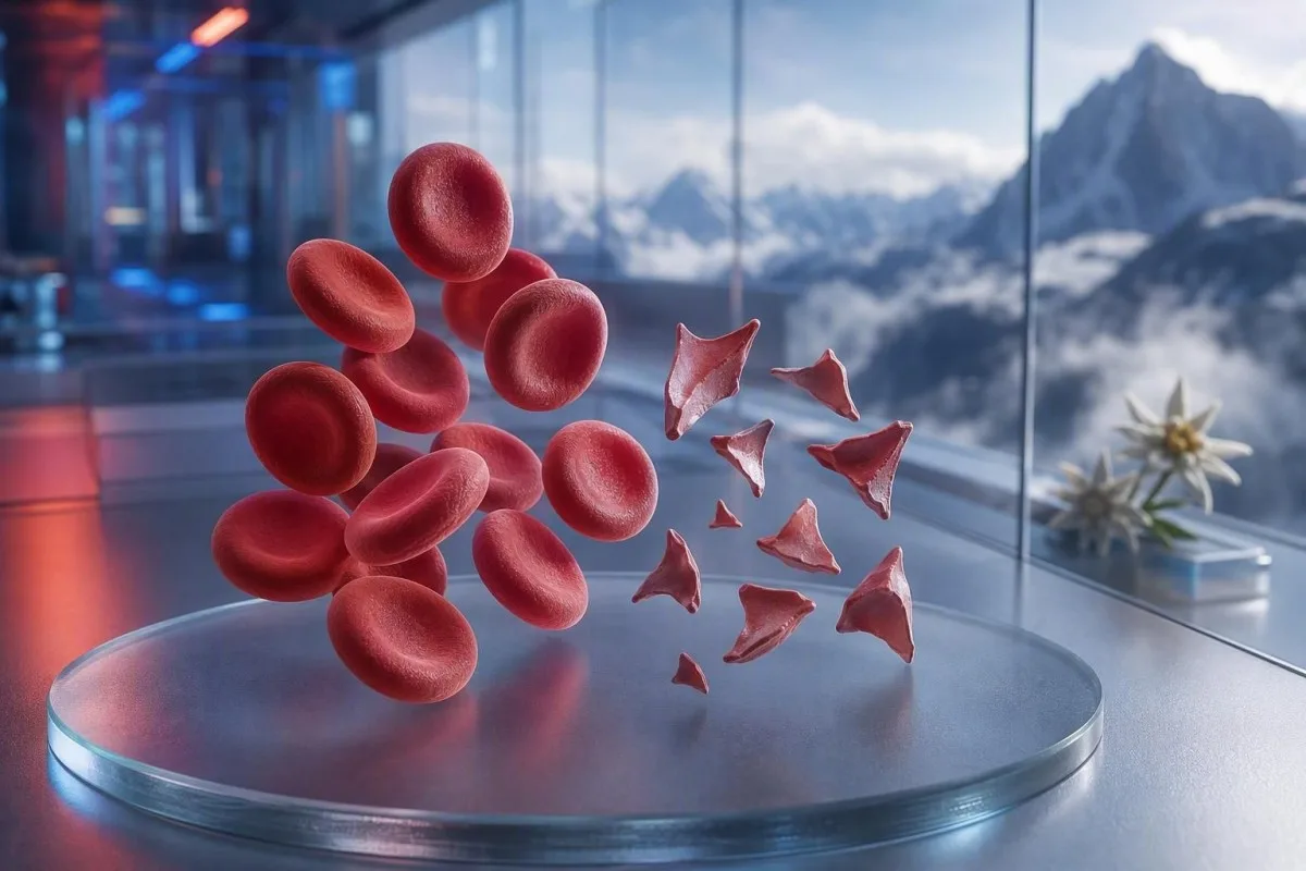
Skistosit adalah sel darah merah yang terfragmentasi yang dapat menandakan penyumbatan berbahaya pada pembuluh darah kecil, tetapi satu komentar pada apusan tidak merupakan diagnosis. Tingkat urgensi bergantung pada persentase fragmen, jumlah trombosit, tren hemoglobin, fungsi ginjal, penanda hemolisis, dan gejala.


- Ambang skistosit: Lebih dari 1% skistosit pada apusan perifer orang dewasa yang dibuat dengan baik bersifat mencurigakan untuk trombotik mikroangiopati bila perubahan bentuk sel darah merah lainnya tidak ada.
- Klaster urgensi: Skistosit disertai trombosit di bawah 150 × 10^9/L, hemoglobin yang menurun, LDH meningkat, dan haptoglobin rendah memerlukan penilaian klinisi pada hari yang sama.
- Petunjuk TTP: Aktivitas ADAMTS13 yang sangat rendah di bawah 10% mendukung purpura trombotik trombositopenik imun, suatu kondisi yang ditangani sebelum hasil tes konfirmasi kembali bila kecurigaan klinis tinggi.
- Tidak semua fragmen berbahaya: Katup jantung mekanis, hipertensi berat, sirkuit dialisis, luka bakar, dan kerusakan sampel dapat menghasilkan sel darah merah yang terfragmentasi.
- Apusan adalah bukti manual: Penganalisis otomatis menandai bahwa skistosit tidak dapat didiagnosis secara andal; seorang morfologis berpengalaman harus memeriksa sediaan.
- Kehamilan mengubah tingkat risikonya: Skistosit dengan tekanan darah tinggi, protein dalam urin, atau trombosit rendah setelah 20 minggu memerlukan penilaian kebidanan segera.
- Pengujian ulang membantu: Sampel EDTA yang baru diambil dan pemeriksaan film ulang dapat memisahkan artefak pengambilan dari fragmentasi sel darah merah yang benar-benar sedang berlangsung.
- Jangan mengobati sendiri: Zat besi, folat, atau suplemen tidak mengobati hemolisis mikroangiopatik dan dapat mengalihkan perhatian dari evaluasi yang mendesak.
Ketika sel darah merah yang terfragmentasi memerlukan peninjauan medis segera
Skistosit memerlukan peninjauan segera pada hari yang sama bila muncul bersama jumlah trombosit rendah, anemia, hemolisis biokimia, cedera ginjal, gejala neurologis, demam, atau hipertensi berat. Komentar pada apusan saja bisa bersifat jinak atau artifaktual; temuan yang berbahaya adalah polanya. Per 7 Agustus 2026, saya akan memperlakukan skistosit baru disertai trombosit di bawah 100 × 10^9/L sebagai hasil “hubungi dokter sekarang”, bukan tindak lanjut rutin.
Penurunan trombosit sering menjadi petunjuk urgensi yang paling cepat. Hemoglobin 92 g/L dengan trombosit 68 × 10^9/L dan skistosit yang baru dilaporkan jauh lebih mengkhawatirkan dibandingkan 1% fragmen dengan hemoglobin 145 g/L dan trombosit yang stabil 260 × 10^9/L. Kantesti adalah penganalisis tes darah berbasis AI yang membaca klaster ini di seluruh hitung darah lengkap, bukan menambahkan komentar morfologi secara terpisah.
Dalam 15 tahun praktik klinis saya, panggilan cepat yang saya buat adalah untuk pasien yang hasilnya berubah dalam beberapa hari: hemoglobin menurun, kreatinin meningkat, dan laktat dehidrogenase meningkat, atau LDH. Aturan praktis Dr. Thomas Klein sederhana: kebingungan, kelemahan baru, nyeri dada, sesak napas, urin sangat sedikit, atau sakit kepala berat bersamaan dengan pola ini berarti penilaian darurat hari ini.
Skistosit bukan alasan untuk menunggu pemeriksaan ulang jika orang tersebut sedang tidak sehat. Kalium normal atau jumlah leukosit normal tidak membatalkan risiko trombotik mikroangiopati. Pasien yang mencoba memahami jumlah yang rendah juga dapat meninjau panduan kami untuk gejala sel darah merah yang rendah, tetapi gejala akut selalu lebih diutamakan daripada interpretasi daring.
Hubungi layanan darurat sekarang untuk
Kelemahan satu sisi yang baru, pingsan, kejang, sesak napas berat, ketidaknyamanan dada seperti tertekan, feses hitam, atau kebingungan yang cepat memburuk memerlukan layanan darurat, bukan pesan ke klinik. Gejala-gejala ini dapat mencerminkan keterlibatan organ, dan tidak ada persentase skistosit yang secara aman dapat menyingkirkannya.
Apa sebenarnya arti skistosit pada apusan perifer
Skistosit adalah fragmen sel darah merah yang tidak teratur yang terbentuk ketika sel-sel yang beredar mengalami pencacahan secara fisik, paling sering oleh untaian fibrin atau permukaan dengan geser tinggi yang abnormal pada pembuluh darah kecil. Biasanya memiliki sudut yang tajam, bentuk seperti helm, atau bentuk segitiga kecil, serta tidak memiliki pucat sentral dari sel darah merah yang utuh.
Skistosit sejati adalah temuan morfologi, bukan nilai yang dihasilkan oleh penganalisis CBC standar. International Council for Standardization in Haematology merekomendasikan menghitung setidaknya 1.000 sel darah merah di zona apusan optimal bila kuantitasi menjadi penting (Zini dkk., 2021). Rincian itu menjelaskan mengapa satu laboratorium mungkin melaporkan “fragmen jarang” sementara laboratorium lain melaporkan persentase.
Kekhawatiran klasik adalah anemia hemolitik mikroangiopatik, di mana sel darah merah bertemu mikro-trombus kaya trombosit dan fibrin dalam sirkulasi. Namun, stenosis aorta berat, katup mekanik, sirkuit ekstrakorporeal, dan cedera termal mayor dapat menciptakan fragmen yang benar-benar nyata melalui mekanisme yang berbeda. Jadi pertanyaannya bukan “Apakah fragmen itu nyata?” melainkan “Apa yang menyebabkan terjadinya fragmentasi?”
Kantesti adalah platform interpretasi tes darah berbasis AI yang dapat menempatkan hitung darah lengkap yang diunggah di samping apa saja yang termasuk dalam CBC dan menyoroti hasil pendamping yang hilang seperti bilirubin, retikulosit, atau haptoglobin. Platform ini tidak dapat memeriksa sediaan itu sendiri, yang tetap menjadi tugas profesional laboratorium terlatih.
Seberapa mengkhawatirkan skistositosis pada apusan orang dewasa
Hitung skistosit di atas 1% pada apusan perifer orang dewasa mencurigakan untuk trombotik mikroangiopati bila fragmen merupakan kelainan utama pada sel darah merah. Hitung di bawah 1% tidak menyingkirkan penyakit, terutama jika film dibuat setelah pemberian cairan, transfusi, atau penyakit yang sedang berkembang.
Batas 1% adalah pemicu diagnostik, bukan skala keparahan. Panduan ICSH menggunakan interval rujukan di bawah 1% untuk orang dewasa sehat dan bayi baru lahir cukup bulan; bayi prematur dapat memiliki hingga 5% fragmen tanpa implikasi yang sama (Zini et al., 2021). Hasil 2% dengan trombositopenia berat lebih mengkhawatirkan daripada 4% setelah pemasangan perangkat dukungan sirkulasi mekanik.
Laboratorium dapat melaporkan “sesekali,” “sedikit,” atau “1–2 per lapang pandang berdaya tinggi” alih-alih persentase. Frasa-frasa tersebut tidak dapat saling dipertukarkan, karena ukuran lapang pandang, ketebalan apusan, dan kebijakan pelaporan berbeda. Menanyakan apakah laboratorium melakukan hitung persentase bergaya ICSH sering kali lebih bermanfaat daripada mencoba mengonversi frasa deskriptif sendiri.
RDW yang tinggi tidak mengukur skistosit. RDW mencerminkan variasi ukuran sel dan dapat meningkat dengan retikulositosis, defisiensi besi, transfusi, atau fragmentasi. Penjelasan rinci kami panduan RDW dan indeks sel darah merah menjelaskan mengapa RDW yang tinggi tidak mendukung TTP maupun artefak semata.
Pola hasil tes darah yang mendukung hemolisis sejati
Fragmentasi sejati sel darah merah biasanya menghasilkan LDH yang tinggi, haptoglobin yang rendah, peningkatan bilirubin indirek, dan respons retikulosit bersamaan dengan anemia. Tidak ada satu penanda yang definitif, karena penyakit hati, inflamasi, transfusi baru-baru ini, dan penyakit sumsum tulang dapat mendistorsikan gambaran.
LDH sensitif tetapi tidak spesifik. LDH di atas 600 IU/L sering mendukung hemolisis aktif pada konteks yang tepat, namun cedera otot, cedera hati, infeksi, dan kanker juga dapat meningkatkannya. Haptoglobin di bawah batas bawah laboratorium, yang umumnya di bawah 0,3 g/L, menambah bobot karena mengikat hemoglobin bebas yang dilepaskan ke plasma.
Hitung retikulosit biasanya meningkat selama destruksi perifer, tetapi dapat tetap rendah pada defisiensi folat, defisiensi besi, penyakit ginjal, atau supresi sumsum tulang akut. Kontradiksi yang tampak ini mudah terlewat: seorang pasien dapat mengalami hemolisis yang nyata dan respons retikulosit yang tidak semestinya rendah. panduan hasil haptoglobin mencakup jebakan interpretasi yang umum.
Kantesti AI menafsirkan pola fragmen sel darah merah dengan membandingkan hemoglobin, MCV, trombosit, LDH, bilirubin, kreatinin, dan retikulosit dari titik waktu yang sama. Penurunan hemoglobin lebih dari 20 g/L dalam 48 hingga 72 jam lebih informatif secara klinis daripada nilai yang sedikit abnormal tanpa perbandingan sebelumnya.
Mengapa bilirubin bisa normal
Bilirubin indirek dapat normal pada awal hemolisis, setelah pengenceran dengan cairan intravena, atau ketika hati membersihkan bilirubin secara efisien. Karena itu, bilirubin normal tidak boleh mengesampingkan fragmen yang meyakinkan, haptoglobin yang rendah, dan hemoglobin yang menurun.
Mengapa TTP ditangani sebagai keadaan darurat hematologi
Purpura trombotik trombositopenik, atau TTP, dicurigai bila skistosit menyertai trombositopenia dan anemia hemolitik mikroangiopatik tanpa alternatif yang lebih jelas. TTP imun yang tidak diobati dapat berkembang cepat, sehingga klinisi dapat memulai pertukaran plasma dan terapi imun sebelum hasil ADAMTS13 kembali.
Aktivitas ADAMTS13 di bawah 10% sangat mendukung TTP, tetapi hasilnya dapat memerlukan waktu berjam-jam atau berhari-hari. Pedoman diagnostik ISTH menyarankan memperkirakan probabilitas pra-uji dengan alat klinis seperti skor PLASMIC sambil mengambil sampel sebelum terapi plasma (Zheng et al., 2020). Skor adalah jembatan menuju tindakan, bukan pengganti penilaian spesialis.
“Pentad” yang sudah dikenal—demam, tanda neurologis, gangguan ginjal, trombositopenia, dan anemia hemolitik—tidak lengkap pada banyak pasien nyata. George dan Nester menggambarkan trombotik mikroangiopati sebagai sindrom dengan penyebab yang saling tumpang tindih, bukan lima kotak wajib (George & Nester, 2014). Dalam praktiknya, menunggu semua lima fitur adalah kebiasaan lama yang berbahaya.
Trombosit di bawah 30 × 10^9/L dengan hemolisis memerlukan diskusi yang sangat mendesak dengan hematologi. Transfusi trombosit biasanya dihindari pada TTP yang dicurigai kecuali ada perdarahan yang mengancam jiwa atau prosedur invasif tidak dapat ditunda. Untuk pengujian terkait, kami panduan tes koagulasi menjelaskan mengapa PT dan aPTT dapat normal pada TTP.
Penyebab berbahaya lain dari skistosit yang dipertimbangkan dokter
Skistosit juga terjadi pada koagulasi intravaskular diseminata, sindrom uremik hemolitik akibat toksin Shiga, hipertensi berat, mikroangiopati terkait kehamilan, obat-obatan tertentu, dan trombotik mikroangiopati terkait transplantasi. Temuan trombosit, koagulasi, ginjal, dan klinis yang menyertainya biasanya memisahkan kondisi-kondisi ini.
DIC sering menggabungkan fragmen dengan PT memanjang, aPTT memanjang, fibrinogen rendah atau menurun, serta D-dimer yang sangat meningkat. TTP lebih sering memiliki waktu koagulasi rutin yang relatif normal. Fibrinogen di bawah 1,5 g/L pada pasien yang sakit menggeser perhatian ke koagulopati konsumsi, meskipun fibrinogen dapat tampak normal pada awalnya karena meningkat selama inflamasi akut.
Hipertensi berat dapat mengoyak sel darah merah ketika tekanan melukai mikrovaskulatur; pembacaan pada atau di atas 180/120 mmHg dengan sakit kepala, perubahan penglihatan, nyeri dada, atau disfungsi ginjal adalah keadaan gawat darurat. Inilah sebabnya pembacaan tekanan darah harus diletakkan berdampingan dengan laporan apusan. Pasien dapat meninjau petunjuk lab untuk hipertensi sekunder setelah bahaya segera telah ditangani.
Diare diikuti penurunan keluaran urine, anemia, dan trombositopenia menimbulkan kekhawatiran akan sindrom uremik hemolitik. Pemeriksaan feses dapat membantu mengidentifikasi toksin Shiga, tetapi status hidrasi, kreatinin, kalium, dan penilaian neurologis memandu penanganan segera terlebih dahulu. Laktat tinggi atau tanda vital yang tidak stabil menuntut evaluasi untuk sepsis dan DIC, bukan pendekatan tunggu-dan-lihat; lihat ini pola penanda sepsis.
Penyebab mekanis sel darah merah terfragmentasi
Penghancuran mekanis dapat menghasilkan skistosit yang benar tanpa TTP, terutama dengan katup jantung prostetik, stenosis aorta berat, perangkat bantu ventrikel, sirkuit dialisis, atau oksigenasi membran ekstrakorporeal. Penyebab-penyebab ini tetap memerlukan peninjauan medis, tetapi terapi menargetkan sumber mekanis, bukan TTP imun.
Pola katup mekanis sering mencakup peningkatan LDH ringan kronis dan haptoglobin rendah dengan trombosit yang stabil. Anemia baru, sesak napas yang memburuk, urine gelap, atau perubahan bunyi katup dapat mengisyaratkan kebocoran paravalvular atau disfungsi perangkat dan harus memicu peninjauan oleh kardiologi. Apusan darah memberikan petunjuk; ekokardiografi menemukan sumbernya.
Atlet kadang bertanya apakah lari maraton dapat menjelaskan fragmen. Hemolisis akibat benturan telapak kaki dapat menyebabkan hemoglobin bebas sementara dan haptoglobin rendah setelah lari jarak jauh, tetapi skistotosis yang meyakinkan bukan sesuatu yang akan saya kaitkan begitu saja dengan olahraga—terutama jika trombosit sedang menurun. Bandingkan dengan panduan panduan laboratorium pelari maraton.
Luka bakar berat dan pembedahan vaskular mayor dapat menghasilkan sel yang terfragmentasi melalui cedera fisik dan cedera endotel. Pada kondisi tersebut, tren hasil laboratorium setiap 6 hingga 24 jam mungkin lebih informatif daripada satu apusan. CBC yang stabil setelah kejadian pemicu menenangkan; hemoglobin yang terus menurun tidak.
Bagaimana artefak pengambilan sampel dan pembuatan sediaan meniru skistosit
Pengambilan sampel yang buruk, penundaan persiapan apusan, tabung antikoagulan yang kurang terisi, dan tepi apusan yang rusak dapat membentuk gambaran yang menyerupai skistosit tanpa mencerminkan hemolisis pada tingkat sirkulasi. Sampel ulang yang dikumpulkan dengan bersih dan ditinjau pada monolayer adalah pemeriksaan pertama yang biasa dilakukan ketika gambaran klinis tenang.
Sel krenasi, distorsi tepi apusan, artefak pengeringan, dan sel yang saling tumpang tindih adalah kemiripan palsu yang umum. Mereka sering tampak menyebar atau hanya pada tepi yang berbulu (feathered edge) daripada merata di zona pembacaan ideal. Morfologis berpengalaman membedakan geometri tajam fragmen yang benar dari sel yang hanya terdistorsi selama persiapan slide.
Pengambilan yang sulit dapat hemolisis tabung dan secara keliru meningkatkan kalium atau LDH, tetapi tidak otomatis menghasilkan skistosit yang benar-benar beredar. Jika suatu hasil mencakup indeks hemolisis atau komentar “sampel terkompromi”, klinisi biasanya mengulang pengambilan sebelum mendiagnosis penyakit. Artikel kami tentang kesalahan pengambilan darah kalium menjelaskan mengapa masalah pra-analitik yang sama dapat menyesatkan beberapa hasil sekaligus.
Penggumpalan trombosit yang bergantung pada EDTA dapat menurunkan hitung trombosit secara keliru dan “menciptakan” kombinasi yang tampak mengkhawatirkan. Hitung trombosit pada tabung sitrat, dikoreksi untuk pengenceran, dan film segar dapat mengungkap hal ini. Nuansanya penting: artefak dapat berbarengan dengan penyakit yang nyata, jadi tes ulang tidak boleh menunda penanganan bagi seseorang yang simptomatik.
Mengapa peringatan analis dan redaksi laboratorium dapat menyesatkan
Penganalisis darah otomatis dapat menandai populasi sel darah merah yang abnormal, tetapi tidak dapat mendiagnosis skistosit secara independen dengan reliabilitas yang cukup untuk menyingkirkan trombotik mikroangiopati. Peninjauan apusan perifer manual tetap diperlukan ketika pertanyaan klinis adalah hemolisis atau TTP.
Komentar “sel darah merah terfragmentasi tampak” harus memicu pertanyaan tentang jumlahnya, kualitas sediaan apus, dan morfologi yang menyertai. Anisopoikilositosis yang nyata akibat defisiensi besi, anemia megaloblastik, atau kelainan herediter sel darah merah membuat aturan 1% menjadi kurang spesifik. Fragmen paling berguna secara diagnostik bila mendominasi morfologi abnormal, bukan muncul di antara banyak bentuk yang tidak terkait.
Kantesti adalah layanan interpretasi hasil uji lab berbasis AI yang dirancang untuk menandai kombinasi yang tidak sesuai, seperti jumlah trombosit rendah disertai komentar yang menyatakan adanya gumpalan trombosit. Layanan ini tidak menggantikan peninjauan mikroskopis dari laboratorium atau dokter yang dapat meminta pengulangan sediaan. Pendekatan kami untuk pemeriksaan kualitas dijelaskan dalam gambaran validasi medis.
Pemeriksaan delta membandingkan hasil saat ini dengan hasil sebelumnya dan dapat mengungkap perubahan mendadak yang tidak masuk akal. Penurunan hemoglobin dari 138 g/L menjadi 87 g/L dalam 24 jam bermakna secara klinis, tetapi pengenceran akibat cairan intravena atau spesimen yang salah label tetap mungkin terjadi. Baca lebih lanjut tentang perubahan mendadak pada hasil laboratorium.
Apa yang harus dilakukan setelah hasil tes darah menyebutkan skistosit
Hubungi dokter atau layanan yang memesan pemeriksaan pada hari yang sama jika skistosit dilaporkan baru, dan cari pertolongan darurat segera bila Anda mengalami gejala neurologis, sesak napas, nyeri dada, urin gelap, sakit kepala berat, demam, atau penurunan keluaran urin. Jangan menafsirkan komentar sediaan apus sebagai diagnosis dengan sendirinya.
Siapkan laporan aktualnya, termasuk hemoglobin, trombosit, kreatinin, bilirubin, LDH, dan setiap indeks hemolisis. Perhatikan juga kehamilan baru-baru ini, penyakit diare, pembacaan tekanan darah yang sangat tinggi, obat baru, operasi katup, dialisis, transfusi, dan penyakit autoimun. Rincian ini mengubah diagnosis banding lebih daripada satu kata tunggal dalam kolom morfologi.
Saya melihat pasien kehilangan waktu berharga karena memulai terapi besi saat mereka membaca “anemia” sebelum siapa pun mengidentifikasi penyebabnya. Defisiensi besi mungkin ada bersamaan, tetapi tidak menjelaskan penurunan trombosit yang cepat disertai fragmen. Dr. Thomas Klein merekomendasikan mengambil foto seluruh laporan, bukan menyalin hanya angka yang disorot; penyalinan sering kali menghilangkan komentar laboratorium yang membuat pola tersebut menjadi koheren.
Hindari aspirin, ibuprofen, dan antikoagulan yang tidak diresepkan sampai dokter menyarankan sebaliknya jika trombosit sangat rendah atau terdapat perdarahan. Jangan menghentikan antikoagulan yang diresepkan tanpa panduan medis yang mendesak, juga. Orang dengan kelelahan atau rasa ringan kepala dapat menggunakan untuk pusing memandu untuk menyiapkan pertanyaan setelah penyebab yang mendesak dikecualikan.
Skistosit pada kehamilan, bayi baru lahir, dan anak-anak
Skistosit selama kehamilan memerlukan penilaian segera bila muncul bersama hipertensi, proteinuria, peningkatan enzim hati, trombosit rendah, sakit kepala, gejala penglihatan, atau nyeri perut bagian atas. Sindrom HELLP, pre-eklampsia, TTP, HUS atipikal, dan DIC dapat saling tumpang tindih, dan waktu di sekitar persalinan membantu tetapi tidak memastikan diagnosis.
Sindrom HELLP secara klasik didefinisikan oleh hemolisis, peningkatan enzim hati, dan trombosit rendah, sering kali setelah 20 minggu kehamilan atau segera setelah persalinan. Jumlah trombosit di bawah 100 × 10^9/L atau AST di atas 70 IU/L dapat mendukung adanya kekhawatiran, tetapi tim obstetri setempat menafsirkan keseluruhan gambaran klinis. Jangan menunggu janji antenatal rutin dengan kombinasi ini.
Bayi baru lahir cukup bulan mungkin memiliki skistosit fewer than 1%, sedangkan bayi prematur dapat memiliki hingga 5% karena morfologi sel darah merah neonatus berbeda (Zini et al., 2021). Namun, pada anak dengan diare, pucat, penurunan keluaran urin, dan trombositopenia, fragmen dapat mengarah ke HUS dan memerlukan penilaian pediatrik segera.
Interval rujukan untuk trombosit dan hemoglobin berubah secara substansial berdasarkan usia dan tahap kehamilan. Hasil yang ditandai “normal” oleh aplikasi generik tetap dapat menjadi perhatian secara klinis dalam konteks tertentu. Sumber daya kami tentang rentang trombosit pada kehamilan Dan penanda lab kehamilan pada hari yang sama berikan latar belakang yang bermanfaat, bukan pengganti triase kebidanan.
Tes yang dipesan klinisi untuk menyelidiki skistosit sejati
Pemeriksaan pertama untuk dugaan hemolisis mikroangiopatik biasanya mencakup CBC yang diulang dan apusan, hitung retikulosit, LDH, haptoglobin, fraksi bilirubin, kreatinin, urinalisis, PT, aPTT, fibrinogen, dan D-dimer. Pemeriksaan aktivitas ADAMTS13 dan inhibitor harus diambil sebelum plasma exchange bila dicurigai TTP, tetapi pengobatan tidak boleh menunggu.
Tes antibodi antieritrosit langsung, juga disebut DAT atau tes Coombs, membantu membedakan hemolisis imun dari fragmentasi non-imun. DAT positif tidak menyingkirkan proses kedua, tetapi TTP klasik biasanya DAT-negatif. Mikroskopi urin dapat menunjukkan silinder protein atau sel ketika cedera mikrovasikular ginjal aktif; lihat penjelasan kami tentang silinder urin granular.
Tes Shiga-toxin pada tinja dipertimbangkan setelah penyakit gastrointestinal yang sesuai, sedangkan tes komplemen, peninjauan obat, dan studi genetik atau fungsional dapat digunakan untuk HUS atipikal. Urutannya bervariasi menurut negara dan rumah sakit. Kenaikan kreatinin sebesar 26,5 µmol/L dalam 48 jam memenuhi salah satu kriteria cedera ginjal akut dan layak mendapat perhatian bahkan sebelum menjadi angka yang dramatis.
Indeks produksi retikulosit di bawah 2 pada anemia bermakna menunjukkan respons sumsum yang tidak adekuat, bahkan jika persentase retikulosit mentah tampak tinggi. Perhitungan itu mengoreksi massa sel darah merah yang lebih rendah pada anemia. Yang panduan retikulosit dan penanda hematologi kami membantu menjelaskan mengapa persentase bisa menyesatkan.
Kapan mengulang apusan itu masuk akal dan kapan tidak
Mengulang apusan dalam beberapa jam hingga 24 jam adalah langkah yang masuk akal bila fragmen sedikit, pasien merasa baik, trombosit dan hemoglobin stabil, serta kualitas sampel dipertanyakan. Pengulangan bukan penundaan yang aman bila terdapat trombositopenia, hemolisis aktif, cedera organ, atau gejala yang mengkhawatirkan.
Sampel ulangan yang baik dikumpulkan segar ke dalam tabung EDTA yang terisi dengan benar dan segera dibuat menjadi sediaan apus. Permintaan harus secara spesifik meminta peninjauan terhadap dugaan skistosit dan penggumpalan trombosit jika salah satunya masuk akal. Laboratorium dapat membandingkan kedua apusan berdampingan, yang jauh lebih bermanfaat daripada sekadar menjalankan ulang CBC otomatis.
Arah tren itu penting. Hitung trombosit yang bergerak dari 210 menjadi 165 lalu 98 × 10^9/L selama tiga hari mengkhawatirkan meskipun dua pembacaan pertama mungkin masih berada dalam interval rujukan lokal. Kantesti AI menggunakan konteks longitudinal untuk menampilkan lintasan tersebut, sebuah prinsip yang dibahas dalam panduan analisis tren hasil tes darah.
Transfusi dapat sementara mengencerkan atau menutupi morfologi asli. Jika Anda menerima sel darah merah sebelum sampel kedua, beri tahu tim tanggal dan jumlah unitnya. Panduan kami untuk perubahan nilai setelah keluar dari rumah sakit menjelaskan mengapa tren setelah pengobatan memerlukan baseline yang berbeda.
Kondisi yang dapat tampak mirip tanpa menjadi penyakit skistosit
Defisiensi besi, defisiensi vitamin B12, penyakit hati, kelainan bawaan sel darah merah, dan infeksi berat dapat membentuk bentuk sel darah merah yang abnormal tanpa pola fragmentasi spesifik dari trombotik mikroangiopati. Morfologi lengkap apusan dan kronologi klinis mencegah penilaian berlebihan terhadap fragmen.
Defisiensi vitamin B12 berat dapat menyebabkan pseudo-trombotik mikroangiopati dengan anemia, LDH tinggi, trombosit rendah, dan fragmen sesekali. Biasanya menghasilkan makrositosis yang nyata, respons retikulosit yang rendah, dan nilai LDH kadang di atas 2.500 IU/L—ciri yang seharusnya memicu pemeriksaan B12 dan asam metilmalonat, bukan pengobatan TTP otomatis. Penjelasan kami tentang panduan B12 vs folat menjelaskan mimic penting ini.
Defisiensi besi biasanya menghasilkan mikrositosis, hipokromia, sel pensil, dan RDW yang tinggi; fragmen dapat dilaporkan pada kasus yang berat, tetapi jarang menjadi fitur dominan. Ferritin di bawah 15 µg/L sangat mendukung cadangan besi yang menurun pada orang dewasa yang tidak memiliki kondisi lain yang menyulitkan, meskipun inflamasi dapat menutupi defisiensi dengan meningkatkan ferritin.
Menurut pengalaman saya, hasil yang paling meyakinkan bukan hanya “tidak ada skistosit” semata, melainkan penjelasan alternatif yang koheren dengan trombosit yang stabil, fungsi ginjal yang stabil, dan tidak ada hemolisis biokimia. Jika temuan tetap tidak selaras, seorang hematolog dapat meninjau slide aktual daripada hanya mengandalkan redaksi laporan akhir.
Menggunakan interpretasi AI tanpa melewatkan keadaan darurat pada apusan
AI dapat membantu mengorganisasi hasil tes darah dan mengidentifikasi kombinasi yang berbahaya, tetapi tidak dapat memverifikasi diagnosis skistosit dari PDF CBC atau menggantikan penilaian klinis yang mendesak. Apusan perifer adalah mikroskopi visual, dan slide laboratorium asli tetap menjadi sumber kebenaran.
Kantesti adalah alat analisis tes darah berbasis AI yang memetakan hasil terkait lintas laporan, termasuk trajektori trombosit, perubahan hemoglobin, kreatinin, bilirubin, dan LDH. Itu dapat mengurangi kesalahan umum membaca setiap penanda abnormal secara terpisah. Alat ini tidak dapat melihat apakah bentuk yang disebut skistosit adalah fragmen yang benar, artefak tepi apusan, atau konvensi pelaporan.
Kualitas unggahan penting karena unit atau desimal yang terlewat dapat membalikkan makna suatu hasil. Sebelum membagikan laporan, periksa identitas pasien, tanggal pengambilan, satuan, rentang rujukan, dan apakah suatu hasil menyatakan “estimated,” “pending,” atau “sample haemolysed.” daftar periksa akurasi laporan AI berguna untuk pemeriksaan manusia terakhir itu.
Metode klinis Kantesti ditinjau dengan masukan dari kami Dewan Penasehat Medis, dan teknologi kami dijelaskan dalam teknologi interpretasi AI menjelaskan bagaimana jaringan saraf Kantesti memberi bobot pada biomarker yang berdekatan, arah tren, dan konsistensi internal. Dari pengalaman saya, hal itu paling penting untuk nilai BUN batas antara. Sebagai Dr. Thomas Klein, saya tetap ingin seorang klinisi menghubungi laboratorium secara langsung untuk pasien dengan fragmen baru, trombosit di bawah 100 × 10^9/L, dan gejala—karena tepat pada saat itulah kecepatan dan pemeriksaan klinis paling penting.
Pertanyaan yang Sering Diajukan
Seberapa tinggi kadar skistosit yang berbahaya?
Lebih dari 1% skistosit pada apusan perifer dewasa yang ditinjau dengan cermat bersifat mencurigakan untuk trombotik mikroangiopati bila sel-sel terfragmentasi merupakan kelainan utama sel darah merah. Persentase skistosit apa pun menjadi mendesak bila terjadi bersamaan dengan jumlah trombosit di bawah 150 × 10^9/L, hemoglobin yang menurun, LDH yang tinggi, haptoglobin yang rendah, cedera ginjal, atau gejala neurologis. Hasil di bawah 1% tidak secara andal mengecualikan kondisi yang sedang berkembang. Pola klinis menentukan tingkat bahaya lebih daripada persentase saja.
Dapatkah skistosit disebabkan oleh pengambilan darah yang buruk?
Kumpulan yang sulit, persiapan yang tertunda, tabung EDTA yang tidak terisi penuh, atau sediaan apus yang dibuat dengan buruk dapat menghasilkan bentuk sel yang meniru skistosit, meskipun hal tersebut tidak menimbulkan fragmentasi sirkulasi yang sesungguhnya. Laboratorium dapat memperjelas hal ini dengan sampel yang baru diambil dan peninjauan manual pada area apus yang optimal. Sampel yang hemolisis juga dapat secara keliru meningkatkan kadar kalium dan LDH, sehingga laporan tampak lebih mengkhawatirkan. Pengujian ulang tidak boleh menunda penilaian emergensi bila terdapat gejala, trombositopenia, atau anemia.
Apakah skistosit selalu berarti TTP?
Schistocytes tidak selalu berarti purpura trombotik trombositopenik, atau TTP. Schistocytes dapat terjadi dengan DIC, hipertensi berat, HUS akibat toksin Shiga, gangguan terkait kehamilan, katup jantung mekanik, sirkuit dialisis, dan cedera jaringan mayor. TTP menjadi perhatian prioritas tinggi ketika schistocytes bersamaan dengan trombositopenia dan anemia hemolitik non-imun, terutama bila aktivitas ADAMTS13 berada di bawah 10%. Karena pemeriksaan ADAMTS13 dapat memerlukan waktu, dugaan TTP memerlukan penilaian spesialis yang mendesak sebelum hasil keluar.
Tes darah apa yang meningkat pada hemolisis dengan skistosit?
Hemolisis fragmentasi sejati yang umum terjadi biasanya meningkatkan LDH dan bilirubin tidak langsung, menurunkan haptoglobin, serta memicu peningkatan hitung retikulosit sementara hemoglobin menurun. LDH dapat melebihi 600 IU/L pada hemolisis aktif, tetapi tidak spesifik karena cedera otot, hati, dan jaringan lain juga dapat meningkatkannya. Haptoglobin di bawah sekitar 0,3 g/L mendukung hemolisis intravaskular bila produksi hati adekuat. Kreatinin, jumlah trombosit, PT, aPTT, fibrinogen, dan D-dimer membantu mengidentifikasi penyebab yang mendasari.
Apakah skistosit normal pada kehamilan?
Schistocytes tidak dianggap sebagai temuan kehamilan normal yang rutin bila muncul bersama hipertensi, proteinuria, peningkatan enzim hati, trombosit yang menurun, sakit kepala, perubahan penglihatan, atau nyeri perut bagian atas. Kombinasi ini dapat menandakan sindrom HELLP, pre-eklampsia, TTP, HUS atipikal, atau DIC dan memerlukan penilaian kebidanan segera. Jumlah trombosit di bawah 100 × 10^9/L pada kondisi ini sangat mengkhawatirkan. Rentang rujukan spesifik kehamilan tidak membuat pola hemolisis aman untuk diabaikan.
Apakah katup jantung mekanis dapat menyebabkan skistosit?
Katup jantung mekanik dan stenosis aorta berat dapat menyebabkan skistosit yang benar karena aliran darah berkecepatan tinggi dengan geser tinggi secara fisik memecah sel darah merah. Hal ini sering menyebabkan hemolisis kronis ringan dengan LDH yang meningkat dan haptoglobin yang menurun, sementara jumlah trombosit tetap stabil. Anemia baru, urin gelap, sesak napas, atau perubahan fungsi katup memerlukan evaluasi jantung karena kebocoran atau masalah perangkat mungkin menjadi penyebabnya. Temuan ini tidak boleh secara otomatis diberi label TTP tanpa mempertimbangkan riwayat jantung.
Dapatkan Analisis Tes Darah Berbasis AI Hari Ini
Bergabunglah dengan lebih dari 2 juta pengguna di seluruh dunia yang mempercayai Kantesti untuk analisis instan dan akurat terhadap tes lab. Unggah hasil tes darah Anda dan terima interpretasi komprehensif biomarker 15,000+ dalam hitungan detik.
📚 Publikasi Riset yang Dirujuk
Klein, T., Mitchell, S., & Weber, H. (2026). Sebuah Benchmark Teknis Otomatis Berbasis Rubrik yang Telah Dipra-Registrasi dari Mesin Interpretasi Tes Darah Kantesti pada 100.000 Kasus Uji Sintetis. Kantesti Penelitian Medis AI.
Klein, T., Mitchell, S., & Weber, H. (2026). Kerangka Validasi Klinis v2.0 (Halaman Validasi Medis). Kantesti Penelitian Medis AI.
📖 Referensi Medis Eksternal
Zheng XL dkk. (2020). Pedoman ISTH untuk diagnosis trombotik trombositopenik purpura. Journal of Thrombosis and Haemostasis.
⚕️ Penafian Medis
Artikel ini hanya untuk tujuan edukasi dan tidak merupakan nasihat medis. Selalu konsultasikan dengan penyedia layanan kesehatan yang berkualifikasi untuk keputusan diagnosis dan perawatan.
Sinyal Kepercayaan E-E-A-T
Pengalaman
Tinjauan klinis yang dipimpin dokter terhadap alur kerja interpretasi hasil lab.
Keahlian
Fokus pada kedokteran laboratorium tentang bagaimana biomarker berperilaku dalam konteks klinis.
Kewenangan
Ditulis oleh Dr. Thomas Klein dengan peninjauan oleh Dr. Sarah Mitchell dan Prof. Dr. Hans Weber.
Kepercayaan
Interpretasi berbasis bukti dengan jalur tindak lanjut yang jelas untuk mengurangi kepanikan.