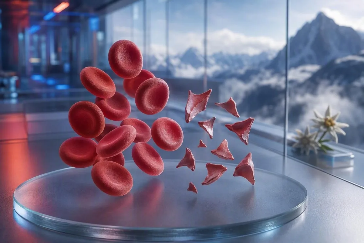
شیستوسیتها گلبولهای قرمزِ قطعهقطعهشدهای هستند که میتوانند نشانهٔ خطرِ لختهشدنِ کوچکعروقی باشند، اما یک اظهار نظرِ تکبرگی در اسمیر بهتنهایی تشخیص نیست. فوریت به درصد قطعات، تعداد پلاکت، روند هموگلوبین، عملکرد کلیه، نشانگرهای همولیز و علائم بستگی دارد.


- آستانهٔ شیستوسیت: بیش از 1% شیستوسیت در یک اسمیر محیطی بزرگسالِ بهخوبی تهیهشده، در نبودِ سایر تغییرات شکل گلبولهای قرمز، برای میکروآنژیوپاتی ترومبوتیک مشکوک است.
- خوشهٔ فوری: شیستوسیتها بهعلاوه پلاکتهای کمتر از 150 × 10^9/L، هموگلوبینِ رو به کاهش، LDH بالا و هاپتوگلوبین پایین نیاز به ارزیابی فوری همانروزهٔ پزشک دارد.
- سرنخ TTP: فعالیت شدید ADAMTS13 کمتر از 10% از پورپورای ترومبوتیک ترومبوسیتوپنی ایمنی حمایت میکند؛ وضعیتی که در صورت بالا بودن شک بالینی، پیش از بازگشت نتایج آزمایش تأییدی درمان میشود.
- هر قطعهای خطرناک نیست: دریچههای مکانیکی قلب، فشار خون شدید، مدارهای دیالیز، سوختگیها و آسیب نمونه میتوانند گلبولهای قرمزِ قطعهقطعهشده ایجاد کنند.
- اسمیر یک شواهد دستی است: آنالایزر خودکار پرچمگذاری میکند که تشخیص قابلاعتمادِ اسکیستوسیتها ممکن نیست؛ یک مورفولوژیست باتجربه باید لام را بررسی کند.
- بارداری ریسکها را بالا میبرد: اسکیستوسیتها همراه با فشار خون بالا، پروتئین در ادرار، یا پلاکتهای پایین پس از ۲۰ هفته نیاز به ارزیابی فوریِ مامایی دارد.
- تکرار آزمایش کمک میکند: یک نمونه تازه جمعآوریشده EDTA و تکرار لام میتواند یک آرتیفکتِ ناشی از جمعآوری را از شکستگیِ واقعیِ در حال وقوعِ گلبولهای قرمز جدا کند.
- خودسرانه درمان نکنید: آهن، فولات یا مکملها، همولیز میکروآنژیوپاتیک را درمان نمیکنند و میتوانند از ارزیابی فوری منحرف کنند.
زمانی که گلبولهای قرمزِ قطعهقطعهشده نیاز به بررسی فوری پزشکی دارند
اسکیستوسیتها وقتی همراه با تعداد پلاکت پایین، کمخونی، همولیز بیوشیمیایی، آسیب کلیه، علائم نورولوژیک، تب، یا فشار خون شدید ظاهر میشوند، نیاز به بررسی فوری همانروزه دارند. صرفِ یک نظر روی اسمیر میتواند خوشخیم یا آرتیفکتی باشد؛ یافته خطرناک، الگو است. از ۷ اوت ۲۰۲۶، من اسکیستوسیتهای جدید همراه با پلاکتهای کمتر از ۱۰۰ × ۱۰^۹/L را «فوراً با پزشک تماس بگیرید» تلقی میکنم، نه پیگیری روتین.
افت پلاکت اغلب سریعترین سرنخِ فوریت است. هموگلوبین ۹۲ g/L با پلاکتهای ۶۸ × ۱۰^۹/L و اسکیستوسیتهای تازه گزارششده، بهطور قابلتوجهی نگرانکنندهتر از ۱TP54T قطعه با هموگلوبین ۱۴۵ g/L و پلاکتهای پایدار ۲۶۰ × ۱۰^۹/L است. ۱TP6T یک آنالایزر تست خونِ مبتنی بر هوش مصنوعی است که این خوشه را در آزمایش خون کامل میخواند، نه اینکه صرفاً بهصورت جداگانه یک نظر مورفولوژی را بالا ببرد.
در ۱۵ سالِ طبابت بالینی من، فراخوانیهایی که سریع انجام میدهم برای بیمارانی است که نتایجشان طی چند روز تغییر کرده است: افت هموگلوبین، افزایش کراتینین، و افزایش لاکتات دهیدروژناز یا LDH. قانون عملیِ دکتر توماس کلاین ساده است: گیجی، ضعف جدید، درد قفسه سینه، تنگی نفس، ادرار بسیار کم، یا سردرد شدید همراه با این الگو یعنی ارزیابی اورژانسی همین امروز.
اسکیستوسیتها دلیلی برای انتظار جهت تکرار آزمایش نیستند، اگر فرد حالش بد است. پتاسیم طبیعی یا شمارش گلبولهای سفید طبیعی، خطر میکروآنژیوپاتی ترومبوتیک را منتفی نمیکند. بیمارانی که میخواهند یک شمارش پایین را درک کنند نیز میتوانند راهنمای ما را مرور کنند تا علائم کمبود گلبولهای قرمز, ، اما علائم حاد همیشه از تفسیر آنلاین پیشی میگیرد.
همین حالا با خدمات اورژانس تماس بگیرید برای
ضعف جدید یکطرفه، غش، تشنج، تنگی نفس شدید، ناراحتی شدید لهکننده در قفسه سینه، مدفوع سیاه، یا گیجیای که بهسرعت بدتر میشود، نیاز به خدمات اورژانس دارد نه یک پیام به کلینیک. این علائم میتوانند درگیریِ اندامها را نشان دهند و هیچ درصدی از اسکیستوسیتها بهطور ایمن آن را رد نمیکند.
شیستوسیتها در اسمیر محیطی دقیقاً چه معنایی دارند
اسکیستوسیتها قطعات نامنظمِ گلبولهای قرمز هستند که وقتی سلولهای در گردش بهصورت فیزیکی بریده میشوند ایجاد میگردند؛ اغلب توسط رشتههای فیبرین یا سطوح غیرطبیعی با برشِ بالا در رگهای کوچک. معمولاً زوایای تیز دارند، شکلهای شبیه کلاهخود، یا شکلهای مثلثی کوچک و فاقد پالور مرکزیِ یک گلبول قرمز سالم هستند.
اسکیستوسیتهای واقعی یک یافته مورفولوژیک هستند، نه مقداری که توسط آنالایزر استاندارد CBC تولید میشود. شورای بینالمللی استانداردسازی در هماتولوژی توصیه میکند وقتی کمّیسازی مهم است، حداقل ۱۰۰۰ گلبول قرمز در ناحیه بهینهِ اسمیر شمرده شود (Zini et al., 2021). این جزئیات توضیح میدهد چرا یک آزمایشگاه ممکن است “قطعات نادر” گزارش کند در حالی که دیگری درصد گزارش میدهد.
نگرانی کلاسیک این است که کمخونی همولیتیک میکروآنژیوپاتیک, ، جایی که گلبولهای قرمز به میکروترومبهای غنی از پلاکت و فیبرین در گردش خون میرسند. با این حال، تنگی شدید آئورت، دریچه مکانیکی، مدارهای خارجبدنی و آسیب حرارتی عمده میتوانند از طریق یک مکانیسم متفاوت، قطعات واقعی ایجاد کنند. بنابراین سؤال نه “آیا قطعات واقعی هستند؟” بلکه “چه چیزی باعث قطعهقطعه شدن میشود؟” است.”
Kantesti یک پلتفرم تفسیر آزمایش خون مبتنی بر هوش مصنوعی است که میتواند شمارش کامل خون بارگذاریشده را کنار اینکه CBC چه چیزهایی را شامل میشود قرار دهد و نتایج همراهِ گمشده مانند بیلیروبین، رتیکولوسیتها یا هاپتوگلوبین را برجسته کند. این پلتفرم نمیتواند خودِ لام را بررسی کند؛ این کار همچنان وظیفهی یک متخصص آموزشدیدهی آزمایشگاه است.
چه میزان شیستوسیتوز در اسمیر بزرگسالان نگرانکننده است
شمارش اسکیستوسیتها بالاتر از 1% در اسمیر محیطی بزرگسالان، زمانی که قطعات، شایعترین ناهنجاری گلبول قرمز هستند، برای میکروآنژیوپاتی ترومبوتیک مشکوککننده است. شمارشهای کمتر از 1% بیماری را رد نمیکنند، بهویژه اگر لام بعد از دریافت مایعات، تزریق خون یا یک بیماری در حال تکامل تهیه شده باشد.
برش 1% یک هشدار تشخیصی است، نه یک مقیاس شدت. راهنمای ICSH از یک بازه مرجع زیر 1% برای بزرگسالان سالم و نوزادان ترم استفاده میکند؛ نوزادان نارس ممکن است تا 5% قطعه بدون همان پیامد داشته باشند (Zini et al., 2021). نتیجهی 2% همراه با ترومبوسیتوپنی شدید، از 4% پس از قرار دادن یک دستگاه حمایت مکانیکی از گردش خون نگرانکنندهتر است.
آزمایشگاهها ممکن است بهجای درصد، “گاهی”، “کم”، یا “1–2 در هر میدان با توان بالا” گزارش کنند. این عبارات قابلجایگزینی نیستند، زیرا اندازه میدان، ضخامت اسمیر و سیاست گزارشدهی متفاوت است. پرسیدن اینکه آیا آزمایشگاه شمارش درصدی به سبک ICSH انجام داده است، اغلب مفیدتر از تلاش برای تبدیل خودِ یک عبارت توصیفی است.
RDW بالا اسکیستوسیتها را اندازهگیری نمیکند. RDW تغییرپذیری در اندازه سلولها را نشان میدهد و ممکن است با رتیکولوسیتوز، کمبود آهن، تزریق خون یا قطعهقطعه شدن افزایش یابد. توضیح دقیق ما راهنمای RDW و شاخصهای گلبول قرمز توضیح میدهد که چرا RDW بالا بهتنهایی نه از TTP حمایت میکند و نه یک آرتیفکت را.
الگوی آزمایش خون که همولیز واقعی را تأیید میکند
قطعهقطعه شدن واقعی گلبول قرمز معمولاً LDH بالا، هاپتوگلوبین پایین، افزایش بیلیروبین غیرمستقیم و پاسخ رتیکولوسیتی را همراه با کمخونی ایجاد میکند. هیچ نشانگر واحدی قطعی نیست، زیرا بیماری کبدی، التهاب، انتقال خون اخیر، و بیماری مغز استخوان میتوانند تصویر را مخدوش کنند.
LDH حساس است اما اختصاصی نیست. LDH بالاتر از 600 IU/L اغلب در شرایط مناسب، همولیز فعال را پشتیبانی میکند؛ با این حال، آسیب عضلانی، آسیب کبدی، عفونت و سرطان نیز میتوانند آن را بالا ببرند. هپتوگلوبین پایینتر از حد پایین آزمایشگاه، که معمولاً کمتر از 0.3 g/L است، وزن بیشتری میدهد زیرا به هموگلوبین آزادِ آزادشده به پلاسما متصل میشود.
شمارش رتیکولوسیتها معمولاً باید در تخریب محیطی افزایش یابد، اما ممکن است در کمبود فولات، کمبود آهن، بیماری کلیوی یا سرکوب حاد مغز استخوان پایین بماند. این تناقض ظاهری بهراحتی نادیده گرفته میشود: بیمار میتواند همولیز واقعی داشته باشد و در عین حال پاسخ رتیکولوسیتی نامناسباً پایین باشد. راهنمای نتیجه هپتوگلوبین دامهای رایج تفسیر را پوشش میدهد.
Kantesti AI الگوهای قطعهقطعهشده گلبولهای قرمز را با مقایسه هموگلوبین، MCV، پلاکتها، LDH، بیلیروبین، کراتینین و رتیکولوسیتها از همان نقطه زمانی تفسیر میکند. افت هموگلوبین بیش از 20 g/L طی 48 تا 72 ساعت از نظر بالینی آموزندهتر از یک مقدار کمی غیرطبیعی بدون مقایسه قبلی است.
چرا بیلیروبین میتواند طبیعی باشد
بیلیروبین غیرمستقیم ممکن است در مراحل اولیه همولیز، پس از رقیقسازی با مایع داخلوریدی، یا زمانی که کبد بیلیروبین را بهطور مؤثر پاکسازی میکند، طبیعی باشد. بنابراین بیلیروبین طبیعی نباید قطعات قانعکننده، هپتوگلوبین پایین و افت هموگلوبین را نادیده بگیرد.
چرا TTP بهعنوان یک فوریت هماتولوژی درمان میشود
پورپورای ترومبوتیک ترومبوسیتوپنیک، یا TTP، زمانی مشکوک میشود که شیستوسیتها همراه با ترومبوسیتوپنی و کمخونی همولیتیک میکروآنژیوپاتیک بدون یک جایگزین روشنتر دیده شوند. TTP ایمنیِ درماننشده میتواند بهسرعت پیشرفت کند، بنابراین پزشکان ممکن است پیش از بازگشت نتایج ADAMTS13، تعویض پلاسما و درمان ایمنی را شروع کنند.
فعالیت ADAMTS13 کمتر از 10% بهشدت از TTP حمایت میکند، اما نتیجه ممکن است طی چند ساعت یا چند روز آماده شود. راهنمای تشخیصی ISTH توصیه میکند هنگام تهیه نمونه پیش از درمان با پلاسما، احتمال پیشآزمون را با ابزارهای بالینی مانند امتیاز PLASMIC برآورد کنید (Zheng et al., 2020). امتیاز پلی به سمت اقدام است، نه جایگزین قضاوت متخصص.
“پنتاد” آشنا شامل تب، علائم نورولوژیک، نارسایی کلیه، ترومبوسیتوپنی و کمخونی همولیتیک در بسیاری از بیماران واقعی کامل نیست. جورج و نستِر، میکروآنژیوپاتی ترومبوتیک را بهعنوان سندرمی با علل همپوشان توصیف کردند، نه پنج جعبه اجباری (George & Nester, 2014). در عمل، منتظر ماندن برای هر پنج ویژگی یک عادت قدیمیِ خطرناک است.
پلاکتهای کمتر از 30 × 10^9/L همراه با همولیز، نیازمند گفتوگوی بهویژه فوری با هماتولوژی است. انتقال پلاکت معمولاً در TTPِ مشکوک اجتناب میشود مگر اینکه خونریزی تهدیدکننده حیات وجود داشته باشد یا یک روش تهاجمی نتواند منتظر بماند. برای آزمایشهای مرتبط، ما راهنمای آزمایش انعقادی توضیح میدهد چرا PT و aPTT ممکن است در TTP طبیعی باشند.
سایر علل خطرناک شیستوسیتها که پزشکان در نظر میگیرند
شیستوسیتها همچنین در انعقاد داخلعروقی منتشر، سندرم همولیتیک اورمیک ناشی از توکسین شیگا، فشار خون شدید، میکروآنژیوپاتی مرتبط با بارداری، برخی داروها، و میکروآنژیوپاتی ترومبوتیک مرتبط با پیوند نیز رخ میدهند. یافتههای همراهِ پلاکتی، انعقادی، کلیوی و بالینی معمولاً این شرایط را از هم جدا میکند.
DIC اغلب قطعات را همراه با PT طولانیشده، aPTT طولانیشده، فیبرینوژن پایین یا رو به کاهش، و D-dimer بهطور واضح بالا ترکیب میکند. TTP بیشتر اوقات زمانهای روتین انعقادی نسبتاً طبیعی دارد. فیبرینوژن کمتر از 1.5 g/L در یک بیمار بدحال، نگرانی را به سمت کوآگولوپاتی مصرفی سوق میدهد، هرچند فیبرینوژن ممکن است در ابتدا طبیعی به نظر برسد چون در التهاب حاد افزایش مییابد.
فشار خون شدید میتواند با آسیب فشار به میکروسیرکولاسیون، گلبولهای قرمز را پاره کند؛ قرائتهایی در سطح یا بالاتر از 180/120 میلیمتر جیوه همراه با سردرد، تغییرات بینایی، درد قفسه سینه یا اختلال عملکرد کلیه یک وضعیت اورژانسی است. به همین دلیل، یک قرائت فشار خون باید کنار گزارش اسمیر قرار بگیرد. بیماران میتوانند بررسی کنند سرنخهای آزمایشگاهی برای پرفشاری خون ثانویه پس از اینکه خطر فوری برطرف شده است.
اسهال به دنبال کاهش برونده ادرار، کمخونی و ترومبوسیتوپنی، نگرانی برای سندرم همولیتیک اورمیک را مطرح میکند. آزمایش مدفوع ممکن است به شناسایی توکسین شیگا کمک کند، اما وضعیت هیدراتاسیون، کراتینین، پتاسیم و ارزیابی نورولوژیک باید پیش از مراقبت فوری هدایتکننده باشند. لاکتات بالا یا علائم حیاتی ناپایدار نیازمند ارزیابی برای سپسیس و DIC است، نه رویکرد «صبر و انتظار»؛ این موارد را ببینید الگوهای نشانگر سپسیس.
علل مکانیکیِ ایجاد گلبولهای قرمزِ قطعهقطعهشده
تخریب مکانیکی میتواند بدون TTP، شیزتوسیتهای واقعی ایجاد کند، بهویژه با دریچههای قلبی پروتزی، تنگی شدید آئورت، دستگاههای کمکبطنی، مدارهای دیالیز یا اکستراکورپورال ممبران اکسیژنیشن. این علل همچنان نیاز به بررسی پزشکی دارند، اما درمان منبع مکانیکی را هدف میگیرد نه TTP ایمنی.
الگوی دریچه مکانیکی اغلب شامل افزایش خفیف مزمن LDH و کاهش هاپتوگلوبین با پلاکتهای پایدار است. کمخونی جدید، بدتر شدن تنگی نفس، ادرار تیره، یا تغییر در صدای دریچه میتواند نشتی پارانوالولار یا اختلال عملکرد دستگاه را مطرح کند و باید باعث ارجاع برای بررسی توسط کاردیولوژی شود. اسمیر سرنخ میدهد؛ اکوکاردیوگرافی منبع را پیدا میکند.
ورزشکاران گاهی میپرسند آیا یک ماراتن میتواند قطعات را توضیح دهد. همولیز ناشی از برخورد پا ممکن است پس از دویدن طولانیمدت، هموگلوبین آزاد گذرا و هاپتوگلوبین پایین ایجاد کند، اما شیزتوسیتوز قانعکننده چیزی نیست که من بهطور معمول به ورزش نسبت بدهم—بهخصوص اگر پلاکتها در حال کاهش باشند. آن را با راهنمای گستردهتر راهنمای آزمایشگاهی دونده ماراتن مقایسه کنید.
سوختگیهای شدید و جراحیهای بزرگ عروقی میتوانند سلولهای قطعهقطعه را از طریق آسیب فیزیکی و آسیب اندوتلیال ایجاد کنند. در این شرایط، روند آزمایشگاهی هر ۶ تا ۲۴ ساعت ممکن است از یک اسمیر منفرد آموزندهتر باشد. CBC پایدار پس از رویداد محرک اطمینانبخش است؛ کاهش مداوم هموگلوبین اینگونه نیست.
چگونه خطاهای جمعآوری نمونه و آرتیفکتهای لام، شیستوسیتها را شبیهسازی میکنند
جمعآوری نامناسب نمونه، آمادهسازی دیرهنگام اسمیر، لولههای ضدانعقاد با حجم ناکافی، و آسیب به لبه اسمیر میتوانند شکلهایی ایجاد کنند که شبیه شیزتوسیتها هستند بدون اینکه همولیز در سطح گردش خون را منعکس کنند. یک نمونه تکراری که تمیز جمعآوری شده و در مونولایر بررسی شود، معمولاً اولین بررسی در زمانی است که تصویر بالینی آرام است.
سلولهای کرناته، اعوجاج لبه اسمیر، آرتیفکت ناشی از خشک شدن، و سلولهای رویهمافتاده از موارد شایع «شبیهسازیهای کاذب» هستند. آنها اغلب بهطور منتشر یا فقط در لبه پرمانند (feathered edge) دیده میشوند، نه بهطور یکنواخت در ناحیه ایدهآل برای مشاهده. یک مورفولوژیست باتجربه، هندسه تیز یک قطعه واقعی را از سلولی که صرفاً در حین آمادهسازی لام دچار اعوجاج شده است، تشخیص میدهد.
یک جمعآوری دشوار میتواند لوله را همولیز کند و بهطور کاذب پتاسیم یا LDH را بالا ببرد، اما بهطور خودکار شیزتوسیتهای واقعیِ در حال گردش ایجاد نمیکند. اگر نتیجه شامل “شاخص همولیز” یا توضیح «نمونه بهطور نامناسب/به خطر افتاده» باشد، پزشکان معمولاً قبل از تشخیص بیماری، نمونه را دوباره جمعآوری میکنند. مقاله ما درباره خطاهای نمونهگیری پتاسیم توضیح میدهد چرا همان مشکل پیشآزمایشگاهی میتواند چندین نتیجه را همزمان گمراه کند.
کلوپ شدن پلاکتها وابسته به EDTA میتواند بهطور کاذب تعداد پلاکت را پایین بیاورد و ترکیبی نگرانکننده از نظر ظاهری بسازد. شمارش پلاکت در لوله سیترات، با اصلاح برای رقیقسازی، و یک فیلم تازه میتواند این موضوع را آشکار کند. این ظرافت مهم است: آرتیفکت میتواند همزمان با یک بیماری واقعی وجود داشته باشد، بنابراین تست تکراری نباید مراقبت را برای فرد علامتدار به تأخیر بیندازد.
چرا هشدارهای دستگاه آنالایزر و واژهگذاری آزمایشگاه میتواند گمراهکننده باشد
آنالایزرهای خودکار خون میتوانند جمعیتهای غیرطبیعی گلبول قرمز را علامتگذاری کنند، اما بهطور مستقل نمیتوانند شیزتوسیتها را با آنقدر قابلیت اطمینان تشخیص دهند که بتوان TMA را قطعی کرد. بررسی اسمیر محیطی دستی همچنان زمانی ضروری است که سؤال بالینی درباره همولیز یا TTP باشد.
یک اظهار “گلبولهای قرمز تکهتکهشده وجود دارد” باید باعث طرح پرسشهایی درباره مقدار، کیفیت اسمیر، و مورفولوژی همزمان شود. آنیزوپوikilوسیتوز واضح ناشی از کمبود آهن، کمخونی مگالوبلاستیک، یا اختلالات ارثی گلبول قرمز، قاعده 1% را کمدقیقتر میکند. قطعات زمانی از نظر تشخیصی بیشترین فایده را دارند که بر مورفولوژی غیرطبیعی غالب باشند، نه اینکه در میان بسیاری از شکلهای بیربط دیده شوند.
Kantesti یک سرویس تفسیر آزمایشگاهی مبتنی بر هوش مصنوعی است که برای مشخصکردن ترکیبهای ناسازگار طراحی شده، مانند تعداد پایین پلاکت در کنار توضیحی که میگوید کلاپهای پلاکتی وجود دارد. این سرویس جایگزین بررسی میکروسکوپی آزمایشگاه یا پزشک/کلینیسینی که میتواند درخواست اسلاید تکراری بدهد نیست. رویکرد ما به کنترل کیفیت در نمای کلی اعتبارسنجی پزشکی ماست.
یک چک دلتا نتیجه فعلی را با نتیجه قبلی مقایسه میکند و میتواند تغییر ناگهانی و غیرقابلباور را آشکار کند. افت هموگلوبین از 138 گرم بر لیتر به 87 گرم بر لیتر طی 24 ساعت از نظر بالینی معنیدار است، اما رقیقشدن ناشی از مایعات داخلوریدی یا نمونه برچسبخورده اشتباه همچنان ممکن است. درباره تغییرات ناگهانی آزمایشگاهی بیشتر بخوانید.
بعد از اینکه نتایج آزمایش خون به وجود شیستوسیتها اشاره میکند چه باید کرد
اگر اسکیستوسیتها برای نخستین بار گزارش شدهاند، همان روز با پزشک یا خدمتی که آزمایش را درخواست کرده تماس بگیرید و اگر علائم نورولوژیک، تنگی نفس، درد قفسه سینه، ادرار تیره، سردرد شدید، تب، یا کاهش برونده ادرار دارید فوراً مراقبت اورژانسی دریافت کنید. توضیح اسمیر را بهتنهایی بهعنوان تشخیص تفسیر نکنید.
گزارش واقعی را در دسترس داشته باشید، شامل هموگلوبین، پلاکتها، کراتینین، بیلیروبین، LDH و هر شاخص همولیز. همچنین بارداری اخیر، بیماری اسهالی، مقادیر شدید فشار خون، داروهای جدید، جراحی دریچه، دیالیز، ترانسفیوژن، و بیماریهای خودایمنی را نیز در نظر بگیرید. این جزئیات بیش از یک کلمه در بخش مورفولوژی، دیفرانسیل را تغییر میدهند.
من میبینم بیماران با شروع آهن، زمان ارزشمند خود را از دست میدهند چون قبل از اینکه کسی علت را مشخص کند، “کمخونی” را میخوانند. کمبود آهن ممکن است همزمان وجود داشته باشد، اما نمیتواند افت سریع پلاکت همراه با قطعات را توضیح دهد. دکتر توماس کلاین توصیه میکند به جای رونویسی فقط اعداد هایلایتشده، از کل گزارش عکس بگیرید؛ رونویسی اغلب توضیح آزمایشگاه را که الگو را منسجم میکند حذف میکند.
تا زمانی که پزشک/کلینیسین در غیر این صورت توصیه نکرده، از آسپیرین، ایبوپروفن و آنتیکوآگولانتهای بدون نسخه خودداری کنید، اگر پلاکتها خیلی پایین هستند یا خونریزی وجود دارد. بدون راهنمایی فوری پزشکی، هیچ آنتیکوآگولانت تجویزی را قطع نکنید. افرادی که خستگی یا سرگیجه دارند میتوانند از TSH، T4 آزاد، گلوکز، آنزیمهای کبدی، عملکرد کلیه و مرور داروها اغلب از آزمایشهای گسترده و بدون انتخاب مفیدتر هستند. یک بیمار ۵۲ ساله با MCV 102 fL و B12 طبیعی ممکن است ماکروسیتوز مرتبط با الکل، کمکاری تیروئید یا اثر دارو داشته باشد، نه یک تشخیص پنهانِ ویتامینی. آزمایشهای خونی برای سرگیجه راهنمای برای آمادهکردن پرسشها استفاده کنند، پس از اینکه علل فوری排除 شدند.
شیستوسیتها در بارداری، نوزادان و کودکان
اسکیستوسیتها در دوران بارداری نیاز به ارزیابی فوری دارند وقتی همراه با هیپرتانسیون، پروتئینوری، افزایش آنزیمهای کبدی، پلاکت پایین، سردرد، علائم بینایی، یا درد قسمت فوقانی شکم رخ میدهند. سندرم HELLP، پرهاکلامپسی، TTP، HUS آتیپیک، و DIC میتوانند همپوشانی داشته باشند و زمانبندی حولوحوش زایمان کمک میکند، اما تشخیص را قطعی نمیکند.
سندرم HELLP بهطور کلاسیک با همولیز، افزایش آنزیمهای کبدی، و پلاکت پایین تعریف میشود که اغلب بعد از 20 هفته بارداری یا خیلی زود پس از زایمان رخ میدهد. تعداد پلاکت کمتر از 100 × 10^9/L یا AST بالاتر از 70 IU/L میتواند نگرانی را پشتیبانی کند، اما تیمهای زنان و زایمان محلی کل تصویر بالینی را تفسیر میکنند. با این ترکیب، منتظر یک ویزیت روتین دوران بارداری (آنتناتال) نمانید.
نوزادان ترم ممکن است کمتر از 1% اسکیستوسیت داشته باشند، در حالی که نوزادان نارس میتوانند تا 5% داشته باشند، چون مورفولوژی گلبول قرمز نوزادی متفاوت است (Zini et al., 2021). با این حال، در کودکی با اسهال، رنگپریدگی، کاهش ادرار، و ترومبوسیتوپنی، قطعات ممکن است به نفع HUS باشد و نیاز به ارزیابی فوری توسط پزشک اطفال دارد.
بازههای مرجع برای پلاکتها و هموگلوبین با سن و مرحله بارداری بهطور قابلتوجهی تغییر میکند. نتیجهای که توسط یک اپلیکیشن عمومی “طبیعی” علامتگذاری میشود، همچنان میتواند در زمینه بالینی نگرانکننده باشد. منابع ما درباره محدودههای پلاکت در بارداری و پرچمهای آزمایشگاهی بارداری همانروزه اطلاعات زمینهای مفید ارائه میدهد و جایگزین تریاژ بارداری نیست.
آزمایشهایی که پزشکان برای بررسی شیستوسیتهای واقعی درخواست میکنند
اولین مجموعه بررسیها برای همولیز میکروآنژیوپاتیکِ مشکوک معمولاً شامل تکرار CBC و اسمیر، شمارش رتیکولوسیت، LDH، هاپتوگلوبین، کسرهای بیلیروبین، کراتینین، آزمایش ادرار، PT، aPTT، فیبرینوژن و D-dimer است. پیش از انجام پلاسمافرزیس، در صورت شک به TTP باید فعالیت ADAMTS13 و آزمونهای وجود مهارکننده انجام شود، اما درمان نباید منتظر بماند.
تست آنتیگلوبولین مستقیم که DAT یا تست کومبس نیز نامیده میشود، کمک میکند همولیز ایمنی از قطعهقطعهشدن غیرایمنی تفکیک شود. مثبت بودن DAT فرایند دوم را رد نمیکند، اما TTP کلاسیک معمولاً DAT-منفی است. میکروسکوپی ادرار میتواند در صورت فعال بودن آسیب میکروواسکولار کلیه، وجود پروتئین یا قالبهای سلولی را نشان دهد؛ توضیح ما را ببینید کستهای دانهای ادراری.
تست شیگا-توکسین در مدفوع پس از یک بیماری گوارشی سازگار در نظر گرفته میشود، در حالی که برای HUS آتیپیک ممکن است از آزمونهای مکمل، بررسی داروها و مطالعات ژنتیکی یا عملکردی استفاده شود. ترتیب انجام آن در کشورهای مختلف و بیمارستانها متفاوت است. افزایش کراتینین به میزان 26.5 میکرومول/لیتر طی 48 ساعت، یکی از معیارهای آسیب حاد کلیه را برآورده میکند و حتی پیش از آنکه به یک عدد چشمگیر تبدیل شود، نیازمند توجه است.
شاخص تولید رتیکولوسیت کمتر از 2 در کمخونی قابلتوجه، پاسخ ناکافی مغز استخوان را نشان میدهد، حتی اگر درصد رتیکولوسیتِ خام بالا به نظر برسد. این محاسبه برای توده کمتر گلبول قرمز در کمخونی اصلاح میکند. رتیکولوسیت و نشانگرهای هماتولوژی ما توضیح میدهد چرا درصدها میتوانند گمراهکننده باشند.
چه زمانی تکرار اسمیر منطقی است و چه زمانی نیست
تکرار اسمیر طی چند ساعت تا 24 ساعت منطقی است وقتی قطعات کم هستند، بیمار حال عمومی خوبی دارد، پلاکت و هموگلوبین پایدارند و کیفیت نمونه مورد تردید است. تکرار کردن، در صورت وجود ترومبوسیتوپنی، همولیز فعال، آسیب اندام، یا علائم نگرانکننده، تأخیر ایمن محسوب نمیشود.
یک نمونه تکراری خوب باید تازه جمعآوری شود، در یک لوله EDTA با پرشدگی صحیح قرار داده شود و بهسرعت به صورت اسمیر تهیه گردد. درخواست باید بهطور مشخص بررسی برای schistocytes مشکوک و کلوخهشدن پلاکت را بخواهد اگر هرکدام محتمل باشد. یک آزمایشگاه ممکن است هر دو اسمیر را کنار هم مقایسه کند که بسیار مفیدتر از صرفاً دوبارهاجرا کردن CBC خودکار است.
جهت روند مهم است. شمارش پلاکت که طی سه روز از 210 به 165 و سپس به 98 × 10^9/L میرسد، نگرانکننده است حتی اگر دو اندازهگیری اول ممکن است در بازه مرجع محلی قرار بگیرند. Kantesti AI از زمینه طولی برای آشکار کردن این مسیرها استفاده میکند؛ اصلی که در توضیح ما مطرح شده است تحلیل روند آزمایش خون.
تزریق خون میتواند بهطور موقت مورفولوژی اصلی را رقیق یا پنهان کند. اگر پیش از نمونه دوم گلبول قرمز دریافت کردهاید، تاریخ و تعداد واحدها را به تیم بگویید. راهنمای ما برای تغییر مقادیر پس از ترخیص از بیمارستان توضیح میدهد چرا روندهای پس از درمان به یک خط پایه متفاوت نیاز دارند.
شرایطی که میتوانند شبیه باشند بدون اینکه بیماری شیستوسیت باشد
کمبود آهن، کمبود ویتامین B12، بیماری کبدی، اختلالات ارثی گلبول قرمز، و عفونت شدید میتوانند بدون الگوی اختصاصی قطعهقطعهشدنِ میکروآنژیوپاتی ترومبوتیک، شکلهای غیرطبیعی گلبول قرمز ایجاد کنند. مورفولوژی کامل اسمیر و زمانبندی بالینی از بیشبرآورد قطعات جلوگیری میکند.
کمبود شدید ویتامین B12 میتواند با کمخونی، LDH بالا، پلاکت پایین و گاهی قطعات، شبه میکروآنژیوپاتی ترومبوتیک ایجاد کند. اغلب ماکروسیتوز واضح ایجاد میکند، پاسخ رتیکولوسیت پایین است و مقادیر LDH گاهی بالاتر از 2,500 IU/L میشود—ویژگیهایی که باید به جای درمان خودکار TTP، تست B12 و اسید متیلمالونیک را فعال کند. توضیح ما را ببینید راهنمای B12 در برابر فولات این تقلید مهم را توضیح میدهد.
کمبود آهن معمولاً میکروسیتوز، هیپوکرومی، سلولهای مدادی و RDW بالا ایجاد میکند؛ در موارد شدید ممکن است قطعات گزارش شوند، اما بهندرت ویژگی غالب هستند. فریتین کمتر از 15 µg/L بهطور قوی از تخلیه ذخایر آهن در یک بزرگسالِ در غیر این صورت بدون عارضه حمایت میکند، هرچند التهاب میتواند با بالا بردن فریتین، کمبود را پنهان کند.
در تجربه من، اطمینانبخشترین نتیجه “بهتنهایی نبودِ اسکیستوسیت” نیست، بلکه یک توضیح جایگزینِ منسجم همراه با پلاکتهای پایدار، عملکرد کلیه پایدار و عدم وجود همولیز بیوشیمیایی است. اگر یافتهها همچنان ناسازگار باشند، یک هماتولوژیست ممکن است به جای اتکا به متن گزارش نهایی، خودِ لام واقعی را بررسی کند.
استفاده از تفسیر مبتنی بر AI بدون از دست دادن یک فوریت در اسمیر
هوش مصنوعی میتواند به سازماندهی نتایج آزمایش خون و شناسایی ترکیبهای خطرناک کمک کند، اما نمیتواند از روی یک PDF مربوط به CBC تشخیص اسکیستوسیت را تأیید کند یا ارزیابی فوری بالینی را جایگزین کند. اسمیر محیطی، مشاهده میکروسکوپی با نور است و لام آزمایشگاهی اصلی همچنان منبع حقیقت است.
Kantesti یک ابزار تحلیل آزمایش خون مبتنی بر هوش مصنوعی است که نتایج مرتبط را در گزارشها نگاشت میکند، از جمله روند پلاکتها، تغییرات هموگلوبین، کراتینین، بیلیروبین و LDH. این کار میتواند اشتباه رایجِ خواندن هر نشانگر غیرطبیعی بهصورت جداگانه را کاهش دهد. اما نمیتواند تشخیص دهد آیا شکلی که «اسکیستوسیت» نامیده میشود واقعاً یک قطعه است، یک آرتیفکتِ لبه اسمیر است یا یک قرارداد گزارشدهی.
کیفیت بارگذاری مهم است، زیرا حذف یک واحد یا یک رقم اعشار میتواند معنیِ نتیجه را معکوس کند. پیش از اشتراکگذاری یک گزارش، شناسههای بیمار، تاریخ نمونهگیری، واحدها، بازههای مرجع و اینکه آیا نتیجه میگوید “estimated”، “pending” یا “sample haemolysed” را بررسی کنید. Our چکلیست دقت گزارش AI برای آن بررسی نهاییِ انسانی مفید است.
روشهای بالینی Kantesti با ورودی از our هیئت مشاوره پزشکی, بازبینی میشود و فناوری ما در فناوری تفسیر AI. توصیف شده است. همانطور که دکتر توماس کلاین میگوید، من همچنان میخواهم یک پزشک برای بیماری که دارای قطعات جدید، پلاکتهای کمتر از 100 × 10^9/L و علائم است، مستقیماً با آزمایشگاه تماس بگیرد—چون دقیقاً در همان زمان است که سرعت و معاینه بالینی بیشترین اهمیت را دارد.
سوالات متداول
چه سطحی از شیستوسیتها خطرناک است؟
بیش از 1% شیستوسیت در یک اسمیر محیطی محیطی بزرگسال که با دقت بررسی شده است، در صورتی که سلولهای قطعهقطعهشده مهمترین ناهنجاری گلبولهای قرمز باشند، برای میکروآنژیوپاتی ترومبوتیک مشکوککننده است. هر درصدی از شیستوسیتها زمانی فوری است که همراه با تعداد پلاکت کمتر از 150 × 10^9/L، کاهش هموگلوبین، افزایش LDH، کاهش هاپتوگلوبین، آسیب کلیه یا علائم نورولوژیک رخ دهد. نتیجه کمتر از 1% بهطور قابلاعتماد وضعیت در حال تکامل را رد نمیکند. الگوی بالینی بیش از خودِ درصد، تعیینکننده میزان خطر است.
آیا اسکیستوسیتها میتوانند به علت یک نمونهگیری نامناسب از خون ایجاد شوند؟
یک مجموعه دشوار، آمادهسازیِ بهتأخیر افتاده، یک لوله EDTA با حجم ناکافی، یا لامِ بدساخت میتواند شکلهای سلولی ایجاد کند که شبیه اسکیستوسیتها هستند، هرچند که باعث ایجاد واقعیِ قطعهقطعهشدنِ در گردش نمیشود. آزمایشگاهها میتوانند این موضوع را با نمونهای تازهجمعآوریشده و بررسی دستیِ ناحیه بهینه لام روشن کنند. نمونه همولیزشده همچنین ممکن است بهطور کاذب پتاسیم و LDH را افزایش دهد که میتواند گزارش را نگرانکنندهتر نشان دهد. تکرار آزمایش نباید ارزیابی فوری را به تأخیر بیندازد، اگر علائم، ترومبوسیتوپنی یا کمخونی وجود داشته باشد.
آیا شیزوسیتها همیشه به معنی TTP هستند؟
اسکیستوسیتها همیشه به معنی پورپورای ترومبوتیک ترومبوسیتوپنیک، یا TTP نیستند. آنها میتوانند در DIC، فشار خون شدید، HUS ناشی از شیگا-توکسین، اختلالات مرتبط با بارداری، دریچههای مکانیکی قلب، مدارهای دیالیز، و آسیب عمده بافتی رخ دهند. TTP زمانی به یک نگرانی با اولویت بالا تبدیل میشود که اسکیستوسیتها همراه با ترومبوسیتوپنی و کمخونی همولیتیک غیرایمنی باشند، بهویژه وقتی فعالیت ADAMTS13 کمتر از 10% باشد. از آنجا که انجام آزمایش ADAMTS13 ممکن است زمانبر باشد، TTPِ محتمل نیازمند ارزیابی فوری تخصصی است پیش از آنکه نتیجه اعلام شود.
چه آزمایشهای خون در همولیز ناشی از شیستوسیتوز (schistocyte) بالا هستند؟
همولیز همتکهای واقعی معمولاً LDH و بیلیروبین غیرمستقیم را افزایش میدهد، هپتوگلوبین را کاهش میدهد و باعث افزایش تعداد رتیکولوسیتها میشود در حالی که هموگلوبین کاهش مییابد. LDH ممکن است در همولیز فعال از 600 IU/L بیشتر شود، اما اختصاصی نیست زیرا آسیب عضله، کبد و سایر بافتها نیز آن را بالا میبرند. هپتوگلوبین کمتر از حدود 0.3 g/L در صورتی که تولید کبدی کافی باشد، همولیز داخلعروقی را حمایت میکند. کراتینین، تعداد پلاکتها، PT، aPTT، فیبرینوژن و D-dimer به شناسایی علت زمینهای کمک میکنند.
آیا اسکیستوسیتها در بارداری طبیعی هستند؟
شیستوسیتها بهعنوان یک یافته طبیعی و روتین در بارداری در نظر گرفته نمیشوند، زمانی که همراه با هیپرتانسیون، پروتئینوری، افزایش آنزیمهای کبدی، کاهش پلاکتها، سردرد، تغییرات بینایی یا درد قسمت فوقانی شکم رخ میدهند. این ترکیب میتواند نشانههای سندرم HELLP، پرهاکلامپسی، TTP، HUS آتیپیک یا DIC باشد و نیاز به ارزیابی فوری توسط متخصص زنان و زایمان دارد. در این شرایط، تعداد پلاکت کمتر از 100 × 10^9/L بهویژه نگرانکننده است. محدودههای مرجع اختصاصی بارداری، الگوی همولیز را ایمن نمیکند که نادیده گرفته شود.
آیا میتواند دریچه مکانیکی قلب باعث شیستوسیتها شود؟
دریچههای مکانیکی قلب و تنگی شدید آئورت میتوانند شیزوسیتهای واقعی ایجاد کنند، زیرا جریان خون پُرشتاب بهصورت فیزیکی گلبولهای قرمز را قطعهقطعه میکند. این موضوع اغلب باعث همولیز مزمن خفیف با افزایش LDH و کاهش هاپتوگلوبین میشود، در حالی که تعداد پلاکتها پایدار میماند. ایجاد کمخونی جدید، ادرار تیره، تنگی نفس، یا تغییر در عملکرد دریچه نیازمند بررسی قلبی است، زیرا ممکن است نشتی یا مشکل دستگاه مسئول باشد. این یافته نباید بهطور خودکار بهعنوان TTP برچسبگذاری شود، بدون در نظر گرفتن سابقه قلبی.
همین امروز آنالیز آزمایش خون با هوش مصنوعی را دریافت کنید
به بیش از 2 میلیون کاربر در سراسر جهان بپیوندید که Kantesti را برای تحلیل فوری و دقیق آزمایشهای آزمایشگاهی مورد اعتماد قرار میدهند. نتایج آزمایش خون خود را بارگذاری کنید و در عرض چند ثانیه، تفسیر جامع 15,000+ از نشانگرهای زیستی را دریافت کنید.
📚 انتشارات پژوهشی ارجاعشده
کلاین، تی.، میچل، اس.، و وبر، اچ. (۲۰۲۶). یک بنچمارک فنی خودکارِ ثبتشده و مبتنی بر روبریک از موتور تفسیر تست خون Kantesti بر روی 100,000 مورد آزمون مصنوعی. پژوهش پزشکی مبتنی بر هوش مصنوعی Kantesti.
کلاین، تی.، میچل، اس.، و وبر، اچ. (۲۰۲۶). چارچوب اعتبارسنجی بالینی v2.0 (صفحه اعتبارسنجی پزشکی). پژوهش پزشکی مبتنی بر هوش مصنوعی Kantesti.
📖 منابع پزشکی خارجی
Zheng XL و همکاران (2020). دستورالعملهای ISTH برای تشخیص پورپورای ترومبوتیک ترومبوسیتوپنیک. مجله ترومبوز و هموستاز.
⚕️ سلب مسئولیت پزشکی
این مقاله فقط برای اهداف آموزشی است و توصیه پزشکی محسوب نمیشود. برای تصمیمهای مربوط به تشخیص و درمان، همیشه با یک ارائهدهنده مراقبتهای بهداشتی واجد شرایط مشورت کنید.
سیگنالهای اعتماد E-E-A-T
تجربه
بازبینی بالینی مبتنی بر نظر پزشک از فرایندهای تفسیر آزمایشگاه.
تخصص
تمرکز بر پزشکی آزمایشگاهی و اینکه نشانگرهای زیستی در زمینه بالینی چگونه رفتار میکنند.
اقتدارگرایی
نوشتهشده توسط دکتر توماس کلاین، با بازبینی توسط دکتر سارا میچل و پروفسور دکتر هانس وبر.
قابل اعتماد بودن
تفسیر مبتنی بر شواهد با مسیرهای پیگیری روشن برای کاهش هشدارها.