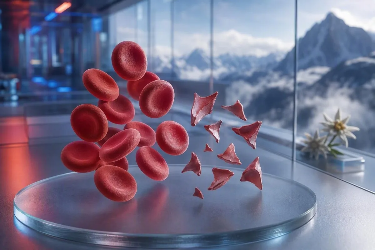
血液检查结果中的裂红细胞:何时涂片需要紧急处理

类别
文章
血液学 实验室解读 2026年更新 面向患者的说明

Schistocytes are fragmented red cells that can signal dangerous small-vessel clotting, but a single smear comment is not a diagnosis. Urgency depends on the fragment percentage, platelet count, haemoglobin trend, kidney function, haemolysis markers, and symptoms.

📖 ~11分钟 📅
📝 发表: 🩺 医学审阅: ✅ 基于证据
⚡ 快速概要 v1.0 —
  1. Schistocyte threshold: More than 1% schistocytes on a well-made adult peripheral smear is suspicious for thrombotic microangiopathy when other red-cell shape changes are absent.
  2. Urgent cluster: Schistocytes plus platelets below 150 × 10^9/L, falling haemoglobin, raised LDH, and low haptoglobin needs same-day clinician assessment.
  3. TTP clue: Severe ADAMTS13 activity below 10% supports immune thrombotic thrombocytopenic purpura, a condition treated before confirmatory testing returns when clinical suspicion is high.
  4. Not every fragment is dangerous: Mechanical heart valves, severe hypertension, dialysis circuits, burns, and sample damage can produce fragmented red blood cells.
  5. A smear is manual evidence: Automated analyser flags cannot reliably diagnose schistocytes; an experienced morphologist should inspect the slide.
  6. Pregnancy changes the stakes: Schistocytes with high blood pressure, protein in urine, or low platelets after 20 weeks require immediate obstetric assessment.
  7. Repeat testing helps: A freshly collected EDTA sample and repeat film can separate a collection artefact from true ongoing red-cell fragmentation.
  8. 不要自行治疗: Iron, folate, or supplements do not treat microangiopathic haemolysis and can distract from urgent evaluation.

When fragmented red cells need urgent medical review

Schistocytes need urgent same-day review when they appear with a low platelet count, anaemia, biochemical haemolysis, kidney injury, neurologic symptoms, fever, or severe hypertension. A smear comment alone can be benign or artifactual; the dangerous finding is the pattern. As of August 7, 2026, I would treat new schistocytes plus platelets below 100 × 10^9/L as a call-the-clinician-now result, not a routine follow-up.

Blood test results visualized as fragmented red cellular elements in a laboratory setting
图1: Fragmented cellular elements can indicate mechanical injury within the circulation.

A platelet fall is often the fastest urgency clue. A haemoglobin of 92 g/L with platelets of 68 × 10^9/L and newly reported schistocytes is substantially more concerning than 1% fragments with haemoglobin 145 g/L and stable platelets of 260 × 10^9/L. Kantesti is an AI blood test analyzer that reads this cluster across the complete blood count rather than elevating a morphology comment in isolation.

In my 15 years of clinical practice, the calls I make quickly are for patients whose results changed over days: falling haemoglobin, rising creatinine, and rising lactate dehydrogenase, or LDH. Dr. Thomas Klein's practical rule is simple: confusion, new weakness, chest pain, breathlessness, very little urine, or severe headache alongside this pattern means emergency assessment today.

Schistocytes are not a reason to wait for a repeat test if the person is unwell. A normal potassium or normal white count does not cancel the risk of thrombotic microangiopathy. Patients trying to understand a low count can also review our guide to low red blood cell symptoms, but acute symptoms always outrank online interpretation.

Call emergency services now for

New one-sided weakness, fainting, seizure, severe breathlessness, crushing chest discomfort, black stools, or rapidly worsening confusion require emergency services rather than a message to a clinic. These symptoms can reflect organ involvement, and no schistocyte percentage safely rules that out.

What schistocytes on a peripheral smear actually mean

Schistocytes are irregular red-cell fragments created when circulating cells are physically sheared, most often by fibrin strands or abnormal high-shear surfaces in small vessels. They typically have sharp angles, helmet-like forms, or small triangular shapes and lack the central pallor of an intact red cell.

Microscopic cell sample slide showing irregular fragmented red cellular elements
图2: Morphologists distinguish true fragments from crenated or overlapping cells.

True schistocytes are a morphology finding, not a value produced by the standard CBC analyser. The International Council for Standardization in Haematology recommends counting at least 1,000 red cells in the optimal smear zone when quantitation matters (Zini et al., 2021). That detail explains why one laboratory may report “rare fragments” while another reports a percentage.

The classic concern is microangiopathic haemolytic anaemia, where red cells meet platelet-rich microthrombi and fibrin in the circulation. Yet severe aortic stenosis, a mechanical valve, extracorporeal circuits, and major thermal injury can create genuine fragments through a different mechanism. The question is therefore not “Are fragments real?” but “What is doing the fragmenting?”

Kantesti is an AI blood test interpretation platform that can place an uploaded full blood count beside CBC 包含哪些内容 and highlight missing companion results such as bilirubin, reticulocytes, or haptoglobin. It cannot inspect the slide itself, which remains a trained laboratory professional's job.

How much schistocytosis is concerning on an adult smear

A schistocyte count above 1% on an adult peripheral smear is suspicious for thrombotic microangiopathy when fragments are the predominant red-cell abnormality. Counts below 1% do not exclude disease, especially if the film was made after fluids, transfusion, or an evolving illness.

Comparison of intact and fragmented red cellular elements for blood test results review
图 3: A one-percent threshold is meaningful only with high-quality morphology review.

The 1% cutoff is a diagnostic prompt, not a severity scale. ICSH guidance uses a reference interval below 1% for healthy adults and term newborns; preterm infants may have up to 5% fragments without the same implication (Zini et al., 2021). A result of 2% with severe thrombocytopenia is more alarming than 4% after placement of a mechanical circulatory support device.

Laboratories may report “occasional,” “few,” or “1–2 per high-power field” rather than a percentage. Those phrases are not interchangeable, because field size, smear thickness, and reporting policy vary. Asking whether the laboratory performed an ICSH-style percentage count is often more useful than trying to convert a descriptive phrase yourself.

High RDW does not measure schistocytes. RDW reflects variability in cell size and may rise with reticulocytosis, iron deficiency, transfusion, or fragmentation. Our detailed RDW and red-cell index guide explains why a high RDW supports neither TTP nor an artefact on its own.

典型的成人参考 <1% schistocytes Usually not significant if platelets and haemolysis markers are normal.
Borderline finding About 1% Review smear quality, other red-cell forms, symptoms, and CBC trend.
Suspicious pattern >1% with predominant fragments Evaluate urgently for thrombotic microangiopathy or a mechanical cause.
High-risk clinical cluster Any percentage plus falling platelets or organ injury Same-day medical assessment is appropriate regardless of the exact percentage.

The blood test pattern that supports true haemolysis

True red-cell fragmentation usually produces high LDH, low haptoglobin, increased indirect bilirubin, and a reticulocyte response alongside anaemia. No single marker is definitive, because liver disease, inflammation, recent transfusion, and marrow illness can distort the picture.

Cellular haemolysis pathway with fragmented red elements and bilirubin laboratory markers
图 4: Haemolysis releases LDH and consumes haptoglobin as fragments circulate.

LDH is sensitive but not specific. An LDH above 600 IU/L often supports active haemolysis in the right setting, yet muscle injury, liver injury, infection, and cancer can also raise it. Haptoglobin below the laboratory lower limit, commonly below 0.3 g/L, adds weight because it binds free haemoglobin released into plasma.

A reticulocyte count should usually rise during peripheral destruction, but it may remain low in folate deficiency, iron deficiency, kidney disease, or acute marrow suppression. That apparent contradiction is easy to miss: a patient can have genuine haemolysis and an inappropriately low reticulocyte response. The haptoglobin result guide covers the common interpretive traps.

Kantesti AI interprets fragmented red blood cell patterns by comparing haemoglobin, MCV, platelets, LDH, bilirubin, creatinine, and reticulocytes from the same time point. A falling haemoglobin of more than 20 g/L in 48 to 72 hours is clinically more informative than a mildly abnormal value with no prior comparison.

Why bilirubin can be normal

Indirect bilirubin may be normal early in haemolysis, after intravenous fluid dilution, or when the liver clears bilirubin efficiently. Normal bilirubin should therefore not override convincing fragments, a low haptoglobin, and falling haemoglobin.

Why TTP is treated as a hematology emergency

Thrombotic thrombocytopenic purpura, or TTP, is suspected when schistocytes accompany thrombocytopenia and microangiopathic haemolytic anaemia without a clearer alternative. Untreated immune TTP can progress rapidly, so clinicians may start plasma exchange and immune treatment before ADAMTS13 results return.

Urgent hematology review of laboratory sample findings associated with schistocytes
图 5: Rapid specialist review matters when thrombocytopenia and haemolysis coexist.

ADAMTS13 activity below 10% strongly supports TTP, but the result may take hours or days. The ISTH diagnostic guideline advises estimating pretest probability with clinical tools such as the PLASMIC score while obtaining a sample before plasma treatment (Zheng et al., 2020). A score is a bridge to action, not a substitute for specialist judgment.

The familiar “pentad” of fever, neurologic signs, renal impairment, thrombocytopenia, and haemolytic anaemia is incomplete in many real patients. George and Nester described thrombotic microangiopathy as a syndrome with overlapping causes rather than five mandatory boxes (George & Nester, 2014). In practice, waiting for all five features is a dangerous old habit.

Platelets below 30 × 10^9/L with haemolysis merit particularly urgent discussion with hematology. Platelet transfusion is usually avoided in suspected TTP unless there is life-threatening bleeding or an invasive procedure cannot wait. For related testing, our CBC explains why PT and aPTT may be normal in TTP.

Other dangerous causes of schistocytes doctors consider

Schistocytes also occur in disseminated intravascular coagulation, Shiga-toxin haemolytic uraemic syndrome, severe hypertension, pregnancy-related microangiopathy, certain medicines, and transplant-associated thrombotic microangiopathy. The associated platelet, coagulation, kidney, and clinical findings usually separate these conditions.

Small-vessel circulation illustration showing fragmented cellular elements and clotting activity
图 6: Different small-vessel disorders can produce similar fragments but different lab patterns.

DIC often combines fragments with prolonged PT, prolonged aPTT, low or falling fibrinogen, and markedly elevated D-dimer. TTP more often has relatively normal routine clotting times. A fibrinogen below 1.5 g/L in a sick patient shifts concern toward consumption coagulopathy, though fibrinogen can initially look normal because it rises during acute inflammation.

Severe hypertension can shear red cells when pressure injures the microvasculature; readings at or above 180/120 mmHg with headache, visual change, chest pain, or kidney dysfunction are an emergency. This is why a blood-pressure reading belongs beside the smear report. Patients can review secondary hypertension lab clues after immediate danger has been addressed.

Diarrhoea followed by reduced urine output, anaemia, and thrombocytopenia raises concern for haemolytic uraemic syndrome. Stool testing may help identify Shiga toxin, but hydration status, creatinine, potassium, and neurologic assessment guide urgent care first. A high lactate or unstable vital signs demand evaluation for sepsis and DIC rather than a wait-and-see approach; see these 脓毒症标志物模式.

Mechanical causes of fragmented red blood cells

Mechanical destruction can create real schistocytes without TTP, particularly with prosthetic heart valves, severe aortic stenosis, ventricular assist devices, dialysis circuits, or extracorporeal membrane oxygenation. These causes still deserve medical review, but the treatment targets the mechanical source rather than immune TTP.

Mechanical heart valve model beside a laboratory slide with fragmented cellular elements
图 7: High-shear cardiac devices can cause genuine red-cell fragmentation.

A mechanical-valve pattern often includes chronic mild LDH elevation and low haptoglobin with stable platelets. New anaemia, worsening breathlessness, dark urine, or a change in the valve sound can suggest paravalvular leak or device dysfunction and should prompt cardiology review. The smear provides a clue; echocardiography finds the source.

Athletes sometimes ask whether a marathon can explain fragments. Foot-strike haemolysis may cause transient free haemoglobin and low haptoglobin after long-distance running, but convincing schistocytosis is not something I would casually attribute to exercise—especially if platelets are falling. Compare it with the broader marathon runner laboratory guide.

Severe burns and major vascular surgery can produce fragmented cells through physical and endothelial injury. In those settings, a laboratory trend every 6 to 24 hours may be more revealing than one smear. A stable CBC after the inciting event is reassuring; a continuously falling haemoglobin is not.

How collection and slide artifacts mimic schistocytes

Poor sample collection, delayed smear preparation, underfilled anticoagulant tubes, and a damaged smear edge can create shapes that resemble schistocytes without reflecting circulation-level haemolysis. A repeat sample collected cleanly and reviewed in the monolayer is the usual first check when the clinical picture is calm.

Laboratory sample collection process showing EDTA handling for reliable blood test results
图 8: Correct anticoagulant fill and prompt slide preparation reduce morphology artefacts.

Crenated cells, smear-edge distortion, drying artefact, and overlapping cells are common false look-alikes. They often appear diffusely or only at the feathered edge rather than evenly in the ideal reading zone. An experienced morphologist distinguishes a true fragment's sharp geometry from a cell merely distorted during slide preparation.

A difficult collection can haemolyse the tube and falsely raise potassium or LDH, but it does not automatically produce true circulating schistocytes. If a result includes a haemolysis index or a “sample compromised” comment, clinicians commonly recollect before diagnosing disease. Our article on 钾采血错误 explains why the same pre-analytical issue can mislead several results at once.

EDTA-dependent platelet clumping can falsely lower the platelet count and manufacture an alarming-looking combination. A citrate tube platelet count, corrected for dilution, and a fresh film can uncover this. The nuance matters: artefact can coexist with a true illness, so a repeat test should never delay care for someone who is symptomatic.

Why analyser flags and laboratory wording can mislead

Automated blood analysers can flag abnormal red-cell populations, but they cannot independently diagnose schistocytes with enough reliability to rule in thrombotic microangiopathy. A manual peripheral smear review remains necessary when the clinical question is haemolysis or TTP.

Automated hematology analyser processing a cell sample slide for blood test results review
图 9: Automation identifies abnormal patterns but morphology confirmation remains manual.

A “fragmented red cells present” comment should prompt questions about quantity, smear quality, and coexisting morphology. Marked anisopoikilocytosis from iron deficiency, megaloblastic anaemia, or hereditary red-cell disorders makes the 1% rule less specific. Fragments are most diagnostically useful when they dominate the abnormal morphology rather than appearing among many unrelated shapes.

Kantesti is an AI lab test interpretation service designed to flag discordant combinations, such as a low platelet count beside a comment that says platelet clumps. It does not replace the laboratory's microscopic review or the clinician who can request a repeat slide. Our approach to quality checks is described in the 医学验证概览.

A delta check compares the current result with a prior result and can expose a sudden implausible change. Haemoglobin falling from 138 g/L to 87 g/L within 24 hours is clinically meaningful, but dilution from intravenous fluids or a mislabeled specimen remains possible. Read more about sudden laboratory changes.

What to do after blood test results mention schistocytes

Contact the clinician or service that ordered the test the same day if schistocytes are newly reported, and seek emergency care immediately if you have neurologic symptoms, breathlessness, chest pain, dark urine, severe headache, fever, or reduced urine output. Do not interpret the smear comment as a diagnosis by itself.

Diagnostic pathway objects arranged after abnormal blood test results showing schistocytes
图 10: A repeat smear and targeted haemolysis tests clarify the next clinical step.

Have the actual report available, including haemoglobin, platelets, creatinine, bilirubin, LDH, and any haemolysis index. Also note recent pregnancy, diarrhoeal illness, severe blood-pressure readings, new medicines, valve surgery, dialysis, transfusion, and autoimmune disease. These details alter the differential more than a single word in the morphology field.

I see patients lose valuable time by starting iron because they read “anaemia” before anyone identifies the cause. Iron deficiency may coexist, but it does not explain a rapid platelet decline with fragments. Dr. Thomas Klein recommends taking a photo of the full report rather than transcribing only highlighted numbers; transcription often drops the laboratory comment that makes the pattern coherent.

Avoid aspirin, ibuprofen, and non-prescribed anticoagulants until a clinician advises otherwise if platelets are very low or bleeding is present. Do not stop a prescribed anticoagulant without urgent medical guidance, either. People with fatigue or light-headedness can use our 用于头晕的血液检测指导 to prepare questions after urgent causes are excluded.

Schistocytes in pregnancy, newborns, and children

Schistocytes during pregnancy require urgent assessment when they occur with hypertension, proteinuria, elevated liver enzymes, low platelets, headache, visual symptoms, or upper abdominal pain. HELLP syndrome, pre-eclampsia, TTP, atypical HUS, and DIC can overlap, and timing around delivery helps but does not settle the diagnosis.

Kidney microvascular anatomy illustration relevant to pregnancy-related schistocyte blood test results
图 11: Kidney and microvascular involvement helps distinguish pregnancy-related microangiopathies.

HELLP syndrome is classically defined by haemolysis, elevated liver enzymes, and low platelets, often after 20 weeks of pregnancy or soon after delivery. A platelet count below 100 × 10^9/L or AST above 70 IU/L can support concern, but local obstetric teams interpret the whole clinical picture. Do not wait for a routine antenatal appointment with this combination.

Term newborns may have fewer than 1% schistocytes, whereas preterm infants can have up to 5% because neonatal red-cell morphology is different (Zini et al., 2021). In a child with diarrhoea, pallor, decreased urine, and thrombocytopenia, however, fragments may point toward HUS and need immediate paediatric assessment.

Reference intervals for platelets and haemoglobin change substantially by age and pregnancy stage. A result flagged “normal” by a generic app can still be clinically concerning in context. Our resources on pregnancy platelet rangessame-day pregnancy lab flags provide useful background, not a substitute for maternity triage.

Tests clinicians order to investigate true schistocytes

The first investigation set for suspected microangiopathic haemolysis usually includes a repeat CBC and smear, reticulocyte count, LDH, haptoglobin, bilirubin fractions, creatinine, urinalysis, PT, aPTT, fibrinogen, and D-dimer. ADAMTS13 activity and inhibitor testing should be drawn before plasma exchange if TTP is suspected, but treatment must not wait.

Haemolysis testing instruments and laboratory samples arranged for schistocyte evaluation
图 12: Complementary haemolysis, kidney, and clotting tests identify the underlying mechanism.

Direct antiglobulin testing, also called DAT or Coombs testing, helps distinguish immune haemolysis from non-immune fragmentation. A positive DAT does not exclude a second process, but classic TTP is typically DAT-negative. Urine microscopy can reveal protein or cellular casts when kidney microvascular injury is active; see our explanation of 颗粒状尿管型.

Stool Shiga-toxin testing is considered after a compatible gastrointestinal illness, while complement testing, drug review, and genetic or functional studies may be used for atypical HUS. The order varies by country and hospital. A creatinine increase of 26.5 µmol/L within 48 hours meets one acute kidney injury criterion and deserves attention even before it becomes a dramatic number.

A reticulocyte production index below 2 in significant anaemia suggests an inadequate marrow response, even if the raw reticulocyte percentage looks high. That calculation corrects for the lower red-cell mass in anaemia. The 网织红细胞与血液学指标解读指南 helps explain why percentages can be deceptive.

When repeating the smear is sensible and when it is not

Repeating a smear within hours to 24 hours is sensible when fragments are scant, the patient feels well, platelets and haemoglobin are stable, and sample quality is questioned. A repeat is not a safe delay when thrombocytopenia, active haemolysis, organ injury, or concerning symptoms are present.

Patient reviewing serial blood test results with a clinician in a laboratory setting
图 13: Serial results distinguish a stable artefact from evolving haemolysis.

A good repeat sample is freshly collected into a correctly filled EDTA tube and made into a film promptly. The request should specifically ask for review of suspected schistocytes and platelet clumping if either is plausible. A lab may compare both films side by side, which is far more useful than simply rerunning the automated CBC.

Trend direction matters. A platelet count moving from 210 to 165 to 98 × 10^9/L over three days is concerning even though the first two readings may fall within a local reference interval. Kantesti AI uses longitudinal context to surface these trajectories, a principle discussed in our 血液检测趋势分析指南.

Transfusion can temporarily dilute or obscure the original morphology. If you received red cells before the second sample, tell the team the date and number of units. Our guide to changing values after hospital discharge explains why post-treatment trends require a different baseline.

Conditions that can look similar without being schistocyte disease

Iron deficiency, vitamin B12 deficiency, liver disease, inherited red-cell disorders, and severe infection can create abnormal red-cell shapes without the specific fragmentation pattern of thrombotic microangiopathy. The smear's full morphology and the clinical timeline prevent overcalling fragments.

Watercolor anatomical illustration of bone marrow red-cell production and fragmented cell forms
图 14: Bone-marrow response and cell shape help separate production problems from destruction.

Severe vitamin B12 deficiency can cause pseudo-thrombotic microangiopathy with anaemia, high LDH, low platelets, and occasional fragments. It often produces marked macrocytosis, a low reticulocyte response, and LDH values sometimes above 2,500 IU/L—features that should trigger B12 and methylmalonic acid testing rather than automatic TTP treatment. Our B12 与叶酸指南中有覆盖。 explains this important mimic.

Iron deficiency usually produces microcytosis, hypochromia, pencil cells, and high RDW; fragments may be reported in severe cases but are rarely the dominant feature. A ferritin below 15 µg/L strongly supports depleted iron stores in an otherwise uncomplicated adult, although inflammation can mask deficiency by elevating ferritin.

In my experience, the most reassuring result is not “no schistocytes” alone but a coherent alternative explanation with stable platelets, stable kidney function, and no biochemical haemolysis. If the findings remain discordant, a hematologist may review the actual slide rather than relying on the final report wording.

Using an AI interpretation without missing a smear emergency

AI can help organize blood test results and identify dangerous combinations, but it cannot verify a schistocyte diagnosis from a CBC PDF or replace an urgent clinical assessment. A peripheral smear is visual microscopy, and the original laboratory slide remains the source of truth.

Kantesti is an AI-powered blood test analysis tool that maps related results across reports, including platelet trajectories, haemoglobin changes, creatinine, bilirubin, and LDH. That can reduce the common mistake of reading each abnormal marker separately. It cannot see whether a shape called a schistocyte is a true fragment, a smear-edge artefact, or a reporting convention.

Upload quality matters because an omitted unit or decimal can reverse the meaning of a result. Before sharing a report, check patient identifiers, collection date, units, reference ranges, and whether a result says “estimated,” “pending,” or “sample haemolysed.” Our AI 报告准确性核对清单 is useful for that final human check.

Kantesti's clinical methods are reviewed with input from our 医疗顾问委员会, and our technology is described in the AI解读技术指南. As Dr. Thomas Klein, I would still want a clinician to call a laboratory directly for a patient with new fragments, platelets below 100 × 10^9/L, and symptoms—because that is exactly when speed and clinical examination matter most.

常见问题

多少水平的裂红细胞是危险的?

在仔细复核的成人外周血涂片中,若见到超过1%的破碎红细胞(schistocytes),当破碎细胞是主要的红细胞异常时,提示血栓性微血管病(thrombotic microangiopathy)的可能性。只要在血小板计数低于150 × 10^9/L时出现任何比例的破碎红细胞,或伴随血红蛋白下降、高LDH、低haptoglobin、肾损伤或神经系统症状,则均属紧急情况。低于1%的结果并不能可靠地排除正在进展的病情。临床表现模式比单纯的百分比更能决定危险程度。.

贫血涂片中的裂细胞(schistocytes)会不会是由于抽血操作不当导致的?

一个困难的标本采集、制备延迟、EDTA管装量不足,或制作不良的涂片都可能产生类似裂红细胞(schistocytes)的细胞形态,尽管这并不会造成真正的循环性碎裂。实验室可通过重新采集的新鲜标本,并对最佳涂片区域进行人工复核来加以澄清。溶血性标本也可能会错误地升高钾和乳酸脱氢酶(LDH),从而使报告看起来更令人警惕。若存在症状、血小板低或贫血,则重复检测不应延误紧急评估。.

破碎红细胞是否总是意味着 TTP?

红细胞碎片(schistocytes)并不总是意味着血栓性血小板减少性紫癜(thrombotic thrombocytopenic purpura,TTP)。它们也可见于弥散性血管内凝血(DIC)、严重高血压、志贺毒素相关的溶血性尿毒综合征(Shiga-toxin HUS)、妊娠相关疾病、机械心脏瓣膜、透析管路以及严重组织损伤。当红细胞碎片与血小板减少和非免疫性溶血性贫血并存时,TTP 才会成为高优先级的关注点,尤其是在 ADAMTS13 活性低于 10% 时。由于 ADAMTS13 检测可能需要时间,疑似 TTP 在结果返回前需要立即进行专科评估。.

与裂红细胞溶血相关的血液检查中,哪些指标会升高?

真性碎片性溶血通常会升高LDH和间接胆红素,降低触珠蛋白,并在血红蛋白下降的同时触发网织红细胞计数增加。在活动性溶血中,LDH可能超过600 IU/L,但它并不特异,因为肌肉、肝脏和其他组织损伤也会升高LDH。当肝脏生成足够时,低于约0.3 g/L的触珠蛋白支持血管内溶血。肌酐、血小板计数、PT、aPTT、纤维蛋白原和D-二聚体有助于识别潜在病因。.

孕期出现裂红细胞正常吗?

当出现高血压、蛋白尿、肝酶升高、血小板下降、头痛、视力改变或上腹部疼痛时,裂红细胞不被视为常规的正常妊娠表现。该组合可能提示 HELLP 综合征、子痫前期、TTP、非典型 HUS 或 DIC,并需要立即进行产科评估。在此情境下,血小板计数低于 100 × 10^9/L 尤其令人担忧。妊娠特异性参考范围并不能使忽视溶血模式变得安全。.

机械心脏瓣膜会导致裂红细胞(schistocytes)吗?

机械心脏瓣膜和重度主动脉狭窄可导致真正的裂红细胞(schistocytes),因为高速剪切血流会在物理上碎裂红细胞。这常常引起轻度的慢性溶血,表现为乳酸脱氢酶(LDH)升高和结合珠蛋白降低,而血小板计数保持稳定。出现新的贫血、尿色变深、气促或瓣膜功能改变时需要进行心脏复查,因为可能是瓣膜渗漏或装置问题所致。该发现不应在未考虑心脏病史的情况下就自动标注为 TTP。.

立即获取AI驱动的血液检测分析

加入全球超过2,000,000名用户,他们信任Kantesti进行即时、准确的检验分析。上传你的血液检查结果,并在几秒内获得对15,000+生物标志物的全面解读。.

📚 参考研究论文

1

Klein, T., Mitchell, S., & Weber, H. (2026). Kantesti 血液检测解读引擎的预注册、基于量规的自动化技术基准评估:在 100,000 例合成测试病例上的表现. Kantesti AI医学研究。.

2

Klein, T., Mitchell, S., & Weber, H. (2026). 临床验证框架 v2.0(医学验证页面). Kantesti AI医学研究。.

📖 外部医学参考资料

3

Zini G et al. (2021). ICSH recommendations for identification, diagnostic value, and quantitation of schistocytes.。 《国际实验室血液学杂志》(International Journal of Laboratory Hematology)。.

4

George JN, Nester CM (2014). Syndromes of thrombotic microangiopathy.。 《新英格兰医学杂志》。.

5

Zheng XL et al. (2020). ISTH guidelines for the diagnosis of thrombotic thrombocytopenic purpura.。 《血栓与止血杂志》(Journal of Thrombosis and Haemostasis)。.

200万+测试分析
127+国家
75+语言

⚕️ 医疗免责声明

E-E-A-T信任信号

经验

由医生主导的临床审阅:实验室解读工作流程。.

📋

专业知识

实验室医学重点:生物标志物在临床情境中的表现。.

👤

权威

由 Thomas Klein 博士撰写,并由 Sarah Mitchell 博士与 Hans Weber 教授审阅。.

🛡️

可信度

基于循证的解读,并提供清晰的后续路径以减少警报。.

🏢 Kantesti LTD 在英格兰和威尔士注册 · 公司编号:. 17090423 英国伦敦 · kantesti.net
blank
作者:Prof. Dr. Thomas Klein

Thomas Klein 博士是获得美国内科医学委员会认证的临床血液科医生,担任 Kantesti AI 的首席医疗官。凭借超过 15 年的实验室医学经验,并对借助 AI 进行血液检查结果解读抱有浓厚兴趣,他致力于将新技术与日常临床实践相连接。他的研究兴趣包括生物标志物分析、临床决策支持研究以及针对特定人群的参考范围优化。作为 CMO,他为平台的内部基准评估提供临床输入,并对 Kantesti 教育报告的医疗质量提供临床监督。.

发表回复

您的邮箱地址不会被公开。 必填项已用 * 标注
