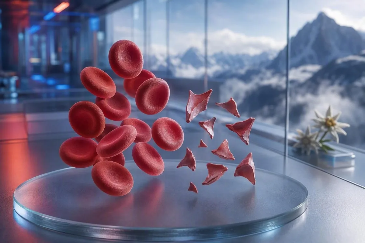
شیسیتسایتها گلبولهای قرمز تکهتکهشدهاند که میتوانند نشانهٔ لختهبندی خطرناک در رگهای کوچک باشند، اما یک اظهار نظر دربارهٔ یک اسمیر بهتنهایی تشخیص نیست. فوریت به درصد تکهتکهشدن، شمار پلاکت، روند هموگلوبین، عملکرد کلیه، نشانگرهای همولیز و علائم بستگی دارد.


- آستانهٔ شیسیتسایت: بیش از 1% شیسیتسایت در یک اسمیر محیطی بزرگسالِ بهخوبی تهیهشده، زمانی که تغییرات دیگری در شکل گلبولهای قرمز وجود ندارد، برای میکروآنژیوپاتی ترومبوتیک مشکوککننده است.
- خوشهٔ فوری: شیسیتسایتها بههمراه پلاکتهای کمتر از 150 × 10^9/L، هموگلوبینِ رو به کاهش، LDH بالا و هاپتوگلوبین پایین نیاز به ارزیابی همانروزهٔ پزشک دارد.
- سرنخ TTP: فعالیت شدید ADAMTS13 کمتر از 10%، پورپورای ترومبوتیک ترومبوسایتوپنی ایمنی را پشتیبانی میکند؛ وضعیتی که پیش از بازگشت نتایج تأییدی، زمانی که شک بالینی زیاد است، درمان میشود.
- هر تکهای خطرناک نیست: دریچههای مکانیکی قلب، فشار خون شدید، مدارهای دیالیز، سوختگیها و آسیب نمونه میتوانند گلبولهای قرمز تکهتکهشده ایجاد کنند.
- اسمیر یک شواهد دستی است: آنالایزر خودکار نمیتواند بهطور قابلاعتماد تشخیص بدهد که شیسیتسایتها واقعی هستند؛ مورفۆلۆژیستِ باتجربه باید لایحه (اسلاید) را بررسی کند.
- بارداری بهای ماجرا را بالا میبرد: شیسیتسایتها همراه با فشارخون بالا، پروتئین در ادرار، یا پایین بودن پلاکتها بعد از ۲۰ هفته نیاز به ارزیابی فوریِ مامایی دارد.
- تکرار آزمایش کمک میکند: نمونه تازه جمعآوریشده EDTA و تکرارِ لایحه میتواند یک آرتیفکتِ جمعآوری را از شکستگیِ واقعیِ در حالِ ادامه در گلبولهای قرمز جدا کند.
- خۆت بەخۆت چارەسەری مەکە: آهن، فولات، یا مکملها میکروآنژیوپاتیک همولیز را درمان نمیکنند و میتوانند از ارزیابی فوری منحرف کنند.
هنگامی که گلبولهای قرمز تکهتکهشده نیاز به بررسی فوری پزشکی دارند
شیسیتسایتها وقتی همراه با پایین بودن تعداد پلاکت، کمخونی، همولیز بیوشیمیایی، آسیب کلیه، علائم نورولوژیک، تب، یا فشارخون شدید ظاهر میشوند، نیاز به بازبینی فوری همانروزه دارند. فقط یک نظر روی اسمیر میتواند خوشخیم یا آرتیفکتی باشد؛ یافته خطرناک الگو است. از ۷ اوت ۲۰۲۶، من شیسیتسایتهای جدید همراه با پلاکتهای کمتر از ۱۰۰ × ۱۰^۹/L را «فوراً با پزشک تماس بگیرید» تلقی میکنم، نه پیگیری روتین.
افت پلاکت اغلب سریعترین سرنخِ فوریت است. هموگلوبین ۹۲ g/L با پلاکتهای ۶۸ × ۱۰^۹/L و شیسیتسایتهای تازه گزارششده بهطور قابلتوجهی نگرانکنندهتر از ۱TP54T قطعه با هموگلوبین ۱۴۵ g/L و پلاکتهای پایدار ۲۶۰ × ۱۰^۹/L است. Kantesti یک آنالایزر تست خونِ AI است که این خوشه را در سراسر CBC میخواند، نه اینکه بهتنهایی یک نظر مورفولوژی را بالا ببرد.
در ۱۵ سالِ طبابت بالینیام، فراخوانیهایی که سریع انجام میدهم برای بیمارانی است که نتایجشان طی چند روز تغییر کرده: افت هموگلوبین، بالا رفتن کراتینین، و بالا رفتن لاکتات دهیدروژناز، یا LDH. قانون عملیِ دکتر توماس کلاین ساده است: گیجی، ضعف جدید، درد قفسه سینه، تنگی نفس، مقدار خیلی کم ادرار، یا سردرد شدید همراه با این الگو یعنی ارزیابی اورژانسی امروز.
شیسیتسایتها دلیلی برای این نیست که منتظر تکرار آزمایش بمانید اگر فرد حالش خوب نیست. پتاسیم طبیعی یا شمارش گلبولهای سفید طبیعی خطر ترومبوتیک میکروآنژیوپاتی را لغو نمیکند. بیمارانی که میخواهند یک شمارش پایین را درک کنند نیز میتوانند راهنمای ما را مرور کنند تا علائمِ کمبود گلبول قرمز, ، اما علائم حاد همیشه از تفسیر آنلاین مهمتر است.
همین حالا با خدمات اورژانس تماس بگیرید برای
ضعف جدیدِ یکطرفه، غش، تشنج، تنگی نفس شدید، ناراحتی لهکننده در قفسه سینه، مدفوع سیاه، یا گیجیای که بهسرعت بدتر میشود نیاز به خدمات اورژانس دارد نه یک پیام به کلینیک. این علائم میتوانند درگیریِ اندامها را نشان دهند، و هیچ درصدی از شیسیتسایتها بهطور ایمن آن را رد نمیکند.
شیسیتسایتها در اسمیر محیطی دقیقاً چه معنایی دارند
شیسیتسایتها قطعات نامنظمِ گلبول قرمز هستند که وقتی سلولهای در گردش بهصورت فیزیکی بریده میشوند ایجاد میگردند؛ بیشتر اوقات توسط رشتههای فیبرین یا سطوح غیرطبیعیِ با برشِ بالا در رگهای کوچک. معمولاً زاویههای تیز دارند، شکلهای شبیه کلاهخود، یا شکلهای کوچکِ مثلثی و فاقد پالور مرکزیِ گلبول قرمزِ سالم هستند.
شیسیتسایتهای واقعی یک یافته مورفولوژی هستند، نه مقداری که توسط آنالایزر استاندارد CBC تولید میشود. شورای بینالمللی استانداردسازی در هماتولوژی توصیه میکند وقتی کمّیسازی مهم است، حداقل ۱۰۰۰ گلبول قرمز را در ناحیه بهینهِ اسمیر بشمارند (Zini et al., 2021). این جزئیات توضیح میدهد چرا یک آزمایشگاه ممکن است “قطعات نادر” گزارش کند در حالی که دیگری یک درصد گزارش میدهد.
نگرانی کلاسیک این است که کمخونی همولیتیک میکروآنژیوپاتیک, ، جێیەوە کە سلولە سوورەکان لەگەڵ میکرۆترۆمبۆسی پڕ لە پلاتێت و فیبرین لە خوێنەکەدا دەکەون. بەڵام توندی تنگی ئاۆرتا، ڤاڵڤێکی مەکانیکی، دەستگاهە دەرهێنراوەکان لە دەورەوەی خوێن (extracorporeal circuits)، و تێکچوونی گەورەی گەرمەیی دەتوانن بە شێوەیەکی جیاواز ڕاستەوخۆ تکهتکهبوونە ڕاستەقینە دروست بکەن. بۆیە پرسیارەکە نە “ئایا تکهکان ڕاستن؟” بەڵکو “چییە کە تکهتکهکردنەکە دەکات؟”
Kantesti پلاتفۆرمێکی تێستکردنی خوێنە بە AI ـە کە دەتوانێت کۆنتڕۆڵی تام وەک full blood count ـەکە لەکنار ئەوەی CBC پێکدێت ڕوون بکات و ئەنجامە هاوڕێیە گمشەکراوەکان وەک bilirubin، reticulocytes، یان haptoglobin پیشان بدات. ناتوانێت خودی شلەیدەکە ببینێت، کە ئەمە کارێکی پڕۆفیشناڵی لابراتۆریای تەمێندراوە.
چه میزان شیسیتوکیتوز در اسمیر بزرگسالان نگرانکننده است
ژمارەی schistocyte ـەکان لەسەر 1% لە پێریفیڕی شلەیدەکەی (peripheral smear) بۆ ئادڵت، کاتێک تکهکان سەرەکیترین کێشەی ناسازگاری سلولی سوورەکان بن، مشکوکە بۆ thrombotic microangiopathy. ژمارە لە خوار 1% نەهێڵێت نەبوونی نەخۆشی ڕەت بکرێت، بە تایبەتی ئەگەر شلەیدەکە لەدوای مایعات، transfusion، یان نەخۆشییەکە کە لە حال گەشەکردنەوە بوو دروست کرا بێت.
کاتئۆف 1% ئاگادارکردنەوەی دایگنۆستیکە، نەوەک کەڵکی سەختی (severity scale). ڕێنمایی ICSH بازەیەکی وەک reference interval ـی لە خوار 1% بۆ ئادڵتێکی تەندروست و نۆزەدی ترم (term newborns) بەکاردێنێت؛ نۆزەدی نێوەترم (preterm infants) دەتوانن تا 5% تکه هەبێت بەبێ ئەوەی هەمان مانای هەستیار هەبێت (Zini et al., 2021). ئەنجامێکی 2% لەگەڵ thrombocytopenia ـی توند، زیاتر هەستیارترە لە 4% دوای دانانی دەستگاهێکی پشتیوانی مەکانیکی بۆ دەوران (mechanical circulatory support device).
لابراتۆریاکان دەتوانن “occasional”، “few”، یان “1–2 per high-power field” بگزارن بەجای ڕێژە. ئەو وشانە یەکسان نییە، چونکە کەڵکی قەبارەی فیلد، قەبارەی نازکی/قەڵەبوونی شلەیم (smear thickness)، و سیاسەتی گەزارشکردن جیاوازن. پرسیارکردن ئەوەی لابراتۆریا ڕێژەیەکی وەک ICSH ـی شێوەیەکی کۆنتڕۆڵی کردووە، زۆرجار بە سودترە لەوەی کە خۆت بە تەنها دەستکاری/گۆڕینی وشەیەک بکەیت.
RDW ـی بەرز schistocytes ناپێمێرێت. RDW دەربڕی گۆڕاوایی لە قەبارەی سلولەکانە و دەتوانێت لەگەڵ reticulocytosis، iron deficiency، transfusion، یان تکهتکهبوون بەرزبێت. ڕوونکردنەوەی راهنمای RDW و red-cell index دەڵێت بۆچی RDW ـی بەرز بە تەنها پشتیوانی ناکات بۆ TTP یان هەروەها artefact.
الگوی آزمایش خون که همولیز واقعی را پشتیبانی میکند
تکهتکهبوونی ڕاستەقینەی سلولە سوورەکان زۆرجار LDH ـی بەرز، haptoglobin ـی کەم، زیادبوونی indirect bilirubin، و وەڵامێکی reticulocyte لەگەڵ anaemia دروست دەکات. هیچ نشانگر واحدی قطعی نیست، چون بیماری کبد، هەڵسوڕان/التهاب، وەرگرتنی تازەی خوێن، و نەخۆشیی مغز استخوان دەتوانن وێنەکە دەستکاری بکەن.
LDH بەهێزە لە حەساسیت، بەڵام تایبەتمەندی نییە. LDH ـی زیاتر لە 600 IU/L زۆرجار پشتیوانی دەکات بۆ هەموڵیزێکی کاریگەر لەو کاتەی ڕاستدا، بەڵام تێکچوونی ماسیچه، تێکچوونی کبد، هەڵچوونی نەخۆشی/هەڵوەشاندن (infection)، و سەرتانیش دەتوانن هەمانە بەرز بکەن. هەپتوگلوبین لە سەرحدی کەمترین لابراتۆریا، زۆرجار لە خوارەوەی 0.3 g/L، گرنگی زیاتر دەدات چونکە دەبەستێت بە هەموگلوبینی ئازاد کە لە پلاسما دەردەکەوێت.
شەماری رتیکولۆسایت (reticulocyte count) زۆرجار دەبێت لە ماوەی نابەدەستبوونی دەوروبەری (peripheral destruction) بەرز بێت، بەڵام دەتوانێت لە کەمبود فۆلات، کەمبود ئایرون، نەخۆشیی کەلیە (kidney disease)، یان سڕینەوەی توندی مغز استخوان (acute marrow suppression) کەم بمێنێت. ئەم ناسازگارییە بەدەر لە هەستیاریدا دەتوانرێت بە ئاسانیت لەبەرچاو بمانێت: نەخۆش دەتوانێت هەموڵیزێکی ڕاستەقینە هەبێت و هەڵسەنگاندنێکی نادروست/نەهەماهنگ لە وەڵامدانەوەی رتیکولۆسایت. ڕێنمایی ئەنجامی هەپتوگلوبین دەربارەی دامەزراندنە زۆر بەکارهاتووەکان لە تێکچوونی تفسیر.
Kantesti AI وێنەی تکهبوونی خولی خوێنی سوور (red blood cell) تێکدەدات بە بەراوردکردنی هەموگلوبین، MCV، پلیتڵت (platelets)، LDH، بیلیروبین، کرێاتینین، و رتیکولۆسایت لە یەک کات/نێوانەوەی هەمان سەردەم. کەمبوونێکی هەموگلوبین بە زیاتر لە 20 g/L لە 48 تا 72 کاتژمێر لە ڕوونکردنەوەی پزیشکییەوە زانیارییەکی گرنگترە لەوەی بەهایەکی تەواو نەگونجاو بەهێزەوە کە پێشتر بەراوردی لەگەڵی نەکراوە.
بۆچی بیلیروبین دەتوانێت ڕاست/نۆرمال بێت
بیلیروبینی ناڕاستەوخۆ (indirect bilirubin) زۆرجار لە سەرەتای هەموڵیزدا دەتوانێت نۆرمال بێت، یان لەدوای رقیقکردنی خوێن بە مایعی وەریدی (intravenous fluid dilution)، یان کاتێک کبد بیلیروبین بە شێوەی بەهێز پاک دەکات. بۆیە بیلیروبینی نۆرمال نابێت تێکچوونی قانعکەر لە تکهکان، هەپتوگلوبینی کەم، و کەمبوونی هەموگلوبین لەبەرچاو بگرێت.
چرا TTP بهعنوان یک فوریت هماتولوژی درمان میشود
پورپورای تومبۆتیک تومبۆسایتۆپێنیک (thrombotic thrombocytopenic purpura) یان TTP پێشنیار دەکرێت کاتێک schistocytes لەگەڵ تومبۆسایتۆپێنیا (thrombocytopenia) هەبن و هەموڵیزێکی نەخۆشیی خوێنی کە بچووک-رەگ/مایکروئانجیوپاتی (microangiopathic haemolytic anaemia) هەبێت بەبێ ئەوەی هۆکارێکی ڕوونتر بۆ دابنرێت. TTP ـی ئیموون (immune) کە نەدرمانکراوە دەتوانێت بە خێرایی پێش بڕوات، بۆیە پزیشکان دەتوانن دەست بە پێداچوون/جێبەجێکردنی پەڕەوەگرتنی پلاسما (plasma exchange) و درمانی ئیموونی بکەن پێش ئەوەی ئەنجامی ADAMTS13 بگەیەنێت.
کارییەتی ADAMTS13 لە خوارەوەی 10% بە شێوەی زۆر قووڵ پشتیوانی دەکات بۆ TTP، بەڵام ئەنجامەکە دەتوانێت ماوەی چەند کاتژمێر یان ڕۆژ بگرێت. ڕێنمایی دۆزینەوەی ISTH دەسپێرێت بۆ برآوردکردنی بە احتمالە پێشینە (pretest probability) بە بەکارهێنانی ئامرازە کلینیکی وەک PLASMIC score لە کاتێکدا نمونە دەگرێت پێش درمانی پلاسما (Zheng et al., 2020). نمرەکە پلەی دەستپێکردنە بۆ کردارە، نەک جێگرەوەی قضاوتی تایبەتمەند.
“پێنجەکە”ی ناسراو (pentad) ـی تێبەردان/تەمەنی (fever)، نیشانەکانی نەورۆلۆجی (neurologic signs)، کەمبوونی کارکردی کەلیە (renal impairment)، تومبۆسایتۆپێنیا، و ئانەمی هەموڵیتیک (haemolytic anaemia) لە زۆربەی نەخۆشە ڕاستەقینەکان ناکاملە. George و Nester توصیف کرد TTP ـی مایکرۆئانجیوپاتی (thrombotic microangiopathy) وەک سەندرومێک کە هۆکارە هاوپێچ/هاوکێشەکان لەگەڵ یەکدی دەبەستێت، نەک پێنج خانەی بەهێز/واجب (George & Nester, 2014). لە کرداردا، وەستان بۆ ئەوەی هەموو پێنج تایبەتمەندییەکە بەدەست بهێنێت عادتێکی خەتاکارەی کۆنە.
پلیتڵت لە سەر 30 × 10^9/L ــەوە کەمتر لەگەڵ هەموڵیز، پێویستە بە تایبەتی گفتوگۆی هەرە گرنگ لەگەڵ هیماتۆلۆژی (hematology) بکرێت. وەرگرتنی پلیتڵت زۆرجار لە کاتی پێشبینی TTP دەبێت بەدوور بخرێت، مەگەر ئەگەر خوێنڕشتنی هەڕەشەدار بژێوی (life-threatening bleeding) هەبێت یان ئەنجامدانی ڕووداوێکی جراحی/دەستکاری (invasive procedure) نەکاتێکی درێژ دەوێت. بۆ ئەزمونە پەیوەندیدارەکان، ئەمانە ڕێنمای تاقیکردنەوەی coagulation دەڵێت بۆچی PT و aPTT دەتوانن لە TTP ـدا نۆرمال بن.
سایر علل خطرناک شیسیتسایتها که پزشکان در نظر میگیرند
Schistocytes هەروەها لە DIC (disseminated intravascular coagulation)، سەندرۆمی هەموڵیتیک یورێمیک بە تۆکسینی Shiga (Shiga-toxin haemolytic uraemic syndrome)، بەرزی توندی خونی (severe hypertension)، مایکرۆئانجیوپاتی پەیوەندیدار بە منداڵبوون/حامڵبوون (pregnancy-related microangiopathy)، بەعضی دارو/پزشکییەکان، و TMA ـی پەیوەندیدار بە وەرگێڕانی ئەنترنەوە (transplant-associated thrombotic microangiopathy) ڕوو دەدەن. ئەو نیشانە کلینیکی و ئەنجامە پەیوەندیدارە لە پلیتڵت، ڕێکخستنی خوێن (coagulation)، کەلیە، و دۆزینەوەی کلینیکی زۆرجار ئەم هەڵکەوتانە جیا دەکاتەوە.
DIC زۆرجار تکهکان لەگەڵ PT ـی درێژکراو، aPTT ـی درێژکراو، fibrinogen ـی کەم یان کەوتوو، و D-dimer ـی بەهێز بەرزبووەوە (markedly elevated) یەکدەگرێت. TTP زۆرجار کاتێکی کەمتر هەیە کە ڕێکخستنی خوێنی روتین (routine clotting times) بە شێوەی نسبی نۆرمال بمێنێت. fibrinogen لە خوارەوەی 1.5 g/L لە نەخۆشێکی نەخۆش/بیمار (sick patient) دڵنیایی زیاتر دەکاتەوە بۆ coagulopathy ـی بەکارهاتوو لە خوارەوەی بەکارهێنانی fibrinogen (consumption coagulopathy)، بەڵام fibrinogen دەتوانێت لە سەرەتادا نۆرمال بنوێنێت چونکە لەدوای هەڵسوڕانی توند (acute inflammation) بەرز دەبێت.
بەرزی توندی خونی دەتوانێت خوێنی سوور تکهتکه بکات کاتێک فشاری خونی زیان بە مایکرۆڤەسکولە (microvasculature) دەگەیەنێت؛ خوێنسنجی لە یان زیاتر لە 180/120 mmHg لەگەڵ سەردرد، گۆڕانی بینایی، دڵدرد/خەنجەرەوەی سینه (chest pain)، یان نەخۆشیی کارکردی کەلیە (kidney dysfunction) هەنگاوێکی هەڕەشەدارە و فوریتە. بۆیە خوێنسنجی دەبێت لەگەڵ ڕاپۆرتی smear ـەکەدا بێت. نەخۆشان دەتوانن لەگەڵ ئەوەدا پێداچوون بکەن سرپۆرتەی پێوەندیدار بە پەرفشارەی دووەم (secondary hypertension) ڕێنمایی لابراتۆری دوای چارەسەری خەتری ڕاستەوخۆ.
ئاسهڵانەوە (Diarrhoea) بەدوای کەمبوونەوەی دەرهێنانی ئاوە (reduced urine output)، هەڵوەشاندنەوەی خونی (anaemia)، و کەمبوونەوەی پلاتێڵت (thrombocytopenia) هەستیار دەکات بۆ هەڵوەشاندن-کلیەیی (haemolytic uraemic syndrome). تاقیکردنەوەی نێوەڕاستی (Stool testing) دەتوانێت یارمەتیدەر بێت بۆ دۆزینەوەی Shiga toxin، بەڵام پێوەری ڕەوشتی ڕوونکردنەوە (hydration status)، کرێاتینین (creatinine)، پۆتاسیم (potassium)، و بەڕێوەبردنی سەرووەری (neurologic assessment) پێشتر ڕێنمایی بۆ خزمەتگوزاری فورس (urgent care) دەکەن. لاکتیاتێکی بەرز (high lactate) یان وەضعی ڤایتال ناپایدار (unstable vital signs) پێویستی بە بەدواداچوون بۆ sepsis و DIC هەیە، نەک چارەسەری “ببینین چی دەبێت”؛ ببینە پاتێرنەکانی نیشانەی sepsis.
علل مکانیکیِ ایجاد گلبولهای قرمز تکهتکهشده
هەڵوەشاندنی مەکانیکی دەتوانێت شێستۆسایتەی ڕاستەقینە دروست بکات بەبێ TTP، بە تایبەتی لەگەڵ ڤاڵڤی دڵی پڕۆستێتیکی، تنگی سەختی ئاۆرتا (severe aortic stenosis)، دەستگاهەکانی یارمەتیدانی وەنتریکڵ (ventricular assist devices)، ڕێگا/سەرکێشی دیالیز (dialysis circuits)، یان ئووکسیجێنەکردنی بەدەستکاری دەرەکی (extracorporeal membrane oxygenation). ئەم هۆکارانە هێشتا پێویستی بە ڕەخنەی پزیشکی هەیە، بەڵام چارەسەکەکە دەبێت سەرچاوەی مەکانیکی هەدف بگرێت نەک TTPی دەستەواژەیی (immune TTP).
ڕێکخستنی (pattern) ڤاڵڤی-مەکانیکی زۆرجار تێکەڵەی بەرزبوونی LDHی کەم و درێژخایەن (chronic mild LDH elevation) و کەمبوونی haptoglobin لەگەڵ پلاتێڵتی بەهێز/پایدار (stable platelets) دەبینێت. ئاناemiaی نوێ، کەمبوونەوەی هەناسەڕەشبوونی (worsening breathlessness)، ئاوەی تاریک (dark urine)، یان گۆڕانکاری لە دەنگی ڤاڵڤ (change in the valve sound) دەتوانێت هەواڵ بدات بۆ نشتکردنی پاراڤاڵڤۆلار (paravalvular leak) یان ناکارایی دەستگاه و پێویستە سەردانی کۆنسڵتاسیۆنی کاردیۆلۆژی (cardiology review) بکات. سمیەر (smear) کلیلێک دەدات؛ ئەکۆکاردیۆگرافی (echocardiography) سەرچاوەکە دەدۆزێتەوە.
وەرزشکاران هەندێک جار پرسیاری ئەوە دەکەن کە آیا ماراتۆن دەتوانێت تکهکان (fragments) ڕوون بکات. هەڵوەشاندنی خۆڵەکە لە پێ (foot-strike haemolysis) دەتوانێت بەردەوامی خۆڵەکەی درێژمدت (long-distance running) هەموارێکی کاتی (transient) لە free haemoglobin و کەمبوونی haptoglobin دروست بکات، بەڵام شێستۆسایتۆزی ڕاستەقینە و قانعکەر (convincing schistocytosis) شتێک نییە کە من بە ئاسانی بە وەرزش نسبت بدەم—بە تایبەتی ئەگەر پلاتێڵت دەکەوێت. لەگەڵ ڕێنمایی لابراتۆریی ماراتۆن-ڕانەر (marathon runner).
سووتاندنی سەخت (severe burns) و جراحی گەورەی وەسەکانی (major vascular surgery) دەتوانن سلۆڵە تکهتکهکراوە (fragmented cells) دروست بکەن لە ڕێگەی زیانی فیزیکی و ئاندۆتێلی (endothelial injury). لە ئەم دۆخەدا، کەشەی ڕێژەیی لابراتۆری (laboratory trend) هەر 6 تا 24 کاتژمێر جارێک، لە یەک سمیەر زیاتر ڕوونکەرەوە دەبێت. CBCی بەهێز/پایدار (stable CBC) دوای ڕوودانی سەرەکی (inciting event) دڵخۆشکەرە؛ هەڵوەشاندنی هەموواری (continuously falling haemoglobin) دڵخۆشکەر نییە.
چطور جمعآوری نمونه و خطاهای اسلاید میتوانند شیسیتسایتها را شبیهسازی کنند
کەمبوونەوەی کۆکردنەوەی نموونە (Poor sample collection)، دوادەستکردنی ئامادەکردنی سمیەر (delayed smear preparation)، تیوپەکان/توبەکانەی پڕنەکردووە لە ئانتیکۆاگولانت (underfilled anticoagulant tubes)، و لەبەرچاوگرتنی لبهکانی سمیەر (damaged smear edge) دەتوانن شێوەهایی دروست بکەن کە شبیه شێستۆسایتەکان بن بەبێ ئەوەی هەڵوەشاندنی خونی لە ڕێگەی سەرکەوتن (circulation-level haemolysis) پیشان بدەن. نموونەی دووبارە کە بە ڕێک و پاکی کۆکراوە و لە monolayerدا سەیری کراوە، زۆرجار یەکەم چێکی ڕاستەقینەیە کاتێک وەضعە بالینی آرامە.
سلۆڵی crenated، کێشانی لبهکانی سمیەر (smear-edge distortion)، ئاساری خشکبوون (drying artefact)، و سلۆڵی یەکدەست/هاوپێچ (overlapping cells) زۆرجار شبیهسازییەی هەڵە (common false look-alikes) دەبن. زۆرجار بە شێوەی گشتی (diffusely) یان تەنها لە لبهکەوتنی پەڕەدار (feathered edge) دەبینن، نەک بە یەکسانی لە ناوچەی خوێندنی ئایدیال. مورفۆلۆژیستێکی بەهێز (experienced morphologist) جیا دەکاتەوە لە نێوەڕاستی هندسیی تکهی ڕاستەقینە (true fragment's sharp geometry) لەوەی کە سلۆڵ تەنها لە کاتی ئامادەکردنی سڵاید (slide preparation) تێکچووە.
کۆکردنەوەی سەخت دەتوانێت تیوپەکە هەڵوەشێنێت (haemolyse) و بە هەڵە پۆتاسیم یان LDH بەرز بکات، بەڵام بە شێوەی خۆکار (automatically) شێستۆسایتەی ڕاستەقینەی سەرکەوتوو (true circulating schistocytes) دروست ناکات. ئەگەر ئەنجامەکە تێکەڵی “haemolysis index” یان وتاری “sample compromised” بێت، پزیشکان زۆرجار نموونەی دووبارە کۆدەکەن پێش ئەوەی نەخۆشی دابنێن. وتارەکەمان لەسەر هەڵەکانی کێشانی potassium ڕوون دەکات کە چۆن ئەو هەمان کێشەی pre-analytical دەتوانێت هەندێک ئەنجامی تر هەمان کات گمراه بکات.
کۆمەڵبوونی پلاتێڵت بەهۆی EDTA (EDTA-dependent platelet clumping) دەتوانێت بە هەڵە پلاتێڵتی کەمتر پیشان بدات و یەک کۆمەڵەی ترسناک لە شێوەی ئەنجام دروست بکات. تاقیکردنەوەی پلاتێڵت لە تیوپی citrate (citrate tube platelet count)، بە پێویستی ڕاستکردنەوە بۆ رقیقبوون (corrected for dilution)، و فلیمێکی نوێ (fresh film) دەتوانێت ئەمە ڕوون بکات. گرنگی ئەو ڕیزەییە (nuance) هەیە: ئاسار (artefact) دەتوانێت لەگەڵ نەخۆشی ڕاستەقینە هاوکات بێت، بۆیە تاقیکردنەوەی دووبارە نابێت چارەسەری کەسێک کە نەخۆشی/نیشانەی هەیە (symptomatic) دوادەخات.
چرا هشدارهای آنالایزر و واژگان آزمایشگاه میتوانند گمراهکننده باشند
ئانالیزەری خوێنی خۆکار (Automated blood analysers) دەتوانن کۆمەڵە/پۆپەڵەی ڕەخنەدار لە سلۆڵی سوور (abnormal red-cell populations) ڕابکەن، بەڵام ناتوانن بە خۆیەوە شێستۆسایتەکان بە ڕێژەی ڕاستییەکی کافی دابنێن بۆ ئەوەی thrombotic microangiopathy (TMA) ڕاست بکەنەوە. سەیری سمیەری دەستی لە قەبارەی دەور (manual peripheral smear review) هێشتا پێویستە کاتێک پرسیارە بالینیەکە لەسەر haemolysis یان TTP ـە.
کامێک “خەڵەکانی سوورە تێکچووە (fragmented red cells present)” دەبێت هۆکار بۆ پرسیارکردن لەسەر کەمیەت، ڕەوانی (quality) لەسەر ڕەسەمی (smear)، و جۆری مۆرفۆلۆژیی هەبوو لەگەڵ خۆیدا دروست بکات. ئانیسۆپویکیلو سیتۆز (anisopoikilocytosis) بەهێز لەبەر کمبودی ئاسن، ئەنێمیای مێگالۆبلاستیک (megaloblastic anaemia)، یان نەخۆشییە ڕەگەکییەکانی خەڵەکانی سوور (hereditary red-cell disorders) دەبێت قاعدەی 1% کەمتر تایبەتمەند بکات. تێکچووەکان (fragments) زۆرترین بەکارهێنانی دیانۆستیکییان هەیە کاتێک دەبن بەسەر مۆرفۆلۆژیی نادرووستدا، نەک ئەوەی لە نێوان زۆر شێوەی جیاوازدا تەنها یەکێک بن.
Kantesti خزمەتگوزاری وەڵامدان/تێکست-خوێندنەوەی تاقیکردنەوەی لابراتۆرییە بە شێوەی AI ــە کە دەتوانێت جۆرە ناسازگارییەکان دەستنیشان بکات، وەک کەمبوونەوەی ژمارەی پلیتڵەکان لەگەڵ وتارێک کە دەڵێت “پلیتڵ کڵامپ (platelet clumps) هەن”. ئەمە جێگای ڕەوی لەسەر میکروسکووپی لابراتۆرییەکە ناکات یان جێگای پزیشک/کلینیسین ناکات کە دەتوانێت داوای دووبارە ڕەسەمی بکات. ڕێوشوێنی چێککردنی ڕەخنە (quality checks) لە ڕوونکردنەوەی تائیدکردنی پزیشکی.
دڵتا چێک (delta check) ئەنجامە ئێستا لەگەڵ ئەنجامێکی پێشوو بەراورد دەکات و دەتوانێت گۆڕانێکی ناگهانی و بێباوەڕ (implausible) ڕوون بکاتەوە. هێموگلوبین کە لە 138 g/L بۆ 87 g/L لە ماوەی 24 کاتژمێر کەم دەبێت، لە ڕووی کلینیکییەوە گرنگە، بەڵام هەڵکەوتن/رقاندن (dilution) لە ڕێگای مایعی ناوەوەی وریدی (intravenous fluids) یان نموونەیەک کە بە نادروستی ناونیشان کراوە (mislabeled specimen) هێشتا دەتوانێت هۆکار بێت. زانیاری زیاتر لەسەر گۆڕانە ناگهانییەکانی لابراتۆری.
بعد از اینکه نتایج آزمایش خون به شیسیتسایتها اشاره کرد چه باید کرد
ئەگەر schistocytes تازە ڕاپۆرت کراون، هەمان ڕۆژ پەیوەندی بکە بە کلینیسین یان خزمەتگوزارییەکە کە تاقیکردنەوەکە داوا کردووە، و ئەگەر هەستیاریت هەیە بە ئامرازەوەی نەورۆلۆجی (neurologic symptoms)، تنگی هەناسە (breathlessness)، دڵدرد/دڵتنگی (chest pain)، ئاوەی تۆخ (dark urine)، سەردردی بەهێز (severe headache)، هەستە (fever)، یان کەمبوونی دەرچوونی ئاو (reduced urine output)، فوراً بە خزمەتی یارمەتیدانی هەنگامی (emergency care) پەیوەندی بکە. ڕەسەمی-وتار (smear comment) بە تەنها وەک دیانۆس (diagnosis) تێنەفسرە.
ڕاپۆرتی ڕاستەقینە هەڵبگرە، لەوانە هێموگلوبین، پلیتڵەکان، کرێاتینین (creatinine)، بیلیروبین (bilirubin)، LDH، و هەر شاخصێکی هەڵهەڵدان/هێماڵیز (haemolysis index). هەروەها سەرنج بدە بە حاڵەتی حەملی تازە، نەخۆشییەکی لەدەستدان/ڕێژەی ڕەش (diarrhoeal illness)، ڕەخنەی بەهێزی فشاری خوون (severe blood-pressure readings)، دارووی نوێ، جراحییەکی ڕێگاکان/ڤاڵڤ (valve surgery)، دیالیز (dialysis)، ترانسفۆژن (transfusion)، و نەخۆشییە خودکار-ئیمون (autoimmune disease). ئەمانە زۆرتر لەوەی یەک وشە لە خانەی مۆرفۆلۆژی دەستکاری دەکەن لەسەر جیاوازیی (differential).
من دەبینم کەسێک زەمانێکی گرنگ لەدەست دەدەن بە دەستپێکردنی ئاسن، چونکە “anaemia” دەخوێنن پێش ئەوەی کەسێک هۆکارەکە دەستنیشان بکات. کمبودی ئاسن دەتوانێت لەگەڵ خۆیدا هەبێت، بەڵام وەک هۆکارێکی ڕوونی کەمبوونەوەی خێرا لە پلیتڵەکان لەگەڵ تێکچووەکان ناکات. د. توماس کلاین (Dr. Thomas Klein) پێشنیار دەکات وێنەی (photo) لەسەر ڕاپۆرتی تەواو بگرێت بەڵام تەنها ژمارە ڕەنگاوە/ڕوونکراوەکان (highlighted numbers) نەنووسێت؛ نووسین/ترانسکرایب (transcription) زۆرجار وتاری لابراتۆری دەکاتەوە کە ڕێکخستنی پاترنەکە (pattern) یەکدەکات.
تا کاتێک کلینیسین ڕێنمایی تر نەدات، ئاسپرین (aspirin)، ئایبوپروفێن (ibuprofen)، و ئانتی-کۆاگولانتی (anticoagulants) بە شێوەی نەخوازراو/بە دەستپێنەکراو (non-prescribed) بەکار مەهێنە ئەگەر پلیتڵەکان زۆر کەم بن یان خوێنڕشتن هەیە. هەرگیز ئانتی-کۆاگولانتییەکەی پێشتر پێنەدراوە (prescribed anticoagulant) بە تەنها بەبێ ڕێنمایییەکی پێشەنگی/هەنگامی (urgent medical guidance) نەبڕە. کەسانە کە هەست بە خەستەگی (fatigue) یان سەرگیجی/سبەیی (light-headedness) دەکەن دەتوانن بەکاربهێنن د سرګرځېدو (dizziness) لپاره د وینې ازموینې لارښود بۆ ئامادەکردنی پرسیارەکان پاش ئەوەی هۆکارە هەنگامییەکان لەسەرەوە ڕەخنە/سەلمێنراون.
شیسیتسایتها در بارداری، نوزادان و کودکان
Schistocytes لە کاتی حەملبوون (pregnancy) پێویستی بە ئارزیابییەکی هەنگامی هەیە کاتێک لەگەڵ هەیبوونی هەڵسەنگاندنی فشاری خوون (hypertension)، پروتێنۆریا (proteinuria)، بەرزبوونی ئەنزایمەکانی کبد (elevated liver enzymes)، پلیتڵە کەمەکان (low platelets)، سەردرد، ئامرازە بینایی (visual symptoms)، یان ئاڵمانی سەرەوەی شکم (upper abdominal pain) ڕوون دەبنەوە. نەخۆشی HELLP، پێش-ئێکلامپسیا (pre-eclampsia)، TTP، HUS ـی تایپیک-نەبوو (atypical HUS)، و DIC دەتوانن یەکدیگر پێکەوە بپۆشن، و کات لە نزیکبوونەوەی لەدایکبووندا یارمەتیدەرە بەڵام دیانۆسەکە ڕوون ناکاتەوە.
نەخۆشی HELLP بە شێوەی کلاسیکی بە هێماڵیز (haemolysis)، بەرزبوونی ئەنزایمەکانی کبد، و پلیتڵە کەمەکان دەستنیشان دەکرێت، زۆرجار پاش 20 هەفتەی حەملبوون یان بە زوو پاش لەدایکبوون. ژمارەی پلیتڵەکان کەمتر لە 100 × 10^9/L یان AST بەرزتر لە 70 IU/L دەتوانێت پشتیوانی بۆ نیگەرانی بکات، بەڵام تیمەکانی مامۆستای/مامایی لۆکال (local obstetric teams) تێکەڵەی تەواوی وەضعی کلینیکی تفسیر دەکەن. بە ئەم جۆرە هەڵسەنگاندنە منتظر مەبە لەبەر نوبەتی ڕێکخراوی پێش-لەدایکبوون (routine antenatal appointment).
نێوەدایکبووە تەواو-ماوە (Term newborns) دەتوانن کەمتر لە 1% schistocytes هەبن، بەڵام نێوەدایکبووە زوو-ماوە (preterm infants) دەتوانن تا 5% هەبن، چونکە مۆرفۆلۆژیی خەڵەکانی سووری نێوەدایکبوو جیاوازە (Zini et al., 2021). بەڵام لە منداڵێک کە نەخۆشییەکی ڕێژەی ڕەش (diarrhoea) هەیە، ڕەنگ-ڕوونی (pallor)، کەمبوونی ئاو (decreased urine)، و ترۆمبۆسایتۆپێنیا (thrombocytopenia) هەیە، تێکچووەکان دەتوانن نیشان بدەن بۆ HUS و پێویستە ئارزیابییەکی فورمی پەیوەندیدار بە منداڵ (immediate paediatric assessment) بکرێت.
ڕێژەی ڕێکخراو بۆ (reference intervals) پلیتڵەکان و هێموگلوبین بە پێوەری تەمەنی (age) و پێلەی حەملبوون (pregnancy stage) بە شێوەی زۆر دەگۆڕێت. ئەنجامێک کە لە ڕێگای ئاپێکی گشتی “normal” (باش/ئاسایی) دەستنیشان دەکرێت، لە کۆنتێکستی (context) کلینیکیدا هێشتا دەتوانێت نیگەرانی هەبێت. سەرچاوەکانمان لەسەر ڕێژەی پلیتڵەکانی حەملبوون û پرچم/هەڵسەنگاندنی لابراتۆریی حەملبوون لە هەمان ڕۆژ پسزمینهٔ سودمند ارائه کنید، نه جایگزین تریاژ زایمان.
آزمایشهایی که پزشکان برای بررسی شیسیتسایتهای واقعی درخواست میکنند
نخستین مجموعهٔ بررسیها برای شک به همولیز میکروآنژیوپاتیک معمولاً شامل CBC تکراری و اسمیر، شمارش رتیکولوسیت، LDH، هاپتوگلوبین، کسرهای بیلیروبین، کراتینین، آزمایش ادرار، PT، aPTT، فیبرینوژن و D-dimer است. پیش از پلاسمااِکسچنج در صورت شک به TTP باید فعالیت ADAMTS13 و آزمون مهارکنندهها گرفته شود، اما درمان نباید منتظر بماند.
آزمون آنتیگلوبولین مستقیم، که DAT یا آزمون کومبس نیز نامیده میشود، کمک میکند همولیز ایمنی از قطعهقطعهشدن غیرایمنی تفکیک شود. DAT مثبت فرایند دوم را رد نمیکند، اما TTP کلاسیک معمولاً DAT منفی است. میکروسکوپی ادرار میتواند در صورت فعال بودن آسیب میکروواسکولار کلیه، پروتئین یا سیلندرهای سلولی را نشان دهد؛ توضیح ما را ببینید دەنەگڕانەکانی یورینی دانهگڕان (granular urinary casts).
آزمون استول Shiga-toxin بعد از یک بیماری گوارشی سازگار در نظر گرفته میشود، در حالی که آزمونهای کمپلمان، بازبینی دارو و مطالعات ژنتیکی یا عملکردی ممکن است برای HUS آتیپیک استفاده شوند. ترتیب آن بسته به کشور و بیمارستان متفاوت است. افزایش کراتینین به میزان 26.5 µmol/L طی 48 ساعت، یکی از معیارهای آسیب حاد کلیه را برآورده میکند و حتی پیش از آنکه به عددی دراماتیک تبدیل شود، نیازمند توجه است.
شاخص تولید رتیکولوسیت کمتر از 2 در کمخونی قابلتوجه، پاسخ ناکافی مغز استخوان را نشان میدهد، حتی اگر درصد رتیکولوسیت خام بالا به نظر برسد. این محاسبه برای تودهٔ کمتر گلبول قرمز در کمخونی اصلاح میکند. reticulocyte و سەرجەمەتی (hematology) ـمان دەردەخات کە حسابەکە چۆنە. توضیح میدهد چرا درصدها میتوانند گمراهکننده باشند.
چه زمانی تکرار اسمیر منطقی است و چه زمانی نیست
تکرار اسمیر طی چند ساعت تا 24 ساعت منطقی است وقتی قطعات کم هستند، بیمار حال عمومی خوبی دارد، پلاکت و هموگلوبین پایدارند، و کیفیت نمونه مورد تردید است. تکرار، تأخیر ایمن نیست وقتی ترومبوسیتوپنی، همولیز فعال، آسیب اندام، یا علائم نگرانکننده وجود دارد.
یک نمونهٔ تکراری خوب باید تازه جمعآوری شود، در یک لولهٔ EDTA با پرشدگی صحیح قرار داده شود و بهسرعت به صورت اسمیر تهیه گردد. درخواست باید بهطور مشخص بخواهد که اسمیرِ شک به شیکستوسیتها و کلوخهشدن پلاکتها بررسی شود، اگر هرکدام محتمل باشد. آزمایشگاه ممکن است هر دو اسمیر را کنار هم مقایسه کند که بسیار مفیدتر از صرفاً دوبارهاجرا کردن CBC خودکار است.
جهت روند مهم است. افت شمارش پلاکت از 210 به 165 و سپس به 98 × 10^9/L طی سه روز نگرانکننده است، حتی اگر دو اندازهگیری اول ممکن است در بازهٔ مرجع محلی قرار بگیرند. Kantesti AI از زمینهٔ طولی برای آشکار کردن این روندها استفاده میکند؛ اصلی که در بخش ڕێنمایی ڕوونکردنەوەی کێشەی تاقیکردنەوەی خوێن.
انتقال خون میتواند بهطور موقت مورفولوژی اصلی را رقیق یا پنهان کند. اگر پیش از نمونهٔ دوم گلبول قرمز دریافت کردهاید، تاریخ و تعداد واحدها را به تیم بگویید. راهنمای ما برای تغییر مقادیر پس از ترخیص از بیمارستان توضیح میدهد چرا روندهای پس از درمان به یک خط پایهٔ متفاوت نیاز دارند.
شرایطی که میتوانند شبیه باشند بدون اینکه بیماری شیسیتسایت باشد
کمبود آهن، کمبود ویتامین B12، بیماری کبدی، اختلالات ارثی گلبول قرمز، و عفونت شدید میتوانند بدون الگوی اختصاصی قطعهقطعهشدنِ میکروآنژیوپاتی ترومبوتیک، شکلهای غیرطبیعی گلبول قرمز ایجاد کنند. مورفولوژی کامل اسمیر و زمانبندی بالینی از بیشازحد برآورد کردن قطعات جلوگیری میکند.
کمبود شدید ویتامین B12 میتواند با کمخونی، LDH بالا، پلاکت پایین و گاهی قطعات، شبه میکروآنژیوپاتی ترومبوتیک ایجاد کند. اغلب ماکروسیتوز واضح ایجاد میکند، پاسخ رتیکولوسیت پایین است و مقادیر LDH گاهی بالاتر از 2,500 IU/L میشود—ویژگیهایی که باید باعث انجام آزمون B12 و اسید متیلمالونیک شود، نه درمان خودکار TTP. راهنمای ما را ببینید. ڕێنمایی [B12] بەرامبەر فۆڵات ئەم گرنگییە ڕوون دەکات کە ئەمە چۆن دەبێت شێوەیەکی هاوبەش/هاوشێوەی فریبکار بێت.
کمبودی ئاسن زۆرجار میکروسیتۆسز دروست دەکات، هایپۆکرۆمیـا، سلولەکانی پەنسیل، و RDW ـی بەرز؛ تکهتکهبوون لە کێشەی سەختدا دەکرێت ڕاپۆرت بکرێت، بەڵام زۆرجار تایبەتمەندی سەرەکی نییە. فێریتین لە خوارەوەی 15 µg/L بەشداری قووڵ دەکات بۆ دڵنیابوون لە خەزنەوەی ئاسنی کەم لە وەستانێکی ئادەمی بەبێ کێشەی تر، بەڵام هەڵسوکەوت/هەڵبژاردنی هەڵبژاردن (inflammation) دەتوانێت کمبودی پۆش بکات بە بەرزکردنەوەی فێریتین.
لە بەرامبەر ئەزموونمدا، دڵخۆشکەرترین ئەنجام ئەوە نییە تەنها “هیچ شێستۆسیت” نییە، بەڵکو وەڵامێکی ڕوون و هاوکۆ/هاوسەنگی هەیە کە بۆ ئەنجامەکە ڕوونکردنەوەیەکی جێگرەوە دەدات: پلاتێڵەکان بێگۆڕن، کارکردی کلیە بێگۆڕن، و هیچ هەڵوەشاندنەوەی بیۆکیمیایی (biochemical haemolysis) نییە. ئەگەر ئەنجامەکان هێشتا ناسازگار بمێنن، پزیشکی خوێن (hematologist) دەتوانێت ڕاستەوخۆ لایەنی ڕاستەقینەی سڵایدەکە سەیری بکات بەڵام پشت بە وشەی ڕاپۆرتی کۆتایی نەبەستێت.
استفاده از تفسیر AI بدون از دست دادن یک فوریت در اسمیر
AI دەتوانێت یارمەتیدەر بێت بۆ ڕێکخستنی ئەنجامەکانی تاقیکردنەوەی خوێن و ناسینی یەکگرتنە خەتەرەکان، بەڵام ناتوانێت دڵنیایی بدات لە سەلماندنی تێشخیصێکی شێستۆسیت لە PDF ـی CBC ـدا، یان جێگەی پێداویستی بەبەردەوامی پێداویستی پزیشکی (urgent clinical assessment) بگرێت. سڵایدی پێرڤێری (peripheral smear) تاقیکردنەوەی بینینی میکروسکووپییە، و سڵایدی سەرەکیی لابراتۆرییەکە هێشتا منبعی ڕاستییە.
Kantesti ئامرازێکی تاقیکردنەوەی خوێنی بەهۆی AI ـە کە ئەنجامە پەیوەندیدارەکان لە نێوان ڕاپۆرتەکاندا مێپێچ دەکات، لەوانەش ڕێژە/کێشانی پلاتێڵەکان، گۆڕانکارییەکانی هێموگلوبین، کرێئاتینین، بیلیروبین، و LDH. ئەمە دەتوانێت هەڵەی زۆر بەکارهاتوو کەم بکات کە هەر مارکەی ناسازگار بە شێوەی جیاواز بخوێنرێت. ناتوانێت ببینێت کە شێوەیەک کە “شێستۆسیت” ناوی لێدەبرێت ڕاستەقینە تکهfragment ـێکە، یان کاریگەری لایەری لەسەر لێپەڕەی سڵاید (smear-edge artefact) ـە، یان ڕێکخستنی ڕاپۆرتە (reporting convention).
ڕێژەی بەکیفییەتی (quality) لە بارکردن گرنگە، چونکە بەجێهێشتنی یەک یەکایەوە (unit) یان دێسیملێک دەتوانێت واتای ئەنجام بگۆڕێت. پێش ئەوەی ڕاپۆرت بڵاو بکەیت، ناسنامەی نەخۆش، ڕێکەوتی کۆکردنەوە، یەکایەکان، ڕێژەی بەراورد (reference ranges)، و ئەوە سەیری بکە کە ئایا ئەنجامەکە دەڵێت “estimated” (بڕیارکراو بە پێشبینی)، “pending” (لە دەرکەوتن/لە دۆزینەوەدا)، یان “sample haemolysed” (نمونە هەڵوەشاوە). چێکلیستی ڕاستی ڕاپۆرتی AI بۆ ئەو کۆتاییەی مرۆڤی دڵنیابوون (human check) بەکاردێت.
ڕێکارە کلینیکییەکانی Kantesti لەگەڵ بەشدارییەوە لە Lijneya Şêwirmendiya Bijîşkî, ـدا ڕێکخراون، و تەکنەلۆژییەکەمان لە ڕێنمایی تەکنیکی تفسیرکردنی AI. ـدا ڕوونکراوە. وەک د. توماس کلاین، من هێشتا دەمەوێت پزیشک/کلینیسین ڕاستەوخۆ لابراتۆرییەکە پەیوەندی بکات بۆ نەخۆشێک کە تازە شێستۆسیت/تکهتکهبوونەکان هەیە، پلاتێڵەکان لە خوارەوەی 100 × 10^9/L ـن، و هەروەها نەخۆشی/نیشانەکان هەن—چونکە ئەوە هەمان کاتەیە کە خێرایی و سەردەمی سەیری کلینیکی زۆرترین گرنگی هەیە.
Pirsên Pir tên Pirsîn
لە چە کەسێکی شیسیتۆسیت چ کەسێکی خەتەرناکه؟
زیاتر لە 1% شیسیتۆسایت لە ڕەخنەکردنی بەدقتی پەردەی دەرەوەیی (peripheral smear)ی منداڵی گەورەسال کە لەگەڵ ئەوەدا شێوە پەرتەبوونەکان (fragmented cells) سەرەکیترین ناهەنجامی سلولی گەورەی ڕەشە (red-cell abnormality) بێت، گومانێکی هەیە بۆ میکرۆئانژیوپاتییەی لەهەڵچووندا (thrombotic microangiopathy). هەر لەسەدێکی شیسیتۆسایت کە بێت، کاتێک گرنگ دەبێت کە لەگەڵ ژمارەی پلیت لەخوار 150 × 10^9/L بێت، یان کەمبوونی هەموگلوبین، بەرزبونی LDH، کەمبوونی haptoglobin، ئاسیب بە کلیە، یان نەخۆشی/نیشانە نێورۆلۆجی (neurologic symptoms). ئەنجامێک لەخوار 1% بە شێوەی ڕێک و ڕاست بەدڵنیایی ناکات کە نەخۆشییەک لە حالەتی هەڵکەوتنەوەدا نییە. ڕەنگی کلینیکی (clinical pattern) زیاتر لە تەنها سەدکەکە گرنگە لە دۆزینەوەی مەترسی.
آیا میتواند شیستوسیتها بهخاطر خونگیریِ نادرست ایجاد شوند؟
کۆمەڵێکی سەخت، ئامادەکردنی دوادەخراو، تیوپێکی EDTA ـی کەم پڕکراو، یان شڵەیەکی بەهێز نەدروستکراو دەتوانن شێوەی سلول دروست بکەن کە شبیه بە شیسـتۆسایتس بن، هەرچەندە ئەمە شێوەی ڕاستەقینەی شکاندنی دەورانی دروست ناکات. لابراتۆریا دەتوانێت ئەمە ڕوون بکاتەوە بە نموونەیەکی تازە کۆکراو و ڕەوانەکردنی دەستی لەسەر بەشە باشترین شڵە (smear) بۆ وەرگرتن. نموونەیەکی هەموڵایزکراو دەتوانێت هەروەها بەهۆی هەڵەیی زیاترکردنی پووتاسیوم و LDH بکات، کە دەتوانێت ڕاپۆرتەکە زیاتر ترسناک بنوێنێت. دووبارە تاقیکردنەوە نابێت ئەگەر نیشانەکان هەن، یان پلاتێت کەمە، یان ئانێمیا هەیە، ئەسەسکردنی هەنگامی (emergency) دوادەخ بکات.
آیا اسکیستوسایتها همیشه به معنی TTP هستند؟
شیسیتۆسیتەکان هەمیشە مانای تومبۆتیک تومبۆسایتۆپێنیک پورپورا نییە، یان TTP. دەتوانن لەگەڵ DIC، بەرزی خونی سەخت، HUS ـی شێگا-توکسین، نەخۆشییە پەیوەندیدار بە منداڵبوون، ڤاڵڤەی دڵی مەکانیکی، سەرکێتێکانی دایالیز، و ئاسیبێکی گرنگی بافتی ڕووبدەن. TTP کاتێک گرنگی بەرز دەبێت کە شیسیتۆسیتەکان لەگەڵ تومبۆسایتۆپێنیا و ئەنێمیا هەڵهەڵنەهێنەی هەڵوەشاندنەوەی خۆ-نەبەستراو (non-immune haemolytic anaemia) یەکدەگرن، بە تایبەتی کاتێک کاری ADAMTS13 لەخوار 10% بێت. چونکە تاقیکردنەوەی ADAMTS13 دەتوانێت کات بگرێت، TTP ـی پێشبینی کراو پێویستی بە بەڕێوەبردنی تایبەتمەند و فورسەی هەیە لە پێش گەیشتنی ئەنجامەکە.
کدام ئازمایشەکانی خوێن بەرز دەبن لەگەڵ هەڵوەشانی خوێن بەهۆی شیسیتۆسیت؟
پارگیِ واقعیِ همولیز معمولاً LDH و بیلیروبین غیرمستقیم را بالا میبرد، هاپتوگلوبین را کاهش میدهد، و باعث افزایش شمارش رتیکولوسیتها میشود در حالی که هموگلوبین افت میکند. LDH ممکن است در همولیز فعال از 600 IU/L بیشتر شود، اما اختصاصی نیست چون آسیب عضله، کبد و سایر بافتها نیز آن را بالا میبرد. هاپتوگلوبین کمتر از حدود 0.3 g/L در صورتی که تولید کبدی کافی باشد، همولیز داخلعروقی را تأیید میکند. کراتینین، شمارش پلاکتها، PT، aPTT، فیبرینوژن و D-dimer به شناسایی علت زمینهای کمک میکنند.
آیا اسکیستوسیتها در بارداری طبیعی هستند؟
شیستۆسایتەکان بە شێوەی ڕوتین و ئاسایی لەگەڵ بەرزبوونەوەی خۆنەخۆری (هێپرتەنشن)، پڕۆتێنوری، بەرزبوونی ئەنزایمێکانی کبد، کەمبوونەوەی پلاتێڵت، سەردرد، گۆڕانکاری بینایی، یان تێکچوونی لاپەڕەی سەرەوەی شکم، بە شێوەی ئاسایی لە بارداری پێناسە ناکرێن. ئەم یەکگرتووییە دەتوانێت ئاگاداری بۆ نەخۆشی HELLP، پێش-ئێکلامپسی (pre-eclampsia)، TTP، HUSی تایبەتمەند (atypical HUS)، یان DIC بکات و پێویستی بە بەدوای هەڵسەنگاندنی فورمی مامانەوەی فوراً هەیە. کەمتر بوونی ژمارهی پلاتێڵت لە 100 × 10^9/L لەم دۆخەدا بە تایبەتی نیگرانکنەرە. ڕێژەی ڕێکخراوی تایبەتمەند بۆ بارداری، الگو/نیشانەی هێمۆلایز (hemolysis) بە شێوەیەکی ئاسایی بێئەوەی سەیرکردن و بەجێهێشتن مەترسیدار ناکات.
آیا میتواند سوپاپ قلبی مکانیکی باعث شیستوسیت شود؟
شیرهای قلبی مکانیکی و تنگی شدید آئورت میتوانند شیزوسیتهای واقعی ایجاد کنند، چون جریان خون با برشبالا بهصورت فیزیکی گلبولهای قرمز را قطعهقطعه میکند. این موضوع اغلب به همولیز مزمن خفیف با افزایش LDH و کاهش هاپتوگلوبین منجر میشود، در حالی که شمارش پلاکتها پایدار میماند. کمخونی جدید، ادرار تیره، تنگی نفس، یا تغییر در عملکرد دریچه نیاز به بررسی قلبی دارد، زیرا ممکن است نشتی یا مشکل دستگاه مسئول باشد. این یافته نباید بهطور خودکار بهعنوان TTP برچسبگذاری شود، بدون در نظر گرفتن سابقه قلبی.
ئەمڕۆ AI-پاوەرد لەسەر تاقیکردنەوەی خوێن بەدەست بهێنە
بە یارمەتی زیاتر لە 2 ملیۆن بەکارهێنەر لە هەموو جیهاندا کە Kantesti دەستپێدەکەن بۆ تاقیکردنەوەی لابراتۆری ڕاست و بەهێز لە کاتێکی کەم. ڕەخنەی تاقیکردنەوەی خوێنت بنێرە و تفسیرێکی تەواو لە 15,000+ نیشانەی زیستی (biomarkers) لە ماوەی چرکەکاندا وەرگرە.
📚 توێژینەوە سەرچاوە پەیوەندیدارەکان
کلاین، ت.، میچێڵ، س.، & وێبەر، ه. (2026). بەرهەمێکی پێشتۆمارکراو، بەستراو بە بنەمای ڕubric، بۆ بەهێزکردنی تەکنیکی ئۆتۆماتیک لە ئێنجینە هەڵسەنگاندنی تێستی خوێنی Kantesti لە 100,000 کەیس تێستی شێوەیی. Kantesti توێژینەوەی پزیشکی AI.
کلاین، ت.، میچێڵ، س.، & وێبەر، ه. (2026). ڕێکخستنی تاقیکردنەوەی کلینیکی v2.0 (لاپەڕەی تاقیکردنەوەی پزیشکی). Kantesti توێژینەوەی پزیشکی AI.
📖 سەرچاوەی پزیشکی دەرەکی
ژینگ ئێکس ئێڵ و هاوبەشیان (2020). ڕێنماییەکانی ISTH بۆ تێشخیصکردنی ترۆمبۆسیتۆپێنیای ترۆمبۆتیکەی پورپورا. ژورنالی Thrombosis and Haemostasis.
⚕️ Daxuyaniya Bijîşkî
ئەم مادەیە تەنها بۆ. I think I must continue but user expects all items.
سەنگەری باوەڕپێکردن E-E-A-T
Tecribe
ڕەوی پشکنینی کلینیکی لەلایەن پزیشکەوە بۆ ڕێکخستنی ڕووداوەکانی لێکدانەوەی ئازمایش.
Pisporî
گرێدانی لابراتۆرییەی پزیشکی بەوەی چۆن بایۆمارکەرەکان لە کۆنتێکستی کلینیکی دەگۆڕن.
Desthilatdarî
نووسراوە لەلایەن د. توماس کلاین بە پشکنینی لەلایەن د. سارا میچێڵ و پڕۆف. د. هانس وێبەر.
Bawerî
لێکدانەوە بە بنەمای دڵنیابوون (Evidence-based) بە ڕێڕەوی دوایینەوەی ڕوون بۆ کەمکردنەوەی هەست بە ترس/هەڵوەشاندن.