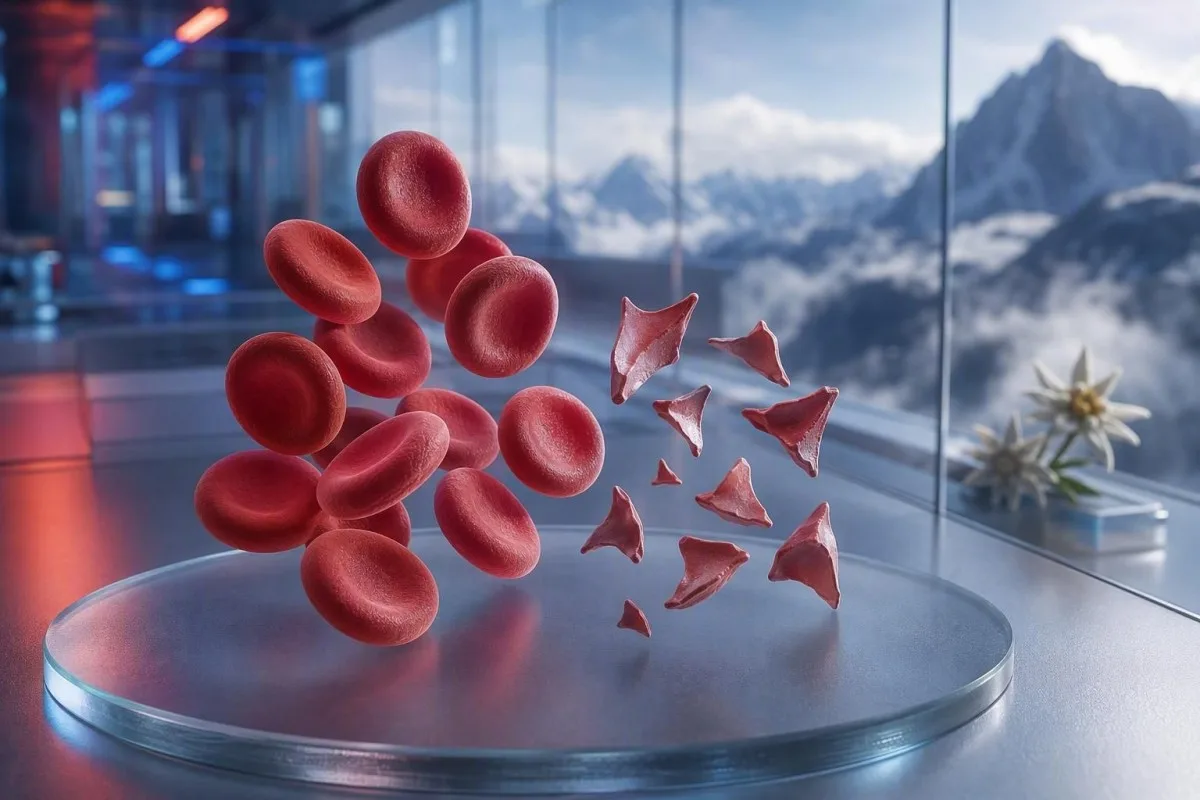
Shistotsitlar — bu bo‘laklangan qizil qon hujayralari bo‘lib, mayda tomirlarda xavfli ivish borligini ko‘rsatishi mumkin, ammo bitta surtma (smear) izohi tashxis emas. Shoshilinchlik fragmentlar foiziga, trombotsitlar soniga, gemoglobin dinamikasiga, buyrak faoliyatiga, gemoliz markerlariga va simptomlarga bog‘liq.


- Shistotsitlar chegarasi: Yaxshi tayyorlangan kattalar periferik surtmasida 1% dan ko‘p shistotsit bo‘lsa, boshqa qizil qon hujayrasi shakli o‘zgarishlari bo‘lmasa, trombotik mikroangiopatiya uchun shubhali hisoblanadi.
- Shoshilinch klaster: Shistotsitlar va trombotsitlar 150 × 10^9/L dan past bo‘lsa, gemoglobin pasayib borayotgan bo‘lsa, LDH ko‘tarilgan bo‘lsa va gaptoglobin past bo‘lsa, o‘sha kunning o‘zida shifokor tomonidan baholanishi kerak.
- TTP belgisi: ADAMTS13 faolligi 10% dan past bo‘lsa, immun trombotik trombotsitopenik purpura (immune thrombotic thrombocytopenic purpura)ni qo‘llab-quvvatlaydi; klinik shubha yuqori bo‘lganda, tasdiqlovchi testlar natijasi qaytib kelguncha ham davolash boshlanadi.
- Har bir bo‘lak xavfli emas: Mexanik yurak klapanlari, og‘ir arterial gipertenziya, dializ tizimlari, kuyishlar va namuna shikastlanishi bo‘laklangan qizil qon hujayralarini keltirib chiqarishi mumkin.
- Surtma — bu qo‘lda olingan dalil (manual evidence): Avtomatlashtirilgan analizator shistotsitlarni ishonchli tarzda aniqlab bera olmaydi; tajribali morfolog slaydni ko‘zdan kechirishi kerak.
- Homiladorlik xavfni oshiradi: 20 haftadan keyin yuqori qon bosimi, siydikda oqsil yoki trombotsitlar pastligi bilan birga shistotsitlar bo‘lsa, darhol akusherlik baholashi talab etiladi.
- Takroriy tekshiruv yordam beradi: Yangi olingan EDTA namunasi va takroriy surtma haqiqiy davom etayotgan eritrotsitlar parchalanishidan yig‘ilish artefaktini ajratishga yordam beradi.
- O‘zingizcha davolamang: Temir, folat yoki qo‘shimchalar mikrovaskulyar angiopatik gemolizni davolamaydi va shoshilinch baholashdan chalg‘itishi mumkin.
Bo‘laklangan qizil qon hujayralari shoshilinch tibbiy ko‘rikni talab qilganda
Shistotsitlar trombotsitlar soni past, anemiya, biokimyoviy gemoliz, buyrak shikastlanishi, nevrologik simptomlar, isitma yoki og‘ir gipertoniya bilan birga paydo bo‘lsa, o‘sha kunning o‘zida shoshilinch ko‘rikdan o‘tishi kerak. Faqat surtma bo‘yicha izoh benign yoki artefakt bo‘lishi mumkin; xavfli topilma esa naqsh (pattern). 2026-yil 7-avgust holatiga ko‘ra, yangi shistotsitlar va trombotsitlar 100 × 10^9/L dan past bo‘lsa, buni odatiy kuzatuv emas, balki “hozir shifokorni chaqiring” natijasi sifatida ko‘rib chiqaman.
Trombotsitlar kamayishi ko‘pincha eng tez shoshilinchlik belgisi bo‘ladi. Gemoglobin 92 g/L, trombotsitlar 68 × 10^9/L va yangi xabar qilingan shistotsitlar bo‘lsa, gemoglobin 145 g/L va trombotsitlar 260 × 10^9/L barqaror bo‘lgan 1% fragmentlarga qaraganda ancha xavotirliroq. Kantesti bu to‘liq qon tahlili (complete blood count) bo‘yicha ushbu klasterni o‘qiydigan va morfologiya izohini alohida ko‘tarib chiqmaydigan AI qon testi analizatoridir.
Klinik amaliyotimdagi 15 yillik tajribamda men tezkor ravishda shuni chaqiraman: natijalari bir necha kun ichida o‘zgargan bemorlar — gemoglobin pasayishi, kreatinin ko‘tarilishi va laktat dehidrogenaza (LDH) ko‘tarilishi. Doktor Tomas Kleinining amaliy qoidasi oddiy: bu naqsh bilan birga chalkashlik, yangi holsizlik, ko‘krak og‘rig‘i, nafas qisishi, juda kam siydik yoki og‘ir bosh og‘rig‘i bo‘lsa, bugun favqulodda baholash kerak.
Shistotsitlar odam o‘zini yomon his qilayotgan bo‘lsa, takroriy testni kutish uchun sabab emas. Kaliy normal bo‘lishi yoki oq qon hujayralari soni normal bo‘lishi trombotik mikrovaskulyar angiopatiya xavfini bekor qilmaydi. Trombotsitlar soni pastligini tushunishga urinayotgan bemorlar ham bizning qo‘llanmamizni ko‘rib chiqishlari mumkin past eritrotsitlar belgilari, ammo o‘tkir simptomlar onlayn talqindan doimo ustun turadi.
Quyidagilar uchun hozir favqulodda xizmatlarga qo‘ng‘iroq qiling:
Yangi bir tomonlama holsizlik, hushdan ketish, tutqanoq, og‘ir nafas qisishi, ko‘krakni ezgandek kuchli noqulaylik, qora najas yoki tez kuchayib borayotgan chalkashlik klinikaga xabar yuborishdan ko‘ra favqulodda xizmatlarni talab qiladi. Bu simptomlar a’zo zararlanishini aks ettirishi mumkin va shistotsit foizining yo‘qligi buni xavfsiz tarzda inkor etmaydi.
Periferik surtma (peripheral smear)da shistotsitlar aslida nimani anglatadi
Shistotsitlar qon aylanishidagi hujayralar jismonan qirqilganda hosil bo‘ladigan notekis eritrotsit fragmentlaridir; ko‘pincha bu fibrin tolalari yoki kichik tomirlardagi g‘ayritabiiy yuqori “shear” (qirqish) yuzalari orqali yuz beradi. Ular odatda o‘tkir burchaklarga ega, dubulg‘aga o‘xshash shakllar yoki kichik uchburchaksimon ko‘rinishlarga ega va butun eritrotsitdagi markaziy xiralik (pallor) yo‘q.
Haqiqiy shistotsitlar morfologik topilma bo‘lib, standart CBC analizator tomonidan ishlab chiqariladigan qiymat emas. Gematologiyada standartlashtirish bo“yicha Xalqaro kengash miqdor aniqlanishi muhim bo”lganda optimal surtma zonasida kamida 1 000 ta eritrotsitni sanashni tavsiya qiladi (Zini va boshq., 2021). Bu tafsilot nega bitta laboratoriya “kam uchraydigan fragmentlar” deb, boshqasi esa foizni xabar qilishi mumkinligini tushuntiradi.
Klassik xavotir shundan iborat: mikrovaskulyar angiopatik gemolitik anemiya, bu yerda qizil qon hujayralari qon aylanishida trombotsitga boy mikrotromblar va fibrin bilan uchrashadi. Biroq og“ir aortal stenoz, mexanik klapan, ekstrakorporal tizimlar va katta termik shikastlanish boshqa mexanizm orqali haqiqiy bo”lakchalar hosil qilishi mumkin. Shuning uchun savol “Bo”lakchalar haqiqiymi?” emas, balki “Bo‘lakchalarni parchalashga nima sabab bo‘lyapti?”
Kantesti — bu AI asosidagi qon tahlili natijalari platformasi bo‘lib, yuklangan to‘liq qon tahlilini yonma-yon joylashtira oladi va CBC tarkibiga nimalar kirishini bilirubin, retikulotsitlar yoki gaptoglobin kabi yetishmayotgan hamroh natijalarni ta’kidlab beradi. U slaydning o‘zini tekshira olmaydi; bu esa o‘qitilgan laboratoriya mutaxassisi vazifasi bo‘lib qoladi.
Kattalarda surtma (adult smear)da shistotsitoz qanchalik darajada xavotirli
Kattalardagi periferik surtma (smear)da shistotsitlar soni 1% dan yuqori bo‘lsa, bo‘lakchalar qizil qon hujayralaridagi asosiy anomaliya bo‘lganda trombotik mikroangiopatiya uchun shubhali hisoblanadi. 1% dan past ko‘rsatkichlar kasallikni istisno qilmaydi, ayniqsa film suyuqliklar yuborilgandan, transfuziyadan yoki kechayotgan kasallikdan keyin tayyorlangan bo‘lsa.
1% kesish nuqtasi diagnostik signal bo‘lib, og‘irlik shkalasi emas. ICSH yo‘riqnomasi sog‘lom kattalar va muddatida tug‘ilgan yangi tug‘ilgan chaqaloqlar uchun 1% dan past bo‘lgan referens intervaldan foydalanadi; muddatidan oldin tug‘ilgan chaqaloqlarda esa bir xil ma’nosiz 5% gacha bo‘lakchalar bo‘lishi mumkin (Zini et al., 2021). Og‘ir trombotsitopeniya bilan 2% natija, mexanik kardiotsirkulyatorni qo‘llab-quvvatlash qurilmasi o‘rnatilgandan keyin 4% ga qaraganda ko‘proq xavotirli.
Laboratoriyalar foiz o“rniga ”vaqti-vaqti bilan“, ”kam“, yoki ”yuqori quvvatli maydon (high-power field)da 1–2 ta” deb xabar berishi mumkin. Bu iboralar bir-birini almashtira olmaydi, chunki maydon o‘lchami, surtma qalinligi va xabar berish siyosati farq qiladi. Laboratoriya ICSH uslubidagi foizli hisobni bajargan-bajarmaganini so‘rash, tasviriy iborani o‘zingiz konvertatsiya qilishga urinishdan ko‘ra ko‘pincha foydaliroq.
Yuqori RDW shistotsitlarni o‘lchamaydi. RDW hujayra o‘lchamidagi o‘zgaruvchanlikni aks ettiradi va retikulotsitoz, temir yetishmovchiligi, transfuzion holat yoki fragmentatsiya bilan birga oshishi mumkin. Bizning batafsil RDW va eritrotsit indeksi bo‘yicha qo‘llanma yuqori RDW o‘zi TTPni ham, artefaktni ham nega qo‘llab-quvvatlamasligini tushuntiradi.
Haqiqiy gemolizni qo‘llab-quvvatlaydigan qon tahlili (blood test) naqshlari
Haqiqiy qizil qon hujayralari fragmentatsiyasi odatda yuqori LDH, past gaptoglobin, ortgan bilvosita bilirubin va anemiya bilan birga retikulotsitlar javobini keltirib chiqaradi. Hech qanday bitta marker yakuniy emas, chunki jigar kasalligi, yallig‘lanish, yaqinda qon quyish va suyak iligi bilan bog‘liq kasalliklar manzarani buzib ko‘rsatishi mumkin.
LDH sezgir, lekin o‘ziga xos emas. LDH 600 IU/L dan yuqori bo‘lishi ko‘pincha to‘g‘ri sharoitda faol gemolizni qo‘llab-quvvatlaydi, ammo mushak shikastlanishi, jigar shikastlanishi, infeksiya va saraton ham uni oshirishi mumkin. Laboratoriyaning pastki chegarasidan past bo‘lgan haptoglobin, odatda 0.3 g/L dan past, plazmaga ajralib chiqqan erkin gemoglobinni bog‘laygani uchun og‘irlik qo‘shadi.
Retikulotsitlar soni odatda periferik destruksiya paytida oshishi kerak, lekin folat yetishmovchiligi, temir yetishmovchiligi, buyrak kasalligi yoki o‘tkir suyak iligi susayishida past bo‘lib qolishi mumkin. Bu ko‘rinadigan qarama-qarshilikni osonlikcha o‘tkazib yuborish mumkin: bemorda haqiqiy gemoliz bo‘lishi va retikulotsit javobi esa noo‘rin past bo‘lishi mumkin. haptoglobin natijasi bo‘yicha yo‘riqnoma keng tarqalgan talqin xatolarini qamrab oladi.
Kantesti AI parchalang an qizil qon hujayralari naqshlarini bir vaqt nuqtasidan olingan gemoglobin, MCV, trombotsitlar, LDH, bilirubin, kreatinin va retikulotsitlarni solishtirish orqali talqin qiladi. 48–72 soatda 20 g/L dan ko‘proq pasaygan gemoglobin, oldingi taqqoslash bo‘lmagan yengil g‘ayritabiiy qiymatdan ko‘ra klinik jihatdan ko‘proq ma’lumot beradi.
Nega bilirubin normal bo‘lishi mumkin
Bilirubinning indirekt fraksiyasi gemolizning boshida, vena ichiga suyuqlik yuborilishi bilan suyultirilgandan keyin yoki jigar bilirubinni samarali tozalaganda normal bo‘lishi mumkin. Shuning uchun normal bilirubin ishonchli fragmentlar, past haptoglobin va pasayayotgan gemoglobinni inkor etmasligi kerak.
Nega TTP gematologik favqulodda holat sifatida davolanadi
Trombotik trombotsitopenik purpura yoki TTP, shistotsitlar trombotsitopeniya bilan birga va aniqroq muqobil bo‘lmagan mikrovaskulyar shikastlanish bilan kechuvchi gemolitik anemiya kuzatilganda gumon qilinadi. Davolanmagan immun TTP tez rivojlanishi mumkin, shuning uchun klinisyenlar ADAMTS13 natijalari qaytib kelguncha plazma almashinuvi va immun davolashni boshlashi mumkin.
ADAMTS13 faolligi 10% dan past bo‘lsa TTP ni kuchli qo‘llab-quvvatlaydi, ammo natija bir necha soat yoki kun talab qilishi mumkin. ISTH diagnostik yo‘riqnomasi, plazma davolashdan oldin namuna olish bilan birga, PLASMIC ball kabi klinik vositalar yordamida testdan oldingi ehtimollikni baholashni tavsiya qiladi (Zheng va boshq., 2020). Ball — harakatga ko‘prik, mutaxassis qarorining o‘rnini bosmaydi.
Isitma, nevrologik belgilar, buyrak yetishmovchiligi, trombotsitopeniya va gemolitik anemiyadan iborat “pentad” ko‘plab real bemorlarda to‘liq bo‘lmaydi. Jorj va Nester trombotik mikroangiopatiyani beshta majburiy quti emas, balki bir-biriga ustma-ust keladigan sabablarga ega sindrom sifatida ta’riflagan (George & Nester, 2014). Amaliyotda beshta belgining barchasini kutish xavfli eski odatdir.
Gemoliz bilan birga trombotsitlar 30 × 10^9/L dan past bo‘lsa, gematolog bilan ayniqsa shoshilinch muhokama talab etiladi. TTP gumon qilinganda trombotsit quyish odatda hayot uchun xavfli qon ketish bo‘lmasa yoki invaziv muolaja kutib bo‘lmasa cheklanadi. Tegishli tekshiruvlar uchun bizning koagulyatsion test bo‘yicha qo‘llanmamiz TTP da PT va aPTT nega normal bo‘lishi mumkinligini tushuntiradi.
Shifokorlar ko‘rib chiqadigan shistotsitlarning boshqa xavfli sabablari
Shistotsitlar tarqalgan tomir ichki ivishida (DIC), Shiga-toksin bilan bog‘liq gemolitik uremik sindromda, og‘ir arterial gipertenziyada, homiladorlik bilan bog‘liq mikrovaskulyar shikastlanishda, ayrim dori vositalarida va transplantatsiya bilan bog‘liq trombotik mikroangiopatiyada ham uchraydi. Odatda trombotsitlar, koagulyatsiya, buyrak va klinik topilmalar bilan bog‘liq belgilar bu holatlarni ajratib beradi.
DIC ko‘pincha fragmentlarni PT ning uzayishi, aPTT ning uzayishi, fibrinogenning past yoki pasayib borishi va D-dimerning keskin oshishi bilan birga olib keladi. TTP da ko‘pincha oddiy koagulyatsiya ko‘rsatkichlari nisbatan normalroq bo‘ladi. Kasal bemorda fibrinogen 1.5 g/L dan past bo‘lsa, iste’mol koagulopatiyasi tomonga xavotirni siljitadi, garchi fibrinogen dastlab normal ko‘rinishi mumkin, chunki u o‘tkir yallig‘lanish paytida ko‘tariladi.
Og‘ir arterial gipertenziya bosim mikrovaskulyaturaga shikast yetkazganda qizil qon hujayralarini “qirqib” yuborishi mumkin; bosh og‘rig‘i, ko‘rish o‘zgarishi, ko‘krak og‘rig‘i yoki buyrak disfunksiyasi bilan birga 180/120 mmHg ga teng yoki undan yuqori ko‘rsatkichlar favqulodda holat hisoblanadi. Shuning uchun qon bosimi ko‘rsatkichi smear hisobotining yonida bo‘lishi kerak. Bemorlar ko‘rib chiqishi mumkin ikkilamchi arterial gipertenziya uchun laborator belgilar darhol xavf bartaraf etilgandan keyin.
Diareya, so‘ngra siydik ajralishining kamayishi, anemiya va trombotsitopeniya gemolitik-uremik sindromdan shubha uyg‘otadi. Najasni tekshirish Shiga toksinini aniqlashga yordam berishi mumkin, ammo birinchi navbatda suvsizlanish holati, kreatinin, kaliy va nevrologik baholash shoshilinch yordamni boshqaradi. Yuqori laktat yoki beqaror hayotiy ko‘rsatkichlar sepsis va DICni baholashni talab qiladi, “kutib turish” yondashuvidan ko‘ra; bularni ko‘ring sepsis markerlari naqshlariga oid alohida qo‘llanmamiz laktat, prokaltsitonin, CRP va CBC o‘zgarishlari nima uchun bir-birini inkor qiladigan testlar sifatida emas, balki birgalikda talqin qilinishini tushuntiradi..
Bo‘laklangan qizil qon hujayralarining mexanik sabablari
Mexanik shikastlanish TTP bo‘lmagan holda ham haqiqiy shistotsitlarni keltirib chiqarishi mumkin, ayniqsa protez yurak klapanlari, og‘ir aorta stenozi, qorincha yordamchi qurilmalari, dializ konturlari yoki ekstrakorporal membrana oksigenatsiyasi (ECMO)da. Bu sabablar baribir tibbiy ko‘rikni talab qiladi, lekin davolash immun TTPga emas, mexanik manbaga qaratiladi.
Mexanik klapan naqshida ko‘pincha surunkali yengil LDH ko‘tarilishi va trombotsitlar barqaror bo‘lganda past haptoglobin kuzatiladi. Yangi anemiya, nafas qisishining kuchayishi, to‘q rangli siydik yoki klapan tovushining o‘zgarishi paravalvulyar oqish yoki qurilma disfunksiyasini ko‘rsatishi mumkin va kardiolog ko‘rigiga turtki bo‘lishi kerak. Smear (surma) ishora beradi; ekokardiografiya manbani topadi.
Sportchilar ba’zan marafon fragmentlarni tushuntira oladimi, deb so‘rashadi. Oyoq urishi bilan bog‘liq gemoliz uzoq masofaga yugurishdan keyin vaqtincha erkin gemoglobin va past haptoglobin keltirib chiqarishi mumkin, ammo ishonchli shistotsitozni men mashq bilan shunchaki bog‘lab qo‘ya olmayman—ayniqsa trombotsitlar kamayib borayotgan bo‘lsa. Buni kengroq bilan solishtiring marafon yuguruvchisi uchun laboratoriya qo‘llanma.
Og‘ir kuyishlar va yirik tomir jarrohligi jismoniy hamda endotelial shikastlanish orqali fragmentlangan hujayralarni keltirib chiqarishi mumkin. Bunday holatlarda har 6–24 soatda laborator trend bitta smeardan ko‘ra ko‘proq ma’lumot berishi mumkin. Qo‘zg‘atuvchi hodisadan keyin CBCning barqaror bo‘lishi taskin beradi; gemoglobin doimiy ravishda pasayib borishi esa emas.
To‘plash (collection) va surtma (slide) artefaktlari shistotsitlarni qanday taqlid qiladi
Noto‘g‘ri namuna olish, smear tayyorlashning kechikishi, antikoagulyant naychalarining kam to‘ldirilishi va smear chetining shikastlanishi qon aylanish darajasidagi gemolizni aks ettirmasdan, shistotsitlarga o‘xshash shakllarni yaratishi mumkin. Toza olingan va monolayerda ko‘rib chiqilgan takroriy namuna klinik manzara tinch bo‘lsa, odatda birinchi tekshiruv hisoblanadi.
Krenatsiyalangan hujayralar, smear cheti buzilishi, qurish artefakti va bir-biriga yopishgan hujayralar tez-tez uchraydigan noto‘g‘ri o‘xshashlardir. Ular ko‘pincha notekis tarqalgan holda yoki ideal o‘qish zonasida bir tekis emas, balki “patli” chetida ko‘proq ko‘rinadi. Tajribali morfolog haqiqiy fragmentning o‘tkir geometriyasini slayd tayyorlash paytida shunchaki buzilgan hujayradan ajratib beradi.
Qiyin yig“ish naychani gemolyz qilishi va kaliy yoki LDHni noto”g‘ri oshirishi mumkin, ammo bu avtomatik ravishda haqiqiy aylanayotgan shistotsitlarni keltirib chiqarmaydi. Agar natijada gemoliz indeksi yoki “namuna buzilgan” degan izoh bo‘lsa, klinisyenlar odatda kasallik tashxisidan oldin qayta namuna olishadi. Bizning maqolamiz kaliy olishdagi xatolar bir xil preanalitik muammo qanday qilib bir vaqtning o‘zida bir nechta natijani chalg‘itishi mumkinligini tushuntiradi.
EDTAga bog‘liq trombotsitlarning klump (uyushma) bo‘lib qolishi trombotsitlar sonini noto‘g‘ri past ko‘rsatishi va xavotirli ko‘rinadigan kombinatsiyani “yaratishi” mumkin. Sitratiy naychadagi trombotsitlar soni (suyultirishga tuzatish kiritilgan holda) va yangi tayyorlangan film buni aniqlab beradi. Nozik jihat muhim: artefakt haqiqiy kasallik bilan birga uchrashi mumkin, shuning uchun takroriy test simptomlari bor odam uchun parvarishni kechiktirmasligi kerak.
Nega analizator ogohlantirishlari va laboratoriya yozuvi chalg‘itishi mumkin
Avtomatlashtirilgan qon analizatorlari g‘ayritabiiy eritrotsit populyatsiyalarini belgilashi mumkin, lekin ular trombotik mikroangiopatiyani istisno qilish uchun shistotsitlarni mustaqil va yetarli ishonchlilik bilan tashxislay olmaydi. Klinik savol gemoliz yoki TTP bo‘lsa, qo‘lda periferik smearni ko‘rib chiqish baribir zarur.
“Maydalangan qizil qon hujayralari mavjud” degan izoh miqdor, surtma sifati va birga uchraydigan morfologiya bor-yo‘qligi bo‘yicha savollar tug‘dirishi kerak. Temir tanqisligi, megaloblastik anemiya yoki irsiy eritrotsit kasalliklaridan kelib chiqqan yaqqol anizopoikilotsitoz 1% qoidasini kamroq o‘ziga xos qiladi. Fragmentlar g‘ayritabiiy morfologiya ustun bo‘lganda eng diagnostik ahamiyatli bo‘ladi, ko‘plab aloqasiz shakllar orasida tasodifiy uchraganda esa kamroq.
Kantesti — bu AI laboratoriya testi talqin qilish xizmati bo‘lib, “trombotsitlar soni past” kabi izoh yonida “trombotsitlar klump(uyum)lari bor” degan izoh kabi mos kelmaydigan kombinatsiyalarni aniqlashga mo‘ljallangan. U laboratoriyaning mikroskopik ko‘rib chiqishini yoki takror surtma so‘rash imkoniga ega bo‘lgan klinitsiyani o‘rnini bosa olmaydi. Sifat tekshiruvlarimizga oid yondashuv quyida tasvirlangan: tibbiy validatsiya bo‘yicha umumiy sharhimizning markazida turadi.
Delta tekshiruv joriy natijani avvalgi natija bilan solishtiradi va to‘satdan mantiqsiz o‘zgarishni aniqlashi mumkin. Gemoglobin 138 g/L dan 87 g/L gacha 24 soat ichida tushishi klinik jihatdan muhim, ammo vena ichiga suyuqliklar bilan suyultirish yoki noto‘g‘ri belgilangan namunani ham ehtimol deb hisoblash kerak. Batafsil: laboratoriyadagi to‘satdan o‘zgarishlar.
Qon tahlili natijalarida shistotsitlar aytilgandan keyin nima qilish kerak
Shistotsitlar birinchi marta qayd etilgan bo‘lsa, o‘sha kuni testni buyurgan klinitsiyaga yoki xizmatga murojaat qiling; nevrologik simptomlar, nafas qisishi, ko‘krak og‘rig‘i, siydikning qorayishi, kuchli bosh og‘rig‘i, isitma yoki siydik ajralishining kamayishi bo‘lsa darhol shoshilinch tibbiy yordamga murojaat qiling. Surtma izohini o‘zi bilan o‘zini tashxis deb talqin qilmang.
Haqiqiy hisobotni, jumladan gemoglobin, trombotsitlar, kreatinin, bilirubin, LDH va har qanday gemoliz indeksi mavjud bo‘lsa, tayyor holda bo‘ling. Shuningdek, yaqinda bo‘lgan homiladorlik, ich ketishi bilan kechgan kasallik, qon bosimining juda yuqori ko‘rsatkichlari, yangi dori vositalari, klapan(ya’ni yurak qopqog‘i) jarrohligi, dializ, transfuzion muolaja va autoimmun kasalliklarni ham qayd eting. Bu tafsilotlar morfologiya bo‘limidagi bitta so‘zdan ko‘ra ko‘proq differensial tashxisni o‘zgartiradi.
Men bemorlar “anemiya” so‘zi sabab aniqlanmasdan oldin o‘qib, temirni boshlash bilan qimmatli vaqtni yo‘qotayotganini ko‘raman. Temir tanqisligi birga uchrashi mumkin, lekin fragmentlar bilan tez trombotsitlar kamayishini tushuntirib bera olmaydi. Doktor Tomas Klein faqat ajratib ko‘rsatilgan raqamlarni ko‘chirib yozish o‘rniga to‘liq hisobotdan rasmga olishni tavsiya qiladi; ko‘chirish ko‘pincha naqshni bir-biriga bog‘lab turadigan laboratoriya izohini tushirib qoldiradi.
Trombotsitlar juda past bo‘lsa yoki qon ketish mavjud bo‘lsa, klinitsiy boshqa narsa aytmaguncha aspirin, ibuprofen va retseptsiz antikoagulyantlardan saqlaning. Belgilangan antikoagulyantni shoshilinch tibbiy yo‘l-yo‘riqsiz to‘xtatmang. Charchoq yoki bosh aylanishi bo‘lgan odamlar bizning bosh aylanishi uchun qon tahlillari shoshilinch sabablar istisno qilingandan keyin savollar tayyorlash uchun foydalanishlari mumkin.
Homiladorlikda, yangi tug‘ilganlarda va bolalarda shistotsitlar
Homiladorlik davrida shistotsitlar, ular gipertenziya, proteinuriya, jigar fermentlari ko‘tarilishi, trombotsitlar pastligi, bosh og‘rig‘i, ko‘rish bilan bog‘liq simptomlar yoki qorin yuqori qismida og‘riq bilan birga uchrasa, shoshilinch baholashni talab qiladi. HELLP sindromi, preeklampsiya, TTP, atipik HUS va DIC bir-birini qisman qoplashi mumkin, tug‘ruq atrofidagi vaqt yordam beradi, lekin tashxisni yakuniy hal qilib bermaydi.
HELLP sindromi klassik tarzda gemoliz, jigar fermentlari ko‘tarilishi va trombotsitlar pastligi bilan belgilanadi; u ko‘pincha homiladorlikning 20 haftasidan keyin yoki tug‘ruqdan ko‘p o‘tmay yuz beradi. Trombotsitlar soni 100 × 10^9/L dan past yoki AST 70 IU/L dan yuqori bo‘lsa xavotirni qo‘llab-quvvatlashi mumkin, ammo mahalliy akusherlik jamoalari butun klinik manzarani talqin qiladi. Bu kombinatsiya bilan oddiy antenatal qabulni kutmang.
Muddatli tug‘ilgan yangi tug‘ilgan chaqaloqlarda 1% shistotsitlar kamroq bo‘lishi mumkin, muddatidan oldin tug‘ilgan chaqaloqlarda esa 5% gacha bo‘lishi mumkin, chunki neonatal eritrotsit morfologiyasi boshqacha (Zini et al., 2021). Biroq ich ketishi bo‘lgan bola, rang oqarishi, siydik kamayishi va trombotsitopeniyada fragmentlar HUS tomon ishora qilishi va darhol pediatrik baholashni talab qilishi mumkin.
Trombotsitlar va gemoglobin uchun mos yozuv (referens) oraliqlari yosh va homiladorlik bosqichiga qarab sezilarli o‘zgaradi. Umumiy app tomonidan “normal” deb belgilangan natija ham kontekstda klinik jihatdan xavotirli bo‘lishi mumkin. Bizning homiladorlikdagi trombotsitlar diapazonlari bo‘yicha resurslarimiz va bir kunda homiladorlik bo‘yicha laboratoriya ogohlantirishlari foydali fon ma’lumotini taqdim etadi, lekin tug‘ruqdan keyingi triageni (maternity triage) o‘rnini bosa olmaydi.
Klinikachilar haqiqiy shistotsitlarni tekshirish uchun buyuradigan testlar
Shubhali mikroangiopatik gemoliz uchun birinchi tekshiruvlar to‘plami odatda takroriy CBC va surtma, retikulotsitlar soni, LDH, gaptoglobin, bilirubin fraksiyalari, kreatinin, siydik tahlili, PT, aPTT, fibrinogen va D-dimerni o‘z ichiga oladi. TTP gumon qilinganda plazma almashinuvidan oldin ADAMTS13 faolligi va ingibitor tekshiruvlari olinishi kerak, ammo davolash kutib qolmasligi lozim.
To‘g‘ridan-to‘g‘ri antiglobulin testi, DAT yoki Kumbs testi deb ham ataladi, immun gemolizni immun bo‘lmagan fragmentatsiyadan farqlashga yordam beradi. Musbat DAT ikkinchi jarayonni istisno qilmaydi, lekin klassik TTP odatda DAT-manfiy bo‘ladi. Siydik mikroskopiyasi buyrak mikrovaskulyar shikastlanish faol bo‘lganda protein yoki hujayraviy silindrlarni ko‘rsatishi mumkin; bizning donador siydik silindrlari (granular urinary casts).
Najasdagi Shiga-toksin testlari mos keladigan gastrointestinal kasallikdan keyin ko‘rib chiqiladi, atipik HUS uchun esa komplement testlari, dori tekshiruvi va genetik yoki funksional tadqiqotlar qo‘llanilishi mumkin. Tartib mamlakat va shifoxonaga qarab farq qiladi. 48 soat ichida kreatininning 26,5 mkmol/L ga oshishi o‘tkir buyrak shikastlanishining bitta mezoniga to‘g‘ri keladi va u keskin raqamga aylanishidan oldin ham e’tiborga loyiq.
Muhim anemiyada retikulotsitlar ishlab chiqarish indeksi 2 dan past bo‘lsa, xom retikulotsit foizi yuqoriga o‘xshasa ham, suyak iligi javobi yetarli emasligini ko‘rsatadi. Bu hisob anemiyada eritrotsitlar massasi pastroq bo‘lishini tuzatadi. The retikulotsit va gematologik markerlar bo‘yicha yo‘riqnomamiz foizlar nega chalg‘itishi mumkinligini tushuntirishga yordam beradi.
Surtmani qayta topshirish qachon mantiqli va qachon emas
Fragmentlar kam bo‘lsa, bemor o‘zini yaxshi his qilsa, trombotsitlar va gemoglobin barqaror bo‘lsa hamda namuna sifati shubha uyg‘otsa, bir necha soat ichida 24 soatgacha surtmani qayta tekshirish mantiqan to‘g‘ri. Trombotsitopeniya, faol gemoliz, organ shikastlanishi yoki xavotirli simptomlar mavjud bo‘lsa, qayta tekshirishni xavfsiz kechiktirish mumkin emas.
Yaxshi qayta olingan namuna yangi olingan bo‘lishi, to‘g‘ri to‘ldirilgan EDTA naychasiga solinishi va filmga tezda tayyorlanishi kerak. So‘rovda, agar ikkalasi ham mumkin bo‘lsa, shubhali shistotsitlar va trombotsitlar klumpalanishini ko‘rib chiqishni aniq so‘rash kerak. Laboratoriya ikkala filmni yonma-yon solishtirishi mumkin, bu esa avtomatlashtirilgan CBCni shunchaki qayta topshirishdan ancha foydaliroq.
Yo‘nalish muhim. Uch kun davomida trombotsitlar soni 210 dan 165 ga, keyin 98 × 10^9/L ga tushsa, bu birinchi ikkita ko‘rsatkich mahalliy mos yozuvlar oralig‘ida bo‘lishi mumkinligiga qaramay, xavotirli. Kantesti AI bu traektoriyalarni yuzaga chiqarish uchun uzunlamasına kontekstdan foydalanadi; bu prinsip bizning qon tahlili trendini tahlil qilish qo‘llanmasi.
Transfuziya original morfologiyani vaqtincha suyultirishi yoki niqoblashi mumkin. Agar ikkinchi namuna olinmasidan oldin eritrotsitlar olgan bo‘lsangiz, jamoaga sana va nechta birlik (unit) bo‘lganini ayting. Bizning kasalxonadan chiqqandan keyin qiymatlarning o‘zgarishi davolashdan keyingi trendlar uchun boshqa bazaviy ko‘rsatkich nega kerakligini tushuntiradi.
Shistotsit kasalligiga o‘xshab ko‘rinishi mumkin bo‘lgan holatlar
Temir yetishmovchiligi, vitamin B12 yetishmovchiligi, jigar kasalligi, irsiy eritrotsit kasalliklari va og‘ir infeksiya trombotik mikroangiopatiyaning o‘ziga xos fragmentatsiya naqshisiz ham g‘ayritabiiy eritrotsit shakllarini keltirib chiqarishi mumkin. Surtmaning to‘liq morfologiyasi va klinik vaqt chizig‘i fragmentlarni ortiqcha baholashning oldini oladi.
Og‘ir vitamin B12 yetishmovchiligi anemiya, yuqori LDH, past trombotsitlar va ba’zan fragmentlar bilan pseudo-trombotik mikroangiopatiyani keltirib chiqarishi mumkin. U ko‘pincha yaqqol makrotsitoz, retikulotsitlar javobining pastligi va LDH ko‘rsatkichlari ba’zan 2,500 IU/L dan yuqoriligini beradi—bu belgilar avtomatik TTP davolashdan ko‘ra B12 va metilmalon kislota testlarini talab qilishi kerak. Bizning B12 va folat bo‘yicha qo‘llanmada yoritilgan buni muhim taqlid (mimika) ekanini tushuntiradi.
Temir yetishmovchiligi odatda mikrositoz, gipoxromiya, qalamsimon hujayralar (pencil cells) va yuqori RDW ni keltirib chiqaradi; og‘ir holatlarda fragmentlar qayd etilishi mumkin, ammo ular kamdan-kam hollarda asosiy belgi bo‘ladi. Ferritin 15 mkg/L dan past bo‘lsa, boshqa jihatdan asoratsiz kattalarda temir zaxiralari kamayganini kuchli qo‘llab-quvvatlaydi, garchi yallig‘lanish ferritinni oshirib, yetishmovchilikni niqoblashi mumkin.
Mening tajribamda eng ishonchli natija faqat “schistotsitlar yo”q” degani emas, balki barqaror trombotsitlar, barqaror buyrak faoliyati va biokimyoviy gemolizning yo‘qligi bilan birga keladigan izchil muqobil tushuntirishdir. Agar topilmalar mos kelmasa, gematolog yakuniy hisobot matniga tayanishdan ko‘ra, haqiqiy slaydni ko‘rib chiqishi mumkin.
Surtma favqulodda holatini o‘tkazib yubormasdan AI talqinidan foydalanish
AI qon tahlili natijalarini tartibga solish va xavfli kombinatsiyalarni aniqlashga yordam berishi mumkin, ammo CBC PDF dan schistotsit tashxisini tasdiqlay olmaydi yoki shoshilinch klinik baholashni o‘rnini bosa olmaydi. Periferik surtma — bu vizual mikroskopiya, va asl laboratoriya slaydi haqiqat manbai bo‘lib qoladi.
Kantesti — AI asosidagi qon tahlili vositasi bo‘lib, hisobotlar bo‘ylab bog‘liq natijalarni, jumladan trombotsitlar dinamikasini, gemoglobin o‘zgarishlarini, kreatinin, bilirubin va LDH ni xaritalaydi. Bu har bir g‘ayritabiiy markerning o‘zini alohida o‘qish bilan bog‘liq keng tarqalgan xatoni kamaytirishi mumkin. U “schistotsit” deb ataladigan shakl haqiqiy fragmentmi, surtma cheti artefaktimi yoki hisobot berish konvensiyasimi — buni ko‘ra olmaydi.
Yuklash sifati muhim, chunki birlik yoki kasrning tushib qolishi natijani ma’nosini butunlay o‘zgartirib yuborishi mumkin. Hisobotni ulashishdan oldin bemor identifikatorlarini, yig“ish sanasini, birliklarni, me’yoriy diapazonlarni va natijada ”estimated“ (taxminiy), ”pending“ (kutilmoqda) yoki ”sample haemolysed” (namuna gemolizlangan) deb yozilgan-yo‘qligini tekshiring. Bizning AI hisobot aniqligi bo‘yicha chek-listimizdan foydalaning. yakuniy insoniy tekshiruv uchun foydali.
Kantesti ning klinik metodlari bizning Tibbiy maslahat kengashi, dan olingan ma’lumotlar bilan ko‘rib chiqilgan va texnologiyamiz AI talqin texnologiyamiz qo‘llanmasi Kantesti ning neyron tarmog‘i yaqin biomarkerlarni, trend yo‘nalishini va ichki izchillikni qanday baholashini tushuntiradi. Mening tajribamda, bu ayniqsa. da tasvirlangan. Doktor Tomas Klein sifatida men baribir shifokor yangi fragmentlar, trombotsitlar 100 × 10^9/L dan past bo‘lsa va simptomlar bo‘lsa, bemor uchun laboratoriyaga bevosita qo‘ng‘iroq qilishini xohlardim — chunki aynan shunday vaziyatda tezlik va klinik ko‘rik eng muhim bo‘ladi.
Tez-tez so'raladigan savollar
Qaysi darajadagi shistotsitlar xavfli hisoblanadi?
Katta yoshdagi periferik surtma ehtiyotkorlik bilan qayta ko‘rib chiqilganda 1% dan ko‘p shistotsitlar trombotik mikroangiopatiya uchun shubhali hisoblanadi, bunda parchalangan hujayralar asosiy eritrotsit anomaliyasi bo‘ladi. Shistotsitlar foizi, agar u trombotsitlar soni 150 × 10^9/L dan past bo‘lsa, gemoglobin pasayayotgan bo‘lsa, LDH yuqori bo‘lsa, gaptoglobin past bo‘lsa, buyrak shikastlanishi bo‘lsa yoki nevrologik simptomlar kuzatilsa, har qanday foiz darhol xavotirli bo‘ladi. 1% dan past natija kechayotgan holatni ishonchli tarzda istisno etmaydi. Klinik ko‘rinish faqat foizga qaraganda ko‘proq xavfni belgilaydi.
Shistotsitlar yomon qon olish (qon namunasi olish) sabab bo‘lishi mumkinmi?
Qiyin yig‘ma, tayyorlashning kechikishi, to‘ldirilmagan EDTA naychasi yoki noto‘g‘ri tayyorlangan surtma hujayra shakllarini shistotsitlarga o‘xshatib ko‘rsatishi mumkin, garchi u haqiqiy aylanma fragmentatsiyani keltirib chiqarmasa ham. Laboratoriyalar buni yangi yig‘ilgan namunani va optimal surtma maydonini qo‘lda ko‘rib chiqish orqali aniqlashtirishi mumkin. Gemolizlangan namuna shuningdek kaliy va LDH ni noto‘g‘ri oshirib ko‘rsatishi, bu esa hisobotni yanada xavotirliroq ko‘rsatishi mumkin. Takroriy tekshiruv simptomlar, trombotsitlar pastligi yoki anemiya mavjud bo‘lsa, favqulodda baholashni kechiktirmasligi kerak.
Shistotsitlar har doim TTPni anglatadimi?
Shistotsitlar har doim ham trombotik trombotsitopenik purpura yoki TTPni anglatmaydi. Ular DIC, og‘ir arterial gipertenziya, Shiga-toksinli HUS, homiladorlik bilan bog‘liq buzilishlar, mexanik yurak klapanlari, dializ konturlari va katta to‘qima shikastlanishi bilan birga uchrashi mumkin. Shistotsitlar trombotsitopeniya va immun bo‘lmagan gemolitik anemiya bilan birga bo‘lganda, ayniqsa ADAMTS13 faolligi 10% dan past bo‘lsa, TTP yuqori ustuvorlikdagi xavotirga aylanadi. ADAMTS13 tahlili vaqt talab qilishi mumkinligi sababli, gumon qilingan TTP natija kelguniga qadar shoshilinch ixtisoslashgan baholashni talab qiladi.
Shistotsit gemolizi bilan qaysi qon tahlillari yuqori bo‘ladi?
To‘g‘ri fragmentatsion gemoliz odatda LDH va indirekt bilirubinni oshiradi, gaptoglobinni pasaytiradi hamda gemoglobin kamayganda retikulotsitlar soni ko‘payishini keltirib chiqaradi. Faol gemolizda LDH 600 IU/L dan oshishi mumkin, lekin u o‘ziga xos emas, chunki mushak, jigar va boshqa to‘qimalarning shikastlanishi ham uni oshiradi. Jigar ishlab chiqarishi yetarli bo‘lganda gaptoglobin taxminan 0,3 g/L dan past bo‘lishi tomir ichidagi gemolizni qo‘llab-quvvatlaydi. Kreatinin, trombotsitlar soni, PT, aPTT, fibrinogen va D-dimer asosiy sababni aniqlashga yordam beradi.
Homiladorlikda shistotsitlar normalmi?
Shistotsitlar gipertenziya, proteinuriya, jigar fermentlari ko‘rsatkichlarining oshishi, trombotsitlarning pasayishi, bosh og‘rig‘i, ko‘rishdagi o‘zgarishlar yoki qorin yuqori qismidagi og‘riq bilan birga uchrasa, ular odatiy normal homiladorlik belgisi hisoblanmaydi. Bu majmua HELLP sindromi, preeklampsiya, TTP, atipik HUS yoki DICni ko‘rsatishi mumkin va darhol akusherlik baholashini talab qiladi. Ushbu holatda trombotsitlar soni 100 × 10^9/L dan past bo‘lishi ayniqsa xavotirli. Homiladorlikka xos ma’lumotnoma diapazonlari gemoliz ko‘rinishini e’tiborsiz qoldirish uchun asos bo‘lmaydi.
Mexanik yurak klapani shistotsitlarni keltirib chiqarishi mumkinmi?
Mexanik yurak klapanlari va og‘ir aortal stenozi yuqori tezlikdagi qon oqimi eritrotsitlarni fizik jihatdan parchalayotgani sababli haqiqiy shistotsitlarni keltirib chiqarishi mumkin. Bu ko‘pincha yengil surunkali gemolizni chaqiradi: LDH ko‘tariladi va gaptoglobin kamayadi, trombotsitlar soni esa odatda barqaror qoladi. Yangi paydo bo‘lgan anemiya, siydikning qorayishi, nafas qisishi yoki klapan funksiyasining o‘zgarishi yurakni qayta ko‘rib chiqishni talab qiladi, chunki oqish yoki qurilma bilan bog‘liq muammo sabab bo‘lishi mumkin. Ushbu topilmani yurak tarixi hisobga olinmasdan avtomatik ravishda TTP deb belgilamaslik kerak.
Bugun AI asosidagi qon tahlilini tahlil qilishni oling
Kantesti’ga tezkor va aniq laboratoriya tahlili uchun ishonadigan butun dunyo bo‘ylab 2 milliondan ortiq foydalanuvchiga qo‘shiling. Qon tahlili natijalaringizni yuklang va soniyalar ichida 15,000+ biomarkerlarining to‘liq talqinini oling.
📚 Havola qilingan ilmiy tadqiqot nashrlari
Klein, T., Mitchell, S., & Weber, H. (2026). Kantesti qon testi talqin qilish dvigateli bo‘yicha 100 000 ta sintetik test holatida Kantesti uchun oldindan ro‘yxatdan o‘tkazilgan, rubrika asosidagi avtomatlashtirilgan texnik benchmark. Kantesti AI tibbiy tadqiqoti.
Klein, T., Mitchell, S., & Weber, H. (2026). Klinik validatsiya asoslari v2.0 (Tibbiy validatsiya sahifasi). Kantesti AI tibbiy tadqiqoti.
📖 Tashqi tibbiy manbalar
Zheng XL va boshq. (2020). ISTH yo‘riqnomalari: trombotik trombotsitopenik purpura tashxisini aniqlash. Qon ivishi va gematologiya bo‘yicha jurnal (Journal of Thrombosis and Haemostasis).
⚕️ Tibbiy ogohlantirish
Ushbu maqola faqat ta’lim maqsadlari uchun mo‘ljallangan va tibbiy maslahatni anglatmaydi. Tashxis va davolash bo‘yicha qarorlar uchun har doim malakali sog‘liqni saqlash mutaxassisiga murojaat qiling.
E-E-A-T ishonch signallari
Tajriba
Shifokor boshchiligidagi laboratoriya talqin qilish ish jarayonlarini klinik ko‘rib chiqish.
Tajriba
Laboratoriya tibbiyoti biomarkerlarning klinik kontekstda qanday o‘zini tutishini yoritadi.
Vakolatlilik
Dr. Tomas Klein tomonidan yozilgan, Dr. Sarah Mitchell va Prof. Dr. Hans Weber tomonidan ko‘rib chiqilgan.
Ishonchlilik
Xavotirni kamaytirish uchun aniq keyingi qadamlar yo‘nalishlari bilan dalillarga asoslangan talqin.