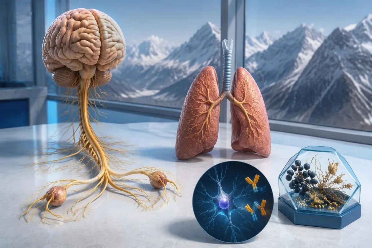
Teigiamas anti-Hu antikūnų tyrimo rezultatas kelia įtarimą dėl su vėžiu susijusio neurologinio imuninio atsako, ypač susijusio su smulkialąsteliniu plaučių vėžiu, tačiau tai nėra vėžio diagnozė. Gydytojai įvertina simptomus, patvirtina laboratorinio tyrimo rezultatą ir organizuoja vėžio paiešką; net jei pirmieji vaizdiniai tyrimai neigiami, gali prireikti pakartotinės patikros.



- Anti-Hu / ANNA-1 yra didelės rizikos paraneoplastinis antikūnas – ši kategorija siejama su vėžiu daugiau nei 70 % nustatytų klinikinių kohortų, bet ne su kiekvienu nepatvirtintu atrankinės patikros rezultatu.
- Vėžio diagnozė nustatoma tik esant pagrindinės piktybinės ligos įrodymų, paprastai ir ištyrus audinį; vienas teigiamas antikūnų tyrimo rezultatas tokiu įrodymu nelaikomas.
- Tyrimo metodu gauto rezultato patvirtinimas dažnai apima 2 vienas kitą papildančius metodus: būdingą audinių dažymosi vaizdą ir antigenui specifinį tyrimą.
- Neurologiniai simptomai pavyzdžiui, progresuojantis jutimų susilpnėjimas, pusiausvyros sutrikimai, traukuliai ar autonominės nervų sistemos disfunkcija keičia rezultato reikšmę ir skubumą.
- Neigiami vaizdiniai tyrimai reiškia, kad tuo metu atliekant tą tyrimą vėžys nebuvo nustatytas; tai neatmeta mažo, iš pradžių neaptinkamo vėžio galimybės.
- Pakartotinė patikra pagal 2021 m. PNS kriterijus rekomenduojama kas 4–6 mėnesius 2 metus, kai didelės rizikos neurologinį fenotipą lydi didelės rizikos antikūnas.
- Ilgesnė stebėsena gali būti tinkama tam tikriems pacientams; 2023 m. anti-Hu kohorta pagrindė svarstymą tęsti patikrą ilgiau nei 2 metus, kartais iki 5 metų.
- Skubūs simptomai apima naujai atsiradusį dusulį, negalėjimą saugiai ryti, greitai stiprėjantį silpnumą, pirmąjį traukulių priepuolį ar staigius neurologinius sutrikimus.
Ką reiškia teigiamas anti-Hu antikūnų tyrimo rezultatas?
An anti-Hu antikūnų teigiamas rezultatas reiškia, kad laboratorija nustatė reaktyvumą prieš Hu neuronų baltymus; vien šis rezultatas nepatvirtina nei vėžio, nei neurologinio sindromo. Interpretaciją sudaro 3 dalys: ar rezultatas tikras, ar neurologinis klinikinis vaizdas atitinka ir ar galima nustatyti susijusį vėžį.
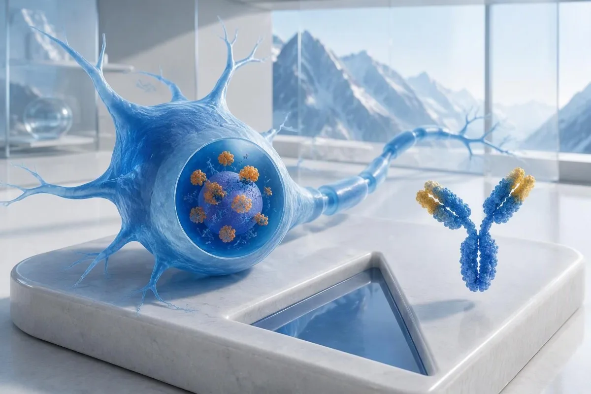
Anti-Hu priskiriamas prie didelės rizikos su vėžiu susijusių antikūnų, o ši kategorija apibrėžiama kaip ryšys su vėžiu, viršijantis 70% atitinkamose klinikinėse populiacijose (Graus ir kt., 2021). Šio procento negalima tiesiog priskirti šiaip sveikam asmeniui, kurio plataus profilio tyrimo rezultatas silpnai teigiamas ir nepatvirtintas: tikimybę lemia tirti pacientai, tyrimo metodas ir neurologinis klinikinis vaizdas.
Hipotetiniam 62 metų žmogui, kuriam 8 savaites blogėja pusiausvyra ir pasireiškia asimetrinis jutimų sutrikimas, reikia kitokio įvertinimo nei žmogui, kurio antikūnas atsitiktinai aptiktas atliekant platų nuovargio tyrimų profilį. Nė vienam iš jų nereikėtų sakyti “Jūs sergate vėžiu” vien pagal laboratorinio tyrimo atsakymą. Mūsų paaiškinimas apie teigiamus antikūnų tyrimų rezultatus padeda atskirti imuninį žymenį nuo galutinės diagnozės.
Kantesti yra AI kraujo tyrimo analizatorius tai gali padėti paaiškinti anti-Hu tyrimo atsakymo formuluotę, tačiau laboratorinio tyrimo patvirtinimą ir vėžio vertinimą turėtų atlikti jus gydantys gydytojai. Kaip Thomas Klein, MD, Kantesti LTD vyriausiasis medicinos pareigūnas, pirmiausia klausčiau, dėl ko buvo paskirtas būtent šis tyrimas, o ne kaip grėsmingai atrodo raudona vėliavėlė; mūsų organizacija ir klinikinė misija paaiškina tą skirtumą.
Ar ANNA-1 yra tas pats antikūnas kaip anti-Hu?
ANNA-1 antikūnas ir anti-Hu antikūnas paprastai reiškia tą pačią antikūnų šeimą. ANNA-1 reiškia 1 tipo antineuroninį branduolinį antikūną; pavadinimas Hu nurodo į ląstelės viduje esančius neuronų baltymus, kuriuos atpažįsta imuninis atsakas, o ne atskirą ligą ar vėžio stadiją.
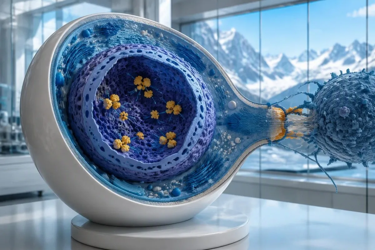
Hu baltymai yra ląstelės viduje esantys RNR jungiantys baltymai, o anti-Hu geriausia suprasti kaip imuninio atsako, kuriame dažnai dalyvauja citotoksinės T ląstelės, žymenį. Tai skiriasi nuo sutrikimų, kai antikūnai jungiasi prie prieinamų neuronų paviršiaus baltymų. Šis skirtumas paaiškina, kodėl vien tik sumažinus antikūnų kiekį ne visada pavyksta panaikinti jau įvykusį anti-Hu sukeltą neurologinį pažeidimą, net jei 2 tyrimų ataskaitose nurodyti titrai labai skiriasi.
Atsakymas “aptiktas ANNA-1” nėra tas pats, kas įprastas teigiamas ANA tyrimas, nepaisant panašių raidžių. ANA paprastai reiškia antinuklearinius antikūnus, naudojamus sisteminių autoimuninių ligų vertinimui, o ANNA-1 žymi neuroninių antikūnų šabloną. Aptardami rezultatus, šių 2 santrumpų nepainiokite; mūsų ANA terminų vadove paaiškinamas labiau įprastas tyrimas.
Anti-Hu galima aptikti kai kuriems smulkialąsteliniu plaučių vėžiu sergantiems žmonėms, kuriems nėra atpažįstamo neurologinio sindromo. Ir atvirkščiai, neurologiniai simptomai gali paskatinti aptikti antikūnus dar prieš vėžiui tampant matomam. Tai 2 skirtingi klinikiniai keliai: vien pagal antikūną gydytojas negali nustatyti, ar šiuo metu pažeidžiami nervai, ar kur gali būti pagrindinis vėžys.
Kaip reikėtų suprasti anti-Hu antikūnų tyrimo ataskaitą?
An anti-Hu antibody test has no universal numerical cutoff that can be applied across laboratories. Read the specimen type, assay method, result category, laboratory reference interval and confirmation comments together; a dilution titer, band intensity and numerical index are 3 different ways of reporting reactivity.
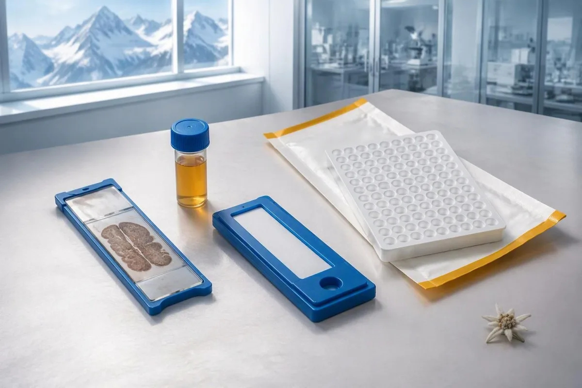
A hypothetical titer of 1:240 describes detectable reactivity after a particular dilution; it is not a cancer probability of 240, nor does it establish cancer stage. A numerical index from another method may use entirely different calibration. Our guide to kokybiniai ir kiekybiniai rezultatai explains why “positive” and “how much” answer different laboratory questions.
Weak or borderline reactivity deserves method-specific interpretation, particularly when a broad panel was ordered without a convincing neurological indication. Ask whether the laboratory observed the characteristic neuronal staining pattern and whether an antigen-specific confirmation was performed. Two tests using the same underlying strip technology may reproduce the same analytical problem; agreement is more informative when the methods are genuinely complementary.
Bring the complete report, not just a screenshot of the word positive. The 5 most useful details are the specimen, collection date, assay name, reported strength or titer and laboratory interpretation. Also list recent intravenous immunoglobulin and other immune treatments, because passive antibodies or treatment effects can complicate some serological results; your laboratory can advise whether that issue plausibly affects this particular finding.
Kada reikia patvirtinti teigiamą anti-Hu rezultatą?
Unexpected, weak or clinically discordant anti-Hu results should be reviewed for confirmation, preferably with an experienced neuroimmunology laboratory. The 2021 PNS criteria emphasize interpreting neuronal antibody testing alongside the phenotype and using appropriate confirmatory methods rather than accepting an isolated commercial-panel result as a diagnosis.
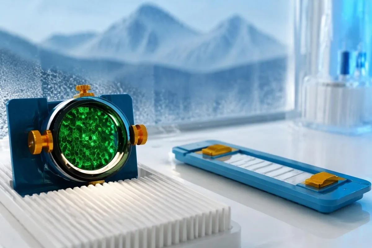
Tissue-based immunohistochemistry or immunofluorescence and an antigen-specific assay provide different information. The first identifies a characteristic distribution of neuronal staining; the second tests reactivity against the relevant antigen. Graus et al. (2021) describe these complementary approaches, with specialist reassessment particularly useful when 1 method gives a result that does not fit the clinical picture.
Repeating a sample can address collection errors or a transient analytical concern, but repeating the same kit is not always enough. In a hypothetical 45-year-old with no objective neurological findings and a faint isolated band, I would want the laboratory’s explanation before treating the report as established anti-Hu autoimmunity. Our GAD65 interpretation guide illustrates another antibody for which context and assay characteristics change meaning.
Confirmation and urgent assessment can happen in parallel. A person who develops seizures, substantial walking difficulty or progressive swallowing impairment should not wait several weeks for a reference-laboratory answer before receiving neurological care. The practical question is not “repeat or investigate?” but which of those 2 tasks can safely proceed together, given the symptoms and the initial laboratory evidence.
Dėl kokių neurologinių simptomų anti-Hu rezultatas kelia didesnį susirūpinimą?
Progressive sensory neuronopathy, encephalomyelitis, limbic encephalitis and marked autonomic dysfunction are recognized anti-Hu presentations. Symptoms can involve more than 1 part of the nervous system, so the pattern and speed of change are usually more informative than a single symptom such as tingling or fatigue.
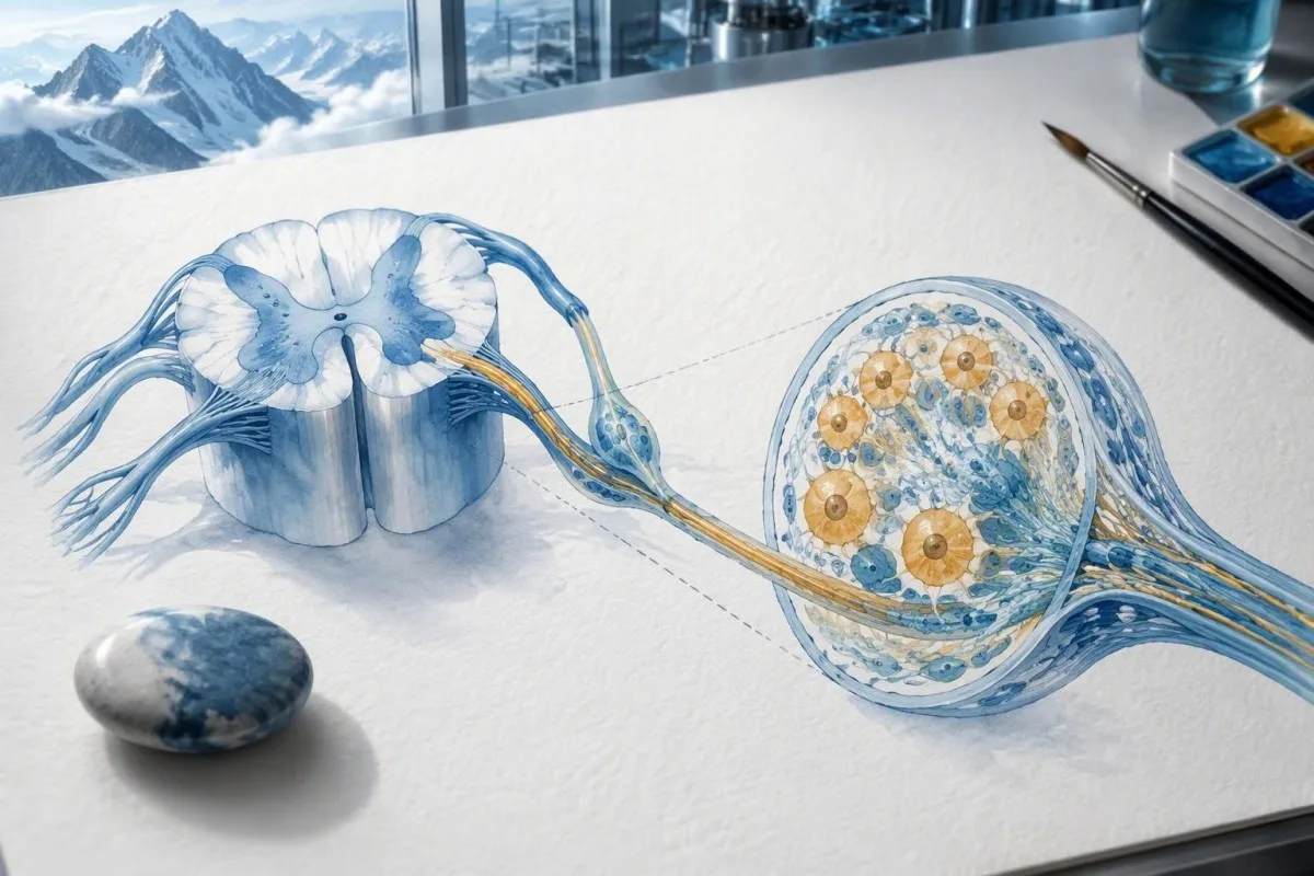
Sensory neuronopathy can cause asymmetric numbness, impaired position sense and sensory ataxia, sometimes involving the hands or trunk early rather than following a simple feet-first pattern. A patient may lose balance in the dark because vision had been compensating for reduced position feedback. That is different from the focal finger distribution discussed in our carpal tunnel symptom guide, although neither pattern can be diagnosed from description alone.
Anti-Hu autonomic involvement can affect gut movement, blood-pressure regulation and bladder function. Severe constipation, unexplained intestinal pseudo-obstruction or dizziness on standing becomes more relevant when accompanied by objective neurological abnormalities. Recording symptoms over 7 days can reveal combinations that otherwise get presented as separate stomach, balance and urinary complaints; sudden severe abdominal symptoms still require assessment for non-neurological emergencies.
New memory impairment, behavioral change or seizures may indicate central nervous system involvement, while rapid weakness has a broader emergency differential. The skubios pagalbos reikalaujančius įspėjamuosius silpnumo požymius in our neurological safety guide apply regardless of the antibody label. Call emergency services for new breathing difficulty, unsafe swallowing, a first seizure or sudden stroke-like symptoms rather than waiting for a scheduled follow-up.
Kokie neurologiniai tyrimai gali būti atliekami gavus teigiamą rezultatą?
Neurological follow-up may include examination, nerve conduction studies, MRI and cerebrospinal fluid testing, selected according to the symptoms. There is no mandatory battery for every anti-Hu-positive person; the purpose is to document which structures are affected and exclude other causes, not merely to collect additional positive tests.
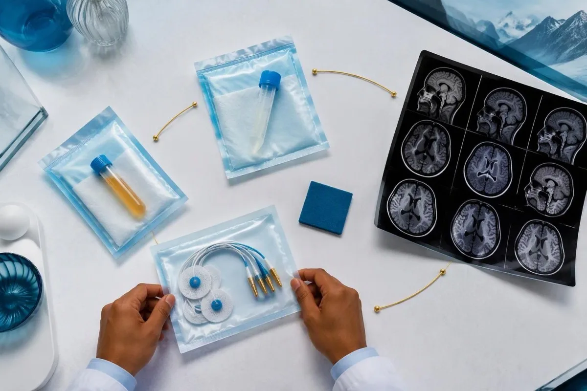
Nerve conduction studies can support a sensory neuronopathy pattern by showing reduced sensory responses across distributions that do not fit a single compressed nerve. Brain or spinal MRI addresses different questions, including structural disease and some inflammatory changes. A normal MRI does not exclude peripheral sensory ganglion involvement; these are 2 distinct anatomical compartments with different diagnostic tools.
Cerebrospinal fluid may show increased cells, protein or evidence of intrathecal immune activity, but none of those findings is specific to anti-Hu. A lumbar puncture is most useful when the neurological syndrome warrants it, rather than as an automatic response to 1 weak serum band. Our discussion of smegenų skysčio oligokloninių juostų interpretavimą explains why immune activity in spinal fluid does not identify a single disease.
Basic investigations still matter: glucose control, vitamin status, renal function and serum protein assessment can uncover treatable contributors to neuropathy. A clinician may request 2 or more of these alongside specialist testing, depending on the examination. The serumo baltymų tyrimo vadove provides background, but normal routine blood work neither confirms nor excludes an anti-Hu-associated neurological process.
Ar teigiamas anti-Hu rezultatas patvirtina paraneoplastinį sindromą?
A positive antibody alone does not confirm anti-Hu paraneoplastic syndrome. The PNS-Care approach combines the neurological phenotype, antibody risk category, cancer findings and follow-up; clinicians also check for direct cancer effects, treatment toxicity and unrelated neurological conditions before attributing symptoms to a paraneoplastic immune response.
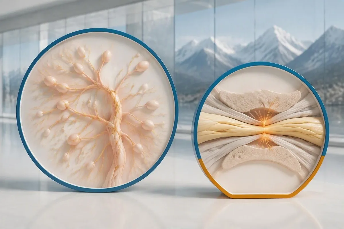
Definite, probable and possible PNS are diagnostic categories, not cancer stages. The 2021 criteria use a structured scoring system rather than allowing 1 antibody to determine the whole diagnosis (Graus et al., 2021). A confirmed anti-Hu result with a strongly compatible syndrome remains clinically significant even before a cancer is identified, but clinicians should state clearly which parts of the assessment remain unresolved.
An older patient can have anti-Hu reactivity and a separate cause of numbness, such as diabetes, nutritional deficiency or a monoclonal protein disorder. Coexistence is possible; either-or reasoning can miss treatable disease. If serum protein studies reveal a band, our immunofixation result guide explains why that finding requires its own interpretation rather than being automatically folded into the anti-Hu diagnosis.
Kantesti yra AI kraujo tyrimo rezultatų interpretavimo platforma that can help organize these distinctions in a laboratory explanation, but it cannot assign a reliable PNS-Care diagnosis from a report alone. Our klinikinio validavimo sistema separates technical evaluation from clinical conclusions. In practice, I would document 3 separate judgments: confidence in the antibody, confidence in the neurological syndrome and the current evidence for cancer.
Kaip gydytojai ieško susijusio vėžio?
Cancer evaluation usually prioritizes the chest because small-cell lung cancer is the strongest adult anti-Hu association. Doctors commonly begin with appropriate chest CT and may add FDG-PET/CT or targeted investigations if suspicion remains; the exact plan depends on age, symptoms, examination, smoking history and previous imaging.
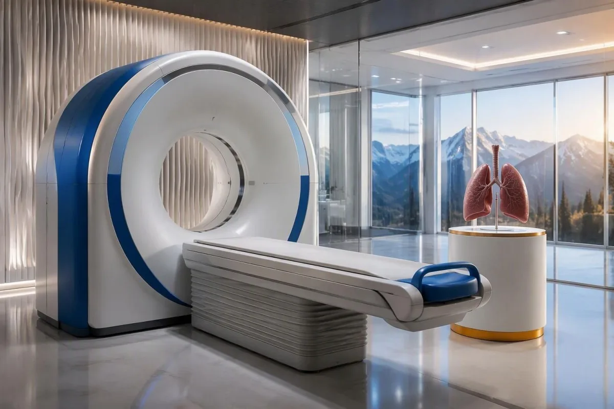
The cancer association is substantial in established anti-Hu clinical cohorts. Villagrán-García et al. (2023) reported identified cancer in 349 of 466 patients, approximately 75%, in a specialist anti-Hu cohort. This is valuable evidence for taking confirmed, clinically relevant anti-Hu seriously; it is not a personalized prediction for an asymptomatic person with an unconfirmed positive screening strip.
A chest X-ray is not equivalent to a chest CT, and routine low-dose screening is not necessarily the correct examination for a suspected paraneoplastic syndrome. The clinician chooses the protocol and body regions based on the diagnostic question. Likewise, neurological symptoms do not automatically mean cancer has spread to the brain; our brain imaging warning-sign guide distinguishes structural investigation from immune-mediated disease.
A suspicious scan often leads to tissue sampling through a method chosen by respiratory or oncology specialists, such as bronchoscopic assessment of an accessible area. Imaging suspicion and cancer confirmation are 2 separate steps. Never-smoking status lowers the baseline probability of small-cell lung cancer but does not cancel a well-confirmed anti-Hu finding with a convincing neurological presentation; other cancers and non-cancer explanations still need consideration.
Kodėl pirmieji vėžio vaizdiniai tyrimai gali būti neigiami?
Initial imaging can be negative because an associated cancer is still too small to detect, lies outside the examined area or is not readily visible with that technique. Neurological autoimmunity may precede cancer detection, so a negative scan reduces concern without necessarily ending follow-up in a high-risk clinical presentation.
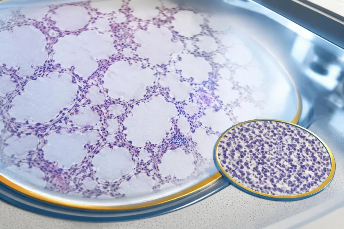
A negative scan is time-specific and method-specific. A normal chest CT performed on 1 date answers whether that examination identified a suspicious chest finding then; it does not guarantee that every possible associated cancer is absent indefinitely. Ask what was actually imaged, whether the protocol was suitable and whether the specialist recommends PET/CT or targeted investigation despite the initial result.
Villagrán-García et al. (2023) described cancers detected beyond the first 2 years in a minority of their anti-Hu cohort, supporting individualized longer surveillance. Their findings also support considering PET-based investigation when initial CT is unrevealing in an appropriate clinical context. This is an escalation decision for the treating team, not a reason to independently purchase repeated whole-body scans without weighing radiation, incidental findings and cost.
Neurological deterioration after negative imaging still needs reassessment. Cancer surveillance and neurological monitoring are 2 parallel tracks; one reassuring scan does not explain new loss of function. A marker such as neurofilament light chain may indicate neuronal injury in selected settings, but our NfL interpretation guide explains why it cannot establish anti-Hu disease, locate cancer or replace examination.
Kiek laiko reikėtų tęsti vėžio patikrą?
The 2021 PNS criteria recommend repeat cancer screening every 4–6 months for 2 years when a high-risk neurological phenotype accompanies a high-risk antibody. Longer surveillance may be appropriate for selected anti-Hu patients; the interval and duration should reflect confirmation, clinical evolution, initial investigations and individual cancer risk.

The 2-year schedule is not an instruction to scan every positive antibody result repeatedly. Graus et al. (2021) link that recommendation to the combination of high-risk phenotype and high-risk antibody. An incidental, weak result without a matching syndrome needs confirmation and individualized assessment first; repeating imaging mechanically can create incidental findings and unnecessary procedures without resolving the original uncertainty.
Older European guidance recommended longer surveillance. The EFNS task force advised repeating screening after 3–6 months and then every 6 months for up to 4 years in relevant antibody-positive paraneoplastic presentations (Titulaer et al., 2011). The 2023 anti-Hu cohort supported considering screening up to 5 years in selected patients, so a specialist may reasonably extend follow-up beyond the newer general 2-year framework.
Keep a dated plan stating the next examination, the clinician responsible and what symptoms should trigger earlier contact. Kantesti explanations can help you understand terminology, but the platform should not be treated as a scan-reminder service or substitute for that plan. Our guide to risk-based cancer screening addresses a different setting: routine prevention is not the same as investigating a possible paraneoplastic syndrome.
Koks gydymas padeda sergant anti-Hu sukelta neurologine liga?
Treatment focuses on an identified cancer, selected immune therapy and symptom-directed rehabilitation. Anti-Hu-associated neurological disease often responds less predictably to immunotherapy than neuronal surface-antibody disorders because intracellular targets and T-cell-mediated injury are involved; treatment may aim to stabilize function rather than promise complete recovery.

Treating an associated cancer is a central part of care, but established neurological injury may persist even when cancer treatment succeeds. Neurology and oncology teams may consider corticosteroids, intravenous immunoglobulin, plasma exchange or other immune treatments in selected situations. There is no single evidence-based dose or 1 universal regimen appropriate for everyone with anti-Hu positivity, especially without a confirmed clinical syndrome.
An antibody titer is not a dependable treatment target on its own. A lower number does not necessarily mean restored nerve function, and a persistent positive result does not by itself prove cancer progression. This differs from some surface-antibody disorders; our MOG antibody diagnosis guide explains another immune neurological condition, but its diagnostic rules and treatment expectations should not be transferred to anti-Hu.
Immune checkpoint cancer therapies deserve an explicit discussion when anti-Hu neurological disease is present, because they can provoke or worsen paraneoplastic neurological autoimmunity. This is a specialist risk-benefit decision, not an automatic prohibition based on 1 laboratory result. Tell the oncologist about previous neurological symptoms and confirmed neuronal antibodies before treatment decisions are finalized; coordinating the teams early can prevent avoidable misunderstanding.
Ką galite saugiai daryti, kol vyksta tyrimai?
While investigations continue, protect mobility, swallowing and medication safety rather than trying to lower anti-Hu antibodies with supplements. Use the neurological symptoms—not the appearance of the laboratory flag—to decide whether ordinary follow-up is sufficient or urgent care is needed; a new functional loss warrants contact before the next planned scan.
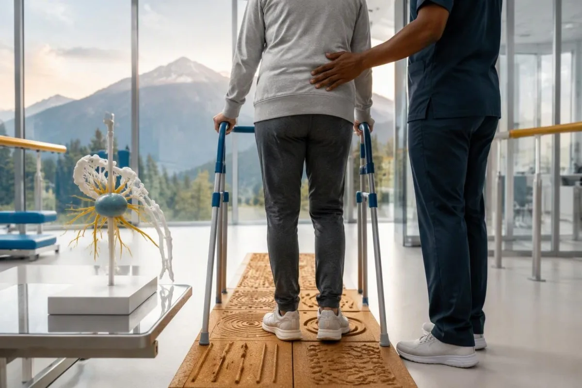
Position-sense loss increases fall risk, particularly in poor lighting or on uneven ground. A physiotherapist may recommend a suitable walking aid and individualized balance strategies; occupational therapy can address hand coordination and home hazards. A short 7-day record of falls, near-falls and walking changes is often more useful at review than the vague statement “my balance is worse,” but stop activities that feel unsafe.
Excess vitamin B6 can itself cause sensory neuropathy, making unsupervised high-dose nerve supplements particularly unhelpful during this assessment. Review every supplement and its daily dose in mg, including overlapping multivitamins and combination products. Our B grupės vitaminų saugos vadovas explains that problem; correcting a documented deficiency is different from taking progressively larger doses in the hope of treating anti-Hu autoimmunity.
Vitamin B12 deficiency can contribute to sensory and gait problems and may need investigation even when anti-Hu is confirmed. There is no universally interchangeable B12 cutoff: results in pg/ml ir pmol/L require their own laboratory context, sometimes with functional testing. Our ribinio B12 stebėjimo vadovas explains those next steps; new choking, breathing difficulty or rapid weakness should be assessed urgently instead of managed with vitamins.
Kaip DI gali saugiai padėti paaiškinti anti-Hu tyrimo ataskaitą?
AI can help explain report terminology and organize questions, but it cannot confirm anti-Hu specificity, diagnose paraneoplastic syndrome or rule out cancer. Safe interpretation preserves the original assay details and separates 3 issues: what the laboratory reported, what the result may mean and what a clinician has actually established.

Kantesti yra AI pagrindu veikiantis kraujo tyrimų analizės įrankis that can provide a plain-language explanation of uploaded laboratory information, including the distinction between an antibody screen and a confirmed clinical diagnosis. A rapid explanation—often around 60 seconds for report processing—is not a reference-laboratory assay. Our AI technologijų gidas describes the interpretation workflow without making anti-Hu-specific diagnostic guarantees.
The complete source report should remain available for verification. An explanation should not silently replace “weakly reactive” with “confirmed,” drop the specimen type or invent a universal reference interval. Our guide to dirbtinio intelekto pateikiamų medicininių paaiškinimų tikrinimą describes practical source checks; for this antibody, compare at least the assay wording, confirmation comment and collection date against the original document.
Kantesti cannot examine sensation, review tissue staining directly through a report explanation or establish that negative imaging has excluded a tiny cancer. Those are 3 different clinical capabilities, not features that follow from understanding laboratory language. Share the explanation with the treating team as a discussion aid, and do not delay urgent assessment while uploading documents or waiting for a second automated interpretation.
Kitas vizitas ir tyrimų skaidrumas
Bring the complete antibody report, a dated symptom history, previous imaging reports and a current medication list to your next appointment. Ask the clinician to clarify antibody confirmation, the neurological diagnosis and cancer surveillance separately; those 3 answers may have different levels of certainty and different follow-up dates.
A useful 1-page symptom history records onset, progression, sensory distribution, walking changes, memory problems and autonomic symptoms. Include prior cancer treatment and any recent immunoglobulin therapy, then ask who will coordinate neurology and oncology if both are involved. As Thomas Klein, MD, I would rather see a clear timeline than 20 disconnected screenshots; it makes discrepancies between the laboratory result and clinical course easier to spot.
Medical authorship is not a substitute for an individual neurological examination. Mūsų medicininės konsultacinės tarybos informacija describes the clinicians associated with our work, while your own specialist determines the appropriate next investigation. As of 2026 m. spalio 25 d., the recommendations discussed here draw on the 2021 international PNS criteria, the 2011 EFNS screening guidance and the 2023 anti-Hu cohort—not an invented 2026 guideline.
The 2 Figshare publications below are platform-related research references, not evidence validating anti-Hu diagnosis or cancer exclusion. Their supplied titles describe hantavirus triage deployment and a synthetic-case technical benchmark; neither title establishes anti-Hu-specific clinical performance. Because publication authors and dates were not supplied for verification, the citations use title-first APA formatting with “n.d.”; the ResearchGate and Academia.edu links are searches, not claims of verified publication copies.
Dažnai užduodami klausimai
0 Ar teigiamas anti-Hu antikūnų tyrimo rezultatas reiškia, kad sergu vėžiu?
0 Anti-Hu antikūnų teigiamas rezultatas pats savaime nereiškia vėžio diagnozės. Anti-Hu yra su didele vėžio rizika susijęs antikūnas; 2023 m. specialistų tirtų pacientų, kuriems nustatyta anti-Hu, grupėje vėžys nustatytas 349 iš 466 pacientų, tačiau šis skaičius nenusako atsitiktinio, nepatvirtinto atrankinio tyrimo rezultato rizikos. Gydytojai įvertina neurologinius simptomus, patvirtina netikėtus laboratorinių tyrimų rezultatus ir ieško galimo vėžio, suaugusiesiems paprastai pirmiausia tirdami krūtinės ląstą. Vėžio diagnozei paprastai reikia atskirų klinikinių, vaizdinimo ir audinių tyrimų duomenų.
Ar anti-Hu ir ANNA-1 yra tas pats?
Anti-Hu ir ANNA-1 paprastai nurodo tą pačią neuronų antikūnų šeimą. ANNA-1 reiškia 1 tipo antineuroninius branduolio antikūnus, o Hu – viduląstelinius neuronų baltymus, kuriuos atpažįsta antikūnas. ANNA-1 nėra tas pats, kas įprastas ANA, naudojamas sisteminių autoimuninių ligų tyrimuose. Patikrinkite tikslų tyrimo metodą ir patvirtinimo formuluotę, nes laboratorijos tą pačią antikūnų šeimą gali pateikti skirtingais formatais.
0 Ar anti-Hu antikūnų tyrimo rezultatas gali būti klaidingai teigiamas?
Anti-Hu antikūnų tyrimas gali sukelti klaidinančią arba nespecifinę reakciją, ypač kai silpnas atrankinio tyrimo rezultatas neatitinka neurologinio klinikinio vaizdo. Patvirtinimui paprastai taikomi 2 vienas kitą papildantys metodai: būdingas audinių dažymo raštas ir antigenui specifinis tyrimas, kurio rezultatus interpretuoja patyrusi laboratorija. Pakartojus tą patį komercinį metodą, ta pati analitinė problema gali pasikartoti, o ne išsispręsti. Sparčiai progresuojančius neurologinius simptomus vis tiek būtina laiku įvertinti, kol laukiama patvirtinimo.
Kaip dažnai reikėtų kartoti tyrimus, jei anti-Hu rezultatas teigiamas?
2021 m. PNS kriterijai rekomenduoja kartoti vėžio patikrą kas 4–6 mėnesius 2 metus, kai didelės rizikos neurologinį fenotipą lydi didelės rizikos antikūnas. Šis tvarkaraštis automatiškai netaikomas kiekvienam atsitiktinai gautam teigiamam anti-Hu rezultatui. Ankstesnėse EFNS gairėse rekomenduota stebėti iki 4 metų, o 2023 m. anti-Hu kohortos tyrimas pagrindė galimybę atrinktiems pacientams stebėjimą tęsti iki 5 metų. Jūsų specialistas turėtų nurodyti vaizdinimo metodą, intervalą, trukmę ir simptomus, dėl kurių reikėtų kreiptis anksčiau.
Ar anti-Hu gali būti teigiamas, kai KT ir PET tyrimų rezultatai yra normalūs?
0 Anti-Hu gali išlikti teigiamas, kai atliekant KT ar PET tyrimą vėžys nenustatomas. Neurologinis autoimunitetas gali pasireikšti anksčiau nei vėžys aptinkamas, o neigiamas tyrimo rezultatas atspindi ištirtas sritis, taikytą metodiką ir tyrimo datą, bet nėra nuolatinė garantija, kad vėžio nėra. Esant suderinamiems didelės rizikos simptomams, 2021 m. kriterijai rekomenduoja kartoti patikrą kas 4–6 mėnesius 2 metus, o tam tikriems anti-Hu pacientams, atsižvelgiant į individualią situaciją, stebėjimą tęsti ilgiau. Taip pat reikėtų iš naujo įvertinti nepatvirtintą antikūnų reaktyvumą ir ne su vėžiu susijusius paaiškinimus, o ne manyti, kad nematomas vėžys tikrai yra.
Ar sumažėjęs anti-Hu antikūnų kiekis reiškia, kad gydymas veiksmingas?
Mažėjantis anti-Hu antikūnų kiekis patikimai neįrodo nei neurologinio atsistatymo, nei sėkmingo vėžio gydymo. Anti-Hu atpažįsta viduląstelinius baltymus, o su juo susijęs neurologinis pažeidimas dažnai apima T ląstelių mechanizmus, o ne vien antikūnų poveikį. Dviejų skirtingose laboratorijose atliktų tyrimų titrai taip pat gali būti nepalyginami dėl skirtingų metodų. Gydytojai vertina funkciją, apžiūros duomenis, aktualių tyrimų rezultatus ir atsaką į vėžio gydymą, užuot laikę antikūnų kiekį savarankišku gydymo tikslu.
Gaukite AI pagrįstą kraujo tyrimo analizę jau šiandien
Prisijunkite prie daugiau nei 2 milijonų naudotojų visame pasaulyje, kurie pasitiki Kantesti dėl momentinės, tikslios laboratorinių tyrimų analizės. Įkelkite savo kraujo tyrimo rezultatus ir per kelias sekundes gaukite išsamią 15,000+ biomarkerių interpretaciją.
📚 Nuorodomis pagrįsti moksliniai leidiniai
Klein, T., Mitchell, S., & Weber, H. (2026). Daugiakalbė AI pagalba klinikiniam sprendimų palaikymui ankstyvai hantaviruso triage: dizainas, inžinerinis validavimas ir realaus pasaulio diegimas per 50 000 interpretuotų kraujo tyrimų ataskaitų. Kantesti AI Medical Research.
Klein, T., Mitchell, S., & Weber, H. (2026). Iš anksto užregistruotas, pagal rubrikas paremtas automatizuotas techninis Kantesti kraujo tyrimo interpretavimo variklio etaloninis vertinimas 100 000 sintetinių testų atvejų. Kantesti AI Medical Research.
📖 Išorinės medicininės nuorodos
Graus F et al. (2021). Updated Diagnostic Criteria for Paraneoplastic Neurologic Syndromes. Neurology: Neuroimmunology & Neuroinflammation.
Titulaer MJ et al. (2011). Screening for tumours in paraneoplastic syndromes: report of an EFNS Task Force. „European Journal of Neurology“.
Villagrán-García D et al. (2023). Revisiting anti-Hu paraneoplastic autoimmunity: phenotypic characterization and cancer diagnosis. „Brain Communications“.
⚕️ Medicininis atsakomybės apribojimas
Šis straipsnis skirtas tik mokymo tikslais ir nesudaro medicininės konsultacijos. Visada kreipkitės į kvalifikuotą sveikatos priežiūros specialistą dėl diagnozės ir gydymo sprendimų.
E-E-A-T patikimumo signalai
Patirtis
Gydytojo vadovaujama klinikinė laboratorinių tyrimų interpretavimo darbo eigų peržiūra.
Ekspertizė
Laboratorinės medicinos dėmesys tam, kaip biomarkeriai elgiasi klinikiniame kontekste.
Autoritetas
Parašyta dr. Thomas Klein, peržiūrėjo dr. Sarah Mitchell ir prof. dr. Hans Weber.
Patikimumas
Įrodymais pagrįsta interpretacija su aiškiais tolesnių veiksmų keliais, siekiant sumažinti nerimą.
