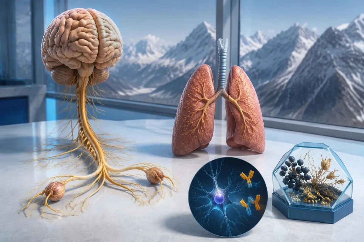
Станоўчы вынік на антыцелы anti-Hu выклікае падазрэнне на імунную рэакцыю нервовай сістэмы, звязаную з ракам, асабліва на рак лёгкага з дробных клетак, але не пацвярджае дыягназ раку. Лекары ацэньваюць сімптомы, пацвярджаюць вынік лабараторнага даследавання і арганізуюць абследаванне на рак; паўторны скрынінг можа быць неабходны, нават калі першыя здымкі не выявілі паталогіі.



- Anti-Hu / ANNA-1 — антыцела высокай рызыкі, звязанае з паранеапластычнымі сіндромамі; гэтая катэгорыя асацыюецца з ракам больш чым у 70% пацверджаных клінічных кагортах, але не абавязкова ў кожным выпадку, калі вынік скрынінгу яшчэ не пацверджаны.
- Дыягназ раку патрабуе пацвярджэння наяўнасці злаякаснай пухліны, звычайна ў тым ліку даследавання тканіны; аднаго станоўчага выніку на антыцелы для гэтага недастаткова.
- Пацвярджэнне выніку аналізу часта аб’ядноўвае 2 узаемадапаўняльныя падыходы: характэрны характар афарбоўвання тканіны і тэст на спецыфічны антыген.
- Неўралагічныя сімптомы такія як прагрэсавальная страта адчувальнасці, парушэнне раўнавагі, прыступы або вегетатыўная дысфункцыя, змяняюць значнасць і тэрміновасць рэагавання на вынік.
- Адмоўныя вынікі візуалізацыйнага абследавання азначаюць, што на момант абследавання рак не быў выяўлены; гэта не выключае магчымасці існавання невялікай пухліны, якую спачатку немагчыма выявіць.
- Паўторны скрынінг рэкамендуецца праводзіць кожныя 4–6 месяцаў на працягу 2 гадоў, калі высокарызыкоўны неўралагічны фенатып спалучаецца з антыцелам высокай рызыкі, згодна з крытэрыямі PNS 2021 года.
- Больш працяглае назіранне можа быць мэтазгодным для асобных пацыентаў; кагорта anti-Hu, апісаная ў 2023 годзе, падтрымала разгляд скрынінгу даўжэй за 2 гады, часам да 5 гадоў.
- Экстраныя сімптомы уключаюць новыя цяжкасці з дыханнем, немагчымасць бяспечна глытаць, хуткае ўзмацненне слабасці, першы эпілептычны прыступ або раптоўныя неўралагічныя парушэнні.
Што азначае станоўчы вынік на антыцелы anti-Hu?
Ан станоўчы вынік на антыцелы anti-Hu азначае, што лабараторыя выявіла рэактыўнасць да нейрональных бялкоў Hu; сам па сабе ён не пацвярджае наяўнасць раку або неўралагічнага сіндрому. Інтэрпрэтацыя мае 3 складнікі: ці з'яўляецца вынік сапраўдным, ці адпавядае яму неўралагічная карціна і ці можна выявіць звязаны з ім рак.
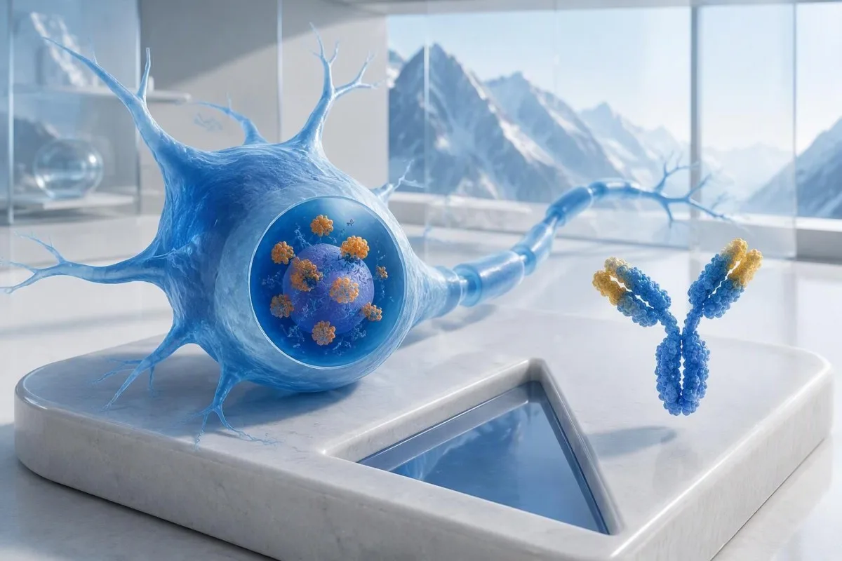
Anti-Hu класіфікуецца як антыцела высокай рызыкі, звязанае з ракам, прычым катэгорыя вызначаецца сувяззю з ракам больш чым у 70% выпадкаў у адпаведных клінічных папуляцыях (Graus et al., 2021). Гэты працэнт нельга проста прыпісваць чалавеку без скаргаў са слабым, непацверджаным вынікам панэльнага аналізу: на верагоднасць уплываюць даследаваная папуляцыя пацыентаў, метад аналізу і неўралагічная карціна.
Гіпатэтычны пацыент ва ўзросце 62 гадоў з 8-тыднёвым пагаршэннем раўнавагі і асіметрычнай стратай адчувальнасці патрабуе іншай ацэнкі, чым чалавек, у якога антыцелы выпадкова выявілі падчас шырокага абследавання з нагоды стомленасці. Ніводнаму з іх не варта казаць: “У вас рак”, абапіраючыся толькі на лабараторны бланк. Наша тлумачэнне станоўчыя вынікі даследавання на антыцелы дапамагае адрозніць імунны маркер ад устаноўленага дыягназу.
Кантэсці — гэта Аналізатар крыві са штучным інтэлектам што можа дапамагчы растлумачыць фармулёўку ў справаздачы аб anti-Hu, але лабараторнае пацвярджэнне і ацэнку на наяўнасць раку павінны праводзіць вашы лечачыя ўрачы. Як Thomas Klein, MD, галоўны медыцынскі дырэктар Kantesti LTD, я найперш спытаў бы, што стала прычынай прызначэння менавіта гэтага аналізу, а не наколькі трывожна выглядае чырвонае папярэджанне; наша арганізацыя і клінічная місія тлумачаць гэта адрозненне.
Ці з’яўляюцца ANNA-1 і anti-Hu аднымі і тымі ж антыцеламі?
антыцелы ANNA-1 і anti-Hu звычайна адносяцца да аднаго сямейства антыцелаў. ANNA-1 расшыфроўваецца як антынейранальныя ядзерныя антыцелы тыпу 1; назва Hu адносіцца да ўнутрыклетачных нейрональных бялкоў, якія распазнае імунны адказ, а не да асобнага захворвання або стадыі раку.
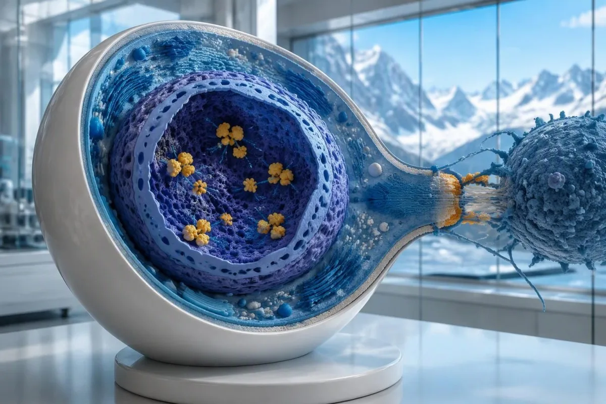
Бялкі Hu — гэта ўнутрыклетачныя РНК-звязвальныя бялкі, і anti-Hu найлепш разумець як маркер імуннага адказу, у якім часта ўдзельнічаюць цытатаксічныя Т-клеткі. Гэта адрозніваецца ад расстройстваў, пры якіх антыцелы звязваюцца з даступнымі вонкавымі бялкамі нейронаў. Гэтае адрозненне тлумачыць, чаму простае зніжэнне ўзроўню антыцелаў не дазваляе надзейна ліквідаваць ужо сфарміраванае неўралагічнае пашкоджанне, звязанае з anti-Hu, нават калі ў 2 справаздачах прыведзены істотна розныя тытры.
Справаздача з вынікам “выяўлена ANNA-1” — гэта не тое самае, што звычайны станоўчы ANA, нягледзячы на падабенства літар. АНА звычайна азначае антынуклеарныя антыцелы, якія выкарыстоўваюцца для ацэнкі сістэмных аўтаімунных захворванняў, тады як ANNA-1 вызначае нейрональны антыцельны патэрн. Абмяркоўваючы вынікі, не блытайце гэтыя 2 скарачэнні; наша кіраўніцтва па тэрміналогіі ANA тлумачыць больш знаёмы аналіз.
Anti-Hu можна выявіць у некаторых людзей з дробнаклетачным ракам лёгкага, у якіх няма распазнавальнага неўралагічнага сіндрому. І наадварот, неўралагічныя сімптомы могуць прывесці да выяўлення антыцелаў яшчэ да таго, як рак стане бачным. Гэта 2 розныя клінічныя сцэнарыі: само па сабе антыцела не паказвае ўрачу, ці пашкоджваюцца цяпер нервы і дзе можа размяшчацца асноўны рак.
Як трэба інтэрпрэтаваць вынік аналізу на антыцелы anti-Hu?
An anti-Hu antibody test has no universal numerical cutoff that can be applied across laboratories. Read the specimen type, assay method, result category, laboratory reference interval and confirmation comments together; a dilution titer, band intensity and numerical index are 3 different ways of reporting reactivity.
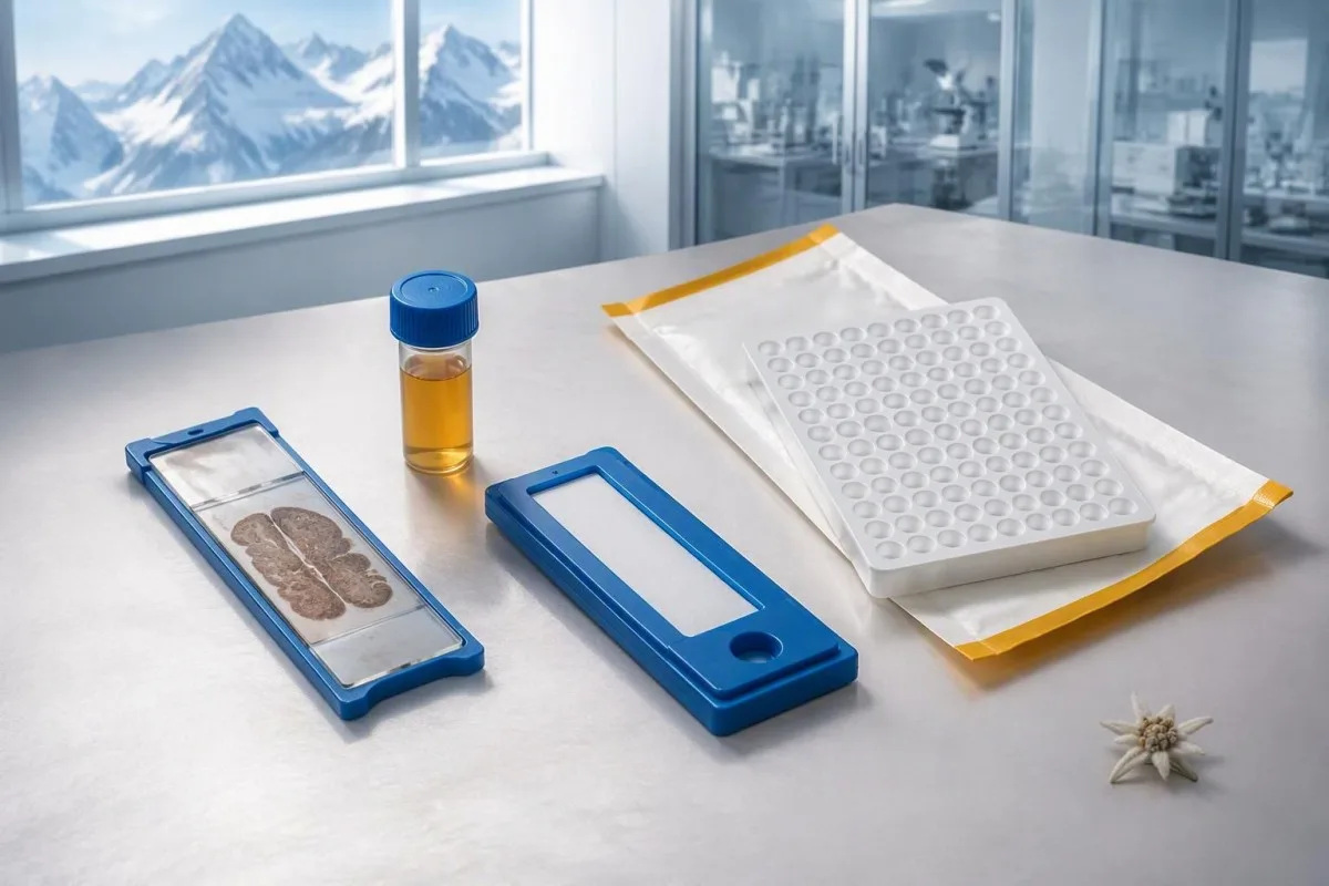
A hypothetical titer of 1:240 describes detectable reactivity after a particular dilution; it is not a cancer probability of 240, nor does it establish cancer stage. A numerical index from another method may use entirely different calibration. Our guide to якасныя і колькасныя вынікі explains why “positive” and “how much” answer different laboratory questions.
Weak or borderline reactivity deserves method-specific interpretation, particularly when a broad panel was ordered without a convincing neurological indication. Ask whether the laboratory observed the characteristic neuronal staining pattern and whether an antigen-specific confirmation was performed. Two tests using the same underlying strip technology may reproduce the same analytical problem; agreement is more informative when the methods are genuinely complementary.
Bring the complete report, not just a screenshot of the word positive. The 5 most useful details are the specimen, collection date, assay name, reported strength or titer and laboratory interpretation. Also list recent intravenous immunoglobulin and other immune treatments, because passive antibodies or treatment effects can complicate some serological results; your laboratory can advise whether that issue plausibly affects this particular finding.
Калі станоўчы вынік на anti-Hu патрабуе пацвярджэння?
Unexpected, weak or clinically discordant anti-Hu results should be reviewed for confirmation, preferably with an experienced neuroimmunology laboratory. The 2021 PNS criteria emphasize interpreting neuronal antibody testing alongside the phenotype and using appropriate confirmatory methods rather than accepting an isolated commercial-panel result as a diagnosis.
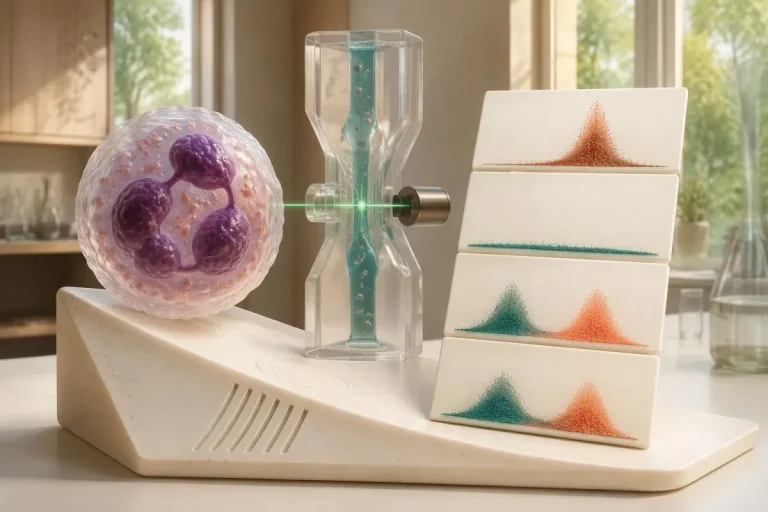
Tissue-based immunohistochemistry or immunofluorescence and an antigen-specific assay provide different information. The first identifies a characteristic distribution of neuronal staining; the second tests reactivity against the relevant antigen. Graus et al. (2021) describe these complementary approaches, with specialist reassessment particularly useful when 1 method gives a result that does not fit the clinical picture.
Repeating a sample can address collection errors or a transient analytical concern, but repeating the same kit is not always enough. In a hypothetical 45-year-old with no objective neurological findings and a faint isolated band, I would want the laboratory’s explanation before treating the report as established anti-Hu autoimmunity. Our GAD65 interpretation guide illustrates another antibody for which context and assay characteristics change meaning.
Confirmation and urgent assessment can happen in parallel. A person who develops seizures, substantial walking difficulty or progressive swallowing impairment should not wait several weeks for a reference-laboratory answer before receiving neurological care. The practical question is not “repeat or investigate?” but which of those 2 tasks can safely proceed together, given the symptoms and the initial laboratory evidence.
Якія неўралагічныя сімптомы робяць вынік на anti-Hu больш трывожным?
Progressive sensory neuronopathy, encephalomyelitis, limbic encephalitis and marked autonomic dysfunction are recognized anti-Hu presentations. Symptoms can involve more than 1 part of the nervous system, so the pattern and speed of change are usually more informative than a single symptom such as tingling or fatigue.

Sensory neuronopathy can cause asymmetric numbness, impaired position sense and sensory ataxia, sometimes involving the hands or trunk early rather than following a simple feet-first pattern. A patient may lose balance in the dark because vision had been compensating for reduced position feedback. That is different from the focal finger distribution discussed in our carpal tunnel symptom guide, although neither pattern can be diagnosed from description alone.
Anti-Hu autonomic involvement can affect gut movement, blood-pressure regulation and bladder function. Severe constipation, unexplained intestinal pseudo-obstruction or dizziness on standing becomes more relevant when accompanied by objective neurological abnormalities. Recording symptoms over 7 days can reveal combinations that otherwise get presented as separate stomach, balance and urinary complaints; sudden severe abdominal symptoms still require assessment for non-neurological emergencies.
New memory impairment, behavioral change or seizures may indicate central nervous system involvement, while rapid weakness has a broader emergency differential. The трывожных прыкметах небяспечнай слабасці in our neurological safety guide apply regardless of the antibody label. Call emergency services for new breathing difficulty, unsafe swallowing, a first seizure or sudden stroke-like symptoms rather than waiting for a scheduled follow-up.
Якія неўралагічныя абследаванні могуць правесці пасля станоўчага выніку?
Neurological follow-up may include examination, nerve conduction studies, MRI and cerebrospinal fluid testing, selected according to the symptoms. There is no mandatory battery for every anti-Hu-positive person; the purpose is to document which structures are affected and exclude other causes, not merely to collect additional positive tests.
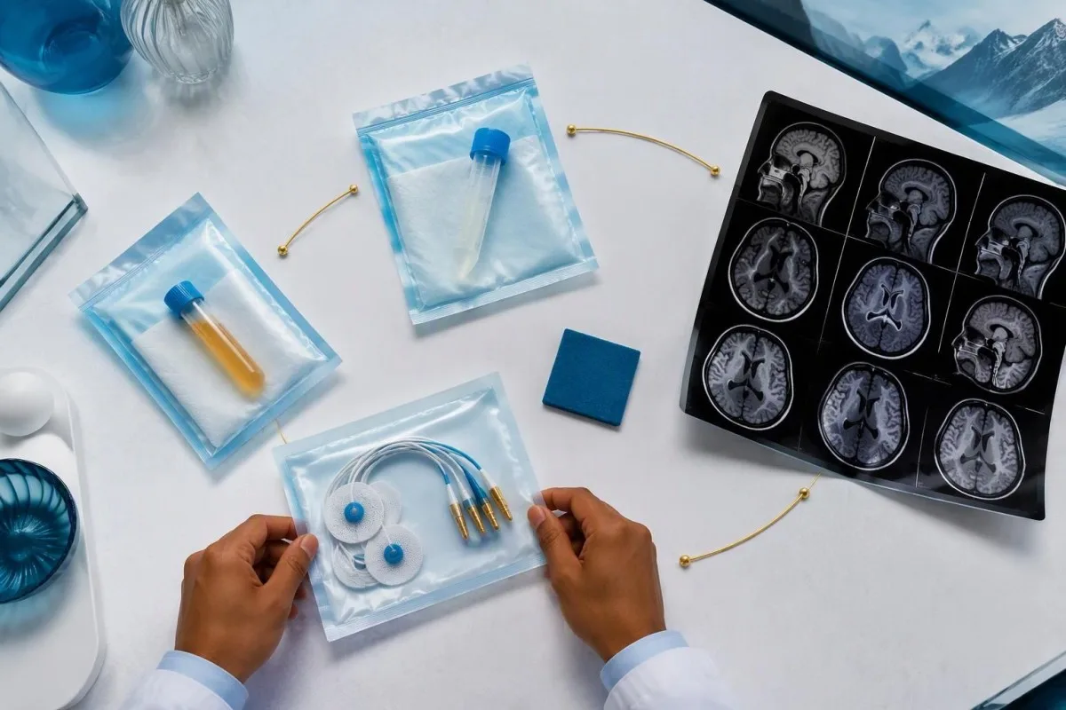
Nerve conduction studies can support a sensory neuronopathy pattern by showing reduced sensory responses across distributions that do not fit a single compressed nerve. Brain or spinal MRI addresses different questions, including structural disease and some inflammatory changes. A normal MRI does not exclude peripheral sensory ganglion involvement; these are 2 distinct anatomical compartments with different diagnostic tools.
Cerebrospinal fluid may show increased cells, protein or evidence of intrathecal immune activity, but none of those findings is specific to anti-Hu. A lumbar puncture is most useful when the neurological syndrome warrants it, rather than as an automatic response to 1 weak serum band. Our discussion of расшыфроўцы вынікаў аналізу СМЖ на алігадыкланальныя палосы explains why immune activity in spinal fluid does not identify a single disease.
Basic investigations still matter: glucose control, vitamin status, renal function and serum protein assessment can uncover treatable contributors to neuropathy. A clinician may request 2 or more of these alongside specialist testing, depending on the examination. The дапаможнік па даследаванні бялкоў сыроваткі provides background, but normal routine blood work neither confirms nor excludes an anti-Hu-associated neurological process.
Ці пацвярджае станоўчы вынік на anti-Hu паранеапластычны сіндром?
A positive antibody alone does not confirm anti-Hu paraneoplastic syndrome. The PNS-Care approach combines the neurological phenotype, antibody risk category, cancer findings and follow-up; clinicians also check for direct cancer effects, treatment toxicity and unrelated neurological conditions before attributing symptoms to a paraneoplastic immune response.
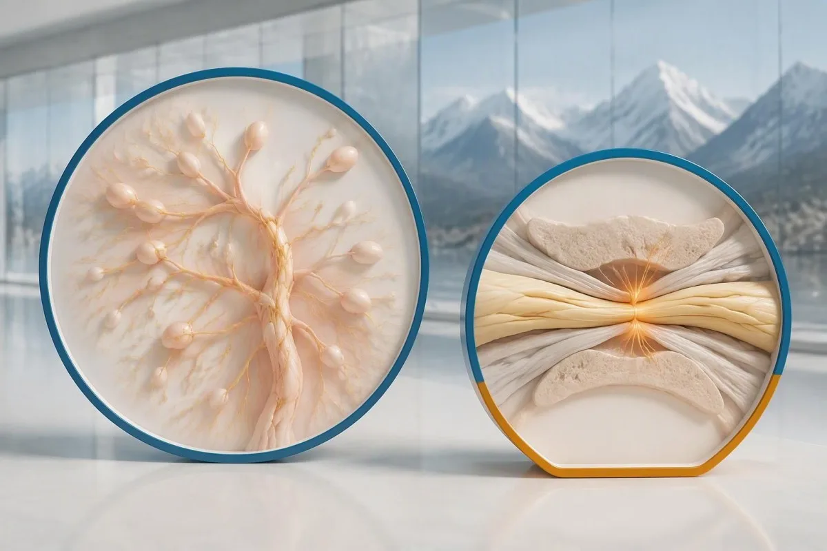
Definite, probable and possible PNS are diagnostic categories, not cancer stages. The 2021 criteria use a structured scoring system rather than allowing 1 antibody to determine the whole diagnosis (Graus et al., 2021). A confirmed anti-Hu result with a strongly compatible syndrome remains clinically significant even before a cancer is identified, but clinicians should state clearly which parts of the assessment remain unresolved.
An older patient can have anti-Hu reactivity and a separate cause of numbness, such as diabetes, nutritional deficiency or a monoclonal protein disorder. Coexistence is possible; either-or reasoning can miss treatable disease. If serum protein studies reveal a band, our immunofixation result guide explains why that finding requires its own interpretation rather than being automatically folded into the anti-Hu diagnosis.
Кантэсці — гэта Платформа AI для расшыфроўкі аналізу крыві that can help organize these distinctions in a laboratory explanation, but it cannot assign a reliable PNS-Care diagnosis from a report alone. Our клінічную рамку валідацыі separates technical evaluation from clinical conclusions. In practice, I would document 3 separate judgments: confidence in the antibody, confidence in the neurological syndrome and the current evidence for cancer.
Як лекары шукаюць звязаны з гэтым рак?
Cancer evaluation usually prioritizes the chest because small-cell lung cancer is the strongest adult anti-Hu association. Doctors commonly begin with appropriate chest CT and may add FDG-PET/CT or targeted investigations if suspicion remains; the exact plan depends on age, symptoms, examination, smoking history and previous imaging.
The cancer association is substantial in established anti-Hu clinical cohorts. Villagrán-García et al. (2023) reported identified cancer in 349 of 466 patients, approximately 75%, in a specialist anti-Hu cohort. This is valuable evidence for taking confirmed, clinically relevant anti-Hu seriously; it is not a personalized prediction for an asymptomatic person with an unconfirmed positive screening strip.
A chest X-ray is not equivalent to a chest CT, and routine low-dose screening is not necessarily the correct examination for a suspected paraneoplastic syndrome. The clinician chooses the protocol and body regions based on the diagnostic question. Likewise, neurological symptoms do not automatically mean cancer has spread to the brain; our brain imaging warning-sign guide distinguishes structural investigation from immune-mediated disease.
A suspicious scan often leads to tissue sampling through a method chosen by respiratory or oncology specialists, such as bronchoscopic assessment of an accessible area. Imaging suspicion and cancer confirmation are 2 separate steps. Never-smoking status lowers the baseline probability of small-cell lung cancer but does not cancel a well-confirmed anti-Hu finding with a convincing neurological presentation; other cancers and non-cancer explanations still need consideration.
Чаму на першых здымках рак можа быць не бачны?
Initial imaging can be negative because an associated cancer is still too small to detect, lies outside the examined area or is not readily visible with that technique. Neurological autoimmunity may precede cancer detection, so a negative scan reduces concern without necessarily ending follow-up in a high-risk clinical presentation.
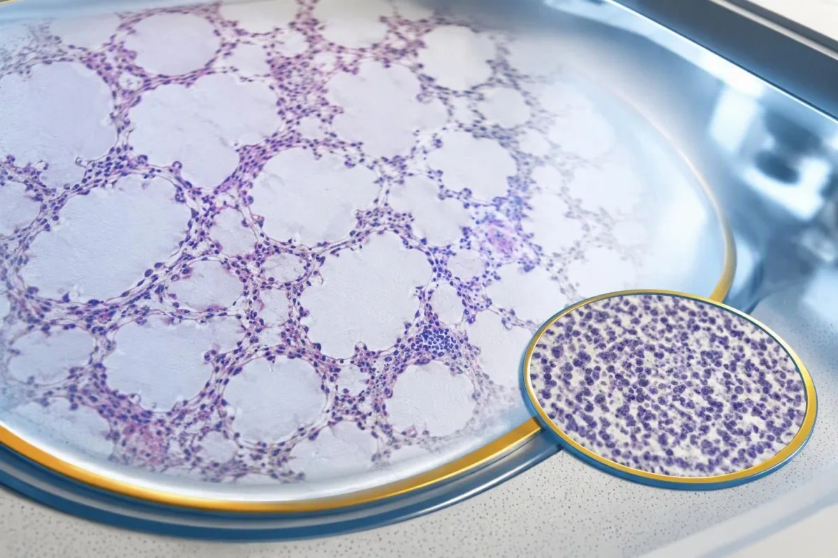
A negative scan is time-specific and method-specific. A normal chest CT performed on 1 date answers whether that examination identified a suspicious chest finding then; it does not guarantee that every possible associated cancer is absent indefinitely. Ask what was actually imaged, whether the protocol was suitable and whether the specialist recommends PET/CT or targeted investigation despite the initial result.
Villagrán-García et al. (2023) described cancers detected beyond the first 2 years in a minority of their anti-Hu cohort, supporting individualized longer surveillance. Their findings also support considering PET-based investigation when initial CT is unrevealing in an appropriate clinical context. This is an escalation decision for the treating team, not a reason to independently purchase repeated whole-body scans without weighing radiation, incidental findings and cost.
Neurological deterioration after negative imaging still needs reassessment. Cancer surveillance and neurological monitoring are 2 parallel tracks; one reassuring scan does not explain new loss of function. A marker such as neurofilament light chain may indicate neuronal injury in selected settings, but our NfL interpretation guide explains why it cannot establish anti-Hu disease, locate cancer or replace examination.
Як доўга трэба працягваць скрынінг на рак?
The 2021 PNS criteria recommend repeat cancer screening every 4–6 months for 2 years when a high-risk neurological phenotype accompanies a high-risk antibody. Longer surveillance may be appropriate for selected anti-Hu patients; the interval and duration should reflect confirmation, clinical evolution, initial investigations and individual cancer risk.

The 2-year schedule is not an instruction to scan every positive antibody result repeatedly. Graus et al. (2021) link that recommendation to the combination of high-risk phenotype and high-risk antibody. An incidental, weak result without a matching syndrome needs confirmation and individualized assessment first; repeating imaging mechanically can create incidental findings and unnecessary procedures without resolving the original uncertainty.
Older European guidance recommended longer surveillance. The EFNS task force advised repeating screening after 3–6 months and then every 6 months for up to 4 years in relevant antibody-positive paraneoplastic presentations (Titulaer et al., 2011). The 2023 anti-Hu cohort supported considering screening up to 5 years in selected patients, so a specialist may reasonably extend follow-up beyond the newer general 2-year framework.
Keep a dated plan stating the next examination, the clinician responsible and what symptoms should trigger earlier contact. Kantesti explanations can help you understand terminology, but the platform should not be treated as a scan-reminder service or substitute for that plan. Our guide to risk-based cancer screening addresses a different setting: routine prevention is not the same as investigating a possible paraneoplastic syndrome.
Якое лячэнне дапамагае пры неўралагічным захворванні, звязаным з anti-Hu?
Treatment focuses on an identified cancer, selected immune therapy and symptom-directed rehabilitation. Anti-Hu-associated neurological disease often responds less predictably to immunotherapy than neuronal surface-antibody disorders because intracellular targets and T-cell-mediated injury are involved; treatment may aim to stabilize function rather than promise complete recovery.

Treating an associated cancer is a central part of care, but established neurological injury may persist even when cancer treatment succeeds. Neurology and oncology teams may consider corticosteroids, intravenous immunoglobulin, plasma exchange or other immune treatments in selected situations. There is no single evidence-based dose or 1 universal regimen appropriate for everyone with anti-Hu positivity, especially without a confirmed clinical syndrome.
An antibody titer is not a dependable treatment target on its own. A lower number does not necessarily mean restored nerve function, and a persistent positive result does not by itself prove cancer progression. This differs from some surface-antibody disorders; our MOG antibody diagnosis guide explains another immune neurological condition, but its diagnostic rules and treatment expectations should not be transferred to anti-Hu.
Immune checkpoint cancer therapies deserve an explicit discussion when anti-Hu neurological disease is present, because they can provoke or worsen paraneoplastic neurological autoimmunity. This is a specialist risk-benefit decision, not an automatic prohibition based on 1 laboratory result. Tell the oncologist about previous neurological symptoms and confirmed neuronal antibodies before treatment decisions are finalized; coordinating the teams early can prevent avoidable misunderstanding.
Што можна бяспечна рабіць, пакуль працягваюцца абследаванні?
While investigations continue, protect mobility, swallowing and medication safety rather than trying to lower anti-Hu antibodies with supplements. Use the neurological symptoms—not the appearance of the laboratory flag—to decide whether ordinary follow-up is sufficient or urgent care is needed; a new functional loss warrants contact before the next planned scan.
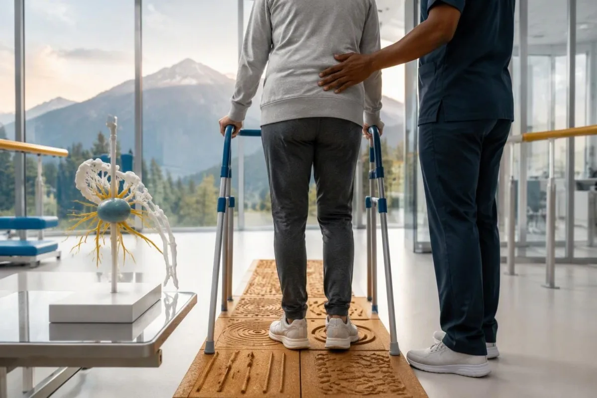
Position-sense loss increases fall risk, particularly in poor lighting or on uneven ground. A physiotherapist may recommend a suitable walking aid and individualized balance strategies; occupational therapy can address hand coordination and home hazards. A short 7-day record of falls, near-falls and walking changes is often more useful at review than the vague statement “my balance is worse,” but stop activities that feel unsafe.
Excess vitamin B6 can itself cause sensory neuropathy, making unsupervised high-dose nerve supplements particularly unhelpful during this assessment. Review every supplement and its daily dose in mg, including overlapping multivitamins and combination products. Our кіраўніцтва па бяспецы B-комплексу explains that problem; correcting a documented deficiency is different from taking progressively larger doses in the hope of treating anti-Hu autoimmunity.
Vitamin B12 deficiency can contribute to sensory and gait problems and may need investigation even when anti-Hu is confirmed. There is no universally interchangeable B12 cutoff: results in пг/мл і пмоль/л require their own laboratory context, sometimes with functional testing. Our кіраўніцтва па наступным назіранні за мяжовым B12 explains those next steps; new choking, breathing difficulty or rapid weakness should be assessed urgently instead of managed with vitamins.
Як ШІ можа дапамагчы бяспечна растлумачыць вынік аналізу на anti-Hu?
AI can help explain report terminology and organize questions, but it cannot confirm anti-Hu specificity, diagnose paraneoplastic syndrome or rule out cancer. Safe interpretation preserves the original assay details and separates 3 issues: what the laboratory reported, what the result may mean and what a clinician has actually established.

Кантэсці — гэта Інструмент аналізу вынікаў аналізу крыві з падтрымкай AI that can provide a plain-language explanation of uploaded laboratory information, including the distinction between an antibody screen and a confirmed clinical diagnosis. A rapid explanation—often around 60 seconds for report processing—is not a reference-laboratory assay. Our кіраўніцтва па тэхналогіі AI describes the interpretation workflow without making anti-Hu-specific diagnostic guarantees.
The complete source report should remain available for verification. An explanation should not silently replace “weakly reactive” with “confirmed,” drop the specimen type or invent a universal reference interval. Our guide to праверцы тлумачэнняў медыцынскіх звестак штучным інтэлектам describes practical source checks; for this antibody, compare at least the assay wording, confirmation comment and collection date against the original document.
Kantesti cannot examine sensation, review tissue staining directly through a report explanation or establish that negative imaging has excluded a tiny cancer. Those are 3 different clinical capabilities, not features that follow from understanding laboratory language. Share the explanation with the treating team as a discussion aid, and do not delay urgent assessment while uploading documents or waiting for a second automated interpretation.
Ваш наступны прыём і празрыстасць даследаванняў
Bring the complete antibody report, a dated symptom history, previous imaging reports and a current medication list to your next appointment. Ask the clinician to clarify antibody confirmation, the neurological diagnosis and cancer surveillance separately; those 3 answers may have different levels of certainty and different follow-up dates.
A useful 1-page symptom history records onset, progression, sensory distribution, walking changes, memory problems and autonomic symptoms. Include prior cancer treatment and any recent immunoglobulin therapy, then ask who will coordinate neurology and oncology if both are involved. As Thomas Klein, MD, I would rather see a clear timeline than 20 disconnected screenshots; it makes discrepancies between the laboratory result and clinical course easier to spot.
Medical authorship is not a substitute for an individual neurological examination. Наш інфармацыя медыцынскай кансультатыўнай рады describes the clinicians associated with our work, while your own specialist determines the appropriate next investigation. As of 25 кастрычніка 2026 года, the recommendations discussed here draw on the 2021 international PNS criteria, the 2011 EFNS screening guidance and the 2023 anti-Hu cohort—not an invented 2026 guideline.
The 2 Figshare publications below are platform-related research references, not evidence validating anti-Hu diagnosis or cancer exclusion. Their supplied titles describe hantavirus triage deployment and a synthetic-case technical benchmark; neither title establishes anti-Hu-specific clinical performance. Because publication authors and dates were not supplied for verification, the citations use title-first APA formatting with “n.d.”; the ResearchGate and Academia.edu links are searches, not claims of verified publication copies.
Часта задаваныя пытанні
Ці азначае станоўчы вынік аналізу на антыцелы anti-Hu, што ў мяне рак?
Станоўчы вынік аналізу на антыцелы anti-Hu сам па сабе не сведчыць пра наяўнасць раку. Anti-Hu — антыцелы высокай рызыкі, звязаныя з ракам; у спецыялізаванай кагорце па anti-Hu за 2023 год рак выявілі ў 349 з 466 пацыентаў, аднак гэтая лічба не вызначае рызыку, звязаную з выпадковым, непацверджаным вынікам скрынінгу. Урачы ацэньваюць неўралагічныя праявы, пацвярджаюць нечаканыя вынікі лабараторных даследаванняў і шукаюць асноўную прычыну ў выглядзе раку, звычайна надаючы першачарговую ўвагу органам грудной клеткі ў дарослых. Для пастаноўкі дыягназу раку звычайна патрэбныя асобныя клінічныя, візуалізацыйныя і гісталагічныя даныя.
Анты-Hu і ANNA-1 — гэта адно і тое ж?
Anti-Hu і ANNA-1 звычайна абазначаюць адно і тое ж сямейства нейрональных антыцелаў. ANNA-1 азначае антынейрональныя ядзерныя антыцелы тыпу 1, тады як Hu адносіцца да ўнутрыклетачных нейрональных бялкоў, якія распазнае антыцела. ANNA-1 — гэта не тое самае, што звычайныя ANA, якія выкарыстоўваюцца пры сістэмным аўтаімуналагічным абследаванні. Праверце дакладную назву аналізу і фармулёўку пацвярджэння, бо лабараторыі могуць падаваць вынікі на тое самае сямейства антыцелаў у розных фарматах.
Ці можа аналіз на антыцелы да Hu быць ілжывададатным?
Тэст на антыцелы да Hu можа даваць зманлівую або неспецыфічную рэактыўнасць, асабліва калі слабы вынік скрынінгу не адпавядае неўралагічнай карціне. Для пацвярджэння звычайна выкарыстоўваюць 2 узаемадапаўняльныя падыходы: характэрны тып афарбоўвання тканін і аналіз, спецыфічны да антыгена, вынікі якога інтэрпрэтуе лабараторыя з адпаведным досведам. Паўторнае выкарыстанне таго ж камерцыйнага метаду можа прывесці да паўтарэння той самай аналітычнай праблемы, а не да яе вырашэння. Хуткае прагрэсаванне неўралагічных сімптомаў па-ранейшаму патрабуе своечасовай ацэнкі, пакуль чакаюць пацвярджэння.
Як часта трэба паўтараць сканаванні, калі вынік аналізу на анты-Hu станоўчы?
Крытэрыі PNS 2021 года рэкамендуюць паўтараць скрынінг на рак кожныя 4–6 месяцаў на працягу 2 гадоў, калі высокарызыкоўны неўралагічны фенатып суправаджаецца высокарызыкоўнымі антыцеламі. Гэты графік не распаўсюджваецца аўтаматычна на кожны выпадкова выяўлены станоўчы вынік аналізу на анты-Hu. Ранейшыя рэкамендацыі EFNS прадугледжвалі назіранне да 4 гадоў, а даследаванне кагорты пацыентаў з анты-Hu, апублікаванае ў 2023 годзе, пацвердзіла мэтазгоднасць разгляду назірання да 5 гадоў для асобных пацыентаў. Ваш спецыяліст павінен вызначыць метад візуалізацыі, інтэрвал, працягласць назірання і сімптомы, пры якіх трэба звярнуцца раней.
Ці можа аналіз на анты-Hu быць станоўчым, калі вынікі КТ і ПЭТ у норме?
0 Антыцелы да Hu могуць заставацца станоўчымі, нават калі пры КТ або ПЭТ-візуалізацыі рак не выяўлены. Неўралагічны аўтаімунітэт можа папярэднічаць яго выяўленню, а адмоўны вынік сканавання адлюстроўвае абследаваныя ўчасткі, метад і дату даследавання, а не з'яўляецца пастаяннай гарантыяй адсутнасці раку. Пры сумяшчальнай клінічнай карціне высокай рызыкі крытэрыі 2021 года рэкамендуюць паўтараць скрынінг кожныя 4–6 месяцаў на працягу 2 гадоў; для асобных пацыентаў з антыцеламі да Hu можна індывідуальна падоўжыць назіранне. Таксама варта паўторна ацаніць непацверджаную рэактыўнасць антыцелаў і прычыны, не звязаныя з ракам, а не меркаваць, што рак, які не ўдаецца выявіць, напэўна ёсць.
Ці азначае зніжэнне ўзроўню антыцелаў да Hu, што лячэнне дзейнічае?
Зніжэнне ўзроўню антыцелаў anti-Hu не з'яўляецца надзейным доказам неўралагічнага аднаўлення або паспяховага лячэння раку. Anti-Hu распазнае ўнутрыклетачныя бялкі, а звязанае з імі неўралагічнае пашкоджанне часта абумоўлена механізмамі з удзелам Т-клетак, а не толькі дзеяннем антыцелаў. Паказчыкі тытраў, атрыманыя ў розных лабараторыях, таксама могуць быць непараўнальнымі з-за адрозненняў у метадах. Замест таго каб выкарыстоўваць колькасны паказчык антыцелаў як самастойную мэту лячэння, клініцысты ацэньваюць функцыянальны стан, вынікі агляду, адпаведныя даследаванні і адказ раку на лячэнне.
Атрымаць аналіз крыві з AI сёння
Далучайцеся больш чым да 2 мільёнаў карыстальнікаў па ўсім свеце, якія давяраюць Kantesti для імгненнага, дакладнага аналізу лабараторных тэстаў. Загрузіце вынікі аналізу крыві і атрымайце поўную інтэрпрэтацыю біямаркераў 15,000+ за лічаныя секунды.
📚 Публікацыі даследаванняў са спасылкамі
Кляйн, Т., Мітчэл, С., і Вебер, Г. (2026). Шматмоўная AI-падтрымка пры прыняцці клінічных рашэнняў для ранняга трыажу хантавірусу: дызайн, інжынерная валідацыя і разгортванне ў рэальных умовах для 50 000 прааналізаваных справаздач аб аналізах крыві. Kantesti AI Medical Research.
Кляйн, Т., Мітчэл, С., і Вебер, Г. (2026). Папярэдне зарэгістраваны, рубрыка-аснаваны аўтаматызаваны тэхнічны бенчмарк інтэрпрэтацыйнага рухавіка Kantesti для аналізу крыві на 100 000 сінтэтычных тэставых выпадках. Kantesti AI Medical Research.
📖 Знешнія медыцынскія спасылкі
Graus F et al. (2021). Updated Diagnostic Criteria for Paraneoplastic Neurologic Syndromes. Neurology: Neuroimmunology & Neuroinflammation.
Titulaer MJ et al. (2011). Screening for tumours in paraneoplastic syndromes: report of an EFNS Task Force. European Journal of Neurology.
Villagrán-García D et al. (2023). Revisiting anti-Hu paraneoplastic autoimmunity: phenotypic characterization and cancer diagnosis. Brain Communications.
⚕️ Медыцынская адмова ад адказнасці
Гэты артыкул прызначаны толькі для адукацыйных мэт і не з’яўляецца медычнай парадай. Заўсёды кансультуйцеся з кваліфікаваным спецыялістам аховы здароўя для прыняцця рашэнняў па дыягностыцы і лячэнні.
Сігналы даверу E-E-A-T
Вопыт
Клінічны агляд, які вядзе лекар, працэсаў інтэрпрэтацыі лабараторных паказчыкаў.
Экспертыза
Фокус лабараторнай медыцыны на тым, як біямаркеры паводзяць сябе ў клінічным кантэксце.
Аўтарытэтнасць
Напісана доктарам Томасам Кляйнам, з аглядам доктара Сара Мітчэл і праф. доктара Ганса Вебера.
Надзейнасць
Інтэрпрэтацыя на аснове доказаў з выразнымі шляхамі далейшых дзеянняў, каб паменшыць трывогу.
