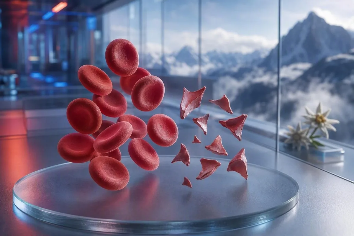
Schistocyty sú fragmentované červené krvinky, ktoré môžu signalizovať nebezpečné zrážanie v malých cievach, ale jediná poznámka z náteru nie je diagnóza. Naliehavosť závisí od percenta fragmentov, počtu trombocytov, trendu hemoglobínu, funkcie obličiek, markerov hemolýzy a príznakov.


- Prahová hodnota schistocytov: Viac než 1% schistocytov na dobre pripravenom periférnom nátere dospelého je podozrivé z trombotickej mikroangiopatie, ak chýbajú iné zmeny tvaru červených krviniek.
- Urgentný zhluk: Schistocyty spolu s trombocytmi pod 150 × 10^9/L, klesajúcim hemoglobínom, zvýšeným LDH a nízkym haptoglobínom vyžadujú vyšetrenie lekárom v ten istý deň.
- Indícia pre TTP: Závažná aktivita ADAMTS13 pod 10% podporuje imunitnú trombotickú trombocytopenickú purpuru, stav, ktorý sa lieči ešte pred návratom výsledkov potvrdzujúcich testov, ak je klinické podozrenie vysoké.
- Nie každý fragment je nebezpečný: Mechanické srdcové chlopne, závažná hypertenzia, dialyzačné okruhy, popáleniny a poškodenie vzorky môžu vytvárať fragmentované červené krvinky.
- Náter je manuálny dôkaz: Automatizovaný analyzátor nedokáže spolehlivě diagnostikovat schistocyty; skúsený morfolog by mal vyšetřit preparát.
- Těhotenství zvyšuje sázky: Schistocyty spolu s vysokým krevním tlakem, bílkovinou v moči nebo nízkými trombocyty po 20. týždni vyžadujú okamžité vyšetrenie pôrodníkom.
- Opakované testovanie pomáha: Čerstvo odobratá vzorka v EDTA a opakovaný náter môžu odlíšiť artefakt z odberu od skutočnej prebiehajúcej fragmentácie červených krviniek.
- Neliečte sa samostatne: Železo, kyselina listová alebo doplnky neliečia mikroangiopatickú hemolýzu a môžu odviesť pozornosť od urgentného vyhodnotenia.
Keď fragmentované červené krvinky vyžadujú urgentné lekárske vyšetrenie
Schistocyty si vyžadujú urgentnú kontrolu v ten istý deň, keď sa objavia spolu s nízkym počtom trombocytov, anémiou, biochemickou hemolýzou, poškodením obličiek, neurologickými príznakmi, horúčkou alebo závažnou hypertenziou. Samotný komentár k náteru môže byť neškodný alebo arteficiálny; nebezpečný je vzorec. K 7. augustu 2026 by som nové schistocyty spolu s trombocytmi pod 100 × 10^9/L považoval za „volajte lekára hneď“, nie za bežné následné sledovanie.
Pokles trombocytov je často najrýchlejším varovným signálom urgentnosti. Hemoglobín 92 g/L pri trombocytoch 68 × 10^9/L a novonahlásených schistocytoch je podstatne znepokojujúcejší než 1% fragmentov s hemoglobínom 145 g/L a stabilnými trombocytmi 260 × 10^9/L. Kantesti je analyzátor AI krvných testov, ktorý číta tento súbor naprieč krvný obraz, namiesto toho, aby izolovane zvyšoval komentár k morfológii.
Za 15 rokov mojej klinickej praxe sú moje rýchle „call“ tie, pri ktorých sa výsledky zmenili v priebehu dní: klesajúci hemoglobín, stúpajúci kreatinín a stúpajúca laktátdehydrogenáza, alebo LDH. Praktické pravidlo doktora Thomasa Kleina je jednoduché: zmätenosť, nová slabosť, bolesti na hrudi, dýchavičnosť, veľmi málo moču alebo silná bolesť hlavy popri tomto vzorci znamená, že dnes treba urgentné vyšetrenie.
Schistocyty nie sú dôvod čakať na opakovaný test, ak je daná osoba chorá. Normálny draslík alebo normálny počet leukocytov nezruší riziko trombotickej mikroangiopatie. Pacienti, ktorí sa snažia pochopiť nízky počet, si môžu tiež pozrieť náš sprievodca k príznakom nízkych červených krviniek, ale akútne príznaky vždy majú prednosť pred interpretáciou online.
Volajte záchrannú službu teraz pre
Novú jednostrannú slabosť, odpadnutie, záchvat, silnú dýchavičnosť, drvivý diskomfort na hrudi, čiernu stolicu alebo rýchlo sa zhoršujúcu zmätenosť treba riešiť záchrannou službou, nie správou do ambulancie. Tieto príznaky môžu odrážať postihnutie orgánov a žiadne percento schistocytov to bezpečne nevylučuje.
Čo schistocyty na periférnom nátere v skutočnosti znamenajú
Schistocyty sú nepravidelné fragmenty červených krviniek vznikajúce vtedy, keď sú cirkulujúce bunky fyzicky „odtrhnuté“ strihom, najčastejšie fibrínovými prameňmi alebo abnormálnymi povrchmi s vysokým strihovým napätím v malých cievach. Typicky majú ostré uhly, formy ako prilby alebo malé trojuholníkové tvary a chýba im centrálne blednutie neporušenej červenej krvinky.
Pravé schistocyty sú morfologický nález, nie hodnota, ktorú vytvára štandardný analyzátor CBC. Medzinárodná rada pre štandardizáciu v hematológii odporúča pri kvantifikácii, keď na tom záleží, spočítať aspoň 1 000 červených krviniek v optimálnej zóne náteru (Zini et al., 2021). Tento detail vysvetľuje, prečo jedna laboratórium môže uviesť “vzácne fragmenty”, zatiaľ čo iné uvedie percento.
Klasická obava je mikroangiopatická hemolytická anémia, kde sa červené krvinky stretávajú s trombocytovo-bohatými mikrotrombmi a fibrínom v krvnom obehu. Napriek tomu však ťažká aortálna stenóza, mechanická chlopňa, mimotelové obehové okruhy a závažné tepelné poranenie môžu vytvoriť skutočné fragmenty iným mechanizmom. Otázka preto nie je “Sú fragmenty reálne?”, ale “Čo spôsobuje fragmentáciu?”
Kantesti je platforma AI na výsledky krvného testu, ktorá dokáže nahraný kompletný krvný obraz umiestniť vedľa čo obsahuje CBC a zvýrazniť chýbajúce sprievodné výsledky, ako je bilirubín, retikulocyty alebo haptoglobín. Nedokáže však priamo skontrolovať samotný náter, čo zostáva úlohou vyškoleného laboratórneho odborníka.
Koľko schistocytózy je znepokojujúce na nátere dospelého
Počet schistocytov nad 1% na periférnom nátere dospelého je podozrivý z trombotickej mikroangiopatie, keď sú fragmenty dominantnou abnormalitou červených krviniek. Počty pod 1% nevylučujú ochorenie, najmä ak bol film pripravený po podaní tekutín, transfúzii alebo pri prebiehajúcom ochorení.
Hraničná hodnota 1% je diagnostický podnet, nie škála závažnosti. Usmernenia ICSH používajú referenčný interval pod 1% pre zdravých dospelých a donosených novorodencov; predčasne narodené deti môžu mať až 5% fragmentov bez rovnakých dôsledkov (Zini et al., 2021). Výsledok 2% pri závažnej trombocytopénii je znepokojujúcejší než 4% po zavedení mechanického zariadenia na podporu obehu.
Laboratóriá môžu uvádzať “príležitostne”, “niekoľko” alebo “1–2 na pole s vysokým zväčšením” namiesto percenta. Tieto formulácie nie sú zameniteľné, pretože veľkosť poľa, hrúbka náteru a pravidlá hlásenia sa líšia. Otázka, či laboratórium vykonalo percentuálne hodnotenie v štýle ICSH, je často užitočnejšia než pokúšať sa sami prepočítať opisnú formuláciu.
Vysoké RDW nemeria schistocyty. RDW odráža variabilitu veľkosti buniek a môže stúpať pri retikulocytóze, nedostatku železa, transfúzii alebo fragmentácii. Naša podrobná Sprievodca RDW a indexmi červených krviniek vysvetľuje, prečo vysoké RDW samo osebe nepodporuje ani TTP, ani artefakt.
Vzor výsledkov krvných testov, ktorý podporuje pravú hemolýzu
Skutočná fragmentácia červených krviniek zvyčajne spôsobuje vysoké LDH, nízky haptoglobín, zvýšený nepriamy bilirubín a retikulocytovú odpoveď popri anémii. Žiadny jediný marker nie je definitívny, pretože ochorenie pečene, zápal, nedávna transfúzia a ochorenie kostnej drene môžu skresliť obraz.
LDH je citlivé, ale nie špecifické. LDH nad 600 IU/L často podporuje aktívnu hemolýzu v správnom kontexte, no zvýšiť ho môžu aj poranenie svalov, poškodenie pečene, infekcia a rakovina. Haptoglobín pod dolnou hranicou laboratória, bežne pod 0,3 g/L, pridáva váhu, pretože viaže voľný hemoglobín uvoľnený do plazmy.
Počet retikulocytov by zvyčajne mal počas periférnej deštrukcie stúpať, ale môže zostať nízky pri nedostatku folátu, nedostatku železa, ochorení obličiek alebo akútnej supresii kostnej drene. Tento zdanlivý rozpor sa ľahko prehliadne: pacient môže mať skutočnú hemolýzu a neprimerane nízku retikulocytovú odpoveď. Ten výsledok haptoglobínu vedie cez bežné interpretačné pasce.
Kantesti AI interpretuje vzorce fragmentovaných červených krviniek porovnaním hemoglobínu, MCV, trombocytov, LDH, bilirubínu, kreatinínu a retikulocytov z rovnakého časového bodu. Pokles hemoglobínu o viac než 20 g/L v priebehu 48 až 72 hodín je klinicky informatívnejší než mierne abnormálna hodnota bez predchádzajúceho porovnania.
Prečo môže byť bilirubín normálny
Nepriamy bilirubín môže byť na začiatku hemolýzy normálny, po zriedení intravenóznymi tekutinami alebo keď pečeň bilirubín účinne odstraňuje. Preto by normálny bilirubín nemal prehlasovať presvedčivé fragmenty, nízky haptoglobín a klesajúci hemoglobín.
Prečo sa TTP lieči ako hematologická urgentná situácia
TTP, trombotická trombocytopenická purpura, sa má podozrievať vtedy, keď schistocyty sprevádzajú trombocytopéniu a mikroangiopatickú hemolytickú anémiu bez jasnejšej alternatívy. Neliečená imunitná TTP môže rýchlo progredovať, takže klinici môžu začať plazmaferézu a imunitnú liečbu ešte pred návratom výsledkov ADAMTS13.
Aktivita ADAMTS13 pod 10% silno podporuje TTP, no výsledok môže trvať hodiny alebo dni. Diagnostické usmernenie ISTH odporúča odhadnúť pravdepodobnosť pred testom pomocou klinických nástrojov, ako je PLASMIC skóre, pričom sa odoberie vzorka ešte pred plazmatickou liečbou (Zheng et al., 2020). Skóre je most k akcii, nie náhrada za odborný úsudok.
Známa “pentáda” horúčky, neurologických príznakov, renálneho postihnutia, trombocytopénie a hemolytickej anémie je u mnohých reálnych pacientov neúplná. George a Nester opísali trombotickú mikroangiopatiu ako syndróm s prekrývajúcimi sa príčinami, nie ako päť povinných „políčok“ (George & Nester, 2014). V praxi je čakanie na všetky päť čŕt nebezpečný starý zvyk.
Trombocyty pod 30 × 10^9/L pri hemolýze si zaslúžia obzvlášť urgentnú diskusiu s hematológiou. Transfúzia trombocytov sa pri suspektnej TTP zvyčajne vyhýba, pokiaľ nejde o život ohrozujúce krvácanie alebo invazívny zákrok, ktorý sa nedá odložiť. Pre súvisiace testovanie naše sprievodcu koagulačnými testami vysvetľuje, prečo môžu byť PT a aPTT v TTP normálne.
Ďalšie nebezpečné príčiny schistocytov, ktoré zvažujú lekári
Schistocyty sa vyskytujú aj pri diseminovanej intravaskulárnej koagulácii, hemolyticko-uremickom syndróme vyvolanom Shiga-toxínom, ťažkej hypertenzii, mikroangiopatii súvisiacej s tehotenstvom, niektorých liekoch a trombotickej mikroangiopatii spojenej s transplantáciou. Súvisiace nálezy trombocytov, koagulácie, obličiek a klinické nálezy tieto stavy zvyčajne odlišia.
DIC často kombinuje fragmenty s predĺženým PT, predĺženým aPTT, nízkym alebo klesajúcim fibrinogénom a výrazne zvýšeným D-dimérom. TTP má častejšie relatívne normálne bežné časy zrážania. Fibrinogén pod 1,5 g/L u chorého pacienta presúva obavy smerom ku konzumpčnej koagulopatii, hoci fibrinogén môže spočiatku vyzerať normálne, pretože pri akútnom zápale stúpa.
Ťažká hypertenzia môže „odtrhnúť“ červené krvinky, keď tlak poškodí mikrocirkuláciu; hodnoty na úrovni alebo nad 180/120 mmHg s bolesťou hlavy, zmenou videnia, bolesťou na hrudníku alebo dysfunkciou obličiek sú urgentné. Preto patrí hodnota krvného tlaku vedľa správy z náteru. Pacienti môžu prehodnotiť sekundárna hypertenzia – laboratórne indície po tom, čo sa rieši bezprostredné ohrozenie.
Hnačka nasledovaná zníženým výdajom moču, anémiou a trombocytopéniou vyvoláva obavy z hemolyticko-uremického syndrómu. Vyšetrenie stolice môže pomôcť identifikovať Shiga toxín, ale stav hydratácie, kreatinín, draslík a neurologické zhodnotenie majú viesť k urgentnej starostlivosti ako prvé. Vysoký laktát alebo nestabilné vitálne funkcie si vyžadujú vyšetrenie sepsy a DIC skôr než prístup „vyčkajte a uvidíte“; pozrite si tieto vzorcom markerov sepsy.
Mechanické príčiny fragmentovaných červených krviniek
Mechanická deštrukcia môže vytvoriť skutočné schistocyty aj bez TTP, najmä pri protetických srdcových chlopniach, ťažkej aortálnej stenóze, ventrikulárnych asistenčných zariadeniach, dialyzačných okruhoch alebo mimotelovej oxygenácii (ECMO). Tieto príčiny si stále zaslúžia lekárske zhodnotenie, ale liečba cieli mechanický zdroj, nie imunitnú TTP.
Vzor pri mechanickej chlopni často zahŕňa chronicky mierne zvýšený LDH a nízky haptoglobín pri stabilných trombocytoch. Nová anémia, zhoršujúca sa dýchavičnosť, tmavý moč alebo zmena zvuku chlopne môžu naznačovať paravalvulárny leak alebo dysfunkciu zariadenia a mali by viesť k vyšetreniu kardiológom. Rozter poskytuje indíciu; echokardiografia nájde zdroj.
Športovci sa niekedy pýtajú, či môže maratón vysvetliť fragmenty. Hemolýza po došľape môže po behu na dlhé trate spôsobiť prechodne voľný hemoglobín a nízky haptoglobín, ale presvedčivá schistocytóza nie je niečo, čo by som bez okolkov pripisoval cvičeniu—najmä ak trombocyty klesajú. Porovnajte to s širším laboratórnym sprievodcom pre maratónskeho bežca.
Ťažké popáleniny a veľké cievne operácie môžu vytvoriť fragmentované bunky prostredníctvom fyzického a endotelového poškodenia. V týchto situáciách môže byť vývoj v laboratórnych parametroch každých 6 až 24 hodín výpovednejší než jeden rozter. Stabilný CBC po vyvolávajúcej udalosti je upokojujúci; kontinuálne klesajúci hemoglobín nie.
Ako chyby pri odbere a artefakty na preparáte napodobňujú schistocyty
Zlá technika odberu vzorky, oneskorená príprava rozteru, nedonaplnené skúmavky s antikoagulantom a poškodený okraj rozteru môžu vytvoriť tvary, ktoré pripomínajú schistocyty, bez toho, aby odrážali hemolýzu na úrovni cirkulácie. Opakovaná vzorka odobratá čistým spôsobom a prehodnotená v monovrstve je zvyčajne prvá kontrola, keď je klinický obraz pokojný.
Zvrásnené (crenated) bunky, deformácia na okraji rozteru, artefakt vysychania a prekrývajúce sa bunky sú časté falošné napodobeniny. Často sa objavujú difúzne alebo len na „pernatej“ (feathered) hranici, nie rovnomerne v ideálnej čítacej zóne. Skúsený morfológ odlíši ostrú geometriu skutočného fragmentu od bunky, ktorá je len deformovaná počas prípravy sklíčka.
Ťažký odber môže hemolyzovať skúmavku a falošne zvýšiť draslík alebo LDH, ale automaticky to nevytvára skutočné cirkulujúce schistocyty. Ak výsledok obsahuje index hemolýzy alebo poznámku “vzorka je kompromitovaná”, klinici zvyčajne odoberú znovu pred stanovením diagnózy ochorenia. Náš článok o chyby pri odbere draslíka vysvetľuje, prečo ten istý predanalytický problém môže naraz zavádzať viacero výsledkov.
Zhlukovanie trombocytov závislé od EDTA môže falošne znížiť počet trombocytov a vyrobiť alarmujúco vyzerajúcu kombináciu. Počet trombocytov v skúmavke so citrátom, korigovaný na riedenie, a čerstvý film to môže odhaliť. Nuansa záleží: artefakt môže koexistovať so skutočným ochorením, takže opakovaný test by nikdy nemal odkladať starostlivosť u niekoho, kto má príznaky.
Prečo môžu upozornenia analyzátora a formulácie v laboratórnej správe zavádzať
Automatizované hematologické analyzátory môžu signalizovať abnormálne populácie červených krviniek, ale nedokážu samostatne diagnostikovať schistocyty s dostatočnou spoľahlivosťou na potvrdenie trombotickej mikroangiopatie. Ručné vyšetrenie periférneho rozteru zostáva potrebné, keď je klinická otázka hemolýza alebo TTP.
Komentár “prítomné fragmentované erytrocyty” by mal vyvolať otázky týkajúce sa množstva, kvality náteru a súčasne prítomnej morfológie. Výrazná anizopoikilocytóza pri nedostatku železa, megaloblastovej anémii alebo hereditárnych poruchách červených krviniek robí pravidlo 1% menej špecifickým. Fragmenty sú diagnosticky najviac užitočné vtedy, keď dominujú abnormálnej morfológii, a nie keď sa objavujú medzi mnohými nesúvisiacimi typmi.
Kantesti je služba interpretácie výsledkov AI z laboratória navrhnutá na upozornenie na nesúladné kombinácie, ako je nízky počet trombocytov spolu s komentárom, že sú prítomné zhluky trombocytov. Nenahrádza mikroskopickú kontrolu laboratória ani lekára, ktorý môže vyžiadať opakovaný náter. Náš prístup ku kontrole kvality je opísaný v prehľad lekárskej validácie.
Delta check porovnáva aktuálny výsledok s predchádzajúcim výsledkom a môže odhaliť náhle, nepravdepodobné zmeny. Pokles hemoglobínu z 138 g/l na 87 g/l v priebehu 24 hodín je klinicky významný, ale riedenie intravenóznymi tekutinami alebo nesprávne označený vzorok zostáva možný. Viac o náhlych zmenách v laboratórnych výsledkoch.
Čo robiť po tom, čo výsledky krvných testov spomínajú schistocyty
Ak sa novozistene uvádzajú schistocyty, kontaktujte ešte v ten istý deň klinika alebo službu, ktorá test objednala, a okamžite vyhľadajte urgentnú starostlivosť, ak máte neurologické príznaky, dýchavičnosť, bolesti na hrudníku, tmavý moč, silnú bolesť hlavy, horúčku alebo znížený výdaj moču. Neinterpretujte komentár k náteru ako diagnózu samotnú o sebe.
Majte k dispozícii skutočnú správu vrátane hemoglobínu, trombocytov, kreatinínu, bilirubínu, LDH a akéhokoľvek indexu hemolýzy. Zároveň zohľadnite nedávne tehotenstvo, hnačkové ochorenie, výrazne zvýšené hodnoty krvného tlaku, nové lieky, operáciu chlopní, dialýzu, transfúziu a autoimunitné ochorenie. Tieto detaily menia diferenciálnu diagnostiku viac než jedno jediné slovo v poli morfológie.
Vidím, že pacienti strácajú cenný čas začatím podávania železa, pretože si prečítajú “anémia”, ešte kým niekto identifikuje príčinu. Nedostatok železa môže koexistovať, ale nevysvetľuje rýchly pokles trombocytov s fragmentmi. Dr. Thomas Klein odporúča urobiť fotografiu celej správy namiesto prepísania iba zvýraznených čísel; pri prepise často vypadne laboratórny komentár, ktorý robí vzorec zrozumiteľným.
Vyhnite sa aspirínu, ibuprofénu a antikoagulanciám, ktoré nie sú predpísané, až kým klinik neodporučí inak, ak sú trombocyty veľmi nízke alebo ak je prítomné krvácanie. Bez urgentného medicínskeho usmernenia neukončujte predpísaný antikoagulant ani tak. Ľudia s únavou alebo závratmi môžu použiť naše krvné testy pri závratoch usmerňujú na prípravu otázok po vylúčení urgentných príčin.
Schistocyty v gravidite, u novorodencov a u detí
Schistocyty počas tehotenstva si vyžadujú urgentné vyšetrenie, keď sa vyskytujú spolu s hypertenziou, proteinúriou, zvýšenými jaternými enzýmami, nízkymi trombocytmi, bolesťou hlavy, zrakovými príznakmi alebo bolesťou v hornej časti brucha. HELLP syndróm, preeklampsia, TTP, atypický HUS a DIC sa môžu prekrývať a načasovanie okolo pôrodu pomáha, ale diagnózu to neuzavrie.
HELLP syndróm je klasicky definovaný hemolýzou, zvýšenými pečeňovými enzýmami a nízkymi trombocytmi, často po 20. týždni tehotenstva alebo krátko po pôrode. Počet trombocytov pod 100 × 10^9/l alebo AST nad 70 IU/l môže podporiť obavy, ale miestne pôrodnícke tímy interpretujú celý klinický obraz. S týmto kombinovaným nálezom nečakajte na rutinnú prenatálnu kontrolu.
Donosené novorodenci môžu mať menej než 1% schistocytov, zatiaľ čo predčasne narodené deti môžu mať až 5%, pretože morfológia erytrocytov u novorodencov je odlišná (Zini et al., 2021). U dieťaťa s hnačkou, bledosťou, zníženým množstvom moču a trombocytopéniou však fragmenty môžu poukazovať na HUS a vyžadujú okamžité pediatrické vyšetrenie.
Referenčné intervaly pre trombocyty a hemoglobín sa výrazne menia podľa veku a štádia tehotenstva. Výsledok označený ako “normálny” všeobecnou aplikáciou môže byť v kontexte stále klinicky znepokojujúci. Naše zdroje o rozsahoch trombocytov v tehotenstve a upozorneniach na laboratórne výsledky v tehotenstve v ten istý deň poskytuje užitočné pozadie, nie je náhradou za pôrodníckú triáž.
Testy, ktoré klinickí lekári objednávajú na vyšetrenie pravých schistocytov
Prvá vyšetrovacia sada pri suspektnej mikroangiopatickej hemolýze zvyčajne zahŕňa opakované vyšetrenie CBC a náteru, počet retikulocytov, LDH, haptoglobín, frakcie bilirubínu, kreatinín, vyšetrenie moču, PT, aPTT, fibrinogén a D-dimér. Odber aktivity ADAMTS13 a testovanie inhibítora sa má urobiť pred plazmaferézou, ak sa uvažuje o TTP, ale liečba sa nesmie odkladať.
Priame antiglobulínové testovanie, tiež nazývané DAT alebo Coombsovo testovanie, pomáha odlíšiť imunitnú hemolýzu od neimunitnej fragmentácie. Pozitívny DAT nevylučuje druhý proces, ale klasická TTP je typicky DAT-negatívna. Mikroskopia moču môže odhaliť proteínové alebo bunkové cylindry, keď je aktívne poškodenie obličkových mikrovaskulárnych ciev; pozri naše vysvetlenie z granulárne močové valy.
Testovanie stolice na Shiga-toxín sa považuje za vhodné po kompatibilnom gastrointestinálnom ochorení, zatiaľ čo doplnkové testovanie komplementu, revízia liekov a genetické alebo funkčné štúdie sa môžu použiť pri atypickom HUS. Poradie sa líši podľa krajiny a nemocnice. Zvýšenie kreatinínu o 26,5 µmol/l do 48 hodín spĺňa jedno kritérium akútneho poškodenia obličiek a zaslúži si pozornosť aj vtedy, keď sa ešte nestane dramatickým číslom.
Index produkcie retikulocytov pod 2 pri významnej anémii naznačuje nedostatočnú odpoveď kostnej drene, aj keď sa zdá, že samotné percento retikulocytov je vysoké. Tento výpočet koriguje nižšiu hmotnosť červených krviniek pri anémii. Ten sprievodné vodítko pre retikulocyty a hematologické ukazovatele pomáha vysvetliť, prečo môžu byť percentá zavádzajúce.
Kedy je rozumné opakovať náter a kedy nie
Opakovanie náteru v priebehu hodín až do 24 hodín je rozumné, keď sú fragmenty skromné, pacient sa cíti dobre, trombocyty a hemoglobín sú stabilné a spochybňuje sa kvalita vzorky. Opakovanie nie je bezpečné odkladanie, ak sú prítomné trombocytopénia, aktívna hemolýza, poškodenie orgánov alebo znepokojujúce príznaky.
Dobrá opakovaná vzorka sa odoberá čerstvo do správne naplneného EDTA tubu a promptne sa z nej pripraví náter. Žiadosť by mala výslovne požiadať o kontrolu suspektných schistocytov a zhlukovania trombocytov, ak je ktorákoľvek z možností pravdepodobná. Laboratórium môže porovnať oba nátery vedľa seba, čo je oveľa užitočnejšie než len opätovné spustenie automatizovaného CBC.
Dôležitý je trend. Trombocytový počet, ktorý sa mení z 210 na 165 na 98 × 10^9/l počas troch dní, je znepokojujúci, aj keď prvé dve hodnoty môžu spadať do lokálneho referenčného intervalu. Kantesti AI používa longitudinálny kontext na zachytenie týchto trajektórií, princíp diskutovaný v našom sprievodnej príručky na analýzu trendov krvných testov.
Transfúzia môže dočasne na riediť alebo zamaskovať pôvodnú morfológiu. Ak ste dostali erytrocyty pred druhou vzorkou, povedzte tímu dátum a počet jednotiek. Náš sprievodca k zmenám hodnôt po prepustení z nemocnice vysvetľuje, prečo si trendy po liečbe vyžadujú iný východiskový bod.
Stavy, ktoré môžu vyzerať podobne, ale nie sú ochorením schistocytov
Nedostatok železa, deficit vitamínu B12, ochorenie pečene, zdedené poruchy červených krviniek a závažná infekcia môžu vytvárať abnormálne tvary červených krviniek bez špecifického fragmentačného vzorca trombotickej mikroangiopatie. Kompletná morfológia v nátere a klinická časová os zabraňujú preceňovaniu fragmentov.
Závažný deficit vitamínu B12 môže spôsobiť pseudo-trombotickú mikroangiopatiu s anémiou, vysokým LDH, nízkymi trombocytmi a občasnými fragmentmi. Často spôsobuje výraznú makrocytózu, nízku retikulocytovú odpoveď a hodnoty LDH niekedy nad 2 500 IU/l—znaky, ktoré by mali spustiť testovanie B12 a metylmalónovej kyseliny, nie automatickú liečbu TTP. Náš príručke B12 vs folát vysvetľuje tento dôležitý napodobňovač.
Nedostatok železa zvyčajne spôsobuje mikrocytózu, hypochrómiu, „pencil cells“ a vysoké RDW; fragmenty sa môžu uvádzať v závažných prípadoch, ale zriedkavo sú dominantným znakom. Ferritín pod 15 µg/L výrazne podporuje vyčerpané zásoby železa u inak nekomplikovaného dospelého, hoci zápal môže maskovať nedostatok tým, že ferritín zvyšuje.
Z mojej skúsenosti je najupokojujúcejší výsledok nie “žiadne schistocyty” samotné, ale ucelené alternatívne vysvetlenie so stabilnými trombocytmi, stabilnou funkciou obličiek a bez biochemickej hemolýzy. Ak nálezy zostávajú nezhodné, hematológ môže skontrolovať samotný sklíčko, namiesto spoliehania sa na znenie konečnej správy.
Použitie interpretácie pomocou AI bez toho, aby sa prehliadla urgentná situácia na nátere
AI môže pomôcť zorganizovať výsledky krvných testov a identifikovať nebezpečné kombinácie, ale nedokáže overiť diagnózu schistocytov z PDF CBC ani nahradiť urgentné klinické vyšetrenie. Periférny náter je vizuálna mikroskopia a pôvodné laboratórne sklíčko zostáva zdrojom pravdy.
Kantesti je nástroj na analýzu krvných testov poháňaný AI, ktorý mapuje súvisiace výsledky naprieč správami, vrátane trajektórií trombocytov, zmien hemoglobínu, kreatinínu, bilirubínu a LDH. To môže znížiť častú chybu čítania každého abnormálneho markera samostatne. Nedokáže však zistiť, či tvar nazývaný schistocyt je skutočný fragment, artefakt na okraji náteru, alebo konvencia uvádzania.
Záleží na kvalite nahrania, pretože vynechaná jednotka alebo desatinná čiarka môže obrátiť význam výsledku. Pred zdieľaním správy skontrolujte identifikátory pacienta, dátum odberu, jednotky, referenčné rozmedzia a či výsledok uvádza “estimated”, “pending” alebo “sample haemolysed”. Naše kontrolný zoznam presnosti AI reportu je užitočné na túto finálnu ľudskú kontrolu.
Klinické metódy Kantesti sú preverené s príspevkom od našich Lekárska poradná rada, a naša technológia je opísaná v sprievodca technológiou AI interpretácie vysvetľuje, ako Kantesti neurónová sieť váži susedné biomarkery, smer trendu a vnútornú konzistentnosť. Z mojej skúsenosti je to najdôležitejšie pri hraničných hodnotách BUN medzi. Ako Dr. Thomas Klein by som aj tak chcel, aby klinik zavolal priamo do laboratória pri pacientovi s novými fragmentmi, trombocytmi pod 100 × 10^9/L a príznakmi—pretože presne vtedy najviac záleží rýchlosť a klinické vyšetrenie.
Často kladené otázky
Aká hladina schistocytov je nebezpečná?
Viac než 1% schistocytov na starostlivo prehodnotenom periférnom nátere dospelého je podozrivé z trombotickej mikroangiopatie, keď sú fragmentované bunky hlavnou abnormalitou červených krviniek. Akékoľvek percento schistocytov sa stáva urgentným, keď sa vyskytuje pri počte krvných doštičiek pod 150 × 10^9/L, poklese hemoglobínu, vysokej LDH, nízkom haptoglobíne, poškodení obličiek alebo neurologických príznakoch. Výsledok pod 1% spoľahlivo nevylučuje prebiehajúci stav. Klinický vzorec určuje nebezpečenstvo viac než samotné percento.
Môžu byť schistocyty spôsobené zlým odberom krvi?
Ťažká vzorka, oneskorená príprava, nedonaplnená EDTA skúmavka alebo zle pripravený náter môžu vytvoriť tvary buniek, ktoré napodobňujú schistocyty, hoci nevytvárajú skutočnú cirkulujúcu fragmentáciu. Laboratóriá to môžu objasniť čerstvo odobratou vzorkou a manuálnou kontrolou optimálnej oblasti náteru. Hemolyzovaná vzorka môže tiež falošne zvýšiť draslík a LDH, čo môže spôsobiť, že správa bude pôsobiť alarmujúcejšie. Opakované testovanie by nemalo odďaľovať urgentné vyšetrenie, ak sú prítomné príznaky, nízke trombocyty alebo anémia.
Znamenajú schistocyty vždy TTP?
Schistocyty neznamenajú vždy trombotickú trombocytopenickú purpuru, ani TTP. Môžu sa vyskytnúť pri DIC, ťažkej hypertenzii, HUS vyvolanom shiga-toxínom, poruchách súvisiacich s tehotenstvom, mechanických srdcových chlopniach, dialyzačných okruhoch a pri závažnom poranení tkaniva. TTP sa stáva otázkou najvyššej priority, keď sa schistocyty vyskytujú spolu s trombocytopéniou a neimunitnou hemolytickou anémiou, najmä keď je aktivita ADAMTS13 nižšia než 10%. Keďže vyšetrenie ADAMTS13 môže trvať, pri suspektnej TTP je potrebné urgentné odborné vyšetrenie ešte pred návratom výsledku.
Aké krvné testy sú zvýšené pri hemolýze so schistocytmi?
Pravá fragmentačná hemolýza často zvyšuje LDH a nepriamy bilirubín, znižuje haptoglobín a vyvoláva zvýšený počet retikulocytov, zatiaľ čo hemoglobín klesá. LDH môže pri aktívnej hemolýze presiahnuť 600 IU/l, ale nie je špecifické, pretože zvýšenie spôsobuje aj poškodenie svalov, pečene a iných tkanív. Haptoglobín pod približne 0,3 g/l podporuje intravaskulárnu hemolýzu, keď je produkcia v pečeni primeraná. Kreatinín, počet trombocytov, PT, aPTT, fibrinogén a D-dimér pomáhajú identifikovať základnú príčinu.
Sú schistocyty normálne v tehotenstve?
Schistocyty sa nepovažujú za bežný normálny nález v tehotenstve, ak sa vyskytujú spolu s hypertenziou, proteinúriou, zvýšenými pečeňovými enzýmami, klesajúcimi trombocytmi, bolesťou hlavy, zmenami videnia alebo bolesťou v hornej časti brucha. Táto kombinácia môže signalizovať HELLP syndróm, preeklampsiu, TTP, atypický HUS alebo DIC a vyžaduje okamžité pôrodnícke vyšetrenie. Počet trombocytov nižší než 100 × 10^9/l v tomto kontexte je obzvlášť znepokojujúci. Tehotensky špecifické referenčné rozmedzia nerobia vzorec hemolýzy bezpečným na ignorovanie.
Môže mechanická srdcová chlopňa spôsobiť schistocyty?
Mechanické srdcové chlopne a závažná aortálna stenóza môžu spôsobiť skutočné schistocyty, pretože prietok krvi s vysokým strihovým (shear) namáhaním fyzicky fragmentuje červené krvinky. Často to spôsobuje miernu chronickú hemolýzu so zvýšeným LDH a zníženým haptoglobínom, pričom počet trombocytov zostáva stabilný. Nová anémia, tmavý moč, dýchavičnosť alebo zmena funkcie chlopne si vyžadujú kardiologické vyšetrenie, pretože za tým môže stáť netesnosť alebo problém so zariadením. Nález by sa nemal automaticky označiť ako TTP bez zohľadnenia kardiálnej anamnézy.
Ako rýchlo by mali schizocyty zmiznúť po liečbe základnej príčiny?
Schizocyty sa môžu znížiť v priebehu dní, keď je základný proces pod kontrolou, ale načasovanie sa líši v závislosti od príčiny a toho, či hemolýza pretrváva. Klinici sledujú počet krvných doštičiek, hemoglobín, LDH, funkciu obličiek a opakované nálezy z rozotretia namiesto toho, aby čakali, kým fragmenty samy zmiznú.
Získajte analýzu krvných testov s podporou AI už dnes
Pridajte sa k viac než 2 miliónom používateľov na celom svete, ktorí dôverujú Kantesti pri okamžitej a presnej analýze laboratórnych testov. Nahrajte svoje výsledky krvných testov a získajte komplexnú interpretáciu biomarkerov 15,000+ v priebehu sekúnd.
📚 Citované publikácie výskumu
Klein, T., Mitchell, S., & Weber, H. (2026). Predregistrovaný, rubrikou riadený automatizovaný technický benchmark interpretácie výsledkov krvných testov pre motor Kantesti na 100 000 syntetických testovacích prípadoch. Kantesti AI Medical Research.
Klein, T., Mitchell, S., & Weber, H. (2026). Rámec klinickej validácie v2.0 (Stránka lekárskej validácie). Kantesti AI Medical Research.
📖 Externé lekárske referencie
Zheng XL a kol. (2020). Odporúčania ISTH pre diagnostiku trombotickej trombocytopenickej purpury. Journal of Thrombosis and Haemostasis.
⚕️ Vyhlásenie o lekárskej starostlivosti
Tento článok je určený len na vzdelávacie účely a nepredstavuje lekárske poradenstvo. Pri rozhodnutiach o diagnostike a liečbe sa vždy poraďte s kvalifikovaným poskytovateľom zdravotnej starostlivosti.
Signály dôvery E-E-A-T Trust Signals
Skúsenosti
Klinické hodnotenie vedené lekárom pracovných postupov interpretácie laboratórnych výsledkov.
Odbornosť
Laboratórna medicína so zameraním na to, ako sa biomarkery správajú v klinickom kontexte.
Autoritatívnosť
Napísané Dr. Thomasom Kleinom, recenzia Dr. Sarah Mitchell a prof. Dr. Hans Weber.
Dôveryhodnosť
Interpretácia založená na dôkazoch s jasnými následnými krokmi na zníženie poplachu.