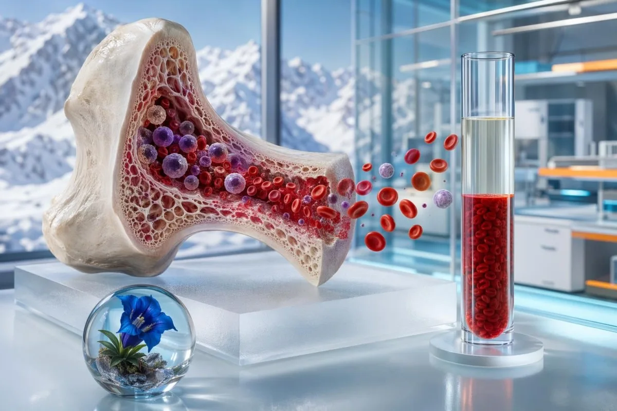
A low hematocrit is not one diagnosis. The pattern around it — hemoglobin, RBC count, MCV, reticulocytes, ferritin, kidney markers and hydration clues — tells you whether red cells are truly low or simply diluted.


- Low hematocrit means red cells occupy a smaller percentage of blood volume; adult ranges are roughly 40-52% for men and 36-46% for non-pregnant women.
- True anemia is likely when hematocrit, hemoglobin and red blood cell count are all low together, especially if symptoms are present.
- Dilutional low hematocrit can happen after IV fluids, heavy fluid intake, pregnancy or overhydration; albumin and sodium may also run low.
- Recent bleeding may not lower hematocrit immediately; the drop often becomes clearer 6-24 hours later after fluid shifts.
- Iron deficiency often shows MCV below 80 fL, ferritin below 30 ng/mL, or transferrin saturation below 20%.
- B12 or folate deficiency often pushes MCV above 100 fL, with B12 below 200 pg/mL or MMA above 0.40 µmol/L supporting deficiency.
- Reticulocytes help separate underproduction from loss; a reticulocyte production index below 2 suggests weak marrow response.
- Pregnancy lowers hematocrit through plasma expansion; hemoglobin below 11.0 g/dL in the 1st or 3rd trimester usually needs review.
- Urgent symptoms include chest pain, fainting, shortness of breath at rest, black stools, heavy ongoing bleeding or a hemoglobin near 7-8 g/dL.
What a low hematocrit result actually means
Low hematocrit usually means one of three things: too few red cells, recent red cell loss, or extra plasma diluting the sample. In practice, I do not interpret hematocrit alone; I compare it with hemoglobin, RBC count, MCV, RDW, reticulocytes and the story around the draw. A low value after 2 litres of IV fluid is a different problem from a low value with ferritin of 9 ng/mL. That distinction changes the next test, the urgency and the treatment.
Hematocrit is the percentage of whole blood volume occupied by red cellular elements. A hematocrit of 33% means about one-third of the sampled volume is cellular elements and about two-thirds is plasma, so shifts in either side of that equation can move the result.
Kantesti is an AI blood test interpretation platform that reads hematocrit beside the rest of the CBC rather than treating the percentage as a standalone diagnosis. Our clinical team, described on About Us, sees the same practical issue every week: a flagged low hematocrit can mean iron deficiency, acute fluid dilution, pregnancy physiology, kidney-related underproduction or a lab timing problem.
The WHO 2011 anemia guidance defines anemia by hemoglobin rather than hematocrit: hemoglobin below 13.0 g/dL in men, below 12.0 g/dL in non-pregnant women and below 11.0 g/dL in pregnancy meets anemia criteria (WHO, 2011). If you are trying to decode the rest of the panel, our guide to CBC components explains where hematocrit sits inside the complete blood count.
Normal hematocrit ranges depend on sex, age and pregnancy
Adult hematocrit is commonly about 40-52% in men and 36-46% in non-pregnant women, although each laboratory sets its own interval. A result just 1-2 percentage points below range is usually less concerning than a sudden fall of 6-10 points from your usual baseline.
Some European labs use slightly narrower intervals, and high-altitude regions may report higher expected hematocrit because chronic lower oxygen pressure stimulates red cell production. In my clinical experience, the most useful comparison is often your own prior result, not the printed range beside today’s number.
Lab ranges differ because hematocrit is influenced by sex hormones, altitude, smoking, pregnancy, hydration and the analyzer method. Our article on sex-specific lab ranges explains why a value that is low for one adult may be normal for another.
A practical rule I use: hematocrit is roughly three times hemoglobin when both are reported in conventional units. If hemoglobin is 10.5 g/dL, a hematocrit near 31-32% is internally consistent; if the pair is wildly mismatched, I check the units, analyzer flags and sample notes before assuming disease.
CBC pattern clues separate anemia from dilution
True anemia is more likely when hematocrit, hemoglobin and red blood cell count are low together. Dilution is more likely when the hematocrit falls after fluids and the pattern is accompanied by low-normal sodium, low albumin or a sudden same-day drop without matching symptoms.
A low red blood cell count with low hemoglobin usually points toward true reduced oxygen-carrying capacity. For a deeper explanation of why RBC count and hemoglobin sometimes disagree, see our guide to RBC versus hemoglobin.
MCV and RDW add the next layer. MCV below 80 fL suggests microcytosis, MCV above 100 fL suggests macrocytosis, and RDW above about 14.5% means cell size variation is high enough to support evolving deficiency or mixed causes; our RDW guide goes into the pattern in more detail.
Kantesti AI interprets hematocrit by checking whether the hemoglobin-to-hematocrit relationship is biologically plausible, then looking for discordant flags such as platelet clumping, sample age or a sudden delta from the last CBC. That kind of pattern check is often what prevents a mild dilutional result from being mislabeled as chronic anemia.
Recent bleeding can hide on the first hematocrit
Acute bleeding may not lower hematocrit immediately because red cells and plasma are lost together at first. The hematocrit often drops more clearly 6-24 hours later as fluid moves into the bloodstream or IV fluids are given.
I have seen patients with obvious heavy nosebleeds or gastrointestinal loss arrive with a surprisingly normal hematocrit, then fall from 39% to 31% by the next morning. The first number was not fake; it was simply drawn before plasma redistribution revealed the red cell deficit.
Menstrual timing matters too. A patient with heavy periods may show hemoglobin 11.2 g/dL and hematocrit 34% during the week after bleeding, while ferritin has been falling for months; our piece on period-related hemoglobin shifts explains that timeline.
Recent bleeding is more concerning when hematocrit falls by more than 3-5 percentage points from baseline, pulse is above 100 beats per minute, stools are black, urine is visibly red or there is fainting. If the story sounds like ongoing loss, waiting a month for a repeat CBC is usually the wrong plan.
Iron deficiency is the common low hematocrit pattern doctors hunt for
Iron deficiency commonly causes low hematocrit with low hemoglobin, low MCV and high RDW. Ferritin below 15 ng/mL is highly specific for depleted iron stores, and ferritin below 30 ng/mL is strongly suggestive in many adult patients.
The British Society of Gastroenterology guideline states that ferritin is the single most useful marker of iron deficiency anemia, but it can look falsely normal when inflammation is present (Snook et al., 2021). In inflammatory states, transferrin saturation below 20% with ferritin below 100 ng/mL can still fit iron-restricted red cell production.
Serum iron swings during the day and after meals, so I rarely use it alone. If your report shows low serum iron, high TIBC and low transferrin saturation, our guide to low serum iron and the deeper iron studies guide can help you interpret the pattern.
In clinic, I ask three unglamorous questions when hematocrit is low and iron looks low: heavy periods, blood donation and gastrointestinal loss. In adults over about 50, new iron deficiency anemia deserves a clinician-led search for GI causes even when diet seems plausible.
High MCV with low hematocrit points toward B12 or folate issues
Low hematocrit with MCV above 100 fL often suggests B12 deficiency, folate deficiency, alcohol effect, liver disease, hypothyroidism or medication interference. B12 below 200 pg/mL usually supports deficiency, while 200-300 pg/mL is a borderline zone where MMA can clarify.
Methylmalonic acid above about 0.40 µmol/L supports functional B12 deficiency, particularly when symptoms include numbness, balance trouble, burning feet or memory change. Homocysteine can rise in both B12 and folate deficiency, so it is less specific by itself.
A patient can have low hemoglobin symptoms before the MCV becomes very high. I have seen B12 deficiency with MCV of only 94 fL when iron deficiency is present at the same time, because microcytosis and macrocytosis partially cancel each other on the average.
If your B12 is borderline, compare units carefully because pg/mL and pmol/L are not interchangeable. Our B12 range guide explains those cutoffs and why MMA sometimes matters more than the headline B12 number.
Kidney disease and inflammation lower red cell production
Low hematocrit from underproduction often shows a low reticulocyte response, normal or small MCV, and chronic inflammatory or kidney markers. CKD-related anemia becomes more common when eGFR falls below 60 mL/min/1.73 m² and is especially frequent below 30.
The kidney produces erythropoietin, the hormone signal that tells marrow to make red cells. KDIGO’s anemia guideline recommends evaluating iron status, B12, folate and inflammation before attributing anemia to chronic kidney disease alone (KDIGO, 2012).
Inflammation changes iron handling by raising hepcidin, which traps iron in storage sites even when total body iron is not truly absent. This is why ferritin may be 150 ng/mL and transferrin saturation 14% in anemia of inflammation — enough stored iron, poor delivery.
Kantesti is an AI-powered blood test analysis tool that reads hematocrit alongside eGFR, creatinine, CRP, albumin and iron indices. If kidney numbers are part of your pattern, our CKD stage guide gives the eGFR and urine ACR context patients often need.
Reticulocytes show whether marrow is responding
Reticulocytes are young red cells, and they tell us whether the marrow is trying to compensate. In anemia, a reticulocyte production index below 2 suggests underproduction, while an index above 3 suggests recent loss or hemolysis if the marrow is healthy.
Hemolysis is suspected when low hematocrit is paired with high reticulocytes, high LDH, indirect bilirubin elevation and low haptoglobin. Haptoglobin below about 25 mg/dL supports red cell breakdown, though liver disease can also lower haptoglobin and complicate the read.
A direct antiglobulin test, sometimes called DAT or Coombs testing, helps detect immune hemolysis. Patients usually notice jaundice, dark urine, back discomfort or sudden fatigue, but mild hemolysis can be surprisingly quiet.
When I review this pattern, I look for the full cluster rather than one dramatic value. Our guide to haptoglobin results and our hematology review of reticulocyte markers explain why the combination is more reliable than any single marker.
Overhydration and IV fluids can dilute hematocrit
Dilutional low hematocrit happens when plasma volume expands faster than red cell mass changes. In hospital charts, 1-2 litres of crystalloid fluid can lower hematocrit by roughly 2-5 percentage points without any new red cell loss.
The clues are not subtle if you know where to look: hematocrit drops quickly, albumin may fall, sodium may drift low and the patient may have received fluids, pregnancy care, endurance-event rehydration or treatment for vomiting. The CBC looks worse, but oxygen delivery may not have changed as much as the percentage suggests.
Endurance athletes are a special case. After long training blocks, plasma volume expansion can create a low-normal hematocrit that looks like anemia on paper, while performance is stable and ferritin is acceptable; hydration markers such as urine concentration add context.
If fluid balance is part of your question, compare the CBC with sodium, albumin, BUN/urea and urine concentration. Our guide to urine osmolality is useful when the question is dehydration, overhydration or a mixed fluid picture.
Pregnancy lowers hematocrit through normal plasma expansion
Pregnancy often lowers hematocrit because plasma volume rises by about 40-50%, while red cell mass rises by roughly 20-30%. This physiologic hemodilution is expected, but hemoglobin below 11.0 g/dL in the 1st or 3rd trimester still deserves review.
The second trimester is where values commonly look lowest. Many clinicians use hemoglobin below 10.5 g/dL in the second trimester as an anemia threshold, though local obstetric protocols differ and ferritin is often checked earlier now.
Postpartum hematocrit is trickier because delivery-related loss, IV fluids and inflammation all overlap. A woman may have hematocrit 29% the morning after delivery, then 32% a week later without iron infusion simply because fluid balance normalized.
As of July 15, 2026, my practical approach is to treat pregnancy hematocrit as a trend plus symptoms, not a single red flag. Our guide to pregnancy blood tests covers the same-day warning patterns that should not wait for routine follow-up.
Low hematocrit symptoms depend on speed, not just the number
Low hematocrit symptoms usually come from reduced oxygen delivery and include fatigue, shortness of breath, dizziness, palpitations, headaches and pale skin. A slow fall to hemoglobin 9.5 g/dL may feel mild, while a rapid fall to 10.5 g/dL can feel frightening.
Low hemoglobin symptoms become more likely as hemoglobin falls below 10 g/dL, especially during exercise or climbing stairs. Below 8 g/dL, many adults feel breathless, weak or lightheaded, and people with heart disease may develop chest pressure at higher levels.
The red flags I do not like are fainting, shortness of breath at rest, chest pain, confusion, black stools, ongoing heavy bleeding, heart rate above 120 beats per minute or oxygen saturation below the patient’s usual baseline. Those are same-day clinical review signs, not lifestyle-adjustment signs.
Symptoms overlap with glucose, thyroid, salt and heart rhythm problems. If dizziness is the complaint rather than the lab flag, our guide to dizziness blood tests explains why a CBC is only one piece of the workup.
Retesting low hematocrit should match the suspected cause
A mildly low hematocrit can often be rechecked in 1-4 weeks if you feel well and the rest of the CBC is stable. A rapid drop, symptoms, pregnancy red flags, suspected bleeding or hemoglobin near 7-8 g/dL needs same-day medical advice.
After heavy fluid intake, IV fluids or a difficult acute illness, a repeat CBC after 48-72 hours can show whether the low hematocrit was transient dilution. After starting oral iron, I usually expect hemoglobin to rise about 1-2 g/dL within 3-4 weeks if absorption and diagnosis are right.
A single abnormal hematocrit should be compared with the prior result, sample notes and units. Our guide to repeating abnormal labs explains why some results need quick confirmation while others can be safely trended.
Delta checks matter. A fall from hematocrit 42% to 36% in one week deserves more attention than someone who has lived at 35-36% for years; our article on sudden lab changes shows how laboratories and clinicians think about unexpected shifts.
How Kantesti reads low hematocrit patterns
Kantesti reads low hematocrit by combining CBC indices, iron markers, kidney function, inflammation markers, hydration clues, sex, pregnancy status and prior trends. The goal is not to diagnose from one number; it is to identify the most plausible pattern and the safest next step.
Kantesti is an AI biomarker interpretation platform that can process uploaded blood test PDFs or photos in about 60 seconds, including CBCs where hematocrit is the flagged result. Our neural network checks over 15,000 biomarkers, but the clinical value comes from combinations — for example, hematocrit 33%, ferritin 7 ng/mL and MCV 76 fL is not the same story as hematocrit 33%, albumin 30 g/L and sodium 130 mmol/L.
Our doctors review the clinical logic used in the system, and the methodology is described in our technology guide. For hematocrit, the model weighs severity, direction of change, internal CBC consistency, unit conversions and whether other markers support iron deficiency, macrocytosis, kidney-related underproduction or dilution.
Accuracy work is not a one-and-done claim. Kantesti’s validation process, including rubric-based checking and medical oversight, is summarized on our medical validation page; in my view as Thomas Klein, MD, this is where AI interpretation earns trust or loses it.
Research notes and when to bring results to a clinician
Bring a low hematocrit result to a clinician promptly if the value is new, falling, symptomatic, pregnancy-related, linked with suspected bleeding or paired with abnormal white cells or platelets. A stable mild low hematocrit still deserves explanation, but the time frame is different.
Thomas Klein, MD, and the Kantesti medical team advise patients to bring the full CBC, prior results and medication list rather than a screenshot of hematocrit alone. If you need human follow-up pathways or medical governance details, our Medical Advisory Board page explains the physician oversight behind our content.
Kantesti LTD. (2026). Clinical Validation Framework v2.0. Zenodo. DOI: 10.5281/zenodo.17993721. ResearchGate listing: clinical validation record; Academia.edu listing: validation framework record.
Kantesti LTD. (2026). AI Blood Test Analyzer: 2.5M Tests Analyzed | Global Health Report 2026. Zenodo. DOI: 10.5281/zenodo.18175532. ResearchGate listing: global health report; Academia.edu listing: AI analyzer report.
If your hematocrit is low and you cannot tell whether the issue is anemia, bleeding or dilution, do not try to fix it with iron blindly. Use the pattern, ask for the missing labs and contact your clinician; if administrative help is needed, Kantesti’s Contact Us page is the safer route than guessing from one flagged percentage.
Frequently Asked Questions
What are the most common low hematocrit causes?
The most common low hematocrit causes are iron deficiency anemia, recent bleeding, pregnancy-related hemodilution, chronic inflammation, kidney disease, B12 or folate deficiency and overhydration. Adult hematocrit is often considered low below about 40% in men and below about 36% in non-pregnant women, but lab ranges vary. The pattern matters: low hemoglobin and low RBC count suggest true anemia, while a sudden fall after IV fluids may be dilution.
Can drinking too much water lower hematocrit?
Yes, heavy fluid intake or IV fluids can temporarily lower hematocrit by expanding plasma volume. In clinical practice, 1-2 litres of crystalloid fluid may lower hematocrit by roughly 2-5 percentage points without new red cell loss. Dilution is more likely when sodium, albumin or BUN/urea also look low and the result normalizes within 48-72 hours.
What low hematocrit symptoms should worry me?
Low hematocrit symptoms that need prompt review include shortness of breath at rest, chest pain, fainting, confusion, black stools, ongoing heavy bleeding or a heart rate above 120 beats per minute. Mild fatigue and dizziness are common when hemoglobin falls below about 10 g/dL, but symptoms depend strongly on how fast the drop occurs. A hemoglobin near 7-8 g/dL is often treated as urgent, especially in older adults or people with heart disease.
Can hematocrit be low while hemoglobin is normal?
Hematocrit can be slightly low while hemoglobin remains normal, especially near the reference range or when hydration status changes. Hematocrit is usually about three times hemoglobin, so a hemoglobin of 12.5 g/dL often pairs with hematocrit around 37-38%. A large mismatch should prompt a check of units, sample quality, analyzer flags and whether plasma volume changed around the test.
How soon should I repeat a low hematocrit test?
A mild, unexpected low hematocrit can often be repeated in 1-4 weeks if you feel well and the rest of the CBC is stable. If the result followed IV fluids, heavy hydration or acute illness, repeating the CBC after 48-72 hours may clarify dilution. Repeat sooner or seek same-day care if hematocrit drops more than 3-5 percentage points from baseline, hemoglobin is near 7-8 g/dL or symptoms are significant.
Does pregnancy always cause low hematocrit?
Pregnancy commonly lowers hematocrit because plasma volume rises about 40-50%, while red cell mass rises about 20-30%. Many pregnant patients have hematocrit around 33-39% without dangerous anemia, especially in the second trimester. Hemoglobin below 11.0 g/dL in the first or third trimester, or below about 10.5 g/dL in the second trimester, usually deserves iron and clinical review.
What labs help explain a low red blood cell count?
The most useful labs for low red blood cell count causes are hemoglobin, hematocrit, MCV, RDW, reticulocyte count, ferritin, transferrin saturation, B12, folate, creatinine/eGFR, CRP and sometimes haptoglobin, LDH and bilirubin. MCV below 80 fL points toward microcytic causes such as iron deficiency, while MCV above 100 fL points toward macrocytic causes such as B12 or folate deficiency. A reticulocyte production index below 2 suggests underproduction, while a higher response suggests recent loss or hemolysis.
Can a low hematocrit delay an elective procedure or surgery?
Possibly. Before an elective procedure, the surgical and anesthesia teams consider the hematocrit, hemoglobin, symptoms, cause of anemia, and expected blood loss; correcting a treatable deficiency beforehand may reduce risk and avoid delays.
Get AI-Powered Blood Test Analysis Today
Join over 2 million users worldwide who trust Kantesti for instant, accurate lab test analysis. Upload your blood test results and receive comprehensive interpretation of 15,000+ biomarkers in seconds.
📚 Referenced Research Publications
Klein, T., Mitchell, S., & Weber, H. (2026). Kantesti LTD. (2026). Clinical Validation Framework v2.0. Zenodo. https://doi.org/10.5281/zenodo.17993721. Kantesti AI Medical Research.
Klein, T., Mitchell, S., & Weber, H. (2026). Kantesti LTD. (2026). AI Blood Test Analyzer: 2.5M Tests Analyzed | Global Health Report 2026. Zenodo. https://doi.org/10.5281/zenodo.18175532. Kantesti AI Medical Research.
📖 External Medical References
World Health Organization (2011). Haemoglobin concentrations for the diagnosis of anaemia and assessment of severity. WHO Vitamin and Mineral Nutrition Information System.
KDIGO Anemia Work Group (2012). KDIGO Clinical Practice Guideline for Anemia in Chronic Kidney Disease. Kidney International Supplements.
⚕️ Medical Disclaimer
This article is for educational purposes only and does not constitute medical advice. Always consult a qualified healthcare provider for diagnosis and treatment decisions.
E-E-A-T Trust Signals
Experience
Physician-led clinical review of lab interpretation workflows.
Expertise
Laboratory medicine focus on how biomarkers behave in clinical context.
Authoritativeness
Written by Dr. Thomas Klein with review by Dr. Sarah Mitchell and Prof. Dr. Hans Weber.
Trustworthiness
Evidence-based interpretation with clear follow-up pathways to reduce alarm.