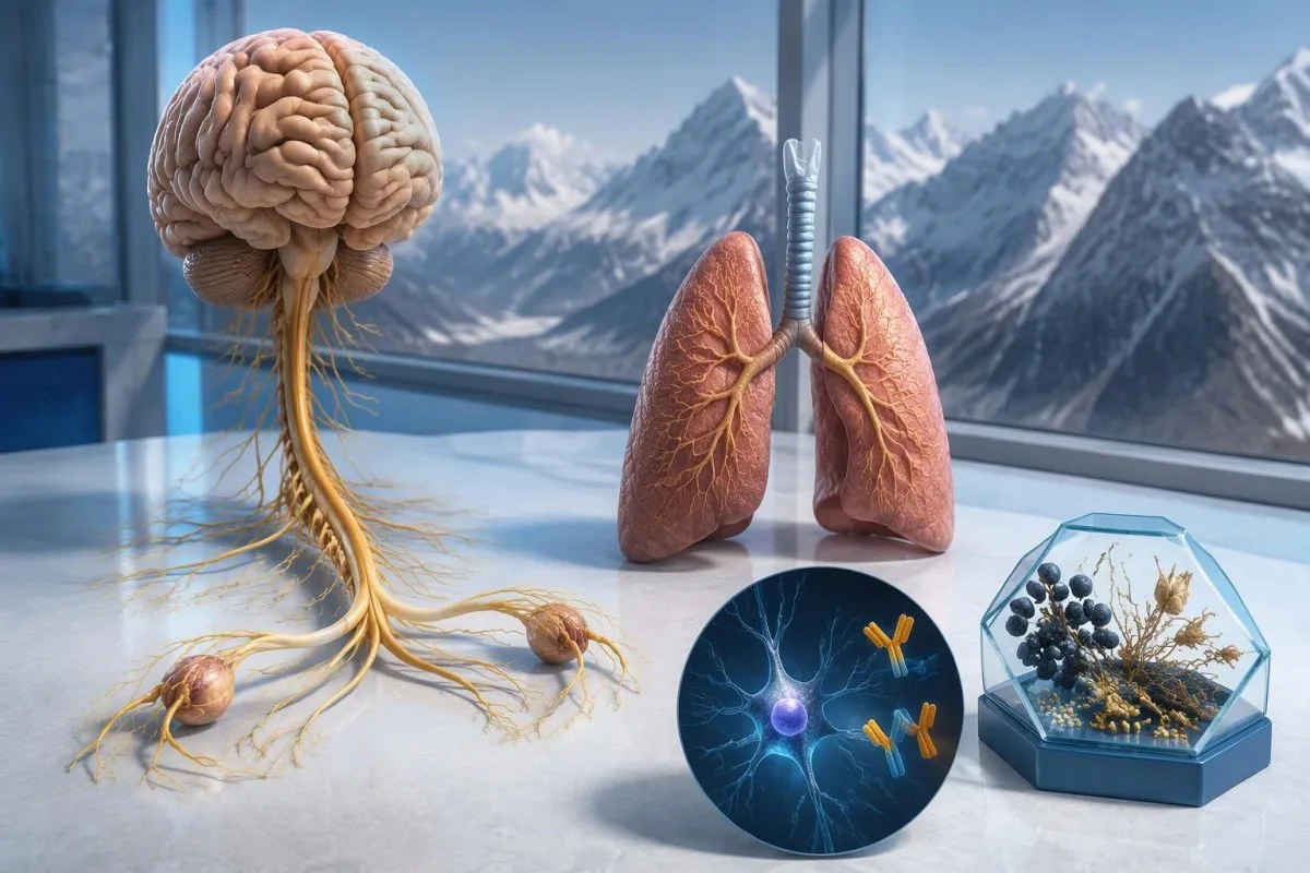
ಆಂಟಿ-Hು ಪ್ರತಿಕಾಯ ಧನಾತ್ಮಕ ಫಲಿತಾಂಶವು ಕ್ಯಾನ್ಸರ್ಗೆ ಸಂಬಂಧಿಸಿದ ನರವೈಜ್ಞಾನಿಕ ರೋಗನಿರೋಧಕ ಪ್ರತಿಕ್ರಿಯೆಯ ಬಗ್ಗೆ, ವಿಶೇಷವಾಗಿ ಸಣ್ಣ-ಕೋಶ ಶ್ವಾಸಕೋಶದ ಕ್ಯಾನ್ಸರ್ಗೆ ಸಂಬಂಧಿಸಿದಂತೆ, ಆತಂಕವನ್ನು ಹುಟ್ಟಿಸುತ್ತದೆ; ಆದರೆ ಅದು ಕ್ಯಾನ್ಸರ್ ಇದೆ ಎಂದು ಪತ್ತೆಹಚ್ಚುವುದಿಲ್ಲ. ವೈದ್ಯರು ಲಕ್ಷಣಗಳನ್ನು ಮೌಲ್ಯಮಾಪನ ಮಾಡುತ್ತಾರೆ, ಪ್ರಯೋಗಾಲಯದ ಫಲಿತಾಂಶವನ್ನು ದೃಢೀಕರಿಸುತ್ತಾರೆ ಮತ್ತು ಕ್ಯಾನ್ಸರ್ಗಾಗಿ ಮೌಲ್ಯಮಾಪನವನ್ನು ಏರ್ಪಡಿಸುತ್ತಾರೆ; ಮೊದಲ ಸ್ಕ್ಯಾನ್ಗಳು ಋಣಾತ್ಮಕವಾಗಿದ್ದರೂ ಮರುಪರಿಶೀಲನೆ ಅಗತ್ಯವಾಗಬಹುದು.



- ಆಂಟಿ-Hು / ANNA-1 ಒಂದು ಹೆಚ್ಚಿನ-ಅಪಾಯದ ಪ್ಯಾರಾನಿಯೋಪ್ಲಾಸ್ಟಿಕ್ ಪ್ರತಿಕಾಯವಾಗಿದೆ; ಇದು ದೃಢಪಟ್ಟ ಕ್ಲಿನಿಕಲ್ ಸಮೂಹಗಳಲ್ಲಿ 70% ಕ್ಕಿಂತ ಹೆಚ್ಚಿನ ಪ್ರಕರಣಗಳಲ್ಲಿ ಕ್ಯಾನ್ಸರ್ಗೆ ಸಂಬಂಧಿಸಿದ ವರ್ಗವಾಗಿದೆ—ದೃಢೀಕರಿಸದ ಪ್ರತಿಯೊಂದು ತಪಾಸಣಾ ಫಲಿತಾಂಶವೂ ಹಾಗಲ್ಲ.
- ಕ್ಯಾನ್ಸರ್ ರೋಗನಿರ್ಣಯ ಆಧಾರದಲ್ಲಿರುವ ಮಾರಕತೆಯ ಸಾಕ್ಷ್ಯವನ್ನು ಅಗತ್ಯಪಡಿಸುತ್ತದೆ; ಸಾಮಾನ್ಯವಾಗಿ ಅಂಗಾಂಶ ಪರೀಕ್ಷೆಯನ್ನೂ ಒಳಗೊಂಡಿರುತ್ತದೆ; ಒಂದು ಧನಾತ್ಮಕ ಪ್ರತಿಕಾಯ ಫಲಿತಾಂಶವೇ ಆ ಸಾಕ್ಷ್ಯವಲ್ಲ.
- ಪರೀಕ್ಷಾ ದೃಢೀಕರಣ ಸಾಮಾನ್ಯವಾಗಿ 2 ಪೂರಕ ವಿಧಾನಗಳನ್ನು ಸಂಯೋಜಿಸುತ್ತದೆ: ವಿಶಿಷ್ಟವಾದ ಅಂಗಾಂಶ-ಬಣ್ಣದ ಮಾದರಿ ಮತ್ತು ಪ್ರತಿಜನಕ-ನಿರ್ದಿಷ್ಟ ಪರೀಕ್ಷೆ.
- ನರವೈಜ್ಞಾನಿಕ ಲಕ್ಷಣಗಳು ಪ್ರಗತಿಶೀಲ ಸಂವೇದನಾ ನಷ್ಟ, ಸಮತೋಲನದ ತೊಂದರೆ, ಸೆಳೆತಗಳು ಅಥವಾ ಸ್ವಾಯತ್ತ ನರಮಂಡಲದ ಕಾರ್ಯವೈಕಲ್ಯದಂತಹವು, ಫಲಿತಾಂಶದ ಮಹತ್ವ ಮತ್ತು ತುರ್ತುತೆಯನ್ನು ಬದಲಾಯಿಸುತ್ತವೆ.
- ಋಣಾತ್ಮಕ ಚಿತ್ರಣ ಪರೀಕ್ಷೆ ಆ ಪರೀಕ್ಷೆಯ ಸಮಯದಲ್ಲಿ ಕ್ಯಾನ್ಸರ್ ಪತ್ತೆಯಾಗಲಿಲ್ಲ ಎಂದರ್ಥ; ಅದು ಆರಂಭದಲ್ಲಿ ಪತ್ತೆಹಚ್ಚಲಾಗದ ಸಣ್ಣ ಕ್ಯಾನ್ಸರ್ ಇರುವ ಸಾಧ್ಯತೆಯನ್ನು ತಳ್ಳಿಹಾಕುವುದಿಲ್ಲ.
- ಪುನಃ ತಪಾಸಣೆ 2021ರ PNS ಮಾನದಂಡಗಳ ಪ್ರಕಾರ, ಹೆಚ್ಚಿನ ಅಪಾಯದ ನರವೈಜ್ಞಾನಿಕ ಲಕ್ಷಣಸಮೂಹದೊಂದಿಗೆ ಹೆಚ್ಚಿನ ಅಪಾಯದ ಪ್ರತಿಕಾಯವಿದ್ದರೆ, 2 ವರ್ಷಗಳ ಕಾಲ ಪ್ರತಿ 4–6 ತಿಂಗಳಿಗೊಮ್ಮೆ ಶಿಫಾರಸು ಮಾಡಲಾಗುತ್ತದೆ.
- ದೀರ್ಘಕಾಲದ ನಿಗಾ ಆಯ್ದ ರೋಗಿಗಳಿಗೆ ಸೂಕ್ತವಾಗಿರಬಹುದು; 2023ರ anti-Hu ಸಮೂಹ ಅಧ್ಯಯನವು 2 ವರ್ಷಗಳಿಗಿಂತ ಹೆಚ್ಚು ಕಾಲ, ಕೆಲವೊಮ್ಮೆ 5 ವರ್ಷಗಳವರೆಗೂ ತಪಾಸಣೆ ಪರಿಗಣಿಸುವುದನ್ನು ಬೆಂಬಲಿಸಿತು.
- ತುರ್ತು ಲಕ್ಷಣಗಳು ಇವುಗಳಲ್ಲಿ ಹೊಸದಾಗಿ ಉಸಿರಾಟದ ತೊಂದರೆ ಕಾಣಿಸಿಕೊಳ್ಳುವುದು, ಸುರಕ್ಷಿತವಾಗಿ ನುಂಗಲು ಆಗದಿರುವುದು, ವೇಗವಾಗಿ ಉಲ್ಬಣಗೊಳ್ಳುವ ದೌರ್ಬಲ್ಯ, ಮೊದಲ ಬಾರಿಗೆ ಸೆಳೆತ ಬರುವುದು ಅಥವಾ ಹಠಾತ್ ನರವೈಜ್ಞಾನಿಕ ಕೊರತೆಗಳು ಸೇರಿವೆ.
ಆಂಟಿ-Hು ಪ್ರತಿಕಾಯ ಧನಾತ್ಮಕ ಫಲಿತಾಂಶದ ಅರ್ಥವೇನು?
ಒಂದು anti-Hu ಪ್ರತಿಕಾಯ ಪಾಸಿಟಿವ್ ಫಲಿತಾಂಶ ಎಂದರೆ ಪ್ರಯೋಗಾಲಯವು Hu ನರಕೋಶದ ಪ್ರೋಟೀನ್ಗಳ ವಿರುದ್ಧ ಪ್ರತಿಕ್ರಿಯಾಶೀಲತೆಯನ್ನು ಪತ್ತೆಹಚ್ಚಿದೆ ಎಂದರ್ಥ; ಇದರಿಂದ ಮಾತ್ರ ಕ್ಯಾನ್ಸರ್ ಅಥವಾ ನರವೈಜ್ಞಾನಿಕ ಲಕ್ಷಣಸಮೂಹ ಇದೆ ಎಂದು ಸಾಬೀತಾಗುವುದಿಲ್ಲ. ಇದರ ಅರ್ಥೈಸುವಿಕೆಯಲ್ಲಿ 3 ಭಾಗಗಳಿವೆ: ಫಲಿತಾಂಶ ನಿಜವಾದದ್ದೇ, ನರವೈಜ್ಞಾನಿಕ ಲಕ್ಷಣಗಳು ಇದಕ್ಕೆ ಹೊಂದಿಕೆಯಾಗುತ್ತವೆಯೇ ಮತ್ತು ಸಂಬಂಧಿತ ಕ್ಯಾನ್ಸರ್ ಪತ್ತೆಹಚ್ಚಬಹುದೇ.
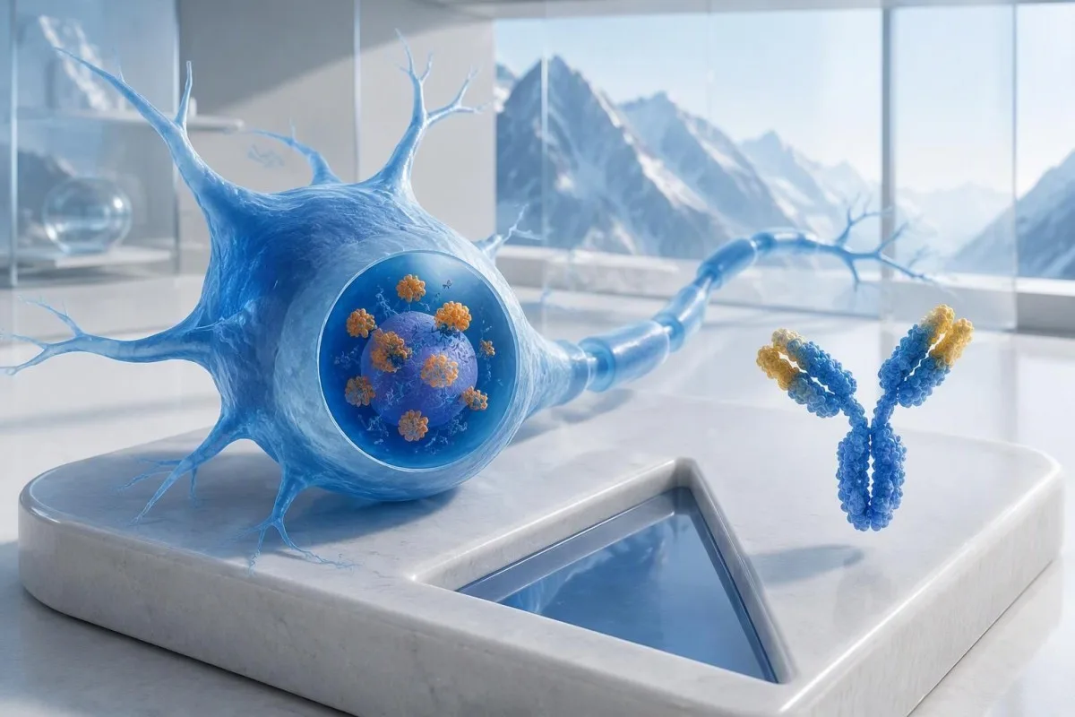
Anti-Hu ಅನ್ನು ಕ್ಯಾನ್ಸರ್ಗೆ ಸಂಬಂಧಿಸಿದ ಹೆಚ್ಚಿನ ಅಪಾಯದ ಪ್ರತಿಕಾಯವೆಂದು ವರ್ಗೀಕರಿಸಲಾಗಿದೆ, ಮತ್ತು ಸಂಬಂಧಿತ ವೈದ್ಯಕೀಯ ಜನಸಂಖ್ಯೆಗಳಲ್ಲಿ ಕ್ಯಾನ್ಸರ್ನೊಂದಿಗಿನ ಸಂಬಂಧ 70%ಗಿಂತ ಹೆಚ್ಚಿರುವ ವರ್ಗವೆಂದು ಇದನ್ನು ವ್ಯಾಖ್ಯಾನಿಸಲಾಗಿದೆ (Graus et al., 2021). ದುರ್ಬಲವಾದ, ದೃಢೀಕರಿಸದ ಪ್ಯಾನೆಲ್ ಫಲಿತಾಂಶವಿರುವ, ಬೇರೆ ರೀತಿಯಲ್ಲಿ ಆರೋಗ್ಯವಾಗಿರುವ ವ್ಯಕ್ತಿಗೆ ಆ ಶೇಕಡಾವಾರು ಪ್ರಮಾಣವನ್ನು ಹಾಗೆಯೇ ಅನ್ವಯಿಸಲಾಗುವುದಿಲ್ಲ: ಅಧ್ಯಯನ ಮಾಡಿದ ರೋಗಿಗಳು, ಪರೀಕ್ಷಾ ವಿಧಾನ ಮತ್ತು ನರವೈಜ್ಞಾನಿಕ ಲಕ್ಷಣಗಳು—ಇವೆಲ್ಲವೂ ಸಂಭವನೀಯತೆಯ ಮೇಲೆ ಪರಿಣಾಮ ಬೀರುತ್ತವೆ.
ಸಮತೋಲನ ಹದಗೆಡುತ್ತಿರುವುದು ಮತ್ತು ಅಸಮಮಿತ ಸಂವೇದನಾ ನಷ್ಟ 8 ವಾರಗಳಿಂದ ಇರುವ ಕಾಲ್ಪನಿಕ 62 ವರ್ಷದ ವ್ಯಕ್ತಿಯ ಮೌಲ್ಯಮಾಪನವು, ವ್ಯಾಪಕ ಆಯಾಸ ಪ್ಯಾನೆಲ್ನ ಭಾಗವಾಗಿ ಆಕಸ್ಮಿಕವಾಗಿ ಅವರ ಪ್ರತಿಕಾಯ ಪತ್ತೆಯಾದ ವ್ಯಕ್ತಿಯ ಮೌಲ್ಯಮಾಪನಕ್ಕಿಂತ ಭಿನ್ನವಾಗಿರಬೇಕು. ಪ್ರಯೋಗಾಲಯದ ವರದಿಯನ್ನು ಮಾತ್ರ ಆಧರಿಸಿ ಈ ಇಬ್ಬರಲ್ಲಿ ಯಾರಿಗೂ, “ನಿಮಗೆ ಕ್ಯಾನ್ಸರ್ ಇದೆ,” ಎಂದು ಹೇಳಬಾರದು. ನಮ್ಮ ಮಾರ್ಗದರ್ಶಿ ರೋಗನಿರೋಧಕ ಗುರುತನ್ನು ಪೂರ್ಣಗೊಂಡ ರೋಗನಿರ್ಣಯದಿಂದ ಬೇರ್ಪಡಿಸಲು ನೆರವಾಗುತ್ತದೆ.
ಕಾಂತೆಸ್ಟಿ ಒಬ್ಬ AI ರಕ್ತ ಪರೀಕ್ಷಾ ವಿಶ್ಲೇಷಕ anti-Hu ವರದಿಯಲ್ಲಿರುವ ಪದಪ್ರಯೋಗವನ್ನು ಅರ್ಥಮಾಡಿಕೊಳ್ಳಲು ಇದು ನೆರವಾಗಬಹುದು; ಆದರೆ ಪ್ರಯೋಗಾಲಯದ ದೃಢೀಕರಣ ಮತ್ತು ಕ್ಯಾನ್ಸರ್ನ ಮೌಲ್ಯಮಾಪನವು ನಿಮ್ಮ ಚಿಕಿತ್ಸೆ ನೀಡುವ ವೈದ್ಯರ ಜವಾಬ್ದಾರಿಯಾಗಿದೆ. Kantesti LTDಯ ಮುಖ್ಯ ವೈದ್ಯಾಧಿಕಾರಿಯಾದ Thomas Klein, MD ಆಗಿ, ನನ್ನ ಮೊದಲ ಪ್ರಶ್ನೆ ಈ ನಿರ್ದಿಷ್ಟ ಪರೀಕ್ಷೆ ಮಾಡಿಸಲು ಕಾರಣವೇನು ಎಂಬುದಾಗಿರುತ್ತದೆ—ಎಚ್ಚರಿಕೆಯ ಸೂಚನೆ ಎಷ್ಟು ಆತಂಕಕಾರಿಯಾಗಿ ಕಾಣುತ್ತದೆ ಎಂಬುದಲ್ಲ; ನಮ್ಮ ಸಂಸ್ಥೆ ಮತ್ತು ಕ್ಲಿನಿಕಲ್ ಕಾರ್ಯಾಚರಣೆ ಆ ವ್ಯತ್ಯಾಸವನ್ನು ವಿವರಿಸುತ್ತವೆ.
ANNA-1 ಮತ್ತು ಆಂಟಿ-Hು ಒಂದೇ ಪ್ರತಿಕಾಯವೇ?
ANNA-1 ಪ್ರತಿಕಾಯ ಮತ್ತು anti-Hu ಪ್ರತಿಕಾಯ ಸಾಮಾನ್ಯವಾಗಿ ಒಂದೇ ಪ್ರತಿಕಾಯ ಕುಟುಂಬವನ್ನು ಸೂಚಿಸುತ್ತವೆ. ANNA-1 ಎಂದರೆ antineuronal nuclear antibody type 1; Hu ಎಂಬ ಪದನಾಮವು ಪ್ರತ್ಯೇಕ ರೋಗವನ್ನಾಗಲಿ ಕ್ಯಾನ್ಸರ್ನ ಹಂತವನ್ನಾಗಲಿ ಸೂಚಿಸುವುದಿಲ್ಲ, ಬದಲಿಗೆ ರೋಗನಿರೋಧಕ ಪ್ರತಿಕ್ರಿಯೆ ಗುರುತಿಸುವ ಒಳಕೋಶೀಯ ನರಕೋಶದ ಪ್ರೋಟೀನ್ಗಳನ್ನು ಸೂಚಿಸುತ್ತದೆ.
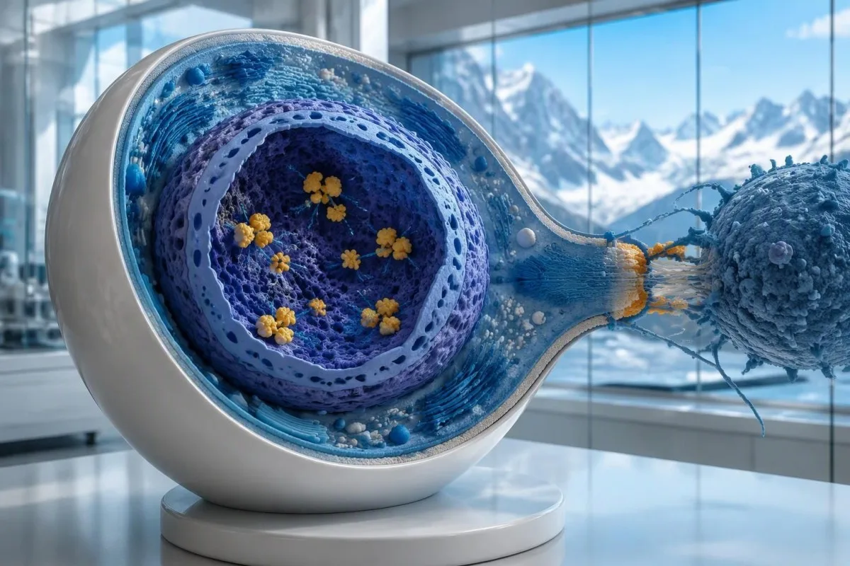
Hu ಪ್ರೋಟೀನ್ಗಳು ಒಳಕೋಶೀಯ RNA-ಬಂಧಕ ಪ್ರೋಟೀನ್ಗಳಾಗಿವೆ, ಮತ್ತು anti-Hu ಅನ್ನು ಸೈಟೋಟಾಕ್ಸಿಕ್ T ಕೋಶಗಳನ್ನು ಹೆಚ್ಚಾಗಿ ಒಳಗೊಳ್ಳುವ ರೋಗನಿರೋಧಕ ಪ್ರತಿಕ್ರಿಯೆಯ ಗುರುತಾಗಿ ಅರ್ಥಮಾಡಿಕೊಳ್ಳುವುದು ಉತ್ತಮ. ನರಕೋಶದ ಮೇಲ್ಮೈಯಲ್ಲಿ ಸುಲಭವಾಗಿ ತಲುಪಬಹುದಾದ ಪ್ರೋಟೀನ್ಗಳಿಗೆ ಪ್ರತಿಕಾಯಗಳು ಬಂಧಿಸುವ ಕಾಯಿಲೆಗಳಿಂದ ಇದು ಭಿನ್ನವಾಗಿದೆ. ಈ ವ್ಯತ್ಯಾಸದಿಂದ, 2 ವರದಿಗಳ ಟೈಟರ್ಗಳು ಬಹಳ ವಿಭಿನ್ನವಾಗಿದ್ದರೂ ಸಹ, ಪ್ರತಿಕಾಯದ ಮಟ್ಟವನ್ನು ಸರಳವಾಗಿ ಕಡಿಮೆ ಮಾಡುವುದರಿಂದ ಈಗಾಗಲೇ ಸ್ಥಾಪಿತವಾಗಿರುವ anti-Hu ನರವೈಜ್ಞಾನಿಕ ಹಾನಿ ವಿಶ್ವಾಸಾರ್ಹವಾಗಿ ಹಿಮ್ಮುಖವಾಗುವುದಿಲ್ಲ ಎಂಬುದು ಸ್ಪಷ್ಟವಾಗುತ್ತದೆ.
“ANNA-1 ಪತ್ತೆಯಾಗಿದೆ” ಎಂದು ಹೇಳುವ ವರದಿಯು, ಅಕ್ಷರಗಳು ಹೋಲುತ್ತಿದ್ದರೂ, ಸಾಮಾನ್ಯ ANA ಪಾಸಿಟಿವ್ ಫಲಿತಾಂಶಕ್ಕೆ ಸಮಾನವಲ್ಲ. ಅನಾ ಸಾಮಾನ್ಯವಾಗಿ ವ್ಯವಸ್ಥಿತ ಸ್ವಪ್ರತಿರಕ್ಷಕ ಮೌಲ್ಯಮಾಪನದಲ್ಲಿ ಬಳಸುವ antinuclear antibodies ಅನ್ನು ಸೂಚಿಸುತ್ತದೆ, ಆದರೆ ANNA-1 ನರಕೋಶದ ಪ್ರತಿಕಾಯ ಮಾದರಿಯನ್ನು ಗುರುತಿಸುತ್ತದೆ. ನಿಮ್ಮ ಫಲಿತಾಂಶಗಳ ಬಗ್ಗೆ ಚರ್ಚಿಸುವಾಗ ಈ 2 ಸಂಕ್ಷೇಪಣಗಳನ್ನು ಪ್ರತ್ಯೇಕವಾಗಿರಿಸಿ; ನಮ್ಮ ANA ಪದಪ್ರಯೋಗದ ಮಾರ್ಗದರ್ಶಿ ಹೆಚ್ಚು ಪರಿಚಿತವಾದ ಪರೀಕ್ಷೆಯನ್ನು ವಿವರಿಸುತ್ತದೆ.
ಸಣ್ಣಕೋಶದ ಶ್ವಾಸಕೋಶದ ಕ್ಯಾನ್ಸರ್ ಇರುವ, ಗುರುತಿಸಬಹುದಾದ ನರವೈಜ್ಞಾನಿಕ ಲಕ್ಷಣಸಮೂಹವಿಲ್ಲದ ಕೆಲವರಲ್ಲಿ anti-Hu ಪತ್ತೆಯಾಗಬಹುದು. ಇದಕ್ಕೆ ವಿರುದ್ಧವಾಗಿ, ಕ್ಯಾನ್ಸರ್ ಗೋಚರಿಸುವುದಕ್ಕೂ ಮುನ್ನ ನರವೈಜ್ಞಾನಿಕ ಲಕ್ಷಣಗಳಿಂದಾಗಿ ಪ್ರತಿಕಾಯ ಪತ್ತೆಯಾಗಬಹುದು. ಇವು 2 ವಿಭಿನ್ನ ವೈದ್ಯಕೀಯ ಮಾರ್ಗಗಳಾಗಿವೆ: ನರಗಳಿಗೆ ಪ್ರಸ್ತುತ ಹಾನಿಯಾಗುತ್ತಿದೆಯೇ ಅಥವಾ ಆಧಾರವಾಗಿರುವ ಕ್ಯಾನ್ಸರ್ ಎಲ್ಲಿರಬಹುದು ಎಂಬುದನ್ನು ಪ್ರತಿಕಾಯವೊಂದೇ ವೈದ್ಯರಿಗೆ ತಿಳಿಸುವುದಿಲ್ಲ.
ಆಂಟಿ-Hು ಪ್ರತಿಕಾಯ ಪರೀಕ್ಷೆಯ ವರದಿಯನ್ನು ಹೇಗೆ ಓದಬೇಕು?
An anti-Hu antibody test has no universal numerical cutoff that can be applied across laboratories. Read the specimen type, assay method, result category, laboratory reference interval and confirmation comments together; a dilution titer, band intensity and numerical index are 3 different ways of reporting reactivity.
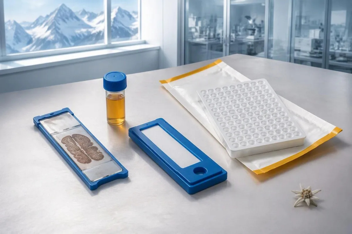
A hypothetical titer of 1:240 describes detectable reactivity after a particular dilution; it is not a cancer probability of 240, nor does it establish cancer stage. A numerical index from another method may use entirely different calibration. Our guide to ಗುಣಾತ್ಮಕ ಮತ್ತು ಪರಿಮಾಣಾತ್ಮಕ ಫಲಿತಾಂಶಗಳು explains why “positive” and “how much” answer different laboratory questions.
Weak or borderline reactivity deserves method-specific interpretation, particularly when a broad panel was ordered without a convincing neurological indication. Ask whether the laboratory observed the characteristic neuronal staining pattern and whether an antigen-specific confirmation was performed. Two tests using the same underlying strip technology may reproduce the same analytical problem; agreement is more informative when the methods are genuinely complementary.
Bring the complete report, not just a screenshot of the word positive. The 5 most useful details are the specimen, collection date, assay name, reported strength or titer and laboratory interpretation. Also list recent intravenous immunoglobulin and other immune treatments, because passive antibodies or treatment effects can complicate some serological results; your laboratory can advise whether that issue plausibly affects this particular finding.
ಧನಾತ್ಮಕ ಆಂಟಿ-Hು ಫಲಿತಾಂಶವನ್ನು ಯಾವಾಗ ದೃಢೀಕರಿಸಬೇಕು?
Unexpected, weak or clinically discordant anti-Hu results should be reviewed for confirmation, preferably with an experienced neuroimmunology laboratory. The 2021 PNS criteria emphasize interpreting neuronal antibody testing alongside the phenotype and using appropriate confirmatory methods rather than accepting an isolated commercial-panel result as a diagnosis.
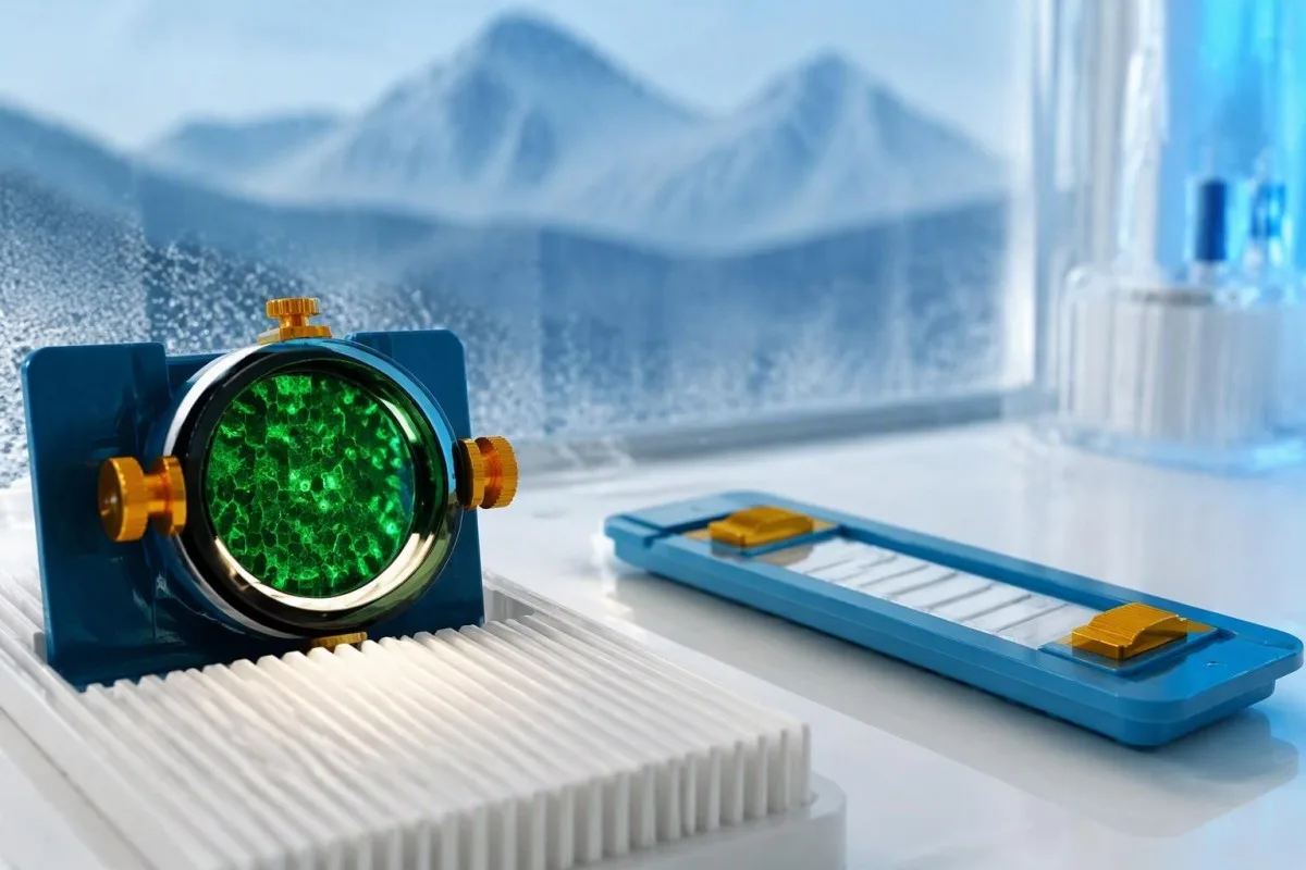
Tissue-based immunohistochemistry or immunofluorescence and an antigen-specific assay provide different information. The first identifies a characteristic distribution of neuronal staining; the second tests reactivity against the relevant antigen. Graus et al. (2021) describe these complementary approaches, with specialist reassessment particularly useful when 1 method gives a result that does not fit the clinical picture.
Repeating a sample can address collection errors or a transient analytical concern, but repeating the same kit is not always enough. In a hypothetical 45-year-old with no objective neurological findings and a faint isolated band, I would want the laboratory’s explanation before treating the report as established anti-Hu autoimmunity. Our GAD65 interpretation guide illustrates another antibody for which context and assay characteristics change meaning.
Confirmation and urgent assessment can happen in parallel. A person who develops seizures, substantial walking difficulty or progressive swallowing impairment should not wait several weeks for a reference-laboratory answer before receiving neurological care. The practical question is not “repeat or investigate?” but which of those 2 tasks can safely proceed together, given the symptoms and the initial laboratory evidence.
ಯಾವ ನರವೈಜ್ಞಾನಿಕ ಲಕ್ಷಣಗಳು ಆಂಟಿ-Hುವಿನ ಬಗ್ಗೆ ಹೆಚ್ಚು ಆತಂಕ ಮೂಡಿಸುತ್ತವೆ?
Progressive sensory neuronopathy, encephalomyelitis, limbic encephalitis and marked autonomic dysfunction are recognized anti-Hu presentations. Symptoms can involve more than 1 part of the nervous system, so the pattern and speed of change are usually more informative than a single symptom such as tingling or fatigue.
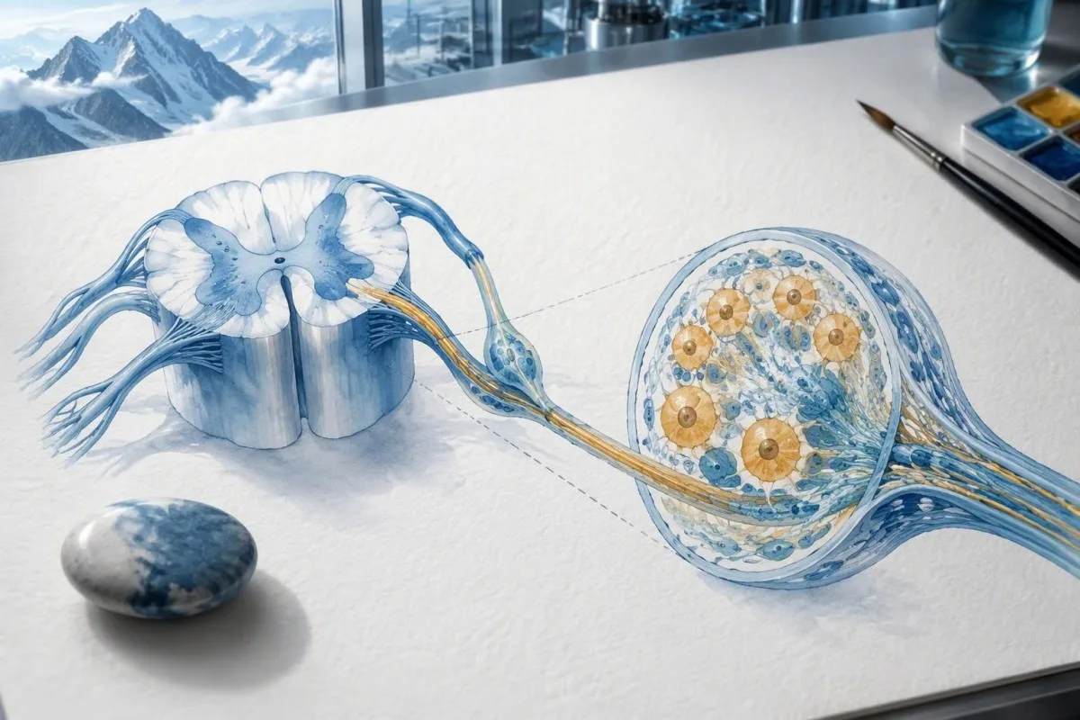
Sensory neuronopathy can cause asymmetric numbness, impaired position sense and sensory ataxia, sometimes involving the hands or trunk early rather than following a simple feet-first pattern. A patient may lose balance in the dark because vision had been compensating for reduced position feedback. That is different from the focal finger distribution discussed in our carpal tunnel symptom guide, although neither pattern can be diagnosed from description alone.
Anti-Hu autonomic involvement can affect gut movement, blood-pressure regulation and bladder function. Severe constipation, unexplained intestinal pseudo-obstruction or dizziness on standing becomes more relevant when accompanied by objective neurological abnormalities. Recording symptoms over 7 days can reveal combinations that otherwise get presented as separate stomach, balance and urinary complaints; sudden severe abdominal symptoms still require assessment for non-neurological emergencies.
New memory impairment, behavioral change or seizures may indicate central nervous system involvement, while rapid weakness has a broader emergency differential. The ದೌರ್ಬಲ್ಯದ ತುರ್ತು ಎಚ್ಚರಿಕೆ ಚಿಹ್ನೆಗಳು in our neurological safety guide apply regardless of the antibody label. Call emergency services for new breathing difficulty, unsafe swallowing, a first seizure or sudden stroke-like symptoms rather than waiting for a scheduled follow-up.
ಧನಾತ್ಮಕ ಫಲಿತಾಂಶದ ನಂತರ ಯಾವ ನರವೈಜ್ಞಾನಿಕ ಪರೀಕ್ಷೆಗಳನ್ನು ಮಾಡಬಹುದು?
Neurological follow-up may include examination, nerve conduction studies, MRI and cerebrospinal fluid testing, selected according to the symptoms. There is no mandatory battery for every anti-Hu-positive person; the purpose is to document which structures are affected and exclude other causes, not merely to collect additional positive tests.
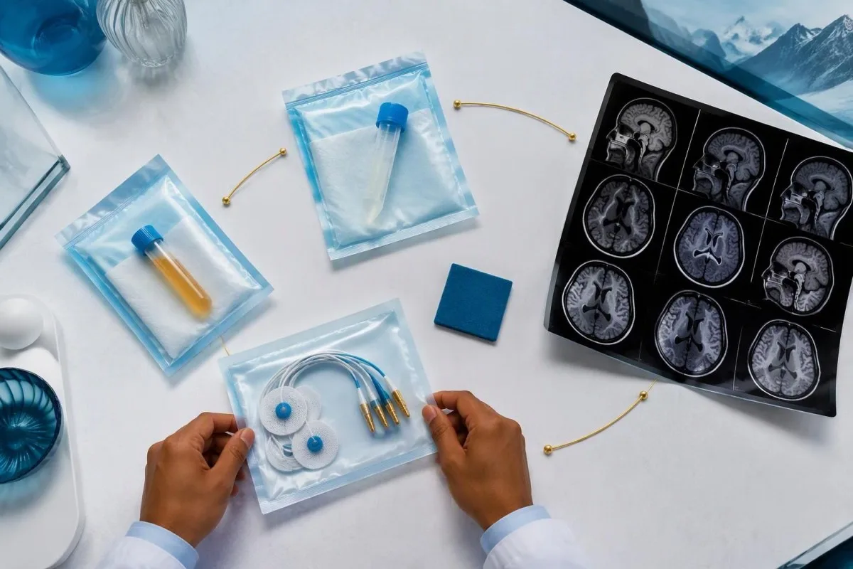
Nerve conduction studies can support a sensory neuronopathy pattern by showing reduced sensory responses across distributions that do not fit a single compressed nerve. Brain or spinal MRI addresses different questions, including structural disease and some inflammatory changes. A normal MRI does not exclude peripheral sensory ganglion involvement; these are 2 distinct anatomical compartments with different diagnostic tools.
Cerebrospinal fluid may show increased cells, protein or evidence of intrathecal immune activity, but none of those findings is specific to anti-Hu. A lumbar puncture is most useful when the neurological syndrome warrants it, rather than as an automatic response to 1 weak serum band. Our discussion of CSF ಆಲಿಗೋಕ್ಲೋನಲ್ ಬ್ಯಾಂಡ್ಗಳ ಫಲಿತಾಂಶ ಅರ್ಥೈಸುವಿಕೆ explains why immune activity in spinal fluid does not identify a single disease.
Basic investigations still matter: glucose control, vitamin status, renal function and serum protein assessment can uncover treatable contributors to neuropathy. A clinician may request 2 or more of these alongside specialist testing, depending on the examination. The ಸೀರಮ್ ಪ್ರೋಟೀನ್ ಮೌಲ್ಯಮಾಪನ ಮಾರ್ಗದರ್ಶಿ provides background, but normal routine blood work neither confirms nor excludes an anti-Hu-associated neurological process.
ಆಂಟಿ-Hು ಧನಾತ್ಮಕತೆ ಪ್ಯಾರಾನಿಯೋಪ್ಲಾಸ್ಟಿಕ್ ಸಿಂಡ್ರೋಮ್ ಅನ್ನು ಖಚಿತಪಡಿಸುತ್ತದೆಯೇ?
A positive antibody alone does not confirm anti-Hu paraneoplastic syndrome. The PNS-Care approach combines the neurological phenotype, antibody risk category, cancer findings and follow-up; clinicians also check for direct cancer effects, treatment toxicity and unrelated neurological conditions before attributing symptoms to a paraneoplastic immune response.
Definite, probable and possible PNS are diagnostic categories, not cancer stages. The 2021 criteria use a structured scoring system rather than allowing 1 antibody to determine the whole diagnosis (Graus et al., 2021). A confirmed anti-Hu result with a strongly compatible syndrome remains clinically significant even before a cancer is identified, but clinicians should state clearly which parts of the assessment remain unresolved.
An older patient can have anti-Hu reactivity and a separate cause of numbness, such as diabetes, nutritional deficiency or a monoclonal protein disorder. Coexistence is possible; either-or reasoning can miss treatable disease. If serum protein studies reveal a band, our immunofixation result guide explains why that finding requires its own interpretation rather than being automatically folded into the anti-Hu diagnosis.
ಕಾಂತೆಸ್ಟಿ ಒಬ್ಬ AI ರಕ್ತ ಪರೀಕ್ಷೆ ವರದಿ ಅರ್ಥ ಪ್ಲಾಟ್ಫಾರ್ಮ್ that can help organize these distinctions in a laboratory explanation, but it cannot assign a reliable PNS-Care diagnosis from a report alone. Our ಕ್ಲಿನಿಕಲ್ ಮಾನ್ಯತಾ ಚೌಕಟ್ಟು separates technical evaluation from clinical conclusions. In practice, I would document 3 separate judgments: confidence in the antibody, confidence in the neurological syndrome and the current evidence for cancer.
ಸಂಬಂಧಿತ ಕ್ಯಾನ್ಸರ್ಗಾಗಿ ವೈದ್ಯರು ಹೇಗೆ ಹುಡುಕುತ್ತಾರೆ?
Cancer evaluation usually prioritizes the chest because small-cell lung cancer is the strongest adult anti-Hu association. Doctors commonly begin with appropriate chest CT and may add FDG-PET/CT or targeted investigations if suspicion remains; the exact plan depends on age, symptoms, examination, smoking history and previous imaging.
The cancer association is substantial in established anti-Hu clinical cohorts. Villagrán-García et al. (2023) reported identified cancer in 349 of 466 patients, approximately 75%, in a specialist anti-Hu cohort. This is valuable evidence for taking confirmed, clinically relevant anti-Hu seriously; it is not a personalized prediction for an asymptomatic person with an unconfirmed positive screening strip.
A chest X-ray is not equivalent to a chest CT, and routine low-dose screening is not necessarily the correct examination for a suspected paraneoplastic syndrome. The clinician chooses the protocol and body regions based on the diagnostic question. Likewise, neurological symptoms do not automatically mean cancer has spread to the brain; our brain imaging warning-sign guide distinguishes structural investigation from immune-mediated disease.
A suspicious scan often leads to tissue sampling through a method chosen by respiratory or oncology specialists, such as bronchoscopic assessment of an accessible area. Imaging suspicion and cancer confirmation are 2 separate steps. Never-smoking status lowers the baseline probability of small-cell lung cancer but does not cancel a well-confirmed anti-Hu finding with a convincing neurological presentation; other cancers and non-cancer explanations still need consideration.
ಮೊದಲ ಕ್ಯಾನ್ಸರ್ ಸ್ಕ್ಯಾನ್ಗಳು ಏಕೆ ಋಣಾತ್ಮಕವಾಗಿರಬಹುದು?
Initial imaging can be negative because an associated cancer is still too small to detect, lies outside the examined area or is not readily visible with that technique. Neurological autoimmunity may precede cancer detection, so a negative scan reduces concern without necessarily ending follow-up in a high-risk clinical presentation.
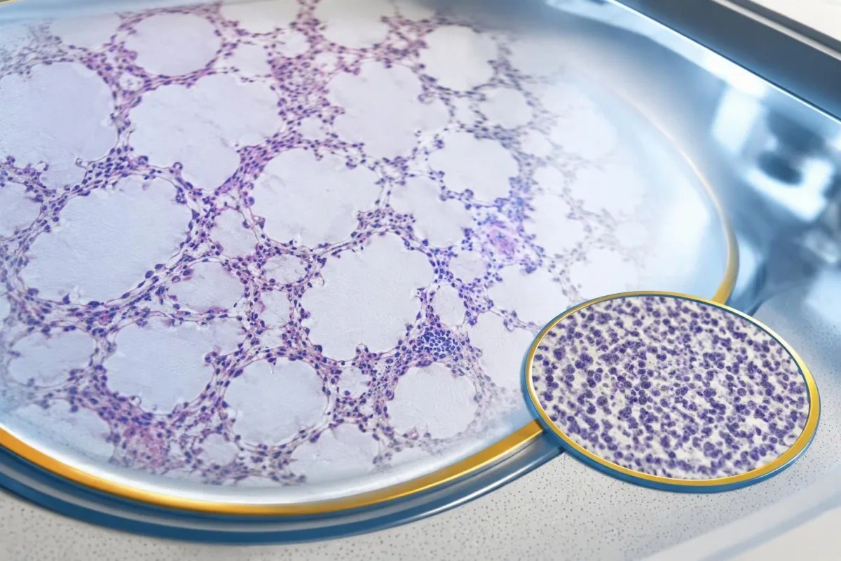
A negative scan is time-specific and method-specific. A normal chest CT performed on 1 date answers whether that examination identified a suspicious chest finding then; it does not guarantee that every possible associated cancer is absent indefinitely. Ask what was actually imaged, whether the protocol was suitable and whether the specialist recommends PET/CT or targeted investigation despite the initial result.
Villagrán-García et al. (2023) described cancers detected beyond the first 2 years in a minority of their anti-Hu cohort, supporting individualized longer surveillance. Their findings also support considering PET-based investigation when initial CT is unrevealing in an appropriate clinical context. This is an escalation decision for the treating team, not a reason to independently purchase repeated whole-body scans without weighing radiation, incidental findings and cost.
Neurological deterioration after negative imaging still needs reassessment. Cancer surveillance and neurological monitoring are 2 parallel tracks; one reassuring scan does not explain new loss of function. A marker such as neurofilament light chain may indicate neuronal injury in selected settings, but our NfL interpretation guide explains why it cannot establish anti-Hu disease, locate cancer or replace examination.
ಕ್ಯಾನ್ಸರ್ ತಪಾಸಣೆಯನ್ನು ಎಷ್ಟು ಕಾಲ ಮುಂದುವರಿಸಬೇಕು?
The 2021 PNS criteria recommend repeat cancer screening every 4–6 months for 2 years when a high-risk neurological phenotype accompanies a high-risk antibody. Longer surveillance may be appropriate for selected anti-Hu patients; the interval and duration should reflect confirmation, clinical evolution, initial investigations and individual cancer risk.

The 2-year schedule is not an instruction to scan every positive antibody result repeatedly. Graus et al. (2021) link that recommendation to the combination of high-risk phenotype and high-risk antibody. An incidental, weak result without a matching syndrome needs confirmation and individualized assessment first; repeating imaging mechanically can create incidental findings and unnecessary procedures without resolving the original uncertainty.
Older European guidance recommended longer surveillance. The EFNS task force advised repeating screening after 3–6 months and then every 6 months for up to 4 years in relevant antibody-positive paraneoplastic presentations (Titulaer et al., 2011). The 2023 anti-Hu cohort supported considering screening up to 5 years in selected patients, so a specialist may reasonably extend follow-up beyond the newer general 2-year framework.
Keep a dated plan stating the next examination, the clinician responsible and what symptoms should trigger earlier contact. Kantesti explanations can help you understand terminology, but the platform should not be treated as a scan-reminder service or substitute for that plan. Our guide to risk-based cancer screening addresses a different setting: routine prevention is not the same as investigating a possible paraneoplastic syndrome.
ಆಂಟಿ-Hು ನರವೈಜ್ಞಾನಿಕ ಕಾಯಿಲೆಗೆ ಯಾವ ಚಿಕಿತ್ಸೆಯು ಸಹಾಯ ಮಾಡುತ್ತದೆ?
Treatment focuses on an identified cancer, selected immune therapy and symptom-directed rehabilitation. Anti-Hu-associated neurological disease often responds less predictably to immunotherapy than neuronal surface-antibody disorders because intracellular targets and T-cell-mediated injury are involved; treatment may aim to stabilize function rather than promise complete recovery.

Treating an associated cancer is a central part of care, but established neurological injury may persist even when cancer treatment succeeds. Neurology and oncology teams may consider corticosteroids, intravenous immunoglobulin, plasma exchange or other immune treatments in selected situations. There is no single evidence-based dose or 1 universal regimen appropriate for everyone with anti-Hu positivity, especially without a confirmed clinical syndrome.
An antibody titer is not a dependable treatment target on its own. A lower number does not necessarily mean restored nerve function, and a persistent positive result does not by itself prove cancer progression. This differs from some surface-antibody disorders; our MOG antibody diagnosis guide explains another immune neurological condition, but its diagnostic rules and treatment expectations should not be transferred to anti-Hu.
Immune checkpoint cancer therapies deserve an explicit discussion when anti-Hu neurological disease is present, because they can provoke or worsen paraneoplastic neurological autoimmunity. This is a specialist risk-benefit decision, not an automatic prohibition based on 1 laboratory result. Tell the oncologist about previous neurological symptoms and confirmed neuronal antibodies before treatment decisions are finalized; coordinating the teams early can prevent avoidable misunderstanding.
ತನಿಖೆಗಳು ಮುಂದುವರಿಯುತ್ತಿರುವಾಗ ನೀವು ಸುರಕ್ಷಿತವಾಗಿ ಏನು ಮಾಡಬಹುದು?
While investigations continue, protect mobility, swallowing and medication safety rather than trying to lower anti-Hu antibodies with supplements. Use the neurological symptoms—not the appearance of the laboratory flag—to decide whether ordinary follow-up is sufficient or urgent care is needed; a new functional loss warrants contact before the next planned scan.
Position-sense loss increases fall risk, particularly in poor lighting or on uneven ground. A physiotherapist may recommend a suitable walking aid and individualized balance strategies; occupational therapy can address hand coordination and home hazards. A short 7-day record of falls, near-falls and walking changes is often more useful at review than the vague statement “my balance is worse,” but stop activities that feel unsafe.
Excess vitamin B6 can itself cause sensory neuropathy, making unsupervised high-dose nerve supplements particularly unhelpful during this assessment. Review every supplement and its daily dose in mg, including overlapping multivitamins and combination products. Our B-ಕಾಂಪ್ಲೆಕ್ಸ್ ಸುರಕ್ಷತಾ ಮಾರ್ಗದರ್ಶಿ explains that problem; correcting a documented deficiency is different from taking progressively larger doses in the hope of treating anti-Hu autoimmunity.
Vitamin B12 deficiency can contribute to sensory and gait problems and may need investigation even when anti-Hu is confirmed. There is no universally interchangeable B12 cutoff: results in pg/mL ನಲ್ಲಿ ವರದಿ ಮಾಡುತ್ತವೆ ಮತ್ತು pmol/L require their own laboratory context, sometimes with functional testing. Our ಗಡಿರೇಖೆಯ B12 ಫಾಲೋ-ಅಪ್ ಮಾರ್ಗದರ್ಶಿ explains those next steps; new choking, breathing difficulty or rapid weakness should be assessed urgently instead of managed with vitamins.
ಆಂಟಿ-Hು ವರದಿಯನ್ನು ಸುರಕ್ಷಿತವಾಗಿ ವಿವರಿಸಲು AI ಹೇಗೆ ಸಹಾಯ ಮಾಡಬಹುದು?
AI can help explain report terminology and organize questions, but it cannot confirm anti-Hu specificity, diagnose paraneoplastic syndrome or rule out cancer. Safe interpretation preserves the original assay details and separates 3 issues: what the laboratory reported, what the result may mean and what a clinician has actually established.
ಕಾಂತೆಸ್ಟಿ ಒಬ್ಬ AI-ಚಾಲಿತ ರಕ್ತ ಪರೀಕ್ಷೆ ವಿಶ್ಲೇಷಣೆ ಸಾಧನ that can provide a plain-language explanation of uploaded laboratory information, including the distinction between an antibody screen and a confirmed clinical diagnosis. A rapid explanation—often around 60 seconds for report processing—is not a reference-laboratory assay. Our AI ತಂತ್ರಜ್ಞಾನ ಮಾರ್ಗದರ್ಶಿ describes the interpretation workflow without making anti-Hu-specific diagnostic guarantees.
The complete source report should remain available for verification. An explanation should not silently replace “weakly reactive” with “confirmed,” drop the specimen type or invent a universal reference interval. Our guide to AI ವೈದ್ಯಕೀಯ ವಿವರಣೆಗಳನ್ನು ಪರಿಶೀಲಿಸುವ ಮಾರ್ಗದರ್ಶಿ describes practical source checks; for this antibody, compare at least the assay wording, confirmation comment and collection date against the original document.
Kantesti cannot examine sensation, review tissue staining directly through a report explanation or establish that negative imaging has excluded a tiny cancer. Those are 3 different clinical capabilities, not features that follow from understanding laboratory language. Share the explanation with the treating team as a discussion aid, and do not delay urgent assessment while uploading documents or waiting for a second automated interpretation.
ನಿಮ್ಮ ಮುಂದಿನ ಅಪಾಯಿಂಟ್ಮೆಂಟ್ ಮತ್ತು ಸಂಶೋಧನೆಯ ಪಾರದರ್ಶಕತೆ
Bring the complete antibody report, a dated symptom history, previous imaging reports and a current medication list to your next appointment. Ask the clinician to clarify antibody confirmation, the neurological diagnosis and cancer surveillance separately; those 3 answers may have different levels of certainty and different follow-up dates.
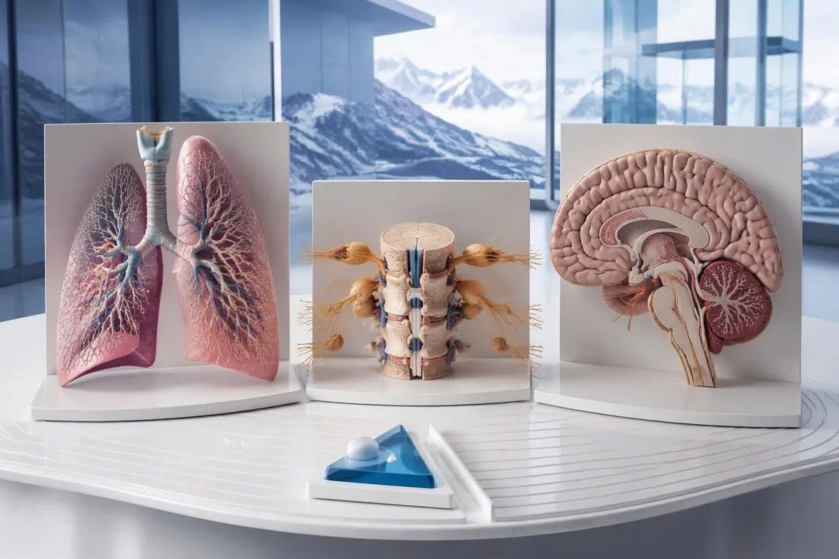
A useful 1-page symptom history records onset, progression, sensory distribution, walking changes, memory problems and autonomic symptoms. Include prior cancer treatment and any recent immunoglobulin therapy, then ask who will coordinate neurology and oncology if both are involved. As Thomas Klein, MD, I would rather see a clear timeline than 20 disconnected screenshots; it makes discrepancies between the laboratory result and clinical course easier to spot.
Medical authorship is not a substitute for an individual neurological examination. ನಮ್ಮ ವೈದ್ಯಕೀಯ ಸಲಹಾ ಮಂಡಳಿಯ ಮಾಹಿತಿ describes the clinicians associated with our work, while your own specialist determines the appropriate next investigation. As of ಅಕ್ಟೋಬರ್ 25, 2026, the recommendations discussed here draw on the 2021 international PNS criteria, the 2011 EFNS screening guidance and the 2023 anti-Hu cohort—not an invented 2026 guideline.
The 2 Figshare publications below are platform-related research references, not evidence validating anti-Hu diagnosis or cancer exclusion. Their supplied titles describe hantavirus triage deployment and a synthetic-case technical benchmark; neither title establishes anti-Hu-specific clinical performance. Because publication authors and dates were not supplied for verification, the citations use title-first APA formatting with “n.d.”; the ResearchGate and Academia.edu links are searches, not claims of verified publication copies.
ಪದೇ ಪದೇ ಕೇಳಲಾಗುವ ಪ್ರಶ್ನೆಗಳು
ಆಂಟಿ-Hು ಆಂಟಿಬಾಡಿ ಪಾಸಿಟಿವ್ ಫಲಿತಾಂಶ ಎಂದರೆ ನನಗೆ ಕ್ಯಾನ್ಸರ್ ಇದೆಯೇ?
0 ಆಂಟಿ-Hu ಪ್ರತಿಕಾಯಕ್ಕೆ ಪಾಸಿಟಿವ್ ಫಲಿತಾಂಶ ಬಂದ ಮಾತ್ರಕ್ಕೆ ಕ್ಯಾನ್ಸರ್ ಇದೆ ಎಂದು ರೋಗನಿರ್ಣಯಿಸಲಾಗುವುದಿಲ್ಲ. ಆಂಟಿ-Hu ಎಂಬುದು ಕ್ಯಾನ್ಸರ್ಗೆ ಸಂಬಂಧಿಸಿದ ಹೆಚ್ಚಿನ ಅಪಾಯದ ಪ್ರತಿಕಾಯವಾಗಿದ್ದು, 2023ರ ತಜ್ಞರ ಆಂಟಿ-Hu ಸಮೂಹದಲ್ಲಿ 466 ರೋಗಿಗಳಲ್ಲಿ 349 ಮಂದಿಗೆ ಕ್ಯಾನ್ಸರ್ ಪತ್ತೆಯಾಗಿತ್ತು; ಆದರೆ ಆ ಅಂಕಿ, ಆಕಸ್ಮಿಕವಾಗಿ ಬಂದಿರುವ ಮತ್ತು ದೃಢೀಕರಿಸದ ತಪಾಸಣಾ ಫಲಿತಾಂಶದ ಅಪಾಯವನ್ನು ನಿರ್ಧರಿಸುವುದಿಲ್ಲ. ವೈದ್ಯರು ನರಮಂಡಲಕ್ಕೆ ಸಂಬಂಧಿಸಿದ ಲಕ್ಷಣಗಳನ್ನು ಮೌಲ್ಯಮಾಪನ ಮಾಡುತ್ತಾರೆ, ಅನಿರೀಕ್ಷಿತ ಪ್ರಯೋಗಾಲಯದ ಫಲಿತಾಂಶಗಳನ್ನು ದೃಢೀಕರಿಸುತ್ತಾರೆ ಮತ್ತು ಆಧಾರವಾಗಿರುವ ಕ್ಯಾನ್ಸರ್ಗಾಗಿ ತನಿಖೆ ನಡೆಸುತ್ತಾರೆ; ವಯಸ್ಕರಲ್ಲಿ ಸಾಮಾನ್ಯವಾಗಿ ಎದೆಯ ಭಾಗಕ್ಕೆ ಆದ್ಯತೆ ನೀಡುತ್ತಾರೆ. ಕ್ಯಾನ್ಸರ್ನ ರೋಗನಿರ್ಣಯಕ್ಕೆ ಸಾಮಾನ್ಯವಾಗಿ ಪ್ರತ್ಯೇಕ ವೈದ್ಯಕೀಯ, ಚಿತ್ರಣ ಮತ್ತು ಅಂಗಾಂಶದ ಸಾಕ್ಷ್ಯಗಳು ಅಗತ್ಯವಾಗಿರುತ್ತವೆ.
ಆಂಟಿ-Hu ಮತ್ತು ANNA-1 ಒಂದೇ ಆಗಿವೆಯೇ?
Anti-Hu ಮತ್ತು ANNA-1 ಸಾಮಾನ್ಯವಾಗಿ ಒಂದೇ ನರಕೋಶ ಪ್ರತಿಕಾಯ ಕುಟುಂಬವನ್ನು ಸೂಚಿಸುತ್ತವೆ. ANNA-1 ಎಂದರೆ ಆಂಟಿನ್ಯೂರೋನಲ್ ನ್ಯೂಕ್ಲಿಯರ್ ಆಂಟಿಬಾಡಿ ಟೈಪ್ 1, ಆದರೆ Hu ಎಂಬುದು ಪ್ರತಿಕಾಯದಿಂದ ಗುರುತಿಸಲ್ಪಡುವ ಅಂತರ್ಜೀವಕೋಶೀಯ ನರಕೋಶ ಪ್ರೋಟೀನ್ಗಳನ್ನು ಸೂಚಿಸುತ್ತದೆ. ವ್ಯವಸ್ಥಿತ ಸ್ವಯಂಪ್ರತಿರಕ್ಷಕ ಪರೀಕ್ಷೆಯಲ್ಲಿ ಬಳಸುವ ಸಾಮಾನ್ಯ ANA ಮತ್ತು ANNA-1 ಒಂದೇ ಅಲ್ಲ. ಪ್ರಯೋಗಾಲಯಗಳು ಒಂದೇ ಪ್ರತಿಕಾಯ ಕುಟುಂಬವನ್ನು ವಿಭಿನ್ನ ರೂಪಗಳಲ್ಲಿ ವರದಿ ಮಾಡಬಹುದಾದ್ದರಿಂದ, ಪರೀಕ್ಷೆಯ ನಿಖರ ವಿಧಾನ ಮತ್ತು ದೃಢೀಕರಣದ ಪದಪ್ರಯೋಗವನ್ನು ಪರಿಶೀಲಿಸಿ.
ಆಂಟಿ-Hu ಪ್ರತಿಕಾಯ ಪರೀಕ್ಷೆಯ ಫಲಿತಾಂಶ ತಪ್ಪಾಗಿ ಪಾಸಿಟಿವ್ ಆಗಿರಬಹುದೇ?
0 ಆಂಟಿ-Hು ಪ್ರತಿಕಾಯ ಪರೀಕ್ಷೆಯು ತಪ್ಪು ದಾರಿಗೆಳೆಯುವ ಅಥವಾ ನಿರ್ದಿಷ್ಟವಲ್ಲದ ಪ್ರತಿಕ್ರಿಯೆಯನ್ನು ನೀಡಬಹುದು, ವಿಶೇಷವಾಗಿ ದುರ್ಬಲ ಸ್ಕ್ರೀನಿಂಗ್ ಫಲಿತಾಂಶವು ನರವೈಜ್ಞಾನಿಕ ಲಕ್ಷಣಗಳಿಗೆ ಹೊಂದಿಕೆಯಾಗದಿದ್ದಾಗ. ದೃಢೀಕರಣಕ್ಕಾಗಿ ಸಾಮಾನ್ಯವಾಗಿ 2 ಪೂರಕ ವಿಧಾನಗಳನ್ನು ಬಳಸಲಾಗುತ್ತದೆ: ವಿಶಿಷ್ಟ ಅಂಗಾಂಶ-ಬಣ್ಣಗೊಳ್ಳುವ ಮಾದರಿ ಮತ್ತು ಅನುಭವಿ ಪ್ರಯೋಗಾಲಯದಿಂದ ವ್ಯಾಖ್ಯಾನಿಸಲಾದ ಪ್ರತಿಜನಕ-ನಿರ್ದಿಷ್ಟ ಪರೀಕ್ಷೆ. ಅದೇ ವಾಣಿಜ್ಯ ವಿಧಾನವನ್ನು ಪುನರಾವರ್ತಿಸುವುದರಿಂದ ವಿಶ್ಲೇಷಣಾತ್ಮಕ ಸಮಸ್ಯೆ ಪರಿಹಾರವಾಗುವ ಬದಲು ಅದೇ ಸಮಸ್ಯೆ ಮತ್ತೆ ಉಂಟಾಗಬಹುದು. ದೃಢೀಕರಣಕ್ಕಾಗಿ ಕಾಯುತ್ತಿರುವಾಗಲೂ, ವೇಗವಾಗಿ ಉಲ್ಬಣಗೊಳ್ಳುವ ನರವೈಜ್ಞಾನಿಕ ಲಕ್ಷಣಗಳಿಗೆ ತ್ವರಿತ ಮೌಲ್ಯಮಾಪನ ಅಗತ್ಯವಿದೆ.
anti-Hu ಧನಾತ್ಮಕವಾಗಿದ್ದರೆ ಸ್ಕ್ಯಾನ್ಗಳನ್ನು ಎಷ್ಟು ಬಾರಿ ಪುನರಾವರ್ತಿಸಬೇಕು?
0 2021ರ PNS ಮಾನದಂಡಗಳ ಪ್ರಕಾರ, ಹೆಚ್ಚಿನ ಅಪಾಯದ ನರವೈಜ್ಞಾನಿಕ ಫೀನೋಟೈಪ್ನೊಂದಿಗೆ ಹೆಚ್ಚಿನ ಅಪಾಯದ ಪ್ರತಿಕಾಯವೂ ಇದ್ದಾಗ, 2 ವರ್ಷಗಳವರೆಗೆ ಪ್ರತಿ 4–6 ತಿಂಗಳಿಗೊಮ್ಮೆ ಕ್ಯಾನ್ಸರ್ಗಾಗಿ ಮರುಪರೀಕ್ಷೆ ಮಾಡಬೇಕು. ಆ ವೇಳಾಪಟ್ಟಿ ಆಕಸ್ಮಿಕವಾಗಿ ಪತ್ತೆಯಾದ ಪ್ರತಿಯೊಂದು anti-Hu-ಪಾಸಿಟಿವ್ ಫಲಿತಾಂಶಕ್ಕೂ ಸ್ವಯಂಚಾಲಿತವಾಗಿ ಅನ್ವಯಿಸುವುದಿಲ್ಲ. ಹಳೆಯ EFNS ಮಾರ್ಗಸೂಚಿಗಳು 4 ವರ್ಷಗಳವರೆಗೆ ಕಣ್ಗಾವಲನ್ನು ಶಿಫಾರಸು ಮಾಡಿದ್ದವು; 2023ರ anti-Hu ಸಮೂಹ ಅಧ್ಯಯನವು ಆಯ್ದ ರೋಗಿಗಳಿಗೆ 5 ವರ್ಷಗಳವರೆಗೆ ಪರಿಗಣಿಸುವುದನ್ನು ಬೆಂಬಲಿಸಿತು. ಯಾವ ಇಮೇಜಿಂಗ್ ವಿಧಾನ, ಮಧ್ಯಂತರ, ಅವಧಿ ಮತ್ತು ಯಾವ ಲಕ್ಷಣಗಳು ಬೇಗನೆ ಮರುಪರಿಶೀಲನೆಗೆ ಕಾರಣವಾಗಬೇಕು ಎಂಬುದನ್ನು ನಿಮ್ಮ ತಜ್ಞರು ನಿರ್ದಿಷ್ಟಪಡಿಸಬೇಕು.
CT ಮತ್ತು PET ಸ್ಕ್ಯಾನ್ಗಳು ಸಾಮಾನ್ಯವಾಗಿದ್ದಾಗ anti-Hu ಪಾಸಿಟಿವ್ ಆಗಿರಬಹುದೇ?
0 ಆಂಟಿ-Hu, CT ಅಥವಾ PET ಇಮೇಜಿಂಗ್ನಲ್ಲಿ ಕ್ಯಾನ್ಸರ್ ಪತ್ತೆಯಾಗದಿದ್ದರೂ ಪಾಸಿಟಿವ್ ಆಗಿಯೇ ಉಳಿಯಬಹುದು. ನರವೈಜ್ಞಾನಿಕ ಸ್ವಪ್ರತಿರಕ್ಷಿತ ಪ್ರತಿಕ್ರಿಯೆಯು ಕ್ಯಾನ್ಸರ್ ಪತ್ತೆಯಾಗುವ ಮೊದಲು ಕಾಣಿಸಿಕೊಳ್ಳಬಹುದು; ಮತ್ತು ನೆಗೆಟಿವ್ ಸ್ಕ್ಯಾನ್, ಕ್ಯಾನ್ಸರ್ ಇಲ್ಲ ಎಂಬ ಶಾಶ್ವತ ಖಾತರಿಯನ್ನು ನೀಡುವುದಿಲ್ಲ, ಬದಲಿಗೆ ಪರೀಕ್ಷಿಸಿದ ಪ್ರದೇಶಗಳು, ತಂತ್ರ ಮತ್ತು ದಿನಾಂಕವನ್ನು ಪ್ರತಿಬಿಂಬಿಸುತ್ತದೆ. ಇದಕ್ಕೆ ಹೊಂದಿಕೆಯಾಗುವ ಹೆಚ್ಚಿನ ಅಪಾಯದ ಲಕ್ಷಣಗಳು ಕಂಡುಬಂದರೆ, 2021ರ ಮಾನದಂಡಗಳು 2 ವರ್ಷಗಳವರೆಗೆ ಪ್ರತಿ 4–6 ತಿಂಗಳಿಗೊಮ್ಮೆ ಮರು ತಪಾಸಣೆಯನ್ನು ಶಿಫಾರಸು ಮಾಡುತ್ತವೆ; ಆಯ್ದ ಆಂಟಿ-Hu ರೋಗಿಗಳಲ್ಲಿ ವೈಯಕ್ತಿಕಗೊಳಿಸಿದ ದೀರ್ಘಕಾಲೀನ ಅನುಸರಣೆಯನ್ನೂ ಶಿಫಾರಸು ಮಾಡುತ್ತವೆ. ದೃಢೀಕರಿಸದ ಪ್ರತಿಕಾಯ ಪ್ರತಿಕ್ರಿಯೆ ಮತ್ತು ಕ್ಯಾನ್ಸರ್ಗೆ ಸಂಬಂಧಿಸದ ವಿವರಣೆಗಳನ್ನು, ಕಾಣದ ಕ್ಯಾನ್ಸರ್ ಖಂಡಿತವಾಗಿಯೂ ಇದೆ ಎಂದು ಊಹಿಸುವ ಬದಲು, ಮರುಪರಿಶೀಲಿಸಬೇಕು.
ಆಂಟಿ-ಹು ಪ್ರತಿಕಾಯದ ಮಟ್ಟ ಕಡಿಮೆಯಾಗುವುದು ಚಿಕಿತ್ಸೆಯು ಪರಿಣಾಮಕಾರಿಯಾಗಿ ಕೆಲಸ ಮಾಡುತ್ತಿದೆ ಎಂದರ್ಥವೇ?
0 ಇಳಿಯುತ್ತಿರುವ anti-Hu ಪ್ರತಿಕಾಯದ ಮಟ್ಟವು ನರವೈಜ್ಞಾನಿಕ ಚೇತರಿಕೆ ಅಥವಾ ಕ್ಯಾನ್ಸರ್ ಚಿಕಿತ್ಸೆಯ ಯಶಸ್ಸನ್ನು ವಿಶ್ವಾಸಾರ್ಹವಾಗಿ ಸಾಬೀತುಪಡಿಸುವುದಿಲ್ಲ. Anti-Hu ಜೀವಕೋಶದೊಳಗಿನ ಪ್ರೋಟೀನ್ಗಳನ್ನು ಗುರುತಿಸುತ್ತದೆ, ಮತ್ತು ಇದಕ್ಕೆ ಸಂಬಂಧಿಸಿದ ನರವೈಜ್ಞಾನಿಕ ಹಾನಿಯಲ್ಲಿ ಸಾಮಾನ್ಯವಾಗಿ ಪ್ರತಿಕಾಯಗಳ ಪರಿಣಾಮ ಮಾತ್ರವಲ್ಲದೆ T-ಕೋಶದ ಕಾರ್ಯವಿಧಾನಗಳೂ ಒಳಗೊಂಡಿರುತ್ತವೆ. ವಿಧಾನಗಳು ಭಿನ್ನವಾಗಿರುವುದರಿಂದ, ಬೇರೆ ಬೇರೆ ಪ್ರಯೋಗಾಲಯಗಳ ಎರಡು ಟೈಟರ್ಗಳನ್ನು ಹೋಲಿಸಲಾಗದಿರಬಹುದು. ಪ್ರತಿಕಾಯದ ಸಂಖ್ಯೆಯನ್ನು ಚಿಕಿತ್ಸೆಯ ಏಕೈಕ ಗುರಿಯಾಗಿ ಬಳಸುವ ಬದಲು, ವೈದ್ಯರು ಕಾರ್ಯಕ್ಷಮತೆ, ಪರೀಕ್ಷೆಯ ಸಂಶೋಧನೆಗಳು, ಸಂಬಂಧಿತ ತನಿಖೆಗಳು ಮತ್ತು ಕ್ಯಾನ್ಸರ್ಗೆ ಚಿಕಿತ್ಸೆಯ ಪ್ರತಿಕ್ರಿಯೆಯನ್ನು ಮೌಲ್ಯಮಾಪನ ಮಾಡುತ್ತಾರೆ.
ಇಂದು AI-ಸಕ್ರಿಯ ರಕ್ತ ಪರೀಕ್ಷೆ ವಿಶ್ಲೇಷಣೆಯನ್ನು ಪಡೆಯಿರಿ
ತಕ್ಷಣದ, ನಿಖರವಾದ ಲ್ಯಾಬ್ ಪರೀಕ್ಷಾ ವಿಶ್ಲೇಷಣೆಗೆ Kantesti ಅನ್ನು ನಂಬುವ ವಿಶ್ವದಾದ್ಯಂತ 2 ಮಿಲಿಯನ್ಗಿಂತ ಹೆಚ್ಚು ಬಳಕೆದಾರರಿಗೆ ಸೇರಿ. ನಿಮ್ಮ ರಕ್ತ ಪರೀಕ್ಷೆ ಫಲಿತಾಂಶಗಳನ್ನು ಅಪ್ಲೋಡ್ ಮಾಡಿ ಮತ್ತು ಕೆಲವೇ ಸೆಕೆಂಡುಗಳಲ್ಲಿ 15,000+ ಬಯೋಮಾರ್ಕರ್ಗಳ ಸಮಗ್ರ ಅರ್ಥೈಸಿಕೆಯನ್ನು ಪಡೆಯಿರಿ.
📚 ಉಲ್ಲೇಖಿತ ಸಂಶೋಧನಾ ಪ್ರಕಟಣೆಗಳು
Klein, T., Mitchell, S., & Weber, H. (2026). ಹಾಂಟಾವೈರಸ್ನ ಆರಂಭಿಕ ತ್ವರಿತ ರೋಗನಿರ್ಣಯಕ್ಕಾಗಿ ಬಹುಭಾಷಾ AI ಸಹಾಯಕ ವೈದ್ಯಕೀಯ ನಿರ್ಧಾರ ಬೆಂಬಲ: ವಿನ್ಯಾಸ, ಎಂಜಿನಿಯರಿಂಗ್ ದೃಢೀಕರಣ ಮತ್ತು 50,000 ರಕ್ತ ಪರೀಕ್ಷೆ ವರದಿಗಳ ನಿಜ-ಜೀವನದ ನಿಯೋಜನೆ. Kantesti AI ವೈದ್ಯಕೀಯ ಸಂಶೋಧನೆ.
Klein, T., Mitchell, S., & Weber, H. (2026). Kantesti ಬ್ಲಡ್-ಟೆಸ್ಟ್ ಇಂಟರ್ಪ್ರಿಟೇಶನ್ ಎಂಜಿನ್ನ 100,000 ಸಿಂಥೆಟಿಕ್ ಟೆಸ್ಟ್ ಕೇಸ್ಗಳ ಮೇಲೆ Kantestiಗಾಗಿ ಪೂರ್ವ-ನೋಂದಾಯಿತ, ರೂಬ್ರಿಕ್-ಆಧಾರಿತ ಸ್ವಯಂಚಾಲಿತ ತಾಂತ್ರಿಕ ಬೆಂಚ್ಮಾರ್ಕ್. Kantesti AI ವೈದ್ಯಕೀಯ ಸಂಶೋಧನೆ.
📖 ಬಾಹ್ಯ ವೈದ್ಯಕೀಯ ಉಲ್ಲೇಖಗಳು
Graus F et al. (2021). Updated Diagnostic Criteria for Paraneoplastic Neurologic Syndromes. ನರವೈಜ್ಞಾನಿಕ: ನ್ಯೂರೋಇಮ್ಯುನಾಲಜಿ & ನ್ಯೂರೋಇನ್ಫ್ಲammation.
Titulaer MJ et al. (2011). Screening for tumours in paraneoplastic syndromes: report of an EFNS Task Force. ಯುರೋಪಿಯನ್ ಜರ್ನಲ್ ಆಫ್ ನ್ಯೂರಾಲಜಿ.
Villagrán-García D et al. (2023). Revisiting anti-Hu paraneoplastic autoimmunity: phenotypic characterization and cancer diagnosis. Brain Communications.
⚕️ ವೈದ್ಯಕೀಯ ಹಕ್ಕುತ್ಯಾಗ
ಈ ಲೇಖನವು ಕೇವಲ ಶಿಕ್ಷಣ ಉದ್ದೇಶಗಳಿಗಾಗಿ이며 ವೈದ್ಯಕೀಯ ಸಲಹೆಯನ್ನು ನೀಡುವುದಿಲ್ಲ. ರೋಗನಿರ್ಣಯ ಮತ್ತು ಚಿಕಿತ್ಸೆ ನಿರ್ಧಾರಗಳಿಗಾಗಿ ಸದಾ ಅರ್ಹ ಆರೋಗ್ಯ ಸೇವಾ ಪೂರೈಕೆದಾರರನ್ನು ಸಂಪರ್ಕಿಸಿ.
E-E-A-T ವಿಶ್ವಾಸ ಸಂಕೇತಗಳು
ಅನುಭವ
ಲ್ಯಾಬ್ ಅರ್ಥೈಸುವ ಕಾರ್ಯಪ್ರವಾಹಗಳ ಮೇಲೆ ವೈದ್ಯರಿಂದ ನೇತೃತ್ವದ ಕ್ಲಿನಿಕಲ್ ವಿಮರ್ಶೆ.
ಪರಿಣಿತಿ
ಕ್ಲಿನಿಕಲ್ ಸಂದರ್ಭದಲ್ಲಿನ ಬಯೋಮಾರ್ಕರ್ಗಳು ಹೇಗೆ ವರ್ತಿಸುತ್ತವೆ ಎಂಬುದರ ಮೇಲೆ ಪ್ರಯೋಗಾಲಯ ವೈದ್ಯಕೀಯದ ಕೇಂದ್ರೀಕರಣ.
ಅಧಿಕಾರಯುತತೆ
ಡಾ. ಥಾಮಸ್ ಕ್ಲೈನ್ ಬರಹ; ಡಾ. ಸಾರಾ ಮಿಚೆಲ್ ಮತ್ತು ಪ್ರೊ. ಡಾ. ಹಾನ್ಸ್ ವೆಬರ್ ಅವರ ವಿಮರ್ಶೆಯೊಂದಿಗೆ.
ವಿಶ್ವಾಸಾರ್ಹತೆ
ಎಚ್ಚರಿಕೆಯನ್ನು ಕಡಿಮೆ ಮಾಡಲು ಸ್ಪಷ್ಟವಾದ ಮುಂದಿನ ಹಂತಗಳೊಂದಿಗೆ ಸಾಕ್ಷ್ಯಾಧಾರಿತ ಅರ್ಥೈಸಿಕೆ.
